Article

Antimicrobial Resistance Pattern of Escherichia coli Isolates from Small Scale Dairy Cattle in Dar es Salaam, Tanzania

by
Rogers R. Azabo
1,2,3,*,
Stephen E. Mshana
4,
Mecky I. Matee
3,5 and
Sharadhuli I. Kimera
3,6
1
Department of Veterinary Microbiology, Parasitology and Biotechnology, College of Veterinary Medicine and Biomedical Sciences, Sokoine University of Agriculture, P.O. Box 3019, Morogoro 67125, Tanzania
2
National Livestock Resources Research Institute, Nakyesasa, Kampala P.O. Box 5704, Uganda
3
SACIDS Africa Centre of Excellence for Infectious Diseases, Sokoine University of Agriculture, P.O. Box 3297, Morogoro 67125, Tanzania
4
Department of Microbiology and Immunology, Weill Bugando School of Medicine, Catholic University of Health and Allied Sciences, P.O. Box 1464, Mwanza 33109, Tanzania
5
Department of Microbiology and Immunology, School of Medicine, Muhimbili University of Health and Allied Sciences, P.O. Box 65001, Dar es Salaam 11103, Tanzania
6
Department of Veterinary Medicine and Public Health, College of Veterinary Medicine and Biomedical Sciences, Sokoine University of Agriculture, P.O. Box 3021, Morogoro 67125, Tanzania
*
Author to whom correspondence should be addressed.
Animals 2022, 12(14), 1853; https://doi.org/10.3390/ani12141853
Submission received: 8 June 2022 / Revised: 3 July 2022 / Accepted: 6 July 2022 / Published: 21 July 2022
(This article belongs to the Section Cattle)

Abstract

:

Simple Summary

Dearth of information on antimicrobial resistance (AMR) in small-scale dairy cattle in Dar es Salaam, the commercial city of Tanzania, prompted us to conduct this study. The objective was to determine the different levels of resistance phenotypical patterns among Escherichia coli (E. coli) isolates from rectal swabs of apparently healthy cattle. Antimicrobial resistance occurs when microorganisms develop the ability to tolerate antimicrobial concentrations to which they were initially susceptible. It is a phenomenon of global concern, which is on the rise due to antimicrobial use in food-producing animals. In dairy farms, cattle carry high levels of AMR Escherichia coli (E. coli), and may act as a potential reservoir. The study revealed that resistance to ampicillin, cefotaxime, tetracycline and trimethoprim/sulfamethoxazole was the most frequent. Resistance to nalidixic acid, ciprofloxacin, chloramphenicol, and gentamycin was also observed among the E. coli isolates, but with lower percentages. E. coli resistant to third generation cephalosporins was also detected. The results of the current study give an insight into the status of antimicrobial resistance and multidrug resistance in small-scale dairy cattle in Dar es Salaam, Tanzania. The findings call for further research, prudent antimicrobial use, and surveillance initiatives.

Abstract

In Tanzania, information on antimicrobial resistance in small-scale dairy cattle is scarce. This cross-sectional study was conducted to determine the different levels and pattern of antimicrobial resistance (AMR), in 121 Escherichia coli isolated from rectal swab of 201 apparently healthy small-scale dairy cattle in Dar es Salaam, Tanzania. Isolation and identification of E. coli were carried out using enrichment media, selective media, and biochemical tests. Antimicrobial susceptibility testing was carried out using the Kirby–Bauer disk diffusion method on Mueller-Hinton agar (Merck), according to the recommendations of Clinical and Laboratory Standards Institute (CLSI). Resistance was tested against ampicillin, gentamicin, chloramphenicol, trimethoprim-sulfamethoxazole, tetracycline, nalidixic acid, ciprofloxacin and cefotaxime. Resistance to almost all antimicrobial agents was observed. The agents to which resistance was demonstrated most frequently were ampicillin (96.7%), cefotaxime (95.0%), tetracycline (50.4%), trimethoprim-sulfamethoxazole (42.1%) and nalidixic acid (33.1%). In this case, 20 extended-spectrum beta-lactamases (ESBLs) producing E. coli were identified. 74.4% (90/121) of the isolates were Multidrug resistant (MDR), ranging from a combination of three to 8 different classes. The most frequently observed phenotypes were AMP-SXT-CTX with a prevalence of 12.4%, followed by the combination AMP-CTX with 10.7% and TE-AMP-CTX and NA + TE + AMP + CTX with 8.3% each. The high prevalence and wide range of AMR calls for prudent antimicrobial use.

1. Introduction

Tanzania is currently ranked as Africa’s second largest source of livestock [1]. Livestock subsector is an important and integral part of Tanzania’s agriculture and a key source of national food security [2]. Cattle are one of the most important livestock species in terms of yield and capital value [2]. Based on the 2016/2017 Livestock Sector Analysis (LSA) report [2], the national herd was 28.4 million. However, according to the latest census of agriculture (livestock) in 2019/2020, the cattle population is 33.9 million [1], an increase of 19.4%. The increasing need for dietary protein for animal-derived foods has led to intensive livestock production in which the use of antimicrobials is unavoidable [3]. Intensive animal husbandry production is widely adopted by both small and large producers as it is considered a more viable way to ensure global food security [4]. In Tanzania, antimicrobials are widely used in the livestock industry for therapeutic and prophylactic purposes as well as growth promotion [5]. Previous studies have confirmed the improper use of antibiotics in cattle in both intensive and large-scale production by herders and nomads [6,7,8]. Indiscriminate use may be due to the availability of antibiotics without a veterinary prescription and the widespread trade of veterinary medicines [7]. The most commonly used antibiotics in livestock are tetracyclines, penicillins, aminoglycosides, macrolides, and sulphonamides [5,8].
Antibiotics have significantly reduced infection-related mortality, however high-dose and repetitive use in animal production is the most important in the selection, emergence and dissemination of antimicrobial resistance (AMR). There is evidence that it is an important factor [9,10]. It also led to the development of bacterial multidrug resistance (MDR) to these drugs [11]. Some of the practices associated with antibiotic abuse are incorrect treatment plans, incorrect diagnoses, incorrect medication, or non-compliance with the required treatment time [12]. AMR has emerged as one of the major threats to human and animal health [13,14], and animal production has been identified as one of the hotspots associated with the emergence and spread of antimicrobial resistance [15,16]. This justifies the need for continuous monitoring of antimicrobial-resistant strains in food producing animals, in order to understand and mitigate AMR. AMR poses a threat to the health and existing drug stockpiles for treating infectious diseases in humans and animals [17,18]. High levels of antimicrobial resistance have been reported in food animals in African countries, with resistance to penicillin and tetracycline being the most frequently observed [19,20].
Antibiotics are commonly used to target pathogenic organisms. However, they also exert selective pressure on commensal gut bacteria, thereby promoting the development and maintenance of antimicrobial resistance in these bacteria [21]. Both antibacterial resistant pathogens and commensal organisms can reach humans through direct contact with animals or animal products [22] or through consumption of animal products [23,24]. Studies have shown that the selection of resistant bacteria can take place at sub-minimum inhibitory concentrations, facilitating the occurrence of antimicrobial resistance [25,26]. Despite the abundant resistance phenotypes observed in the bacteria, their mechanism of action is a specific gene on any mobile genetic elements such as plasmids, transposons, and integrons, which are either transmitted vertically or horizontally [27]. Recently, Tanzanian authorities established a comprehensive national action plan on antibiotic resistance, year 2017 to 2022, to tackle resistance nationwide including animal husbandry among their targets.
Escherichia coli (E. coli), though a commensal bacterium of the intestinal flora of humans and animals, they are commonly used as indicator organisms for antimicrobial resistance [28]. This is because they are extensively distributed in the gut and easily acquire those genes that encode antimicrobial resistance due to their genomic plasticity [29]. Therefore, it serves as a reservoir for lifelong antimicrobial resistance genes and exerts pressure on the intestinal flora of the organism exposed to the pressures applied on the gut flora of the organism [30].
In Tanzania, several studies have been conducted on poultry and pig production [31,32,33,34,35,36,37]. However, there is dearth of information on antimicrobial resistance profiles of isolates from healthy small-scale dairy cattle to antibiotics commonly used in humans and livestock. The recent sharp rise in antibiotic usage in livestock from 176,932 kgs in 2010 to 419,957 kgs in 2017 in Tanzania [38] and Dar es Salaam region from 55.3 tonnes in 2016 to 60.4 tonnes in 2018 [39] calls the urgent need for monitoring of antimicrobial use in small scale cattle production. Such information is required by policymakers to guide the prudent use of antimicrobials in animal production.

2. Materials and Methods

2.1. Ethical Considerations

The Medical Research Coordinating Committee of the National Institute for Medical Research (NIMR) of Tanzania gave ethical approvals for (Reference No. NIMR/HQ/R.8a/Vol. IX /3233) and Sokoine University of Agriculture (Permit No. DPRTC/186/3). The permission was sought from the relevant authorities which are the municipal directors of the two districts. Then verbal consent from the participating farmers.

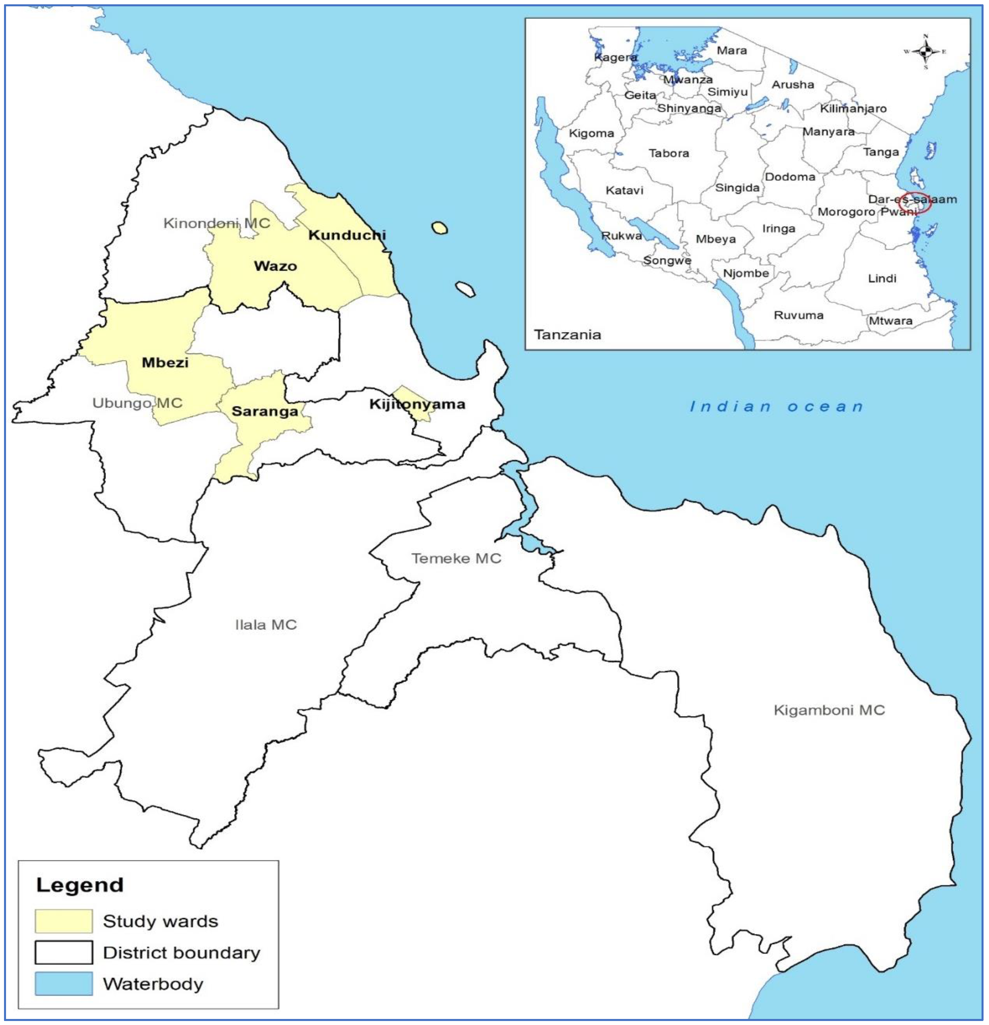
2.2. Study Design, Area and Cattle Farm Selection

This was a cross-sectional study conducted from September to October 2021 in Kinondoni and Ubungo districts, which are part of the city of Dar es Salaam in eastern, Tanzania. It included five wards: Kijitonyama, Kunduchi and Wazo in Kinondoni and Mbezi and Saranga in Ubungo (Figure 1). The selection of cattle farms included in this study was randomized based on a list of cattle herders provided by the local livestock officers in the study area. However, the selection was not totally random, as verbal consent from the herd owners was needed. 54 farms with a total herd of 201 participated in the study. As part of the city, most small-scale dairy farms had up to four heads of cattle as stipulated by the city bylaws, but some farms had more than 10 heads of cattle. Of the 54 small-scale dairy cattle farms, 39 farms each had four heads of cattle, all of which were sampled. The rest of the farm had more than ten heads of cattle. At each of the latter farms, 1 to 3 three heads of cattle were randomly selected and sampled according to the milk yield. In almost all of these 15 farms, cattle were high producers, so only low-yield cattle were allowed to be sampled.

2.3. Sample Collection

Briefly, cattle were restrained in a pen. Using gloved hands, the tail was raised using one hand and the other for swabbing. Approximately one gram of cattle faecal material was aseptically swabbed from the rectum using a sterile cotton swab (Himedia, Mumbai, India). The swab was then placed in sterile tubes filled with 3 mL Stuart’s transport medium, cap tightened, and shaken well. All samples were transported in a clean cool box with ice packs at a temperature of 2 to 8 °C and processed within 48 h of collection at the Microbiology Teaching Laboratory at the College of Veterinary Medicine and Biomedical Sciences, Sokoine University of Agriculture (SUA), Morogoro, Tanzania.

2.4. Bacterial Isolation and E. coli Identification

Looped rectal swab specimens from Stuart transport medium, were streaked directly onto the MacConkey agar (Oxoid, Basingstoke, UK) without antibiotics and aerobically incubated at 37 °C for 24 h. On incubation, for the case of mixed growth, a single colony from almost morphologically similar colonies (deep pinkish, round, mid-sized, and flat) was selected for the purity-plate, from each plain MacConkey agar plate and aerobically sub-cultured on nutrient agar medium (Hi Media, Mumbai, India) at 37 °C for 18–24 h, as previously reported [37]. Pure colonies were gram stained and subjected to biochemical tests to identify E. coli according to the manufacturer’s instructions using indole methyl red, Voges–Proskauer, and citrate utilization test (IMViC; Sigma-Aldrich Co., Zurich, Switzerland). Isolates that indicated indole positive, methyl-red positive, Voges–Proskauer negative, and citrate negative were identified as E. coli [40]. The identified E. coli isolates were subjected to an antibiotic susceptibility test (AST) and phenotypic confirmation of ESBL production.

2.5. Antimicrobial Susceptibility Testing

Antimicrobial susceptibility test was performed using the Kirby–Bauer disk diffusion method on Mueller-Hinton agar (Merck), according to the recommendations of the Clinical and Laboratory Standards Institute (CLSI) [41]. The E. coli isolates were tested using the following antibiotics shown in Table 1.
These antibiotics have been selected because are considered to be clinical and useful in animal production by the World Health Organization (WHO) [42]. Single well-isolated colonies from the identified pure lactose fermenter cultures were emulsified into 5 mL of sterile saline and adjusted to that of standard 0.5 McFarland (Oxoid Ltd., Basingstoke, UK). A sterile cotton swab was dipped in a standardized bacterial (E. coli) suspension and evenly streaked or spread over the entire surface of Mueller Hinton agar medium. Impregnated paper discs with constant concentration of antibiotics were placed on the surface of the agar and incubated at 37 °C for 24 h. The zone of inhibition interpretation was performed according to the CLSI 31st Edition guidelines [41]. All the E. coli isolates that showed intermediate susceptibility to the antibiotics tested were not regarded as resistant to such particular antibiotics. A double-disk synergistic test of cefotaxime and cefotaxime-clavulanate was used to identify the extended-spectrum ß-lactamases (ESBL) phenotype [41]. E. coli ATCC 25922 was used as a control strain. Percentages of isolates resistant to any number of antibiotics were reported. If the isolate was resistant to three or more drugs from different classes of antimicrobials, it was considered MDR [43].

2.6. Statistical Analysis

Statistical analysis was performed using IBM SPSS Statistics for windows version 20.0. The graphical representation was performed using a program (Microsoft Office Excel, 2019). The Kruskal–Wallis test was used with a significance value of 0.05 to compare the co-resistance of different isolates. This is a non-parametric statistical test that evaluates differences between three or more independently sampled groups on a single, non-normally distributed continuous variable [44].

3. Results

This study evaluated the antimicrobial resistance of E. coli isolates from healthy cattle rectal swabs. A total of 201 rectal swab samples were examined and 121 E. coli isolates were isolated. This corresponds to an isolation rate of 60.2%.

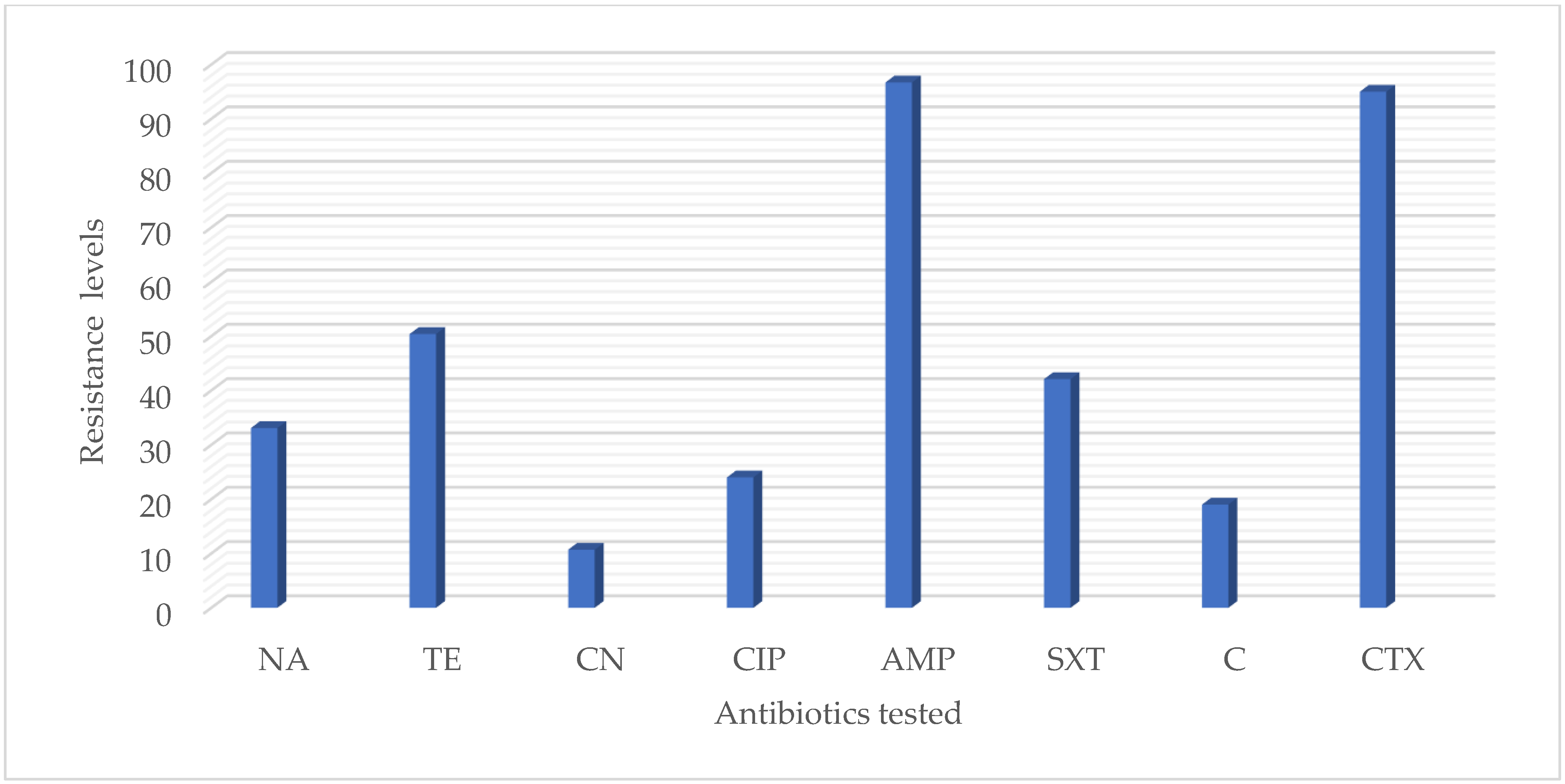
3.1. Antimicrobial Susceptibiliy

The prevalence of antimicrobial resistance in 121 E. coli isolates at the isolate level is presented in Table 2.
The majority of isolates (Table 2 and Figure 2) have shown resistance to ampicillin (96.7%), followed by cefotaxime (95.0%), tetracycline (50.4%), trimethoprim/sulfamethoxazole (42.1%) and nalidixic acid (33.1%). Moderate resistance rates were observed for ciprofloxacin (20.7). However, low resistance was observed with chloramphenicol (19.0%) and gentamycin (10.7%). In this case, 20 extended-spectrum beta-lactamases (ESBLs) that produce E. coli were detected (Table 3).

3.1.1. Coresistances and AMR Profile

Analysis of coresistance for the E. coli isolates is shown in Table 4.
A Kruskal-Wallis H test showed that there was no statistically significant difference in isolates across the categories of resistance, x2 = 3.049, p = 0.384, with a mean rank isolate of 19.50 for category of resistance to One antimicrobial agent, 15.80 for category of resistance to Two antimicrobial agents, 26.43 for category of resistance to Three antimicrobial agents and 19.88 for category of resistance to More than three agents.
On the other hand, 90 isolates (74.4%) were MDR, which means resistant to three or more families of antibiotics, as we considered beta-lactams and cephalosporins as two different families.

3.1.2. MDR Patterns and Antimicrobial Resistance Phenotype

Depending on their antibiotic resistance phenotypes, the 121 isolates of E. coli belong to 41 different phenotypes, showing a large variety of resistances, ranging from one antimicrobial to a combination of 8 (Table 5).
The most frequently observed phenotypes were AMP-SXT-CTX with a prevalence of 12.4%, followed by AMP-CTX (10.7%), TE-AMP-CTX and NA + TE + AMP + CTX with a prevalence of 8.3% each. In this case, 15 different phenotypes that produce extended-spectrum ß-lactamases (ESBLs) were detected and had a prevalence of 42.9%. They were all MDR ranging from three antimicrobials to a combination of 8. ESBL phenotypes were resistant to ampicillin and cefotaxime.

4. Discussion

In this study, 60.2% of the isolates from 201 samples of rectal swabs from small scale dairy cattle were recovered. Overall, 74.4%, which is close to three quarters of all E. coli isolates from the small-scale dairy cattle, exhibited multidrug resistance against three to eight classes of antimicrobial, with the most resistant pattern being AMP-SXT-CTX. The 121 E. coli isolates belonged to 41 phenotypes ranging from one antimicrobial agent to a combination of 8. 20 E. coli isolates were ESBLs producers with a prevalence rate of 16.5%. There were 15 different phenotypes detected with a prevalence of 42.9% producing extended-spectrum ß-lactamases and all of them were MDR ranging from three antimicrobials to a combination of 8. The occurrence of a high-level antibiotic resistance obtained in E. coli isolates from the healthy dairy cattle studied gives an indication of the possible roles these bacteria play in the transfer of the observed resistance among pathogenic E. coli from livestock in Tanzania. The resistance levels identified in this study are comparable with the findings reported by other studies in E. coli isolates from healthy cattle elsewhere in Africa. For instance, Ogunleye et al. [45] reported 96.7% (232/240) resistance for ampicillin among E. coli isolated from rectal swab from apparently healthy cattle from a major cattle market in Ibadan, Oyo State, South West Nigeria, 59.1% (117/198) from rectal swab from healthy cattle in Eastern Algeria, Barour et al. [46], 54.8% (23/42) from apparently healthy cattle in greater part of Kumasi, Ghana, Rita et al. [47], while in the current study, 96.7% (117/121) resistance was obtained for ampicillin. However, the resistance percentage reported in this study was higher than the 21.4% (29/135) in Madoshi et al. [48] in Tanzania.
It was expected from the start of the study for a possibility of at least observing some level of resistance to cephalosporins. A study by Katakweba et al. [49] reported some level of resistance by cefotaxime (57.1% (40/70), a 3rd generation cephalosporin, in Enterobacteriaceae isolated from dairy cattle in Tanzania. The percentage resistance in the current study for cefotaxime were also comparable to the findings of Olowe et al. [50] in Nigeria. Cefotaxime had 95.0% (115/121) resistance in the current study and 92.1% (105/114) as reported by Olowe et al. [50]. However, it was in contrast with the findings of Barour et al. [46] who reported a low resistance rate of 4.5% (9/198). Cephalosporins are not readily available for veterinary use and are rarely used on small scale in farms in Tanzania [48]. Surprisingly, the current study showed widespread occurrence of resistance to cephalosporins in E. coli from farm animals. This could be explained by cross transmission of resistant strains between cattle and humans where there is widespread use of cephalosporin Although in this study, we did not address antibiotic use in humans, the exchange of resistance genes between humans and animals is likely to occur during the rainy season when cattle drinks contaminated water or pastures. Therefore, the 95.0% resistance to cefotaxime could imply a potentially serious threat to public health. Remarkable resistance in the current study was also observed against tetracycline and trimethoprim/sulfamethoxazole which is comparable to the findings of Abbassi et al. [51] in Tunisia but lower than the findings of Gupta et al. [52] in Bangladesh. For tetracycline, there was 50.4% (61/121) in the current study; 33.3% (20/60) for Abbassi et al. [51] and 83.3% (70/84) for Gupta et al. [52]. For trimethoprim/sulfamethoxazole, there was 42.1% (51/121) resistance in the current study, 30% (18/60) in Abbasi et al. [51] and 83.3% (70/84) from the work of Gupta et al. [52]. Resistance to tetracycline and trimethoprim/sulfamethoxazole might have relation with the wide use of these antibiotics in livestock production over along period on farms in Tanzania. In the current study, a high resistance level of cefotaxime was observed as compared to that of tetracycline and trimethoprim/sulfamethoxazole. The contrast in cefotaxime resistance is difficult to explain, as the antibiotic was not commonly used on animals. Further studies are probably required to address this observation. However, this antibiotic could possibly serve as an indicator for monitoring drug resistance in animals and humans.
In this study, 16.5% (20/121) of the E. coli isolates were found to be ESBLs producers, which is comparable to other studies in Tanzania and elsewhere. For instance, Seni et al. [53], reported 10.8% (14/130) of the E. coli isolates from cattle as ESBL producers in Tanzania; Chishimba et al. [54] reported 20.1% (77/384) of E. coli isolates in market-ready broiler chicken in Zambia; Reuben et al. [55] reported 10% (20/200) of E. coli isolates from cattle in Tanzania; Okpara et al. [56], reported 10.6% (53/457) of E. coli isolates from animals and humans in households in Nigeria. However, the prevalence reported in the current study is lower than the levels reported by Montso et al. [57], of 66.3% (69/104) in E. coli isolates from cattle on farms and raw beef in South Africa and Kimera et al. [37] of 65.3% (301/461) in broiler chicken in Tanzania. Almost all the ESBL producers were resistant to most of the tested antimicrobials. This is probably indicative of the selection pressure exerted by excessive use of beta-lactams and cephalosporins in animal production, and presence of various resistance mechanisms from inappropriate use of veterinary drugs [58].
The MDR levels in the current study are comparable with the findings reported by other studies in E. coli isolates from clinically health cattle elsewhere. For instance, Ajayi et al. [59], reported 81.0% (851/1051) MDR level from E. coli isolates from clinically health cattle in Nigeria, 61.1% (99/162) in Nariman et al. [60] from rectal swabs of healthy cattle from the suburban and farming areas in Tripoli, Libya, 64.3% (27/42) in Rita et al. [47] in Kumasi, Ghana from fresh droppings from clinically health cattle at the abattoir, and 44.2% (77/174) in Abbasi et al. [51], in Tunisia, from clinically healthy cattle while in the current study, 74.4% (90/121) MDR level was obtained. However, it was lower than the findings of Donkor et al. [61], in Ghana of 97.7% (262/268). The possible explanation of MDR level in this study could be attributed to indiscriminate exploitation of antimicrobial agents in small scale dairy cattle thus providing selective pressure favouring the emergence of resistant strains. That is to say, the MDR reported in this study can probably be the result of an independent resistance for each antimicrobial agent or coresistance. MDR bacteria presents an impending therapeutic impasse to human and animal health. Some major factors which may contribute to the increase of bacterial MDR are: transfer of resistance determinants by genetic elements such as plasmids, transposons, and gene cassettes into integrons and changing regulation in mar locus [62]. The high MDR level we recorded in the current study calls for monitoring of MDR E. coli strains.
The antimicrobial susceptibility patterns observed in the isolates from the small- scale dairy cattle towards one to a combination of 8 antimicrobials in this study shows that the isolates were diverse in their antimicrobial resistance spectrum. This is comparable to the findings of Barour et al. [46], who reported a large variety of resistances, ranging from one antimicrobial to a combination of 10. MDR patterns in the current study were comparable to some studies in E. coli isolates from apparently health cattle elsewhere. For instance, Amosun et al. [63] reported 41 MDR patterns from E. coli isolates from apparently health on-farm cattle, in Ibadan, Nigeria, 31 MDR patterns in Barour et al. [46], in Algeria from rectal swab E. coli isolates from health cattle and 33- MDR patterns in the current study. However, it was lower than the 71 MDR patterns in Ajayi et al. [59] in Ado-Ekiti, Nigeria from E. coli isolated from feces of apparently healthy ready to slaughter cattle. MDR patterns may harbor resistance genes that may be transferred to humans via the food chain. There is need for further studies on identifying the resistance genes and their ability to mobilize. All the MDR phenotypes in the current study were AMP resistant. This was comparable with the findings by Barour et al. [46], who reported that all the MDR phenotypes were AMP resistant. This implies that E. coli strains resistant to this antibiotic have an increased ability to be resistant to other antimicrobials.
In this case, 15 out of the 20 ESBL phenotypes in the current study were MDR. This was comparable with the studies in E. coli isolates from clinically health cattle carried out elsewhere. For instance, Olowe et al. [50] in Nigeria, reported 63.2% (72/114) as ESBL phenotypes and MDR, 4.5% (9/198) in Barour et al. [46], in Algeria and in the current study was 16.5% (20/121). ESBL phenotype includes resistance to ampicillin and cefotaxime, which is the cause of many therapeutic failures [62]. This requires the surveillance of strains with this type of phenotype.
The high variability of resistance phenotypes in this study can be explained by the acquisition of resistance to several antibiotics of different classes (coresistance) since the plasmids exchanged usually have several resistance genes such as the coresistance of E. coli to cephalosporins, penicillins, chloramphenicol, tetracyclines, and fluoroquinolones Barour et al. [46].
Despite the above observations, the current study had some limitations. In the first place, there could be other antibiotics used among the small-scale dairy cattle in Tanzania, which were not tested in this study. Secondly, we did not perform molecular characterization of resistant isolates. The high resistance observed could be explained based on standardized Clinical Laboratory Standard Institute (CLSI) 2021 protocol and guidelines [41] which are internationally recognized, thus depicting the real magnitude and pattern of AMR in the study setting.

5. Conclusions

The study has revealed levels of AMR as well as ESBL producers, of E. coli isolates from the rectal swab of apparently healthy animals to commonly used antimicrobials in small-scale dairy cattle production. This renders the antimicrobials inactive to their intended use. The results of the current study call for prudent antimicrobial use in cattle to create a better picture of the situation and advice policymakers on their decisions.

Author Contributions

Conceptualization, R.R.A.; methodology, R.R.A., S.E.M. and M.I.M.; software, R.R.A. and S.I.K.; validation, S.I.K.; formal analysis, R.R.A. investigation, R.R.A.; data curation, R.R.A.; writing—original draft preparation, R.R.A.; writing—review and editing, R.R.A., S.I.K., M.I.M. and S.E.M.; visualization, all authors; supervision, S.I.K., M.I.M. and S.E.M. All authors have read and agreed to the published version of the manuscript.

Funding

This study was funded by the Government of the United Republic of Tanzania and the World Bank [WB-ACE II Grant PAD1436, IDA [Credit 5799-TZ] through SACIDS Foundation for One Health, Sokoine University of Agriculture, Morogoro, Tanzania.

Institutional Review Board Statement

The ethical clearance was provided by the Medical Research Coordinating Committee of the National Institute of Medical Research of Tanzania (NIMR), (Ref #. NIMR/HQ/R.8a/Vol.IX/3233), and the ethical committee of Sokoine University of Agriculture (Ref # DPRTC/186/3). The permission was sought from the relevant authorities which are the municipal directors of the two districts.

Informed Consent Statement

Not applicable.

Data Availability Statement

The data generated is contained within the article.

Acknowledgments

The authors wish to thank the Government of the United Republic of Tanzania and World Bank through SACIDS Foundation for One Health for providing a scholarship grant to R.R.A. We appreciate the technical assistance provided by the local government authorities and the SACIDS staff during data collection and the technicians at the Veterinary Microbiology Laboratory at the College of Veterinary Medicine and Biomedical Sciences and colleagues in Antimicrobial Community of Practice (CoP) at the SACIDS Foundation for One Health for their continuous encouragement during the study.

Conflicts of Interest

The authors declare no conflict of interest.

References

  1. Tanzania UR National Bureau of Statistics (2021). National Sample Census of Agriculture 2019/20 -ATLAS-. Available online: https://www.nbs.go.tz/index.php/en/census-surveys/agriculture-statistics/669-2019-20-national-sample-census-of-agriculture-atlas (accessed on 5 May 2022).
  2. Tanzania UR Ministry of Livestock and Fisheries: Tanzania Livestock Master Plan 2017 (2017/2018–2021/2022). Available online: www.mifugouvuvi.go.tz/uploads/projects/1553601703-TANZANIALIVESTOCKMASTERPLAN.pdf (accessed on 18 May 2021).
  3. Van Boeckel, T.P.; Pires, J.; Silvester, R.; Zhao, C.; Song, J.; Criscuolo, N.G. Global trends in antimicrobial resistance in animals in low- and middle- income countries. Science 2019, 365, eaaw1944. [Google Scholar] [CrossRef] [PubMed] [Green Version]
  4. Robinson, T.P.; Thornton, P.K.; Franceschini, G.; Kruska, R.; Chiozza, F.; Notenbaert, A.M. Global Livestock Production Systems; FAO: Rome, Italy; ILRI: Nairobi, Kenya, 2011. [Google Scholar]
  5. Azabo, R.; Mshana, S.; Matee, M.; Kimera, S.I. Antimicrobial usage in cattle and poultry production in Dar es Salaam, Tanzania: Pattern and quantity. BMC Vet. Res. 2022, 18, 7. [Google Scholar] [CrossRef] [PubMed]
  6. Karimuribo, E.D.; Mdegela, R.H.; Kusiluka, L.J.M.; Kambarage, D.M. Assessment of drug usage and antimicrobial residues in milk on smallholder farms in Morogoro, Tanzania. Bull. Anim. Health Prod. Afr. 2005, 53, 234–241. [Google Scholar]
  7. Katakweba, A.A.S.; Mtambo, M.M.A.; Olsen, J.E.; Muhairwe, A.P. Awareness of human health risks associated with the use of antimicrobials among livestock keepers and factors that contribute to selection of antibiotic resistance bacteria within livestock in Tanzania. Livest. Rural. Res. Dev. 2012, 24, 1. [Google Scholar]
  8. Caudell, M.A.; Quinlan, M.B.; Subbiah, M.; Call, D.R.; Roulette, C.J.; Roulette, J.W.; Roth, A.; Matthews, L.; Quinlan, R.J. Antimicrobial use and veterinary care among agro-pastoralists in northern Tanzania. PLoS ONE 2017, 12, e0170328. [Google Scholar]
  9. Sayah, R.S.; Kaneene, J.B.; Johnson, Y.; Miller, R. Patterns of antimicrobial resistance observed in Escherichia coli isolates obtained from domestic- and wild-animal fecal samples, human septage, and surface water. Appl. Environ. Microbiol. 2005, 71, 1394–1404. [Google Scholar] [CrossRef] [Green Version]
  10. Tempini, P.N.; Aly, S.S.; Karle, B.M.; Pereira, R.V. Multidrug residues and antimicrobial resistance patterns in waste milk from dairy farms in Central California. J. Dairy Sci. 2018, 101, 8110–8122. [Google Scholar] [CrossRef]
  11. Berghiche, A. Special attention is needed for reduce antibiotic residue risk in the white meat produced in Algeria. J. Food Qual. Hazards Control 2019, 6, 44. [Google Scholar] [CrossRef]
  12. Maron, D.; Smith, T.J.; Nachman, K.E. Restrictions on antimicrobial use in food animal production: An international regulatory and economic survey. Glob. Health 2013, 9, 48. [Google Scholar] [CrossRef] [Green Version]
  13. Gottlieb, T.; Nimmo, G.R. Antibiotic resistance is an emerging threat to public health: An urgent call to action at the antimicrobial resistance summit. Med. J. Aust. 2011, 194, 281–283. [Google Scholar] [CrossRef]
  14. Stella, P.; Beloeil, P.A.; Guerra, B.; Hugas, M.; Liebana, E. The role of the European Food Safety Authority (EFSA) in the fight against antimicrobial resistance (AMR). Food Prot. Trends 2018, 38, 72–80. [Google Scholar]
  15. Mathew, A.G.; Cissell, R.; Liamthong, S. Antibiotic resistance in bacteria associated with food animals: A United States perspective of livestock production. Foodborne Pathog. Dis. 2007, 4, 115–133. [Google Scholar] [CrossRef] [PubMed] [Green Version]
  16. Merle, R.; Hajek, P.; Kasbohrer, A.; Hegger-Gravenhorst, C.; Mollenhauer, Y.; Robanus, M.; Ungemach, F.-R.; Kreienbrock, L. Monitoring of antibiotic consumption in livestock: A German feasibility study. Prev. Vet. Med. 2012, 104, 34–43. [Google Scholar] [CrossRef] [PubMed]
  17. Acar, J.F.; Moulin, G.; Page, S.W.; Pastoret, P. Antimicrobial resistance in animal and public health: Introduction and classification of antimicrobial agents. Rev. Sci. Tech. OIE 2012, 31, 15–21. [Google Scholar] [CrossRef] [PubMed]
  18. WHO. Global Antimicrobial Resistance Surveillance System (GLASS) Report Early Implementation; World Health Organization: Geneva, Switzerland, 2017. [Google Scholar]
  19. Founou, L.L.; Amoako, D.G.; Founou, R.C.; Essack, S.Y. Antibiotic Resistance in Food Animals in Africa: A Systematic Review and Meta-Analysis. Microb. Drug Resist. 2018, 24, 648–665. [Google Scholar] [CrossRef] [PubMed]
  20. Kimera, Z.I.; Mshana, S.E.; Rweyemamu, M.M.; Mboera, L.E.G.; Matee, M.I.N. Antimicrobial Use and Resistance in FoodProducing Animals and the Environment: An African Perspective. Antimicrob. Resist. Infect. Control 2020, 9, 37. [Google Scholar] [CrossRef] [Green Version]
  21. Barbosa, T.M.; Levy, S.B. The impact of antibiotic use on resistance development and persistence. Drug Resist. Updates 2000, 3, 303–311. [Google Scholar] [CrossRef] [Green Version]
  22. Price, L.B.; Graham, J.P.; Lackey, L.G.; Roess, A.; Vailes, R.; Silbergeld, E. Elevated risk of carrying gentamicin-resistant Escherichia coli among U.S. poultry workers. Environ. Health Perspect. 2007, 115, 1738–1742. [Google Scholar] [CrossRef] [Green Version]
  23. Silbergeld, E.K.; Graham, J.; Price, L.B. Industrial food animal production, antimicrobial resistance, and human health. Annu. Rev. Public Health 2008, 29, 151–169. [Google Scholar] [CrossRef]
  24. Van den Bogaard, A.E.; Stobberingh, E.E. Epidemiology of resistance to antibiotics. Links between animals and humans. Int. J. Antimicrob. Agents 2000, 14, 327–335. [Google Scholar] [CrossRef]
  25. Andersson, D.I.; Hughes, D. Microbiological effects of sublethal levels of antibiotics. Nat. Rev. Microbiol. 2014, 12, 465–478. [Google Scholar] [CrossRef] [PubMed]
  26. Pereira, R.V.V.; Siler, J.D.; Bicalho, R.C.; Warnick, L.D. In vivo selection of resistant E. coli after ingestion of milk with added drug residues. PLoS ONE 2014, 9, e115223. [Google Scholar] [CrossRef] [PubMed]
  27. Harbottle, H.; Thakur, S.; Zhao, S.; White, D.G. Genetics of antimicrobial resistance. Anim. Biotechnol. 2006, 17, 111–124. [Google Scholar] [CrossRef] [PubMed]
  28. Moreno, M.A.; Domínguez, L.; Teshager, T.; Herrero, I.A.; Porrero, M.C. Antibiotic resistance monitoring: The Spanish programme. The VAV Network. Red de Vigilancia de Resistencias Antibioticas en Bacterias de Origen Veterinario. Int. J. Antimicrob. Agents 2000, 14, 285–290. [Google Scholar] [CrossRef]
  29. Darwich, L.; Vidal, A.; Seminati, C.; Albamonte, A.; Casado, A.; López, F.; Molina-López, R.A.; Migura-Garcia, L. High prevalence and diversity of extended-spectrum β-lactamase and emergence of OXA-48 producing Enterobacterales in wildlife in Catalonia. PLoS ONE 2019, 14, e0210686. [Google Scholar]
  30. Pedersen, K. DANMAP; National Food Institute, Technical University of Denmark: Copenhagen, Denmark, 2014. [Google Scholar]
  31. Rugumisa, B.T.; Call, D.R.; Mwanyika, G.O.; Mrutu, R.I.; Luanda, C.M.; Lyimo, B.M.; Subbiah, M.; Buza, J.J. Prevalence of Antibiotic-Resistant Fecal Escherichia coli Isolates from Penned Broiler and Scavenging Local Chickens in Arusha, Tanzania. J. Food Prot. 2016, 79, 1424–1429. [Google Scholar] [CrossRef]
  32. Bernadether, T.R.; Douglas, R.C.; Gaspary, O.M.; Murugan, S.; Joram, B. Comparison of the prevalence of antibiotic-resistant Escherichia coli isolates from commercial-layer and free-range chickens in Arusha district, Tanzania. Afr. J. Microbiol. Res. 2016, 10, 1422–1429. [Google Scholar] [CrossRef] [Green Version]
  33. Maganga, R. Antimicrobial Resistance in Commensal Escherichia coli Isolated from Poultry along a Gradient of Intensification of Poultry Production in the Northern Zone of Tanzania. Master’s Thesis, University of Glasgow, Glasgow, UK, 2019. [Google Scholar]
  34. Sonola, V.S.; Katakweba, A.S.; Misinzo, G.; Matee, M.I.N. Occurrence of Multi-Drug-Resistant Escherichia coli in Chickens, Humans, Rodents and Household Soil in Karatu, Northern Tanzania. Antibiotics 2021, 10, 1137. [Google Scholar] [CrossRef]
  35. Mgaya, F.X.; Matee, M.I.; Muhairwa, A.P.; Hoza, A.S. Occurrence of Multidrug Resistant Escherichia coli in Raw Meat and Cloaca Swabs in Poultry Processed in Slaughter Slabs in Dar es Salaam, Tanzania. Antibiotics 2021, 10, 343. [Google Scholar] [CrossRef]
  36. Kiiti, R.W.; Komba, E.V.; Msoffe, P.L.; Mshana, S.E.; Rweyemamu, M.; Matee, M.I.N. Antimicrobial Resistance Profiles of Escherichia coli Isolated from Broiler and Layer Chickens in Arusha and Mwanza, Tanzania. Int. J. Microbiol. 2021, 2021, 6759046. [Google Scholar] [CrossRef]
  37. Kimera, Z.I.; Mgaya, F.X.; Misinzo, G.; Mshana, S.E.; Moremi, N.; Matee, M.I.N. Multidrug-Resistant, Including Extended-Spectrum Beta Lactamase-Producing and Quinolone-Resistant, Escherichia coli Isolated from Poultry and Domestic Pigs in Dar es Salaam, Tanzania. Antibiotics 2021, 10, 406. [Google Scholar] [CrossRef] [PubMed]
  38. Sangeda, R.Z.; Baha, A.; Erick, A.; Mkumbwa, S.; Bitegeko, A.; Sillo, H.B.; Fimbo, A.M.; Chambuso, M.; Mbugi, E.V. Consumption Trends of Antibiotic for Veterinary Use in Tanzania: A Longitudinal Retrospective Survey From 2010–2017. Front. Trop. Dis. 2021, 2, 694082. [Google Scholar] [CrossRef]
  39. Azabo, R.; Matee, M.; Kimera, S. Assessment of antimicrobial consumption in food animals in Dar es Salaam, Tanzania. J. Anim. Sci. Med. 2021, 6, 159–170. [Google Scholar] [CrossRef]
  40. Mahe, A.; Sabui, B.; Adam, A.A.; Abdullahi, U.Z.; Ado, B.A. Effect of citric acid at different Ph on the survival of Escherichia coli. Bayero J. Pure Appl. Sci. 2021, 14, 79–84. [Google Scholar] [CrossRef]
  41. CLSI. Performance Standards for Antimicrobial Susceptibility Testing, 31st ed.; CLSI Supplement M100; Clinical and Laboratory Standards Institute: Malvern, PA, USA, 2021. [Google Scholar]
  42. WHO. Integrated Surveillance of Antimicrobial Resistance—Guidance from WHO Advisory Group; WHO: Geneva, Switzerland, 2013; pp. 1–100. Available online: http://apps.who.int/iris/bitstream/10665/91778/1/9789241506311_eng.pdf?ua=1 (accessed on 3 September 2018).
  43. Magiorakos, A.; Srinivasan, A.; Carey, R.B.; Carmeli, Y.; Falagas, M.E.; Giske, C.G.; Harbarth, S.; Hindler, J.F.; Kahlmeter, G.; Olsson-Liljequist, B.; et al. Multidrug-resistant, extensively drug-resistant and pandrug-resistant bacteria: An internatiojnal expert proposal for interim standard definitions for acquired resistance. Clin. Microbiol. Infect. 2012, 18, 268–281. [Google Scholar] [CrossRef] [PubMed] [Green Version]
  44. Kruskal, W.H.; Wallis, W.A. Use of ranks in one-criterion variance analysis. J. Am. Stat. Assoc. 1952, 47, 583–621. [Google Scholar] [CrossRef]
  45. Ogunleye, A.O.; Okunlade, A.O.; Jeminlehin, F.O.; Ajuwape, A.T. Antibiotic resistance in Escherichia coli isolated from healthy cattle at a major cattle market in Ibadan, Oyo State, South-Western, Nigeria. Afr. J. Microbiol. Res. 2013, 7, 4572–4575. [Google Scholar] [CrossRef] [Green Version]
  46. Barour, D.; Berghiche, A.; Boulebda, N. Antimicrobial resistance of Escherichia coli isolates from cattle in Eastern Algeria. Vet. World 2019, 12, 1195–1203. [Google Scholar] [CrossRef]
  47. Rita, O.L.; Linda, A.O.; Augustina, A.S.; Matilda, A.-A.; Kwasi, O.-D. Antimicrobial Resistance of Escherichia coli from Broilers, Pigs, and Cattle in the Greater Kumasi Metropolis, Ghana. Int. J. Microbiol. 2021, 2021, 5158185. [Google Scholar]
  48. Madoshi, B.P.; Kudirkiene, E.; Mtambo, M.M.A.; Muhairwa, A.P.; Lupindu, A.M.; Olsen, J.E. Characterisation of Commensal Escherichia coli Isolated from Apparently Healthy Cattle and Their Attendants in Tanzania. PLoS ONE 2016, 11, e0168160. [Google Scholar] [CrossRef]
  49. Katakweba, A.A.; Muhairwa, A.P.; Lupindu, A.M.; Damborg, P.; Rosenkrantz, J.T.; Minga, U.M.; Mtambo, M.M.; Olsen, J.E. First Report on a Randomized Investigation of Antimicrobial Resistance in Fecal Indicator Bacteria from Livestock, Poultry, and Humans in Tanzania. Microb. Drug Resist. 2018, 24, 260–268. [Google Scholar] [CrossRef] [PubMed]
  50. Olowe, O.A.; Adewumi, O.; Odewale, G.; Ojurongbe, O.; Adefioye, O.J. Phenotypic and Molecular Characterisation of Extended-Spectrum Beta-Lactamase Producing Escherichia coli Obtained from Animal Fecal Samples in Ado Ekiti, Nigeria. J. Environ. Public Health 2015, 2015, 497980. [Google Scholar] [CrossRef] [Green Version]
  51. Abbassi, M.S.; Kilani, H.; Zouari, M.; Mansouri, R.; El Fekih, O.; Hammami, S.; Ben Chehida, N. Antimicrobial Resistance in Escherichia Coli Isolates from Healthy Poultry, Bovine and Ovine in Tunisia: A Real Animal and Human Health Threat. J. Clin. Microbiol. Biochem. Technol. 2017, 3, 19–23. [Google Scholar] [CrossRef] [Green Version]
  52. Gupta, M.D.; Islam, M.; Sen, A.; Sarker, M.S.; Das, A. Prevalence and antibiotic susceptibility pattern of Escherichia coli in cattle on Bathan and intensive rearing system. Microbes Health 2017, 6, 2226. [Google Scholar] [CrossRef] [Green Version]
  53. Seni, J.; Falgenhauer, L.; Simeo, N.; Mirambo, M.M.; Imirzalioglu, C.; Matee, M.; Rweyemamu, M.; Chakraborty, T.; Mshana, S.E. Multiple ESBL-producing Escherichia coli sequence types carrying quinolone and aminoglycoside resistance genes circulating in companion and domestic farm animals in Mwanza, Tanzania, harbor commonly occurring plasmids. Front. Microbiol. 2016, 7, 142. [Google Scholar] [CrossRef] [PubMed]
  54. Chishimba, K.; Hang’ombe, B.M.; Muzandu, K.; Mshana, S.E.; Matee, M.I.; Nakajima, C.; Suzuki, Y. Detection of Extended Spectrum Beta-Lactamase-Producing Escherichia coli in Market-Ready Chickens in Zambia. Int. J. Microbiol. 2016, 2016, 5275724. [Google Scholar] [CrossRef] [PubMed] [Green Version]
  55. Reuben, S.M.; Kaunara, A.A. Prevalence and antimicrobial resistance patterns of Extended Spectrum Beta-Lactamase producing enterohemorrhagic Escherichia coli strain O157:H7 from cattle and humans in Moshi, Northern Tanzania. Microbiol. Res. J. Int. 2017, 19, 1–10. [Google Scholar]
  56. Okpara, E.O.; Ojo, O.E.; Awoyomi, O.J.; Dipeolu, M.A.; Oyekunle, M.A.; Schwarz, S. Antimicrobial usage and presence of extended-spectrum β-lactamase-producing Enterobacteriaceae in animal-rearing households of selected rural and peri-urban communities. Vet. Microbiol. 2018, 218, 31–39. [Google Scholar] [CrossRef]
  57. Montso, K.P.; Dlamini, S.B.; Kumar, A.; Ateba, C.N. Antimicrobial Resistance Factors of Extended-Spectrum Beta-Lactamases Producing Escherichia coli and Klebsiella pneumoniae Isolated from Cattle Farms and Raw Beef in North-West Province, South Africa. Biomed. Res. Int. 2019, 2019, 4318306. [Google Scholar] [CrossRef] [Green Version]
  58. Sadiq, M.B.; Syed-Hussain, S.S.; Ramanoon, S.Z.; Saharee, A.A.; Ahmad, N.I.; Zin, N.M.; Khalid, S.F.; Naseeha, D.S.; Syahirah, A.A.; Mansor, R. Knowledge, attitude and perception regarding antimicrobial resistance and usage among ruminant farmers in Selangor, Malaysia. Prev. Vet. Med. 2018, 156, 76–83. [Google Scholar] [CrossRef]
  59. Ajayi, A.O.; Oluyege, A.O.; Olowe, O.A.; Famurewa, O. Antibiotic resistance among commensal Escherichia coli isolated from faeces of cattle in Ado-Ekiti, Nigeria. J. Anim. Vet. Adv. 2011, 10, 174–179. [Google Scholar] [CrossRef]
  60. Nariman, F.A.; Murad, A.H.; Ahmed, S.A.; Mohamed, S.A.; Ahmed, A.E.; Yousef, M.A.; Mohamed, O.A. Antimicrobial resistance among commensal enteric bacteria isolated from healthy cattle in Libya. Pan Afr. Med. J. One Health 2020, 1, 3. [Google Scholar]
  61. Donkor, E.S.; Newman, M.J.; Yeboah-Manu, D. Epidemiological aspects of non-human antibiotic usage and resistance: Implications for the control of antibiotic resistance in Ghana. Trop. Med. Int. Health 2012, 17, 462–468. [Google Scholar] [CrossRef]
  62. Sáenz, Y.; Briñas, L.; Domínguez, E.; Ruiz, J.; Zarazaga, M.; Vila, J.; Torres, C. Mechanisms of resistance in multiple-antibiotic-resistant Escherichia coli strains of human, animal, and food origins. Antimicrob. Agents Chemother. 2004, 48, 3996–4001. [Google Scholar] [CrossRef] [PubMed] [Green Version]
  63. Amosun, E.A.; Ojo, O.E.; Alao, I.K.; Ajuwape, A.T.P. Antimicrobial resistance among commensal Escherichia coli from cattle faeces and beef in Ibadan, Nigeria. Afr. J. Biotechnol. 2012, 11, 12240–12245. [Google Scholar] [CrossRef]
Figure 1. Map of the study districts (wards) in Dar- es- Salaam, Tanzania.
Animals 12 01853 g001
Figure 2. Frequencies of antibiotic resistance in 121 E. coli isolates: Nalidixic acid (NA), Tetracycline (TE), Gentamycin (CN), Ciprofloxacin (CIP), Ampicillin (AMP), Trimethoprim/sulfamethoxazole (SXT), Chloramphenicol (C), Cefotaxime (CTX).
Animals 12 01853 g002
Table 1. Antimicrobial concentrations and interpretation breakpoints of the various antimicrobial agents used in this study to interpret the results (CLSI, 2021).
Antimicrobial Agent (Code)Disc Drug Concentration (µg)Breaking Point (mm)
Sensitive (S)Intermediate (I)Resistant (R)
AMP10 µg≥1714–16≤13
CTX30 µg≥2623–25≤22
CN10 µg≥1513–14≤12
TE30 µg≥1512–14≤11
NA30 µg≥1914–18≤13
CIP5 µg≥2116–20≤15
SXT1.25/23.75 µg≥1611–15≤10
C30 µg≥1813–17≤12
AMP: Ampicillin, CTX: Cefotaxime, CN: Gentamycin, TE: Tetracycline, NA: Nalidixic acid, CIP: Ciprofloxacin, SXT: Trimethoprim/sulfamethoxazole, C: Chloramphenicol.
Table 2. Susceptibility to Antibiotics of E. coli Isolates (n = 121) from Small-Scale Dairy Cattle in Dar es Salaam, Tanzania.
ATBRISDistribution (Number) in Each inhibition zone Diameter (mm)
%%%078910111213141516171819202122232425>25
NA33.125.641.333 61 32151113211112 2
TE50.47.442.1361096 4323879710 51 1
CN10.713.276.05 4 431313151718131411
CIP24.026.449.691135 2 17546710712691016
AMP96.71.71.711111 2 211 1 1
SXT42.114.043.82534712525146410107 15433
C19.014.066.920 1 2 1423777209137747
CTX95.04.10.8251 1213 31476121120362121
E. coli isolated from plain MacConkey agar. ATB: antibiotic, R: resistance, I: intermediate, S: susceptible, NA: nalidixic acid, TE: tetracycline, CN: gentamycin, CIP: ciprofloxacin, AMP: ampicillin, SXT: trimethoprim/sulfamethoxazole, C: chloramphenicol, CTX: Cefotaxime. Dark grey fields present frequencies of resistant isolates, fields in light grey with borders present frequencies of intermediate isolates, and white fields present frequencies of susceptible isolates.
Table 3. Phenotypic pattern of 20 ESBL producing E. coli isolates from rectal swab of small-scale dairy cattle.
No of Antibiotic ClassesResistance PatternNo. of IsolatesPrevalence (%)
2AMP + CTX315.0
3AMP + SXT + CTX210.0
TE + AMP + CTX315.0
CN + AMP + CTX15.0
4NA + AMP + SXT + CTX15.0
NA + TE + AMP + CTX315.0
TE+ AMP + SXT + CTX210.0
TE + CIP + AMP + CTX15.0
5CIP + AMP + SXT + C +CTX15.0
NA +TE + AMP + SXT + CTX15.0
6NA + TE + CIP + AMP + C + CTX210
Table 4. Co-resistance of the E. coli isolates from cattle.
Number (n) and Percentages (%) of Isolates Resistant toKruskal-Wallis H (p-Value)
Categories of resistance to antimicrobial agentsOneTwoThreeMore than three agentsx2 = 3.049
p = 0.384
df = 3
n (%)n (%)n (%)n (%)
2 (5.0)5 (12.5)7 (17.5)26 (65.0)
Mean ranks19.5015.8026.4319.88
E. coli: Escherichia coli.
Table 5. Patterns of antimicrobial resistance phenotypes for Escherichia coli strains isolated in the study, with antibiogram pattern codes.
Number of ResistancesAntibiogram PatternsCodes of PatternNumber of Isolates
1AMP13
CTX21
2AMP + CTX313
NA + CTX41
SXT + CTX51
TE + CTX61
CN + AMP71
3NA + AMP + CTX86
AMP + SXT + CTX915
TE + AMP + CTX1010
AMP + C + CTX111
CN + AMP + CTX123
CIP + AMP + CTX132
NA + CIP + AMP 141
4NA + TE + AMP + CTX1510
TE + CIP + AMP + CTX165
AMP + SXT + C + CTX171
TE + AMP + SXT + CTX185
NA + CN + AMP + CTX191
CN + CIP + AMP + CTX201
NA + CIP + AMP + CTX212
TE + CIP + AMP + SXT 221
TE + AMP + C + CTX233
NA + AMP + SXT + CTX242
CN + AMP + SXT + CTX251
5TE + CIP + AMP + SXT + CTX263
NA + TE+ AMP + SXT+ CTX272
TE + CIP + AMP + C+CTX281
TE + CN + AMP + SXT + CTX291
NA+ CIP + AMP + SXT + CTX303
TE + AMP + SXT + C + CTX315
CIP + AMP + SXT + C + CTX321
NA + TE + CIP + AMP + CTX331
NA + TE + AMP + C + CTX341
6TE + CN + AMP + SXT + C + CTX351
NA + TE + CIP + AMP + C+CTX372
NA + TE + AMP + SXT + C+CTX383
NA + TE + CIP + AMP + SXT + CTX392
7TE + CN + CIP + AMP + SXT + C + CTX401
8NA + TE + CN + CIP + AMP + SXT + C + CTX413
Publisher’s Note: MDPI stays neutral with regard to jurisdictional claims in published maps and institutional affiliations.

