Article

In-Depth Analysis of an Obligate Anaerobe Paraclostridium bifermentans Isolated from Uterus of Bubalus bubalis

1
Gujarat Biotechnology Research Centre, Gandhinagar 382011, India
2
Department of Microbiology, Gujarat Vidyapith, Gandhinagar 382320, India
3
Directorate of Research, Kamdhenu University, Gandhinagar 382010, India
*
Author to whom correspondence should be addressed.
Animals 2022, 12(14), 1765; https://doi.org/10.3390/ani12141765
Submission received: 23 March 2022 / Revised: 12 May 2022 / Accepted: 18 May 2022 / Published: 9 July 2022
(This article belongs to the Collection Veterinary Microbiology in Farm Animals)

Abstract

:

Simple Summary

Non-specific uterine infections in bovine (uterine line inflammation) are a significant issue for the dairy industry. These infections are responsible for significant financial losses all over the world. Paraclostridium bifermentans is an obligate anaerobe, gram-positive rod-shaped bacteria belonging to the Clostridia class and the Peptostreptococcaceae family produces endospores. This bacterium has the ability to infiltrate bovine uterine endometrial epithelial cells and cause infection in the endometrium epithelial cells. Our study found that an examination of a buffalo uterus with yellowish purulent discharge reported the presence of pathogenic bacteria Paraclostridium bifermentans, where its genomic characterization, substrate utilization, and antibiotic susceptibility potentiality was studied. This discovery indicates the presence of virulence genes as well as pathogenic features. This is the first report of P. bifermentans from the bovine uterus environment.

Abstract

Chronic non-specific contamination of the reproductive tract in animals is a major issue during early postpartum, natural coitus, or artificial insemination. Uterine infection is one of the major concerns reducing fertility, production loss, and early culling of the animals. Therefore, the aim of this study was to identify any novel bacterium if present in the uterine environment of Bubalus bubalis causing infections. A strictly anaerobic bacterial strain designated as Paraclostridium bifermentans GBRC was isolated and characterized. Bacterium was found to be Gram positive moderate rod with motility. The optimum growth was observed at 40 ± 2 °C. The pathogenic characteristics of the GBRC strain, such as hemolysis, gelatin hydrolysis, and the production of volatile sulfur compounds, were similar to those seen in the epithelial layer invading pathogenic strains. Assembled genome size was 3.6 MB, with 78 contigs, and a G + C content of 28.10%. Furthermore, the whole genome sequence analysis confirmed the presence of genes encoding virulence factors and provided genomic insights on adaptation of the strain in the uterine environment. Based on the phenotypic and genetic differences with phylogenetic relatives, strain GBRC is proposed to represent a first reported species of the genus Paraclostridium with potential pathogenic character, from the buffalo uterine environment. This study analysis of the GBRC strain serves as a key reference point for the investigation of potential pathogenic strains that may cause endometritis and metritis in bovine.

1. Introduction

The Bubalus bubalis species is a significant participant in the Indian dairy sector. Optimum fertility of dairy buffaloes is inevitably vital for economically sustainable dairy production. Therefore, maintaining the reproduction performance at an optimal level is a priority. The non-specific uterine dysfunction causes a reduction in reproductive efficiency, animal production, as well as increasing the diagnostic and treatment costs, which ultimately results in the culling of the animal from the herd. Farmers continue to bear the brunt of this issue, which is one of their most expensive problems. Many opportunistic organisms from natural flora or from the environment may infiltrate the reproductive system during postpartum, particularly in buffaloes with low immunity, causing infertility in buffaloes. Apart from non-specific contamination during calving, other pathogens can gain access to the reproductive organs of the buffaloes during natural service or artificial insemination via contaminated sources [1,2].
Many studies and reviews have reported metritis, clinical endometritis, and subclinical endometritis ranging from 8 to 40%, 12 to 35% and 7 to 35%, respectively in dairy bovines [2,3,4,5]. In Indian cows and buffaloes, the reported ranges for metritis, clinical endometritis, and subclinical endometritis are from 7 to 40%, 8 to 38% and 13 to 27%, respectively [6,7,8]. The pathogens such as Trueperella pyogenes, Escherichia coli, Prevotella melaninogenica, and Fusobacterium necrophorum are recognized as uterine pathogens responsible for metritis, endometritis, and subclinical endometritis. Many studies and reviews have reported and described common opportunistic pathogens and their plausible mechanisms of action in depth [6,8,9]. Nonetheless, the literature on the connections of these four pathogens with uterine disease has not always been consistent, especially with regards to the involvement of Escherichia coli and Prevotella melaninogenica [9].
Much has been undertaken to explore uterine normal flora, opportunistic organisms, and infections; nevertheless, there are still species that have not been identified that need to be investigated. There are still many unexplored/unknown pathogens that need to be investigated for their involvement in the development of uterine endometrial disease. Therefore, the aim of this study was to identify any novel bacterium if present in the uterine environment of Bubalus bubalis causing infections.

2. Materials and Methods

2.1. Collection of Samples

Our team routinely inspects the uterine organ of adult buffalos at the slaughterhouse for education purposes. In one case, a buffalo (apparently looking multiparous) uterus with unidentified reproductive history and yellowish purulent discharge from the uterus was taken into considerations. Both horns were symmetric, flaccid, and revealed fluid on palpation. The cervix had a cylindrical shape and fully revealed an involuted uterus with mild inflammation. The right ovary had a corpus luteum and multiple small and medium follicles on the left ovary. NS (sterile normal saline) (0.85 percent NaCl) was used to clean the uterus’ surface aseptically shortly after slaughter at the Ahmedabad Municipal Corporation slaughterhouse in Gujarat. Within 3 h, the slaughtered uterus was transported to the laboratory at 4 °C using a transport sipper. The samples were obtained aseptically from the uterus’s left and right horns by flushing using a sterile 18 G needle and a 20 mL syringe in a sterile environment. For flushing, uterine horns were punctured from the uppermost area near the body of the uterus. In both horns, 40 mL NS was introduced and recovered back in the syringe within 30–40 s. For both horns, separate sterile needles and syringes were used. The flushed NS was collected in the 10 mL vacutainers. When collecting the samples, every precaution, including aseptic techniques, was followed.

2.2. Bacterial Isolation and Culture Condition

The anaerobic culture techniques as described by Hungate was used for the enrichment of the anaerobic bacteria [10]. The flushed NS sample was enriched in modified Mah et al. medium [11] and Anaerobic Enrichment Medium supplemented with peptone, and yeast extract. To isolate obligate anaerobes, serial dilutions [diluent contained: 0.5% NaCl and 0.2% Cysteine HCl, pre-gassed with N2:H2 (80:20)] were prepared from the enriched samples and 0.1 mL from each dilution was streaked on solid medium in anaerobic glove box (Don Whitely, Bingley, UK) and incubated in anaerobic glove box maintaining strict anaerobic conditions by purging N2: CO2 (80:20). Plates were incubated at 40 ± 2 °C, and colony morphology was noted. Subsequently, three sequential transfers were performed for each well-isolated colony to ensure the purity of culture.

2.3. Morphological and Biochemical Characterization

The isolate’s growth, colony characteristics, and morphology were studied using the methods outlined in Bergey’s Manual [12]. General characteristics of P. bifermentans JCM 1386T [13]; P. benzoelyticum JC272T [14]; P. bifermentans subsp. muricolitidis [13]; and P. bifermentans GBRC were compared. To evaluate the isolate’s substrate utilization ability, the carbon and nitrogen source utilization ability of the isolate was studied using the AN-Biolog® microplate technique. After 72 h of incubation, the OD595 was determined using a microtiter plate reader. Antibiotic sensitivity test of the isolate against 16 antibiotics was carried out using HiMedia 8 *G-I-plus.

2.4. DNA Extraction and Library Preparation

For DNA extraction, isolate grown in roll tube was washed with Phosphate Buffered Saline (PBS) and CO₂ gas purged at low pressure at the same time. PBS containing suspended cells was collected using a sterile syringe and transferred into falcon tubes. Genomic DNA was extracted as described by Wilson [15]. The quality (260/280 and 260/230 ratio) of the DNA was evaluated using a QIAexpert system (QIAGEN, Hilden, Germany) as well as with electrophoresis on 1% agarose gel. DNA quantification was carried out using Qubit dsDNA HS Assay Kit with Qubit 4.0 (Thermo Fisher Scientific, Waltham, MA, USA). Before going for the whole genome sequencing, the culture was identified using 16S rDNA gene sequencing. For library preparation, Ion Plus Fragment Library Kit (Thermo Fisher Scientific, Waltham, MA, USA) was used. The quality of the library was assessed using Bioanalyzer (Agilent, CA, USA) and the library concentration was estimated using the Qubit Fluorometer (Thermo Fisher Scientific). Emulsion PCR of the finally diluted library (8 pM) was carried out using the Ion OneTouch 2 system and sequencing was performed on Ion GeneStudio™ S5 System using 520 chip and 400 bp sequencing chemistry.

2.5. De Novo Genome Assembly and Annotation

The raw sequence reads were assessed for quality using FastQC [16]. Low-quality reads were filtered using PRINSEQ 0.20.4 where reads with an average quality score <20 and length <50 bases were discarded. Various assemblers were used for genome assembly, including the ABySS assembler [17], Velvet de novo assembler [18], SOAPdenovo2 [19], SPAdes [20], and Newbler 2.7 [21]. For the gene prediction, we used Prodigal [22], Glimmer3 [23], the NCBI prokaryotic genome annotation pipeline which uses GenemarKs + 2 s [24], and MetaGeneAnnotator [25], and finally, the genes were predicated by the NCBI prokaryotic genome annotation pipeline for further annotation. Annotation was accomplished using the Rapid Automated Annotation using the Subsystem Technology (RAST) server v.2.0 [26]. Similarly, the NCBI prokaryotic genome annotation pipeline was also used for detailed gene analysis, where the best-placed reference protein set method was used for annotation; DFAST [27] and comparative genome analysis was carried out using the PATRIC annotation tool v.3.6.9. To obtain genomic insights on pathogenicity and virulence, the virulence factors of pathogenic bacteria (VFDB) web tool was used [28]. Proteome comparison was carried out by using PATRIC’s Circos tool which uses CIRCUS plots for the analysis of genomic structural variants with sets of closely related organisms’ genomes. To determine identity between genomes, an average nucleotide identity (ANI) was calculated using the ANI/AAI matrix from the Kostas laboratory between the de novo assembled genome with closely related species. The same platform was used for the phylogenetic analysis using the neighbor-joining method from whole genome sequences [29], and the iTOl interactive tree of life was used to visualize the phylogenetic tree [30].

2.6. Identification of Antimicrobial Drug Resistance Genes and Biochemical Pathway

The PATRIC specialty gene tool was used to curate antimicrobial genes from the PATRIC AMR database. The Comprehensive Antibiotic Resistance Database (CARD) was used to study antimicrobial drug resistance genes present in the isolate. The Promoter 2.0 server [31] was used for finding transcription starting sites from the sequences. Predicted proteins from the genome were uploaded to Blast KOALA, which is an online automated annotation program that generates KEGG orthology identifier numbers (KO IDs). The KO ID identifications were further used to map pathways from the Kyoto Encyclopedia of Genes and Genomes (KEGG) database, where KEGG mapper tool uses the KEGG2 database.

3. Results and Discussion

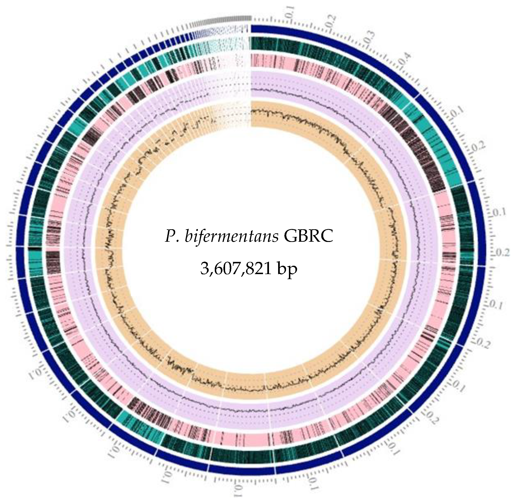
3.1. Morphological Characteristics and Genome Assembly Statistics

Morphologically, the organism was found to be obligate anaerobes and forms ceramist white colonies on modified Mah et al. medium [11]. Microscopic examination showed the organism to be spore-forming Gram positive, with moderate rods, motile, and found to be singly or in pair. A total of 3,116,578 clean reads were generated after trimming and used for de novo assembly. The Newbler assembler generated a minimum of 78 contigs from the genome, with higher N50, which were further selected for whole genome analysis. The assembly results with different assembler’s packages are summarized in Table 1.

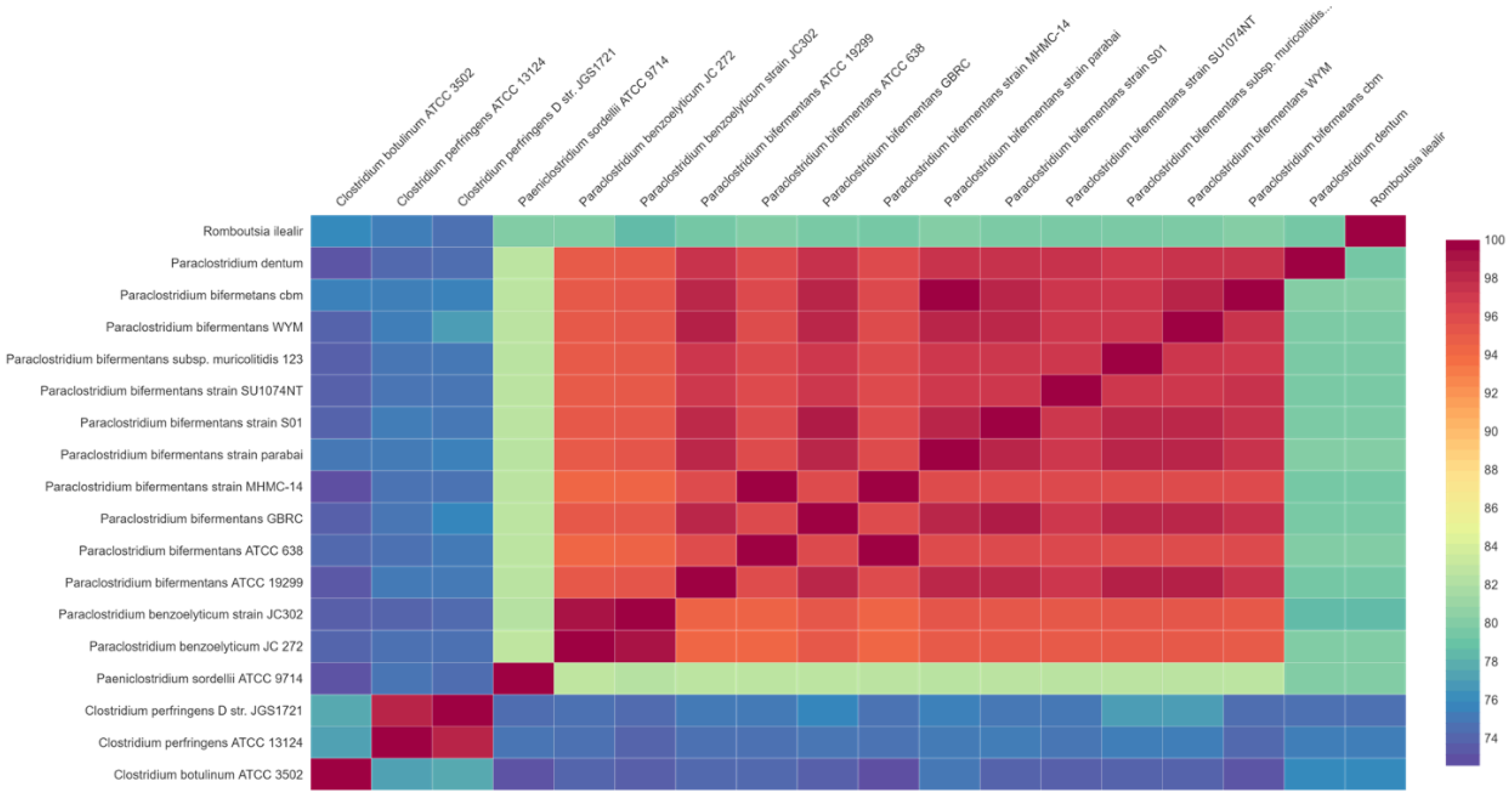
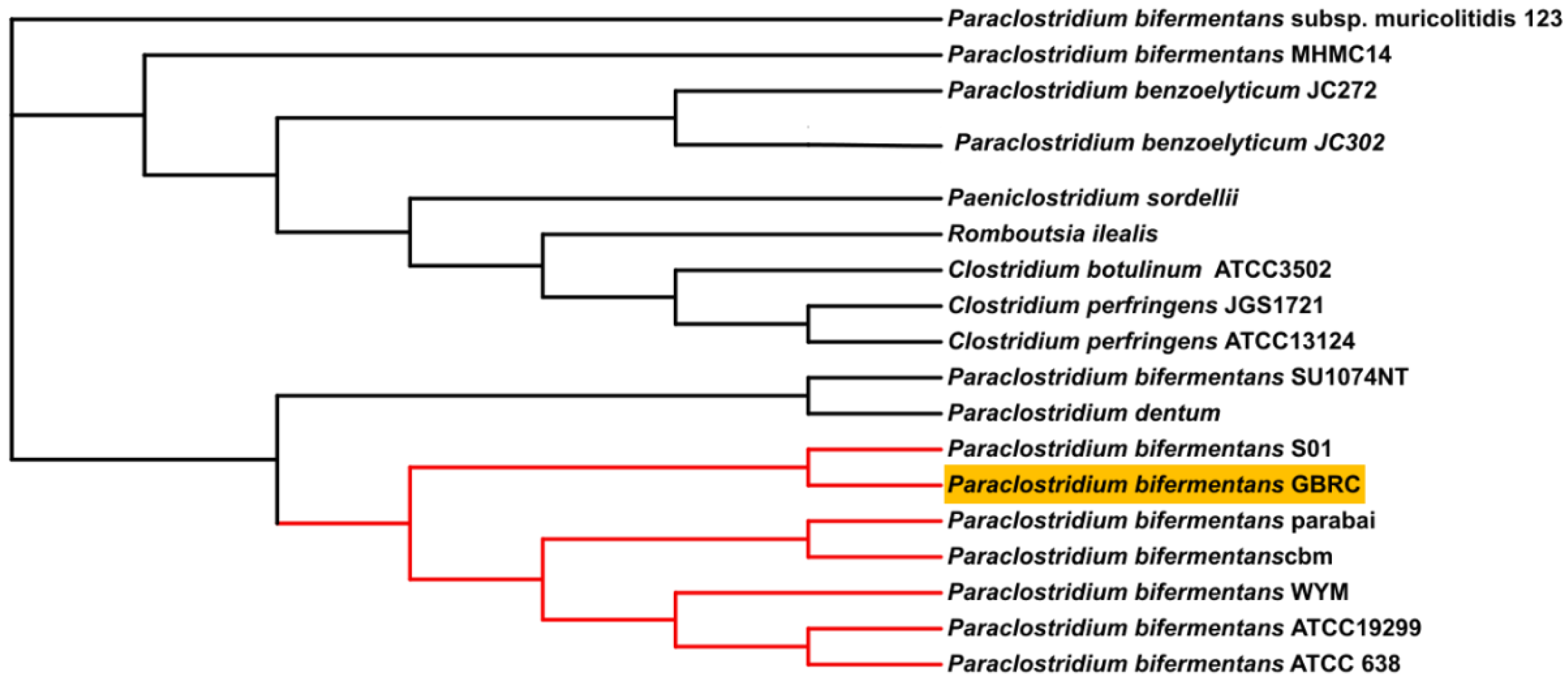
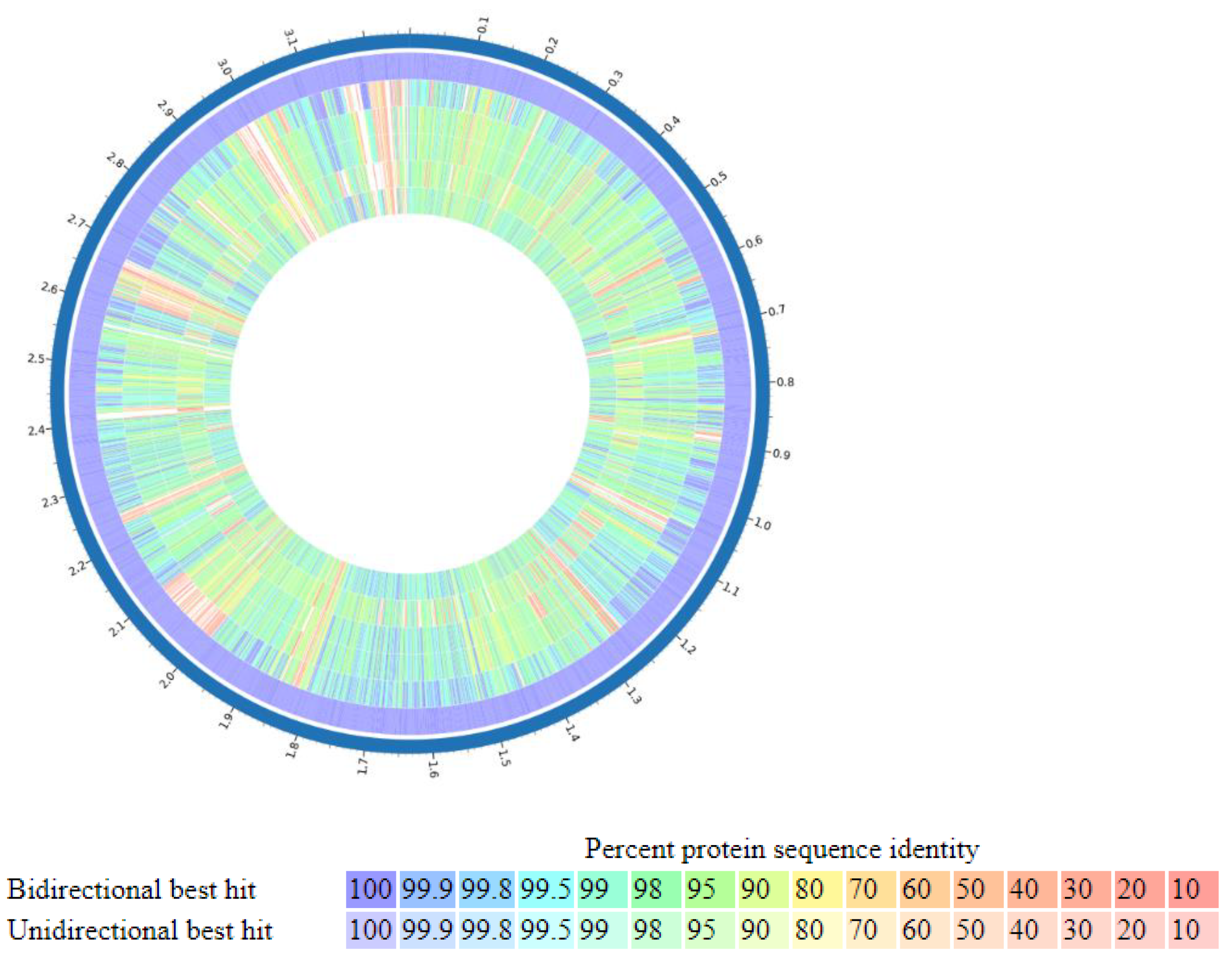
3.2. Comparative Genome Analysis of P. bifermentans GBRC with Closely Related Species

Species identification from the public database for molecular typing and microbial genome identification PubMLST web tool [32] predicted the taxa of the isolate was P. bifermentans with 100% support. Figure 1 represents genome visualization of the strain GBRC. ANI analyses are commonly used to compare bacterial strains based on whole genome sequencing [33]. Usually, ANI values between genomes of the same species are above 95%. Average nucleotide identity was found to be 98.5% with P. bifermentans S01 (Figure 2). The digital DNA (DNA hybridization (dDDH) type (strain) genome server), provides a genome-based delineation of species [34]. The dDDH values <70% considered as an indication of separate species. The P. bifermentans GBRC shows the highest dDDH percentage (82.7%) with the P. dentum SKVG24 T. The dDDH values with nearer species are described in Supplementary Table S3. The evolutionary distance with close species inferred with the neighbor-joining method using [35] whole genome sequences from selected genomes is shown in Figure 3; the strain GBRC shows more relatedness with the strain P. bifermentans S01. Figure 4 shows the percent identity across all the proteins in the compared genomes, where strain GBRC shows the highest similarity with the strain ATCC 638 and strain S01.

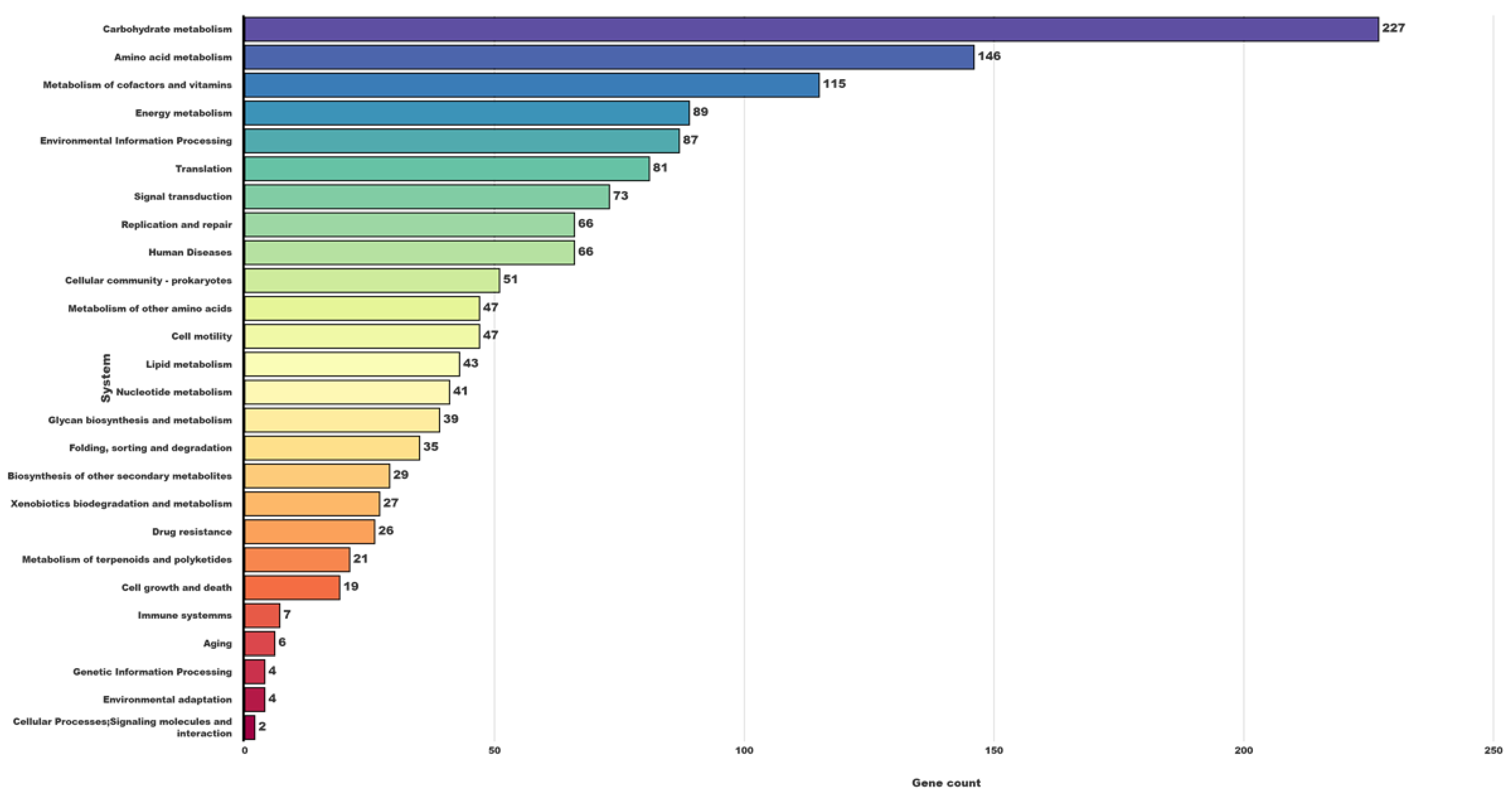
3.3. Gene Predictions and Genome Annotation

The NCBI prokaryotic genome annotation pipeline (PGAP) generated 3511 genes using GeneMarkS + 2, which were then chosen for genome annotation. After predicting the genes, genome annotation was carried out using multiple platforms (Supplementary Tables S1 and S2). The salient features of the genome are depicted in Table 2. The isolate included a total of 37 genes associated with virulence factors, including four toxin-related genes. (Table 3). Other genes were mainly pertaining to adherence, regulation, and immune evasion classes. Toxin-encoding genes included hemolysin, perfringolysin O (PFO), alpha toxin, and kappa toxin. The translated protein sequence of the hemolysin encoding gene sequence showed 99.07% identity with hemolysin found in P. bifermentans. kappa toxin displayed 100% identity with collagenase found in Clostridium thiosulfatireducens. Perfringolysin O had a 99.60% matched identity to the thiol-activated cytolysin family P. bifermentans. Other putative virulence factors (non-toxins) found in strain GBRC include those that may play a role in adherence and colonization such as capsule, fibronectin-binding proteins, flagella, and the heat shock proteins, Catalase-peroxidase for stress adaptation. GroEL, for example, is a heat shock protein family chaperon discovered alongside the heat-inducible transcription repressor HrcA, as is heat shock protein GrpE [36]. Fibronectin-binding protein, capsule encoding genes, flagella, and LPS OAntigen genes were among the additional adherence factors.

3.4. Substrate Utilization and Metabolic Pathway Analysis

As shown in Table 4, different general characteristics for strain GBRC and their closely related species were compared. Strain GBRC differs from the other three isolates in that it lacks lysine decarboxylase. Starch, gelatine, and casein hydrolysis, the ability to produce H2S and indole all seem to be characteristics of strain GBRC. The results of biochemical characterization using BioLOg are shown in (Table 5). The isolate can hydrolyze gelatine, starch, and casein and also utilize various carbon and nitrogen sources as a substrate. Genomic profiles to metabolize these substrates were also studied to confirm its substrate utilization capability (Table 5). KEGG computerizes data knowledge on pathway databases and chemical reactions that are responsible for the various cellular processes [37]. Major biochemical pathway groups include cellular processes (124), metabolism (1560), human diseases (34), genetic information processing (149), organizational system (41), environment information processing (160), and drug resistance (26), (Figure 5). P. bifermentans is found to damage the intestinal mucosa layer and induce inflammatory response by the invasion of the tissue in the mouse [14]. Strain GBRC is found in the uterus of the buffalo; a few specific genes are present that may damage the uterus epithelial layer and cause uterine infection. For example, the internalin A gene (inlA) mainly involved in the bacterial invasion of epithelial cells further binds to the E-cadherin and MET hepatocyte growth factor receptor and invades epithelial cells [38]. In addition, pathways related to flagellar assembly, including bacterial chemotaxis (Supplementary Figure S1), two-component system, and cationic antimicrobial peptide (CAMP) resistance were found in strain GBRC. Chemotaxis has been found to have a role in biofilm development in numerous microorganisms [39,40,41] and it may guide a bacterium to swim toward nutrients (hydrophobic pollutants) adsorbed to a surface, followed by flagella attachment. Flagella have been indicated as a significant organelle responsible for bacterial adhesion, biofilm formation, and invasion into host cells [42]. In various pathogens, flagellin and/or the distally located flagellar cap protein have been reported to function as adhesins [43]. The biofilm form by pathogenic bacteria helps to sequester antimicrobial agents and makes it difficult to clear the infections. P. melaninogenica, A. pyogenes, and E. coli have all been reported to infect uterine linings in cattle, and have also been found to produce biofilm in uterine linings, suggesting that biofilm formation could be a major cause of persistent uterine infections [44,45].

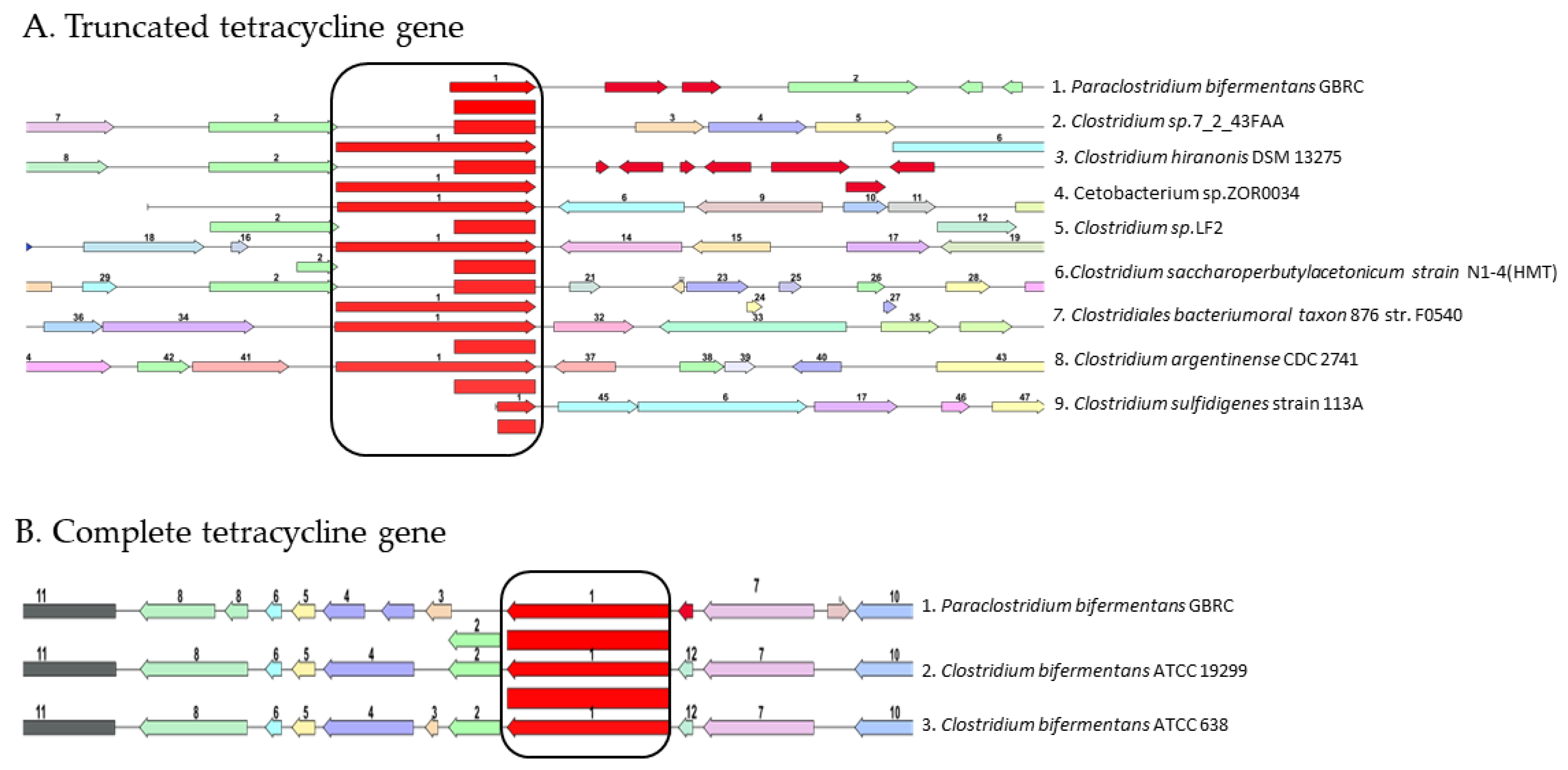
3.5. Antibiotic Sensitivity

P. bifermentans GBRC is resistant to erythromycin and co-trimoxazole (Sulpha/Trimethoprim). In several bacteria, efflux has been discovered to be the primary mechanism of erythromycin resistance [46,47]. The fact that co-trimoxazole resistance is linked to decreased susceptibility to antibiotics from many structural families reveals the overexpression of efflux pumps capable of extruding various substrates and could be the cause of the observed resistance [48]. Tetracycline resistance genes are present in the genome, although antibiotic susceptibility testing has revealed the isolate to be susceptible to this antibiotic. Further analysis showed there to be the presence of two copies of the TetM/TetW/TetO/TetS tetracycline family resistance ribosomal protection protein in different locations of the genome. When these genes were compared with gene of closely related genera, the one Tet gene was found to be truncated of having 801 base pairs (Figure 6A), another Tet gene with 1983 bp was not found to be located under the promoter (Figure 6B). This could be the possible reason for the isolate to be sensitive to tetracycline although it has the tetracycline resistance genes. Furthermore, there are several reports on the presence of tetracycline resistance genes in the environment pathogenic bacteria as a result of horizontally-acquired gene mechanisms [49,50] which may be the reason for the presence of the TetM/TetW/TetO/TetS tetracycline family resistance ribosomal protection protein in non-functional form in various locations of the genome. The KEGG pathway identified eight genes for the vancomycin resistance from the genome (Supplementary Figure S2); however, strain GBRC was found to be sensitive towards the vancomycin antibiotic. To address this question further, its pathway was studied in detail with the vanRB gene acting as a response regulator for the vanHAX gene cassette, which led to the resistance of the vancomycin. However, in the P. bifermentans GBRC, no significant match with the vanRB gene was found. However, the vanRB gene BLAST result shares 100% similarity with the response regulator from P. bifermentans, which may have led to the sensitivity of the vancomycin sensitivity to the strain GBRC.
Antibiotic resistant pathogens are responsible for the persistent endometritis in bovines due to the presence of constant bovine uterine inflammation co-infection with other pathogenic species; Escherichia coli or Fusobacterium necrofurom may increase the severity of the diseases and persistence of pathogens in the uterus [51,52]. There is evidence that P. bifermentans PB produces a neurotoxic that has insecticidal properties, which selectively targets anopheline mosquitoes [53]. In addition, one report revealed that P. bifermentans PAGU1678 causes intestinal damage in mouse models of UC and induces an inflammatory response by invading the tissues [13]. This is the first report on P. bifermentans GBRC isolated from the inflamed uterus lining of B. bubalis, and it has genomic features that allow it to infect the uterus endometrium lining. Inflammation of the lining can be caused by vaginal bacteria entering the uterus after the birth and causing infection within six weeks of the birth. Postpartum endometritis occurs after about 1% to 3% of vaginal births in humans, and in up to 27% of cesarean births. Retention of membranes and multiple vaginal examinations appear to increase the risk [54]. Endometritis and metritis are nowadays major causes of the decline in the fertility of bovines that causes great economic loss in dairy herds [55]. Findings from this study suggest a potential risk of infection by P. bifermentans GBRC and cause endometritis and metritis; this may raise an alarm for bovine health.

4. Conclusions

The newly isolated obligate anaerobe P. bifermentans GBRC from the buffalo uterus environment from the family Peptostreptococcaceae suggests a potential risk of infections in bovines. The anamnesis of P. bifermentans GBRC presence in the slaughtered buffalo uterine organ is unknown and might be of environment origin. We used a whole genomic sequencing platform for genetic evidence on ecological significance and niche adaptive strategies for the microorganisms befitting into the current knowledge and understanding of periodontal microbiology, the reported species appears to be a late colonizer of uterine environment and has significant virulence potential to contribute towards the causation and persistence of uterine non-specific infections.

5. Nucleotide Sequence Accession Numbers

The assembled whole genome is deposited at ta NBCI GenBank under the accession no. JAIRBK000000000.

Supplementary Materials

The following supporting information can be downloaded at: https://www.mdpi.com/article/10.3390/ani12141765/s1, Figure S1. Flagellar assembly of strain GBRC in a hydrophobic environment where cheW is purine-binding chemotaxis protein, MCP; aerotaxis receptor, CheA; two-component system, CheR; chemotaxis protein methyl transferase, CheB; Protein-glutamate, CheV; two-component system; Figure S2. KEGG pathway for vancomycin resistance mechanism; Table S1. Predicted genes from the whole genome with different gene-finding tools; Table S2. Genome annotation results with different platforms; Table S3. Digital DDH values for pairwise comparisons of user genomes vs. type strain genomes.

Author Contributions

The experimental scheme was designed by S.P., S.D. and V.S.; V.S. and S.P. participated in the experimental study and assisted in sampling. The analysis of experimental data and the creation of charts were completed by P.G., K.P., R.P., S.P. and S.D., P.G. completed the initial draft. R.P., V.S. and M.J. completed the overall modification of the manuscript. C.J., S.D. and D.P. improved the language of the article. S.D., D.P., M.J. and C.J. provided the necessary experimental equipment and key guidance during the experiment process. All authors have read and agreed to the published version of the manuscript.

Funding

This study was funded by the Gujarat State Biotechnology Mission, Department of Science and Technology, Government of Gujarat, India (Grant No. 201885Y0K83).

Institutional Review Board Statement

Not applicable.

Data Availability Statement

The data will be available with the corresponding author upon request.

Acknowledgments

The authors thank Gujarat State Biotechnology Mission, Gandhinagar, Gujarat, India, for providing funding support. The authors also thank Kamdhenu University and thank the Department of Microbiology, Gujarat Vidyapith, India for providing technical support and logistics for conducting the experiment.

Conflicts of Interest

The authors declare no conflict of interest.

References

  1. Philpott, M. The dangers of disease transmission by artificial insemination and embryo transfer. Braz. Vet J. 1993, 149, 339–369. [Google Scholar] [CrossRef]
  2. Noakes, E.D.; Parkinson, T.J.; England, G.C.W. Post parturient prolapse of the uterus. Arthur’s Vet. Reprod. Obstet. 2001, 8, 333–338. [Google Scholar]
  3. Sheldon, I.M.; Lewis, G.S.; LeBlanc, S.; Gilbert, O.R. Defining postpartum uterine disease in cattle. Theriogenology 2006, 65, 1516–1530. [Google Scholar] [CrossRef] [PubMed]
  4. Sheldon, I.M.; Cronin, J.; Goetze, J.; Donofrio, G.; Schuberth, H. Defining postpartum uterine disease and the mechanisms of infection and immunity in the female reproductive tract in Cattle. Biol. Reprod. 2009, 81, 1025–1032. [Google Scholar] [CrossRef]
  5. Sheldon, I.M.; Owen, S.E. Postpartum uterine infection and endometritis in dairy cattle. In Proceedings of the 33rd Annual Scientific Meeting of the European Embryo Transfer Association (AETE); Anim. Reprod: Bath, UK, 2017; pp. 622–629. [Google Scholar] [CrossRef]
  6. Azaw, O.I. Uterine infection in buffalo cows; A review. Buffalo Bull 2010, 29, 151–171. Available online: https://ibic.lib.ku.ac.th/e-bulletin/2010-154.pdf (accessed on 3 October 2021).
  7. Mandhwani, B.A.; Kumar, S.; Shivhare, M.; Aich, R. Insights into bovine endometritis with special reference to phytotherapy. VetWorld 2017, 10, 1529–1532. [Google Scholar] [CrossRef] [Green Version]
  8. Parmar, K. Endometritis in Bovine: A Review. Agricul. Rev. 2021, 42, 342–347. [Google Scholar] [CrossRef]
  9. Galvão, K.N.; Bicalho, R.C.; Jeon, S.J. Symposium review: The uterine microbiome associated with the development of uterine disease in dairy cows. J. Dairy Sci. 2019, 102, 11786–11797. [Google Scholar] [CrossRef]
  10. Hungate, R.E.; Smith, W.; Clarke, R.T. Suitability of butyl rubber stoppers for closing anaerobic roll culture tubes. J. Bacteriol. 1966, 91, 908–909. [Google Scholar] [CrossRef] [Green Version]
  11. Mah, R.A.; Smith, M.R.; Baresi, L. Studies on an acetate-fermenting strain of Methanosarcina. Appl. Environ. Microbiol. 1978, 35, 1174–1184. [Google Scholar] [CrossRef] [Green Version]
  12. Holt, J.G.; Krieg, N.R.; Sneath, P.H.A.; Stanley, J.T.; William, S.T. Bergey’s Manual of Determinative Bacteriology. Williams Wilikins Baltim. 1994, 9, 786–788. [Google Scholar]
  13. Kutsuna, R.; Miyoshi-Akiyama, T.; Mori, K.; Hayashi, M.; Tomida, J.; Morita, Y.; Tanaka, K.; Kawamura, Y. Description of P. bifermentans subsp. muricolitidis subsp. emended description of P. bifermentans and creation of P. bifermentans subsp. Bifermentans subsp. nov. Microbiol. Immunol. 2019, 63, 1–10. [Google Scholar] [CrossRef] [PubMed] [Green Version]
  14. Jyothsna, T.S.; Tushar, L.; Sasikala, C.; Ramana, C.V. Paraclostridium benzoelyticum gen. nov., sp. nov., isolated from marine sediment and reclassification of Clostridium bifermentans as Paraclostridium bifermentans comb. nov. Proposal of a new genus Paeniclostridium gen. nov. to accommodate Clostridium sordellii and Clostridium ghonii. Int. J. Syst. Evol. Microbiol. 2016, 66, 1268–1274. [Google Scholar] [CrossRef]
  15. Wilson, K. Preparation of genomic DNA from bacteria. Curr. Protoc. Mol. Biol. 2001, 56, 2–4. [Google Scholar] [CrossRef] [PubMed]
  16. Andrews, S. A Quality Control Tool for High throughput Sequence Data. FastQC. 2014. Available online: http://www.bioinformatics.babraham.ac.uk/projects/fastqc/ (accessed on 6 October 2021).
  17. Simpson, J.T.; Wong, K.; Jackman, S.D.; Schein, J.E.; Jones, S.J.; Birol, I. ABySS: A parallel assembler for short read sequence data. Genome Res. 2009, 19, 1117–1123. [Google Scholar] [CrossRef] [PubMed] [Green Version]
  18. Zerbino, D.R. Using the Velvet de novo assembler for short-read sequencing technologies. Curr. Protoc. Bioinform. 2010, 11, Unit 11.5. [Google Scholar] [CrossRef] [PubMed] [Green Version]
  19. Luo, R.; Liu, B.; Xie, Y.; Li, Z.; Huang, W.; Yuan, J.; He, G.; Chen, Y.; Pan, Q.; Liu, Y.; et al. SOAPdenovo2: An empirically improved memory-efficient short-read de novo assembler. GigaScience 2012, 1, 18. [Google Scholar] [CrossRef]
  20. Bankevich, A.; Nurk, S.; Antipov, D.; Gurevich, A.A.; Dvorkin, M.; Kulikov, A.S.; Lesin, V.M.; Nikolenko, S.I.; Pham, S.; Prjibelski, A.D.; et al. SPAdes: A new genome assembly algorithm and its applications to single-cell sequencing. J. Comp. Biol. 2012, 19, 455–2477. [Google Scholar] [CrossRef] [Green Version]
  21. Zheng, Y.; Zhao, L.; Gao, J.; Fei, Z. I Assembler: A package for de novo assembly of Roche-454/Sanger transcriptome sequences. BMC Bioinform. 2011, 12, 1–8. [Google Scholar] [CrossRef] [Green Version]
  22. Hyatt, D.; Chen, G.L.; LoCascio, P.F.; Miriam, L.; Frank, W.; Hauser, L.J. Prodigal: Prokaryotic gene recognition and ranslation initiation site identification. BMC Bio. 2010, 11, 119. [Google Scholar] [CrossRef] [Green Version]
  23. Kelley, D.R.; Liu, B.; Delcher, A.L.; Pop, M.; Salzberg, S.L. Gene prediction with Glimmer for metagenomics sequences augmented by classification and clustering. Nucleic Acids Res. 2012, 40, e9. [Google Scholar] [CrossRef] [PubMed]
  24. Besemer, J.; Lomsadze, A.; Borodovsky, M. GeneMarkS: A self-training method for prediction of gene starts in microbial genomes. Implications for finding sequence motifs in regulatory regions. Nucleic Acids Res. 2001, 29, 2607–2618. [Google Scholar] [CrossRef] [PubMed] [Green Version]
  25. Noguchi, H.; Taniguchi, T.; Itoh, T. MetaGeneAnnotator: Detecting species-specific patterns of ribosomal binding site for precise gene prediction in anonymous prokaryotic and phage genomes. DNA Res. Int. J. Rap. Pub. Repo Genes Genomes 2008, 15, 387–396. [Google Scholar] [CrossRef] [Green Version]
  26. Overbeek, R.; Olson, R.; Pusch, G.D.; Olsen, G.J.; Davis, J.J.; Disz, T.; Edwards, R.A.; Gerdes, S.; Parrello, B.; Shukla, M.; et al. The SEED and the rapid annotation of microbial genomes using subsystems technology (RAST). Nucleic Acids Res. 2014, 42, D206–D214. [Google Scholar] [CrossRef] [PubMed]
  27. Tanizawa, Y.; Fujisawa, T.; Nakamura, Y. DFAST: A flexible prokaryotic genome annotation pipeline for faster genome publication. Bioinformatics 2018, 34, 1037–1039. [Google Scholar] [CrossRef] [Green Version]
  28. Liu, B.; Zheng, D.; Jin, Q.; Chen, L.; Yang, J. VFDB 2019: A comparative pathogenomic platform with an interactive web interface. Nucleic Acids Res. 2019, 47, D687–D692. [Google Scholar] [CrossRef]
  29. Rodriguez-R, L.M.; Konstantinidis, K.T. The Enveomics Collection: A Toolbox for Specialized Analyses of Microbial Genomes and Metagenomes (No. e1900v1). PeerJ Prepr. 2016, 4, e1900v1. [Google Scholar]
  30. Letunic, I.; Bork, P. Nucleic Acids Res Interactive Tree Of Life v2: Online annotation and display of phylogenetic trees made easy. Nucleic Acids Res. 2011, 39, W475–W478. [Google Scholar] [CrossRef]
  31. Knudsen, S. Promoter2.0: For the recognition of PolII promoter sequences. Bioinformatics 1999, 15, 356–361. [Google Scholar] [CrossRef]
  32. Jolley, K.A.; Bray, J.E.; Maiden, M. Open-access bacterial population genomics: BIGSdb software, the PubMLST.org website and their applications. Wellcome Open Res. 2018, 3, 124. [Google Scholar] [CrossRef]
  33. Fujikawa, T.; Sawada, H. Genome analysis of the kiwifruit canker pathogen Pseudomonas syringae pv actinidiae biovar 5. Sci. Rep. 2016, 6, 21399. [Google Scholar] [CrossRef] [PubMed] [Green Version]
  34. Meier-Kolthoff, J.P.; Göker, M. TYGS is an automated high-throughput platform for state-of-the-art genome-based taxonomy. Nat. Commun. 2019, 10, 2182. [Google Scholar] [CrossRef] [PubMed]
  35. Simonsen, M.; Mailund, T.; Pedersen, C.N. Rapid neighbour-joining. In International Workshop on Algorithms in Bioinformatics; Springer: Berlin/Heidelberg, Germany, 2008; pp. 113–122. [Google Scholar]
  36. Minder, A.C.; Fischer, H.M.; Hennecke, H.; Narberhaus, F. Role of HrcA and CIRCE in the heat shock regulatory network of Bradyrhizobium japonicum. J. Bacteriol. 2000, 182, 14–22. [Google Scholar] [CrossRef] [PubMed] [Green Version]
  37. Kanehisa, M.; Furumichi, M.; Sato, Y.; Ishiguro-Watanabe, M.; Tanabe, M. KEGG: Integrating viruses and cellular organisms. Nucleic Acids Res. 2021, 49, D545–D551. [Google Scholar] [CrossRef]
  38. Martens, E.C.; Neumann, M.; Desai, M.S. Interactions of commensal and pathogenic microorganisms with the intestinal mucosal barrier. Nat. Rev. Microbiol. 2018, 16, 457–470. [Google Scholar] [CrossRef]
  39. O’Toole, G.A.; Kolter, R. Flagellar and twitching motility are necessary for Pseudomonas aeruginosa biofilm development. Mol. Microbiol. 1998, 30, 295–304. [Google Scholar] [CrossRef]
  40. Pratt, L.A.; Kolter, R. Genetic analysis of Escherichia coli biofilm formation: Roles of flagella, motility, chemotaxis and type I. pili. Mol. Microbiol. 1998, 30, 285–293. [Google Scholar] [CrossRef]
  41. Prigent-Combaret, C.; Vidal, O.; Dorel, C.; Lejeune, P. Abiotic surface sensing and biofilm-dependent regulation of gene expression in Escherichia coli. J. Bacteriol. 1999, 181, 5993–6002. [Google Scholar] [CrossRef] [Green Version]
  42. Watnick, P.I.; Kolter, R. Steps in the development of a Vibrio cholerae El Tor biofilm. Mol. Microbiol. 1999, 34, 586–595. [Google Scholar] [CrossRef] [Green Version]
  43. Haiko, J.; Westerlund-Wikström, B. The role of the bacterial flagellum in adhesion and virulence. Biology 2013, 2, 1242–1267. [Google Scholar] [CrossRef] [Green Version]
  44. Raheel, I.A.E.R.; Hassan, W.H.; Salem, S.S.R.; Salam, H.S.H. Biofilm forming potentiality of Escherichia coli isolated from bovine endometritis and their antibiotic resistance profiles. J. Adv. Vet. Anim. Res. 2020, 7, 442. [Google Scholar] [CrossRef] [PubMed]
  45. Gurunathan, S.; Choi, Y.J.; Kim, J.H. Antibacterial efficacy of silver nanoparticles on endometritis caused by Prevotella melaninogenica and Arcanobacterum pyogenes in dairy cattle. Int. J. Mol. Sci. 2018, 19, 1210. [Google Scholar] [CrossRef] [PubMed] [Green Version]
  46. Braoudaki, M.; Hilton, A.C. Mechanisms of resistance in Salmonella enterica adapted to erythromycin, benzalkonium chloride and triclosan. Int. J. Antimicrob. Agents 2005, 25, 31–37. [Google Scholar] [CrossRef] [PubMed]
  47. Ravanel, N.; Gestin, B.; Maurin, M. In vitro selection of fluoroquinolone resistance in Brucella melitensis. Int. J. Antimicrob. Agents 2009, 34, 76–81. [Google Scholar] [CrossRef] [PubMed] [Green Version]
  48. Sánchez, M.B.; Martínez, J.L. Overexpression of the efflux pumps SmeVWX and SmeDEF is a major cause of resistance to co-trimoxazole in Stenotrophomonas maltophilia. Antimicrob. Agents Chemother. 2018, 62, e00301-18. [Google Scholar] [CrossRef] [Green Version]
  49. Akhtar, M.; Hirt, H.; Zurek, L. Horizontal transfer of the tetracycline resistance gene tetM mediated by pCF10 among Enterococcus faecalis in the house fly (Musca domestica L.) alimentary canal. Microbiol. Ecol. 2009, 58, 509–518. [Google Scholar] [CrossRef]
  50. Von Wintersdorff, C.J.; Penders, J.; Van Niekerk, J.M.; Mills, N.D.; Majumder, S.; Van Alphen, L.B.; Wolffs, P.F. Dissemination of antimicrobial resistance in microbial ecosystems through horizontal gene transfer. Front. Microbiol 2016, 7, 173. [Google Scholar] [CrossRef] [Green Version]
  51. Farca, A.M.; Nebbia, P.; Robino, P.; Re, G. Effects of the combination antibiotic—edta–tris in the treatment of chronic bovine endometritis caused by antimicrobial-resistant bacteria. Pharm. Res. 1997, 36, 3539. [Google Scholar] [CrossRef]
  52. Bicalho, M.L.; Machado, V.S.; Oikonomou, G.; Gilbert, R.O.; Bicalho, R.C. Association between virulence factors of Escherichia coli, Fusobacterium necrophorum, and Arcanobacterium pyogenes and uterine diseases of dairy cows. Vet. Microbiol. 2012, 157, 125–131. [Google Scholar] [CrossRef]
  53. Contreras, E.; Masuyer, G.; Qureshi, N.; Chawla, S.; Dhillon, H.S.; Lee, H.L.; Chen, J.; Stenmark, P.; Gill, S.S. A neurotoxin that specifically targets Anopheles mosquitoes. Nat. Commun. 2019, 10, 2869. [Google Scholar] [CrossRef]
  54. Mackeen, A.D.; Packard, R.E.; Ota, E.; Speer, L. Antibiotic regimens for postpartum endometritis. Cochrane Database Syst. Rev. 2015. [Google Scholar] [CrossRef]
  55. LeBlanc, S.J. Postpartum uterine disease and dairy herd reproductive performance: A review. Vet. J. 2008, 176, 102–114. [Google Scholar] [CrossRef] [PubMed]
Figure 1. Graphical circular representation of the genome of the strain GBRC circles from interior to exterior represent GC skew, GC content, CDS on reverse strand, and CDS on forward strand.
Animals 12 01765 g001
Figure 2. Relationship of P. bifermentans GBRC among other 17 closely related organisms. The average nucleotide identity (ANI) matrix was generated using ANI calculator and the enveomics collection toolbox genome was used for distance matrix calculation.
Animals 12 01765 g002
Figure 3. Phylogenetic tree reconstructed with the neighbor-joining method based on the whole genome sequence of the Paraclostridium bifermentans strains and related genus of Paraclostridium. P. bifermentans GBRC showing the phylogenetic relationship with the related species.
Animals 12 01765 g003
Figure 4. Proteome comparison: The entire proteome for each different strain is aligned against the P. bifermentans GBRC proteome. Track order from outside to inside is shown in the top right part of the figure where the reference strain, GBRC, is the most outside circle. The color key indicates the percentage of protein sequence similarity over the entire genome. The list of tracks, from outside to inside includes P. bifermentans GBRC, P. bifermentans strain Cbm, P. bifermentans strain MHMC-14, P. bifermentans strain DSM 14991, P. benzoelyticum strain JC302, and P. bifermentans subsp. muricolitidis 123.
Animals 12 01765 g004
Figure 5. Kyoto Encyclopedia of Genes and Genomes (KEGG) classification of metabolic pathways P. bifermentans GBRC.
Animals 12 01765 g005
Figure 6. (A) Comparison of the truncated gene (1) TetM/TetW/TetO/TetS family tetracycline resistance ribosomal protection protein with different genera; (B) comparison of full gene (red colored) (1) TetM/TetW/TetO/TetS tetracycline family resistance ribosomal protection protein with different bacteria.
Animals 12 01765 g006
Table 1. De novo genome assembly statistics for P. bifermentans GBRC with different assembly software.
Assembler PlatformTotal Number of ScaffoldsN50 Size (bp)Longest Contig’s Length
ABySS73,2752401237
Velvet44515023135
SOAPdenovo239686644829
SPAdes178165,665517,124
Newbler 2.778177,236477,527
Table 2. General feature of P. bifermentans GBRC draft genome.
FeatureParaclostridium bifermentans GBRC
DomainBacteria
TaxonomyFirmicutes; Clostridia; Eubacteriales; Peptostreptococcaceae; Paraclostridium;
Paraclostridium bifermentans
Genome size3,607,821 bp
N50 size(bp)177,236 bp
Total Num. of contigs78
G + C content28.10%
Contamination0%
Completeness0.99
Genes3511
CDSs (Total)3441
Genes (Coding)3.395
Genes(RNA)70
rRNAs3, 3, 5 (5 S, 16 S, 23 S)
Complete rRNAs3 (5 S)
Partial rRNAs3, 5 (16 S, 23 S)
tRNAs55
ncRNAs4
Table 3. Virulence factors present in the P. bifermentans GBRC genome.
Virulence ClassVirulence FactorsRelated GenesPredicated Gene ID
AdherenceFibronectin-binding proteinfbpA/fbp68gene_12_113
GroELgroELgene_11_64
Flagella (Pseudomonas)flipgene_3_40
LPS O-antigen (P. aeruginosa) gene_1_129, gene_1_130
Listeria adhesion proteinLapgene_7_23
RegulationCheA/CheY (Listeria)cheYgene_3_167
LisR/LisK (Listeria)lisRgene_19_8, gene_8_135
Sigma A (Mycobacterium)sigA/rpoVgene_9_93
ToxinAlpha-toxinPlcgene_18_29
Hemolysin gene_11_116
gene_8_36
Kappa-toxin (collagenase)colAgene_11_57
Perfrigolysin O (theta-toxin/PFO) /botulinolysinpfoAgene_6_144
CytolysincylR2gene_10_57
AntiphagocytosisCapsular polysaccharidermlBgene_12_15
Capsular polysaccharidewcaJgene_8_159
Capsule gene_3_150
Cell surface ComponentsTrehalose-recycling ABC transportersugCgene_13_45
Immune evasionCapsulecps4Igene_26_21
gene_8_154
LPSacpXLgene_26_5
Polysaccharide capsule gene_8_145
gene_8_146
gene_8_155
gene_8_161
Polysaccharide capsuleepsEgene_12_3
manAgene_12_22
Iron uptakeHeme biosynthesishemBgene_25_23
Periplasmic binding proteinvctCgene_13_103
Nutritional virulencePyrimidine biosynthesis gene_11_52
Secretoin systemT6SS-II gene_29_17
Serum resistance and immune evastionCapsule gene_12_130
LPSwbtEgene_8_165
LPSwbtFgene_8_166
LPSwbtIgene_3_25
Table 4. General characteristics of Taxa: 1, P. bifermentans JCM 1386T [12]; 2, P. benzoelyticum JC272T [13]; 3, P. bifermentans subsp. Muricolitidis [12]; 4, P. bifermentans GBRC +, positive; −, negative.
Characteristic1234
Gram-stainGram-PositiveGram-PositiveGram-PositiveGram-Positive
ShapeRod ShapedRod ShapedRod ShapedRod Shaped
SporulationCentral to TerminalTerminalTerminal to Sub terminalTerminal
MotilityMotileMotileMotileMotile
Obligate AnaerobesObligate AnaerobeObligate AnaerobeObligate AnaerobeObligate Anaerobe
Catalase
Oxidase
Indole+++
H2S production+/−++
Starch hydrolysis++
Gelatin hydrolysis++++
Nitrate reduction
Lysine Decarboxylase+++
Casein++++
Table 5. Substrate utilization profile of the P. bifermentans GBRC as obtained by AN-Biolog® Plate as well as genomic approach.
Classification SubstratePredicted KEGG Pathway ID
SystemSubsystem
CarbohydratesKetosed-fructosemap00051, map00052, & map00500
Monosaccharideα–d-Glucosemap00010, map00052, & map00520
d-Mannosemap00051
α–d-Glucose 6 phosphatemap00010, map00030, & map00052
Galactosemap00052
HexosamineN-Acetyl-d-Galactosaminemap00052
Sugar AlcoholSorbitolmap00051
NucleosideInosinemap00230
Amino acids l-Asparaginemap00250, & map00460
l-Glutaminemap00250
l-Threoninemap00260
l-Methioninemap00270
l-Serinemap00260, & map00270
l-Prolinemap00330, & map00470
l-Phenylalaninemap00360
Publisher’s Note: MDPI stays neutral with regard to jurisdictional claims in published maps and institutional affiliations.

