Article

Effects of Dietary Fucoidan Supplementation on Serum Biochemical Parameters, Small Intestinal Barrier Function, and Cecal Microbiota of Weaned Goat Kids

1
College of Coastal Agriculture Science, Guangdong Ocean University, Zhanjiang 524088, China
2
The Key Laboratory of Animal Resources and Breed Innovation in Western Guangdong Province, Zhanjiang 524088, China
*
Authors to whom correspondence should be addressed.
Animals 2022, 12(12), 1591; https://doi.org/10.3390/ani12121591
Submission received: 18 April 2022 / Revised: 18 June 2022 / Accepted: 19 June 2022 / Published: 20 June 2022

Abstract

:

Simple Summary

Weaning stress has negative effects on the growth and intestinal health of goat kids. The application of antibiotics can relieve weaning stress; however, their prophylactic application has motivated researchers to find alternatives to antibiotics for mitigating the weaning stress. Fucoidan is a natural polysaccharide with antioxidant and immune-modulatory properties, and it has shown beneficial effects on intestinal barrier function. In this study, dietary supplementation of fucoidan improved the small intestinal barrier function and cecal microflora in weaned goat kids. These results suggested that fucoidan can be used as a promoter of gut health in weaned goat kids.

Abstract

The purpose of this study was to evaluate the effects of fucoidan supplementation on serum biochemical parameters, small intestinal barrier function, and cecal microbiota of weaned goat kids. A total of 60 2-month-old weaned castrated male goat kids (Chuanzhong black goat) were used in this 30-day experiment. The goat kids were randomly divided into four groups: a control group (CON) fed the basal diet, and three other groups supplemented with 0.1%, 0.3%, and 0.5% fucoidan in the basal diet (denoted as F1, F2, and F3 groups, respectively). The results indicated that dietary fucoidan supplementation decreased (p < 0.05) the activity of lactate dehydrogenase (LDH) and the content of glucose (GLU) as measured on day 15. As measured on day 30, dietary fucoidan increased (p < 0.05) the content of total protein (TP) and decreased the activity of aspartate aminotransferase (AST), and supplementation with 0.3% and 0.5% fucoidan decreased (p < 0.05) the activity of LDH. Dietary fucoidan decreased (p < 0.05) the content of D-lactic acid (D-LA) and the activity of diamine oxidase (DAO). Dietary fucoidan increased (p < 0.05) the activity of catalase (CAT) in the duodenum. Dietary 0.3% and 0.5% fucoidan enhanced (p < 0.05) the activity of glutathione peroxidase (GSH-Px) in the ileum, the activity of total superoxide dismutase (T-SOD) in the jejunum and ileum, and the activity of CAT in the ileum. Dietary 0.3% and 0.5% fucoidan reduced the contents of malondialdehyde (MDA) in the duodenum, jejunum, and ileum and the content of hydrogen peroxide (H2O2) in the duodenum. Dietary fucoidan increased (p < 0.05) the content of secretory immunoglobulin A (sIgA) in the duodenum. Supplementation of 0.3% and 0.5% fucoidan upregulated (p < 0.05) the gene expression of ZO-1 and claudin-1 in the duodenum, jejunum, and ileum, and dietary supplementation of 0.3% and 0.5% fucoidan upregulated (p < 0.05) the gene expression of occludin in the jejunum and ileum. The 16S rRNA high-throughput sequencing results showed that at the phylum level, dietary fucoidan increased (p < 0.05) the abundance of Bacteroidetes while decreasing (p < 0.05) the abundance of Firmicutes. At the genus level, dietary 0.3% and 0.5% fucoidan increased (p < 0.05) the abundances of Unspecified_Ruminococcaceae, Unspecified_Bacteroidale, Unspecified_Clostridiales, and Akkermansia. In conclusion, dietary fucoidan supplementation had positive effects on intestinal permeability, antioxidant capacity, immunity function, tight junctions, and the cecal microflora balance in weaned goat kids.

1. Introduction

The weaning transition is a critical period for goat kid growth. Previous studies have confirmed that weaning stress has negative consequences for growth performance, intestinal barrier function, and the balance of intestinal microflora [1,2]. Moreover, it has been suggested that weaning stress causes intestinal barrier disruption leading to diarrhea and endangering the health of the goat kids [3,4]. Thus, gut health is critical for goat kids’ growth and welfare. Formerly, antibiotics were widely used to alleviate weaning stress [5]. However, the ban on antibiotic use in feed worldwide has forced researchers to find alternatives to antibiotics. At present, increasing attention is being paid to seaweed polysaccharides due to their bioactivities in animals [6,7,8,9].
Fucoidan, a macromolecular polysaccharide rich in sulfate groups, is found in the cell walls of brown seaweed (Pelvetia canaliculate, Fucus evanescens, and Cladosiphon okamuranus), the mucous matrix, and several marine invertebrates [10,11]. Recent studies have shown that dietary fucoidan supplementation could maintain intestinal health by repairing the intestinal mucosa, enhancing mucosal immune function, and regulating intestinal microflora in mice and pigs [12,13,14]. In general, fucoidan has protective effects on the intestinal physical, chemical, immune, and microbial barriers [15,16]. Thus, fucoidan has been widely used as a functional feed supplement. Furthermore, dietary inclusion of fucoidan has been suggested to improve intestinal mucosal antioxidant capacity, immune function, tight junctions, and gut microbiota balance in weaning pigs [17,18,19,20]. Similar results have also been reported in chickens and fish [21,22]. However, to the best of our knowledge, reports concerning the application of fucoidan in weaned goat kids are limited. In a companion study, fucoidan dietary supplementation increased growth performance, antioxidant capacity, immune function, and intestinal morphology in weaned goat kids [23]. However, there is at present no information concerning changes in serum biochemical parameters, small intestinal barrier function, or the cecal microbial community of goat kids after fucoidan feeding. Therefore, the purpose of this study was to fill this knowledge gap.

2. Materials and Methods

2.1. Source of Fucoidan

The fucoidan used in this study was provided by Mingyue Hailin Fucoidan Biotechnology Co., Ltd. The fucoidan was a pale-yellow powder with the odor of seaweed. The fucoidan purity was determined to be 98%; the overall sugar content was 70.5%; the fucose content was 24.9%; and the sulfate ion content was 28.9%.

2.2. Kids, Diet and Experimental Design

The design of this study was described in an earlier paper [23]. Briefly, a total of 60 2-month-old weaned castrated male goat kids (Chuanzhong black goat) with a mean body weight of 12.5 ± 0.5 kg were used in a 30-day experiment. The control group (CON) was fed a basal diet, while the other three groups were fed the same diet supplemented with fucoidan at 0.1% (F1 group), 0.3% (F2 group), and 0.5% (F3 group). The basal diet (Table 1) was formulated to meet or exceed the nutrient requirements from the Feeding Standards of Goats, China (NY/T 861-2004). The roughage consisted of Pennisetum purpureum. The goat kids were fed twice daily (8:30 am and 5:30 pm). Water was available ad libitum. Fucoidan was manually mixed into the concentrate. The goat kids were first fed the concentrate and then the roughage.

2.3. Sample Collection

On days 15 and 30, blood samples were collected from the jugular vein. The blood samples were centrifuged at 3500× g for 10 min at 4 °C, and the serum was collected and stored at −80 °C for later analysis.
Goat kids were fasted for 12 h prior to slaughter at the end of the trial. Six goat kids of similar weight per group were selected for slaughter. To isolate the gut tissue, approximately 3 cm segments of the midpoint of the duodenum, jejunum, and ileum were collected and stored at −80 °C for gene expression analysis. The mucosal samples were collected by the method described by Wang et al. [24]. Mucosal samples were stored at −80 °C.
Finally, the cecum was opened using a sterile scalpel, and the cecal contents were separated into 15 mL tubes using sterile pipette tips. Cecal content samples were stored at −80 °C for microbiota analysis.

2.4. Serum Biochemical Parameters

The contents of total glyceride (TG), total cholesterol (T-CHO), glucose (GLU), total protein (TP), and the activities of alanine aminotransferase (ALT), aspartate aminotransferase (AST), and lactate dehydrogenase (LDH) were examined using commercial kits (Nanjing Jiancheng Bioengineering Institute, Nanjing, China).

2.5. Intestinal Permeability

The content of D-lactic acid (D-LA) and the activity of diamine oxidase (DAO) were measured with enzyme immunoassay kits (Jiangsu Meimian Industrial Co., Ltd., Nanjing, China).

2.6. Antioxidant Capacity of Intestinal Mucosa

The contents of malondialdehyde (MDA), hydrogen peroxide (H2O2), and the activities of total antioxidant capacity (T-AOC), total superoxide dismutase (T-SOD), catalase (CAT), and glutathione peroxidase (GSH-Px) in the intestinal mucosa were measured using commercial kits (Nanjing Jiancheng Bioengineering Institute, Nanjing, China).

2.7. Intestinal Mucosal Immunity

The content of secretory immunoglobulin A (sIgA) in the intestinal mucosa was estimated using an enzyme immunoassay kit (Jiangsu Meimian Industrial Co., Ltd., Nanjing, China).

2.8. RT-qPCR Analysis

The total RNA extraction, reverse transcription to cDNA, and real-time quantitative polymerase chain reaction analysis employed the methods described by Guo et al. [25].
The primers were designed by Primer Premier 6.0 software (Table 2). β-actin mRNA expression was used to calculate and normalize the values, and the relative mRNA expression was calculated by the 2∆∆Ct method.

2.9. DNA Extraction

The total genomic DNA from cecal content samples was extracted using the cetyltrimethylammonium bromide (CTAB) method [26]. The concentration and quality of the DNA were estimated using 1% agarose gels. Finally, samples were diluted to 1 ng/µL with sterile water.

2.10. 16S rRNA Gene Amplification and Illumina novaSeq Sequencing

The distinct regions (16S rRNA V3-V4) were amplified by the specific primers 341F (5′-CCTAYGGRBGCASCAG-3′) and 806R (5′-GGACTACNNGGGTATCTAAT-3′). The PCR reactions were performed with Phusion High-Fidelity PCR Master Mix® (New England Biolabs) using 2 µM primers (forward and reverse) and 10 ng template DNA. Finally, purifying the pooled PCR products was performed using a Qiagen Gel Extraction Kit (Qiagen, Germany).
Sequence libraries were generated and indexed by library construction kits (Illumina, San Diego, CA, USA). The quality of the library was assessed by a Qubit 2.0 fluorometer (Thermo Scientific, Waltham, MA, USA). Then, the library was sequenced on an Illumina NovaSeq platform, and 250 bp paired-end reads were generated.

2.11. Statistical Analysis

The operational taxonomic units (OTUs) were clustered with 97% similarity (3% cutoff) using Uparse software [27] (v7.0.1), and chimeric sequences were identified and removed. The phylogenetic affiliation of each 16S rRNA gene sequence was analyzed by the RDP classifier against the Database software (v1.3.1) 16S rRNA database using a confidence threshold of 70%. Alpha diversity indices and beta diversity distances were calculated by QIIME software (v1.9.1). The microbial diversity of samples was estimated by alpha diversity indices (Chao1, Shannon, and Simpson indices).
All statistical analyses were performed using SPSS 26.0. One-way ANOVA and Duncan’s multi-range test were used to analyze group differences. The results were expressed as the mean ± standard error of the mean (SEM), and differences were considered significant at p < 0.05.

3. Results

3.1. Serum Biochemical Parameters

As shown in Table 3, on day 15 the goat kids fed diets with fucoidan had decreases in the activity of LDH and the content of GLU (p < 0.05). Goat kids fed the F3 diet had increased TP content compared to goat kids fed CON, F1, and F2 diets (p < 0.05). No significant differences were observed in the activities of ALT, AST, or the contents of TG and T-CHO among treatments.
On day 30, goat kids fed diets with fucoidan had decreased activity of AST (p < 0.05). Goat kids fed F2 and F3 diets had decreased activity of LDH compared to goat kids fed CON and F1 diets (p < 0.05). Dietary inclusion of fucoidan increased (p < 0.05) the content of TP compared to goat kids fed the CON diet. No significant differences were observed in the activity of ALT or the contents of TG, T-CHO, and GLU among the treatments.

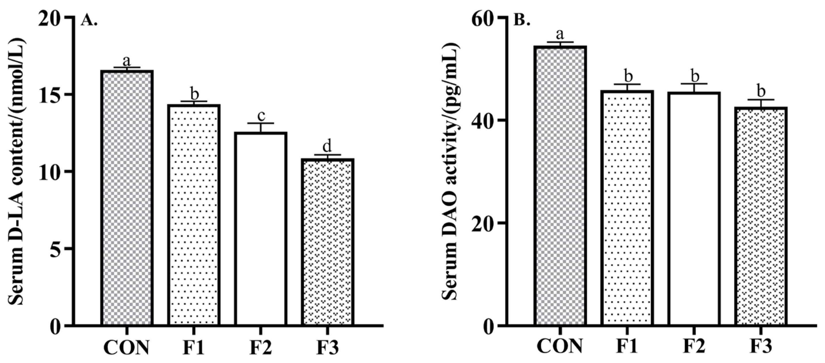
3.2. Intestinal Permeability

As shown in Figure 1, dietary supplementation of fucoidan decreased the content of D-LA and the activity of DAO compared to goat kids fed the CON diet (p < 0.05).

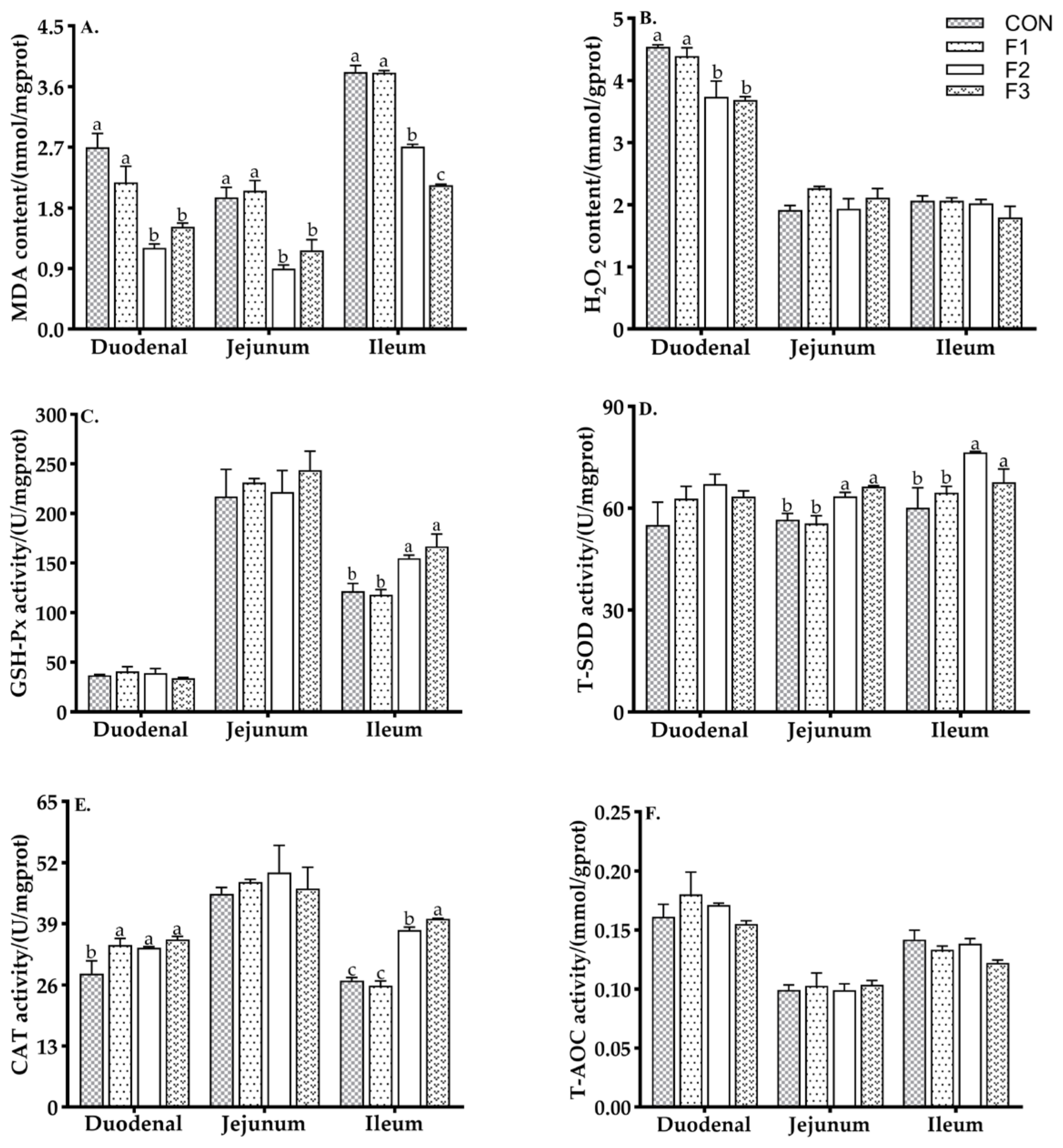
3.3. Antioxidant Capacity of Intestinal Mucosa

As shown in Figure 2, goat kids fed the F2 and F3 diets had decreased MDA content in the duodenum, jejunum, and ileum and H2O2 in the duodenum compared to goat kids fed the CON and F1 diets (p < 0.05). Goat kids fed diets with fucoidan had increased activity of CAT in the duodenum compared to goat kids fed the CON diet. Goat kids fed the F2 and F3 diets had increased activities of T-SOD in the jejunum and ileum and CAT in the ileum compared to goat kids fed the CON and F1 diets (p < 0.05).

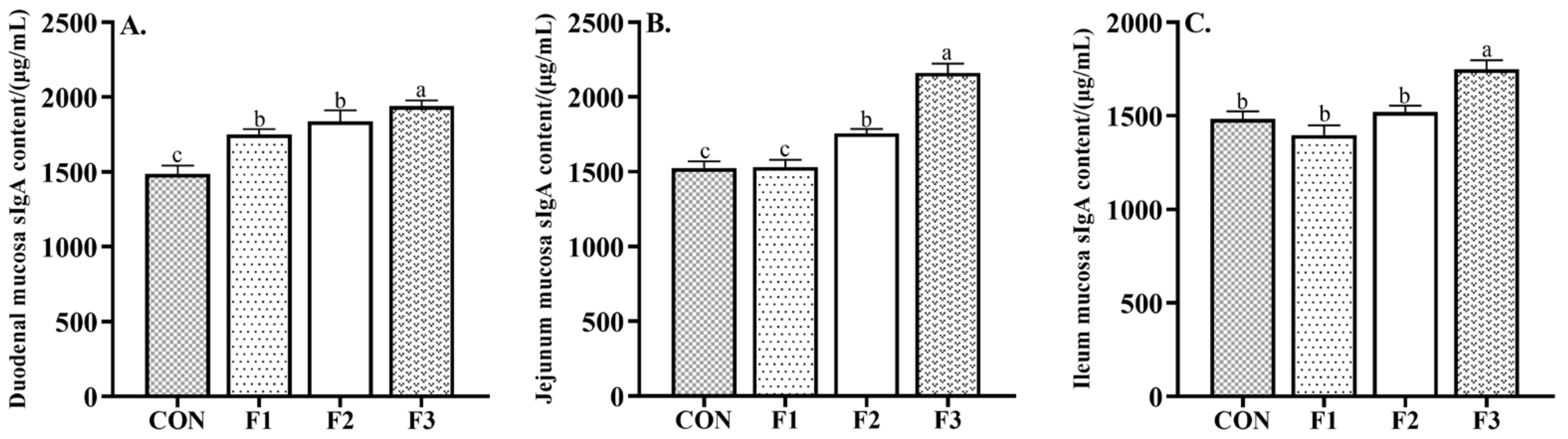
3.4. Intestinal Mucosal Immunity

As shown in Figure 3, goat kids fed diets with fucoidan had increased content of sIgA in the duodenal mucosa compared to goat kids fed the CON diet. Goat kids fed F2 and F3 diets had increased content of sIgA in the jejunum mucosa compared to goat kids fed the CON and F1 diets (p < 0.05). Goat kids fed the F3 diet had increased content of sIgA in the ileum mucosa compared to goat kids fed the CON, F1, and F2 diets (p < 0.05).

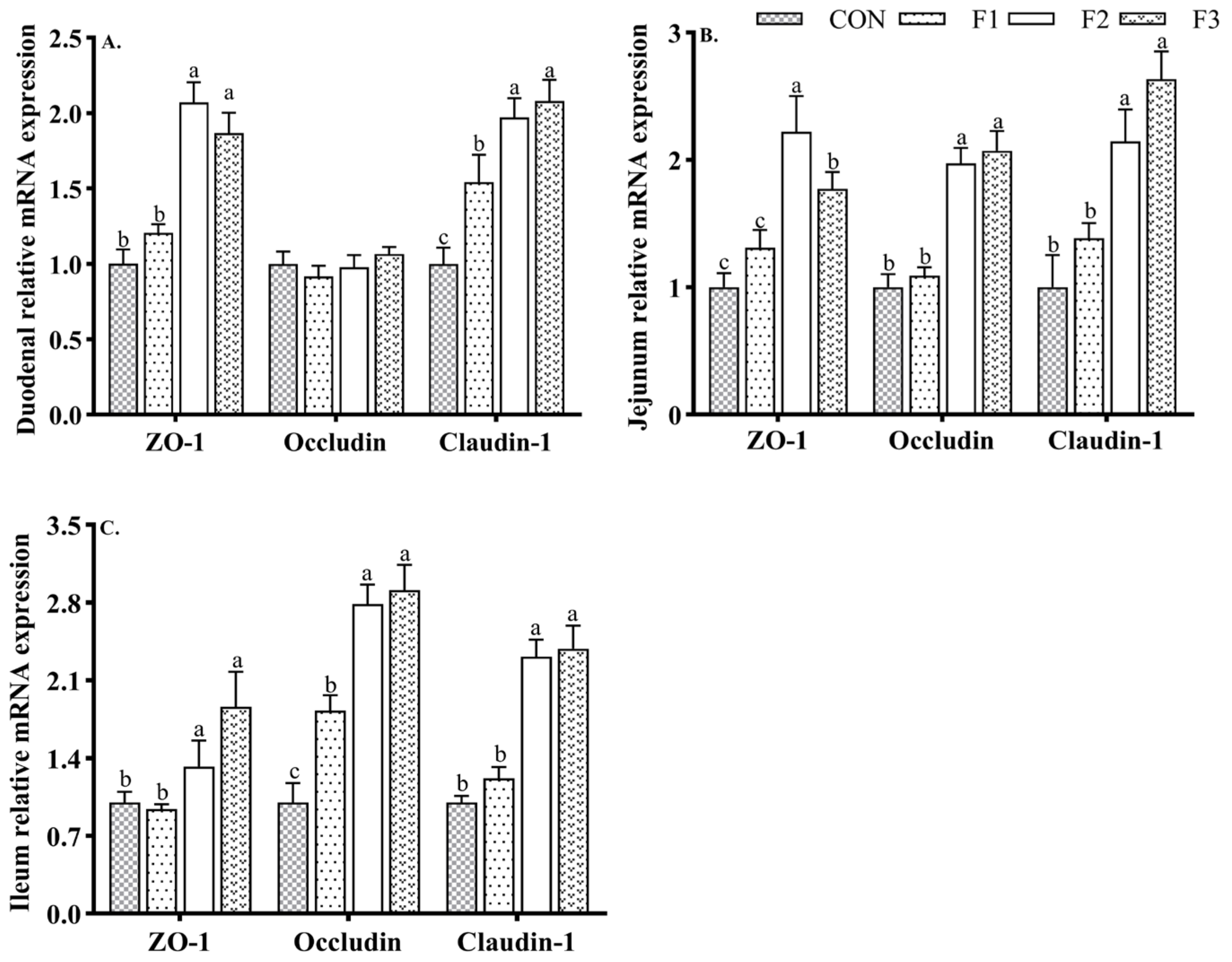
3.5. Intestinal Tight Junction Protein mRNA Expression

As shown in Figure 4, goat kids fed diets with fucoidan upregulated the gene expression of claudin-1 in the duodenal mucosa compared to those fed the CON diet. Goat kids fed the F2 and F3 diets displayed upregulated (p < 0.05) the gene expression of ZO-1 in the duodenum, and the gene expression levels of ZO-1, occludin, and claudin-1 in the jejunum and ileum were higher than in goat kids fed the CON and F1 diets.

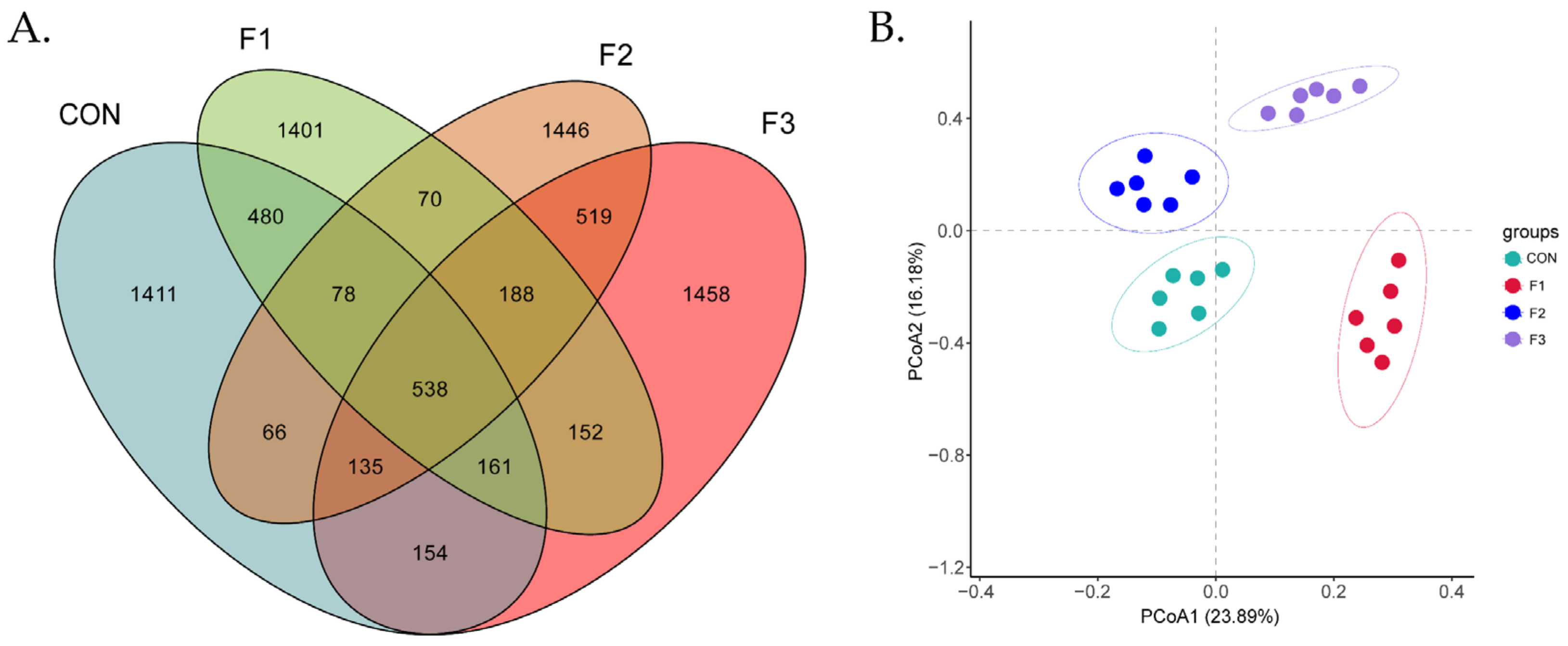
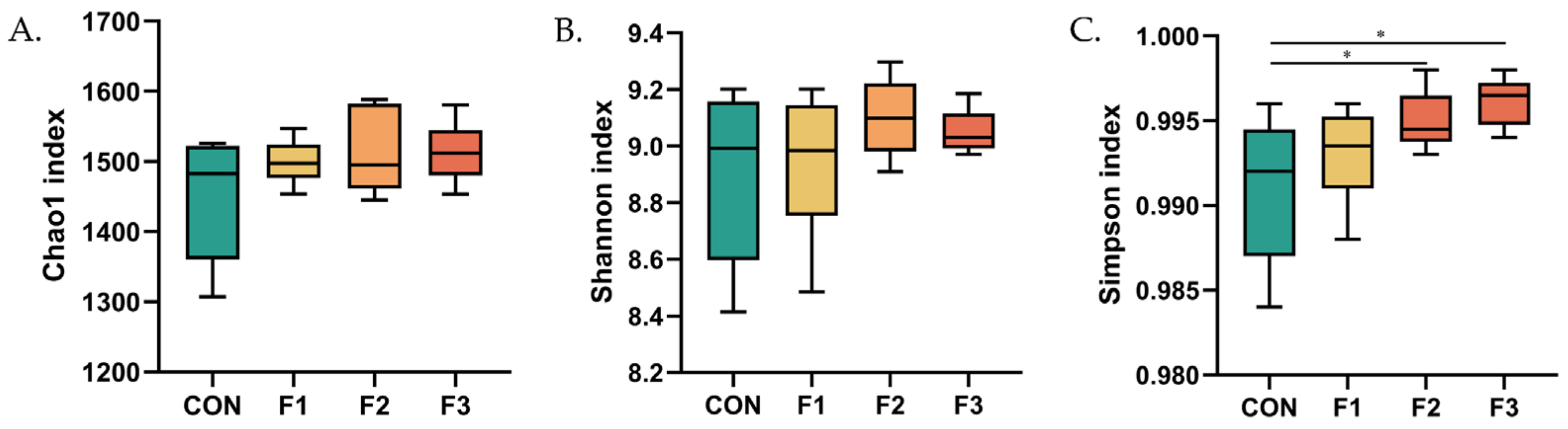
3.6. Alteration in the Composition of the Cecal Bacterial Community

Operational taxonomic units were identified using the criterion of 97% identity. The CON group contained 3023 OTUs; the F1 group contained 3068 OTUs; the F2 group contained 3040 OTUs, and the F3 group contained 3170 OTUs. There were 538 shared OTUs and 1141, 1401, 1446, and 1458 OTUs specific to the respective four groups. The distance between points represented the difference in the composition of the flora (Figure 5A). The principal coordinate 1 explained 23.89% of the variation among microbial colonies in all cecal contents, and the principal coordinate 2 explained 16.18% of the variation among microbial colonies in all cecal contents. The figure shows that the samples from the F1, F2, and F3 groups were clearly separated from the CON samples. Thus, fucoidan affected the cecal contents in goat kids (Figure 5B).
As shown in Figure 6, goat kids fed with the F2 and F3 diets had increased values of the Simpson index compared to goat kids fed the CON and F1 diets. No significant differences were observed in the values of the Chao1 or Shannon indices among treatments.

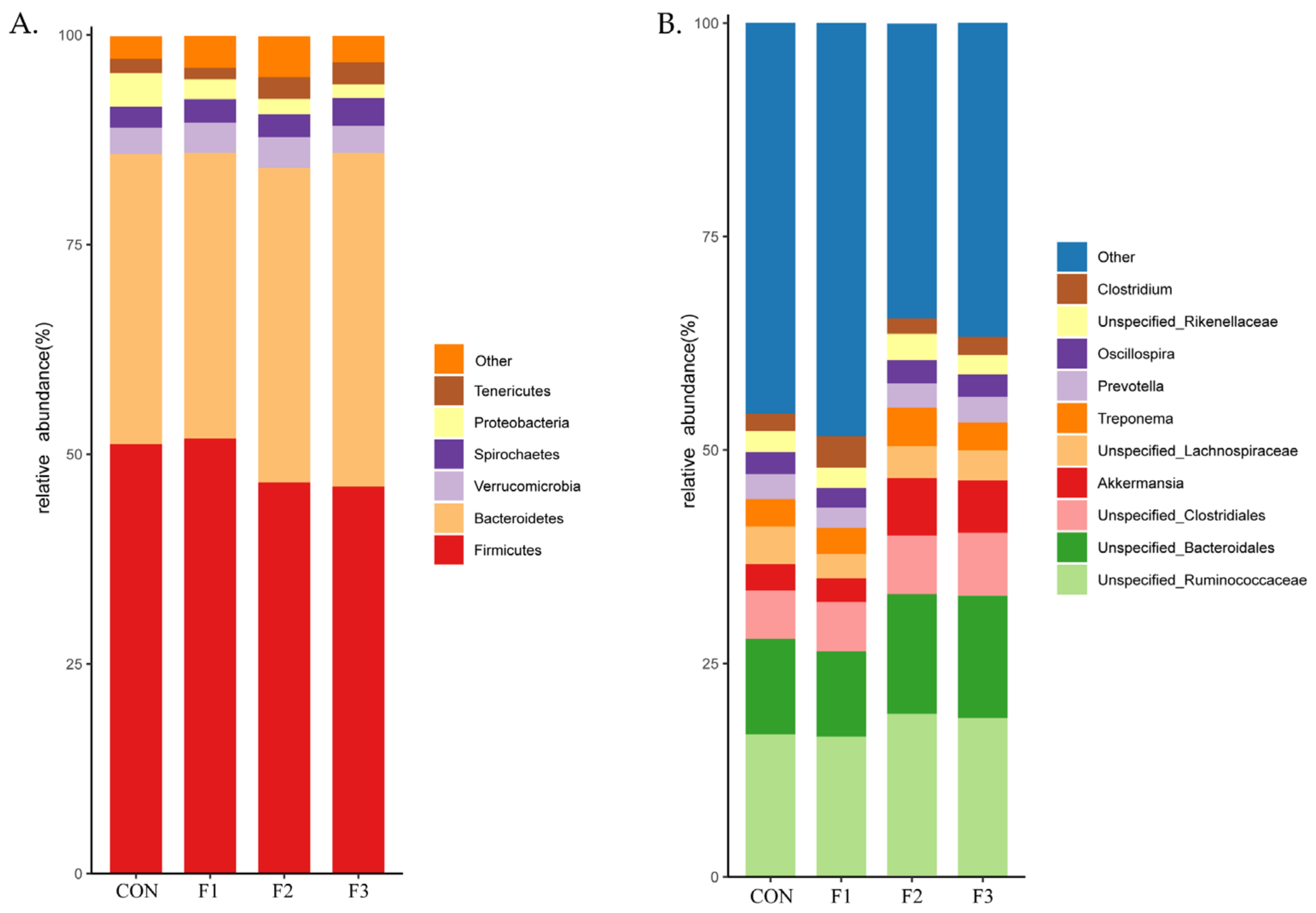
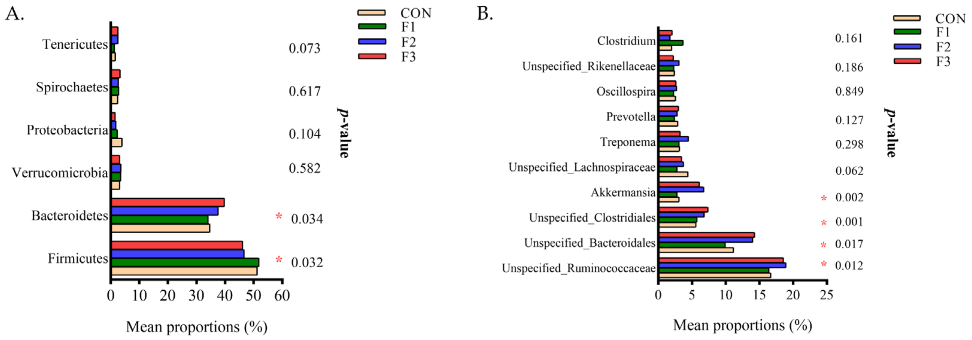
3.7. Alteration of Cecal Microbiota Composition Caused from Fucoidan

At the phylum level (Figure 7A), species annotation results showed that the abundances of Firmicutes and Bacteroidetes accounted for more than 70% of the cecal microbiota composition, and these were the dominant phyla in the cecal microbiota of goat kids. At the genus level (Figure 7B), species annotation results showed that the abundance of Unspecified_Ruminococcaceae, Unspecified_Bacteroidale, and Unspecified_Clostridiales accounted for more than 30% of the genera in the cecal microbiota in weaned goat kids.
At the phylum level (Figure 8A), compared to the CON group, goat kids fed diets with fucoidan supplementation showed increased (p < 0.05) the abundance of Bacteroidetes and decreased (p < 0.05) the abundance of Firmicutes. At the genus level (Figure 8B), compared with the CON group, goat kids fed the F2 and F3 diets had increased abundances of Unspecified_Ruminococcaceae, Unspecified_Bacteroidale, Unspecified_Clostridiales, and Akkermansia (p < 0.05).

4. Discussion

4.1. Serum Biochemical Indices

Serum biochemical parameters are important indices that reflect the metabolism and health status of goat kids [28]. In this study, biochemical parameters and enzyme activities in the serum were within the reference value ranges established for goat kids [29]. LDH is a key enzyme that catalyzes pyruvate to lactate and has been demonstrated to be upregulated in hepatitis and kidney disease [30]. In our study, dietary fucoidan reduced the activity of LDH. Similarly, Piner et al. [31] reported that the administration of 50 mg/kg fucoidan for 7 days decreased the activity of LDH and exhibited a protective role as an antioxidant against sulfoxaflor-induced oxidative stress. In addition, transaminases are enzymes that catalyze the transformation of amino acids to keto acids, and ALT and AST are two of the most important transaminases [32]. The activity of AST in serum is increased in liver damage. In this study, dietary fucoidan supplementation reduced the activity of AST as measured on day 30. In accordance with our study, Choi et al. [33] reported that dietary supplementation with 0.02 g/kg fucoidan improved the aspirin-induced stomach ulceration in rats and reduced the AST activity. The contents of TP, GLU, TG, and T-CHO in serum indicate the metabolic status of proteins, sugars, and fats in ruminants [34,35]. Our results have shown that dietary fucoidan administration reduced the content of GLU but increased the content of TP. Similarly, Abdel et al. [36] reported that dietary fucoidan protected Nile tilapia from the toxicity of waterborne atrazine and increased the content of TP in the serum. In summary, dietary fucoidan does not adversely affect the liver or kidneys of goat kids. According to our previous results [23], we speculate that dietary fucoidan supplementation increases the utilization of TP, thus promoting the growth of goat kids.

4.2. Small Intestinal Barrier Function

The small intestine is not only an important site for digestion and absorption but also a barrier to pathogens and toxins [37]. The maintenance of gut integrity is necessary for normal functioning. Previous research has shown that the effects of weaning on the intestinal barrier function of goat kids may be due to stress [38]. D-lactic acid (D-LA) is an indicator of bacterial metabolites and bacterial lysis. The content of D-LA increases in serum with increased intestinal mucosal permeability [39]. DAO is a highly active intracellular enzyme that is a reliable serum marker for the indirect assessment of the mucosal epithelial cell layer [40]. In this study, dietary fucoidan administration decreased the content of D-LA and the activity of DAO in the serum of goat kids. In accordance with our study, Xue et al. [41] indicated that fucoidan reduced the content of D-LA and the activity of DAO in the serum of rats and alleviated intestinal barrier injury caused by breast cancer. Weaning led to the metabolic disorders of oxygen-free radicals, resulting in oxidative stress [42]. The SOD, GSH-Px, and CAT enzymes are the first line of defense against oxidative injury, and MDA and H2O2 are the final products of lipid peroxidation [42,43]. In this study, dietary fucoidan administration decreased the contents of MDA and H2O2 and increased the activities of GSH-Px, SOD, and CAT, resulting in increased intestinal antioxidant function. Similarly, Li et al. [44] reported that fucoidan improved the oxidative damage to the porcine intestinal epithelial cells by increasing the activities of GSX and GSH and decreasing the content of MDA. The protein sIgA is the most abundant immunoglobulin and the principal regulator of adaptive defenses on mucosal surfaces [45]. In our study, fucoidan increased the contents of sIgA of the duodenum, jejunum, and ileum mucosa in weaned goat kids. Tight junctions are the structural barriers that prevent antigens from moving from the intestinal lumen to the body tissues via the paracellular pathway [46]. Tight junctions are composed of a complex of proteins including claudin, occludin, and zonula occludens. Research has confirmed that weaning stress resulted in damage to the intestinal barrier in goat kids and reduced the mRNA expression of tight junction proteins in the small intestine [47]. In this study, dietary fucoidan supplementation increased the mRNA expression levels of ZO-1, claudin-1, and occludin in the duodenum, jejunum, and ileum. Our results were in line with those of Iraha et al. [48], who reported that fucoidan enhanced the intestinal barrier function by upregulating the expression of Claudin-1. Taken together, the results suggest that improvement of intestinal barrier function by dietary fucoidan may be related to increases in the physical barrier, immunological barrier, and mucosal antioxidant capacity.

4.3. Cecal Microbial Community

The gut microbiome in ruminants has been shown to play an important role in the extraction of valuable nutrients from feed and thereby preserving host health [49]. The richness, diversity, and evenness of microbial communities of samples can be suggested by OTUs and alpha-diversity analysis. In our study, the samples from the fucoidan treatment groups had higher numbers of OTUs and higher values of the Simpson index than samples from the CON group. These results suggest that dietary fucoidan supplementation may increase the diversity of the cecum microflora of weaned goat kids. In addition, we explored the impact of fucoidan on the composition of the cecal bacterial community. Previous studies have suggested that the dominant microflora in the ruminant gut at the phylum level were Bacteroidetes and Firmicutes and that the relative abundances could vary [50]. Consistent with previous studies, we found that the phyla Firmicutes and Bacteroidetes dominated the cecal core microbiome in the goat kids. Previous research has shown that Firmicutes play a major role in fiber degradation and that Bacteroidetes are involved in fermenting carbohydrates [51]. However, Proteobacteria includes a variety of opportunistic pathogens, and metabolic disorders and the intestinal inflammatory response may be due to the increased abundance of Proteobacteria [52]. Our study found that fucoidan increased the abundance of the phyla Bacteroidetes, Tenericutes, and Fibrobacteres and decreased the abundances of the phyla Firmicutes, Proteobacteria, and TM7 in the cecum. At the genus level, Unspecified_Ruminococcaceae, Unspecified_Bacteroidales, and Unspecified_Clostridiales were the dominant bacterial genera in the cecum of weaned goat kids, consistent with the previous findings by Gu et al. [53]. Previous research has shown that some genera in Ruminococcaceae could produce cellulase and amylase [54]. The genus Clostridium are beneficial bacteria commonly found in the intestine that ferment fibrous material to produce butyric acid in stimulating the mucosal immune response [55]. In our study, dietary supplementation with fucoidan increased the abundances of Unspecified_Ruminococcaceae, Unspecified_Bacteroidales, Unspecified_Clostridiales, and Akkermansia in the cecum. These results suggest that active carbohydrate fermentation may be present in the cecum of goat kids and that fucoidan played a regulatory role in the intestinal microflora of the goats.

5. Conclusions

In this study, it was found that dietary fucoidan supplementation improved the small intestinal barrier function and cecal microflora balance in weaned goat kids. This suggested that the supplements could be used to promote the gut health of juvenile goats.

Author Contributions

Conceptualization, F.Y., Z.Z. and W.Y.; methodology, F.Y. and W.Y.; software, W.Y. and G.G.; investigation, J.C. and S.W.; resources, F.Y. and Z.Z.; data curation, W.Y.; writing—original draft preparation, W.Y.; writing—review and editing, W.Y. and F.Y.; supervision, Z.G. and F.Y.; project administration, F.Y. All authors have read and agreed to the published version of the manuscript.

Funding

This research was funded by the Fundamental Research Funds for the Guangdong Province (2020A03025), the Fundamental Research Funds for the Agriculture and Rural Affairs of Guangdong Department (2019KJ127), the Fundamental Research Funds for Education Department of Guangdong (2021003).

Institutional Review Board Statement

This research was approved by the Animal Care and Use Committee of Guangdong Ocean University (SYXK-2018-0147, 2018).

Data Availability Statement

The data presented in this study were available on request from the corresponding author.

Acknowledgments

We would like to thank all people who were involved in the trial. We grateful to the financial support by the Agriculture and Rural Affairs of Guangdong Department.

Conflicts of Interest

The authors declare no conflict of interest.

References

  1. Han, Y.S.; Tang, C.H.; Li, Y.; Yu, Y.N.; Zhan, T.F.; Zhao, Q.Y.; Zhang, J.M. Effects of Dietary Supplementation with Clostridium butyricum on Growth Performance, Serum Immunity, Intestinal Morphology, and Microbiota as an Antibiotic Alternative in Weaned Piglets. Animals 2020, 10, 2287. [Google Scholar] [CrossRef] [PubMed]
  2. Sun, Y.Y.; Li, J.; Meng, Q.S.; Wu, D.L.; Xu, M. Effects of butyric acid supplementation of acidified milk on digestive function and weaning stress of cattle calves. Livest. Sci. 2019, 225, 78–84. [Google Scholar] [CrossRef]
  3. Chen, K.; Liu, Y.; Cheng, Y.; Yan, Q.; Zhou, C.; He, Z.; Zeng, J.; He, J.; Tan, Z. Supplementation of Lactobacillus plantarum or Macleaya cordata Extract Alleviates Oxidative Damage Induced by Weaning in the Lower Gut of Young Goats. Animals 2020, 10, 548. [Google Scholar] [CrossRef] [PubMed] [Green Version]
  4. Jiao, J.Z.; Wu, J.; Wang, M.; Zhou, C.S.; Zhong, R.Z.; Tan, Z.L. Rhubarb Supplementation Promotes Intestinal Mucosal Innate Immune Homeostasis through Modulating Intestinal Epithelial Microbiota in Goat Kids. J. Agric. Food Chem. 2018, 66, 1047–1057. [Google Scholar] [CrossRef]
  5. Saleem, A.M.; Zanouny, A.I.; Singer, A.M. Growth performance, nutrients digestibility, and blood metabolites of lambs fed diets supplemented with probiotics during pre- and post-weaning period. Asian Australas. J. Anim. Sci. 2017, 30, 523–530. [Google Scholar] [CrossRef] [Green Version]
  6. Liu, W.C.; Ou, B.H.; Liang, Z.L.; Zhang, R.; Zhao, Z.H. Algae-derived polysaccharides supplementation ameliorates heat stress-induced impairment of bursa of Fabricius via modulating NF-κB signaling pathway in broilers. Poult. Sci. 2021, 100, 101139. [Google Scholar] [CrossRef]
  7. Guo, Y.; Zhao, Z.H.; Pan, Z.Y.; An, L.L.L.; Balasubramanian, B.; Liu, W.C. New insights into the role of dietary marine-derived polysaccharides on productive performance, egg quality, antioxidant capacity, and jejunal morphology in late-phase laying hens. Poult. Sci. 2020, 99, 2100–2107. [Google Scholar] [CrossRef]
  8. Zhao, Y.; Balasubramanian, B.; Guo, Y.; Qiu, S.J.; Jha, R.; Liu, W.C. Dietary Enteromorpha polysaccharides supplementation improves breast muscle yield and is associated with modification of mRNA transcriptome in broiler chickens. Front. Vet. Sci. 2021, 8, 663988. [Google Scholar] [CrossRef]
  9. Liu, W.C.; Zhu, Y.R.; Zhao, Z.H.; Jiang, P.; Yin, F.Q. Effects of Dietary Supplementation of Algae-Derived Polysaccharides on Morphology, Tight Junctions, Antioxidant Capacity and Immune Response of Duodenum in Broilers under Heat Stress. Animals 2021, 11, 2279. [Google Scholar] [CrossRef]
  10. Zhu, Y.L.; Liu, L.B.; Sun, Z.Y.; Ji, Y.J.; Wang, D.Y.; Mei, L.; Shen, P.L.; Li, Z.X.; Tang, S.; Zhang, H.; et al. Fucoidan as a marine-origin prebiotic modulates the growth and antibacterial ability of Lactobacillus rhamnosus. Int. J. Biol. Macromol. 2021, 180, 599–607. [Google Scholar] [CrossRef]
  11. Liu, X.; Xi, X.Y.; Jia, A.R.; Zhang, M.S.; Cui, T.T.; Bai, X.F.; Shi, Y.P.; Liu, C.H. A fucoidan from Sargassum fusiforme with novel structure and its regulatory effects on intestinal microbiota in high-fat diet-fed mice. Food Chem. 2021, 358, 129908. [Google Scholar] [CrossRef]
  12. Zuo, T.; Li, X.M.; Chang, Y.G.; Duan, G.F.; Yu, L.; Zheng, R.; Xue, C.H.; Tang, Q.J. Dietary fucoidan of Acaudina molpadioides and its enzymatically degraded fragments could prevent intestinal mucositis induced by chemotherapy in mice. Food Funct. 2015, 6, 415–422. [Google Scholar] [CrossRef]
  13. Shi, H.J.; Chang, Y.G.; Yuan, G.; Wang, X.; Tang, Q.J. Dietary fucoidan of Acaudina molpadioides alters gut microbiota and mitigates intestinal mucosal injury induced by cyclophosphamide. Food Funct. 2017, 8, 3383–3393. [Google Scholar] [CrossRef]
  14. Rattigan, R.; Sweeney, T.; Vigors, S.; Thornton, K.; Rajauria, G.; O’Doherty, J.V. The Effect of Increasing Inclusion Levels of a Fucoidan-Rich Extract Derived from Ascophyllum nodosum on Growth Performance and Aspects of Intestinal Health of Pigs Post-Weaning. Mar. Drugs 2019, 17, 680. [Google Scholar] [CrossRef] [Green Version]
  15. Sun, T.; Xue, M.L.; Yang, J.; Pei, Z.Q.; Zhang, N.; Qin, K.P.; Liang, H. Metabolic regulation mechanism of fucoidan via intestinal microecology in diseases. J. Sci. Food Agric. 2021, 101, 4456–4463. [Google Scholar] [CrossRef]
  16. Luo, J.M.; Wang, Z.; Fan, B.; Wang, L.; Liu, M.Y.; An, Z.Z.; Zhao, X. A comparative study of different fucoidan on the cefoperazone-induced gut microbiota disturbance and intestinal inammation. Food Funct. 2021, 12, 9087–9097. [Google Scholar] [CrossRef]
  17. Nagamine, T.; Nakazato, K.; Tomioka, S.; Iha, M.; Nakajima, K. Intestinal Absorption of Fucoidan Extracted from the Brown Seaweed, Cladosiphon okamuranus. Mar. Drugs 2014, 13, 48–64. [Google Scholar] [CrossRef]
  18. Walsh, A.M.; Sweeney, T.; O’Shea, C.J.; Doyle, D.N.; O’Doherty, J.V. Effect of dietary laminarin and fucoidan on selected microbiota, intestinal morphology and immune status of the newly weaned pig. Br. J. Nutr. 2013, 110, 1630–1638. [Google Scholar] [CrossRef] [Green Version]
  19. Lynch, M.B.; Sweeney, T.; Callan, J.J.; O’Sullivan, J.T.; O’Doherty, J.V. The effect of dietary Laminaria-derived laminarin and fucoidan on nutrient digestibility, nitrogen utilisation, intestinal microflora and volatile fatty acid concentration in pigs. J. Sci. Food Agric. 2010, 90, 430–437. [Google Scholar] [CrossRef]
  20. Wu, Q.F.; Wu, S.Y.; Cheng, Y.; Zhang, Z.S.; Mao, G.X.; Li, S.J.; Yang, Y.; Zhang, X.; Wu, M.J.; Tong, H.B. Sargassum fusiforme fucoidan modifies gut microbiota and intestinal metabolites during alleviation of hyperglycemia in type 2 diabetic mice. Food Funct. 2021, 12, 3572–3585. [Google Scholar] [CrossRef]
  21. Shokaiyan, M.; Ashayerizadeh, O.; Shargh, M.S.; Dastar, B. Algal Crude Fucoidan Alone or with Bacillus subtilis DSM 17299 in Broiler Chickens Diet: Growth Performance, Carcass Characteristics, Blood Metabolites, and Morphology of Intestine. Poult. Sci. J. 2019, 7, 87–94. [Google Scholar]
  22. Ikeda-Ohtsubo, W.; Nadal, A.L.; Zaccaria, E.; Iha, M.; Kitazawa, H.; Kleerebezem, H.; Brugman, S. Intestinal Microbiota and Immune Modulation in Zebrafish by Fucoidan from Okinawa Mozuku (Cladosiphon okamuranus). Front. Nutr. 2020, 7, 67. [Google Scholar] [CrossRef]
  23. Yang, W.G.; Chen, J.Y.; Guo, G.Z.; Wang, S.N.; Peng, S.; Gao, Z.H.; Zhao, Z.H.; Lan, R.X.; Yin, F.Q. The Effects of Fucoidan Dietary Supplementation on Growth Performance, Serum Antioxidant Capacity, Immune Function Indices and Intestinal Morphology in Weaned Kids. Animals 2022, 12, 574. [Google Scholar] [CrossRef] [PubMed]
  24. Wang, X.; Hu, C.; Ding, L.; Tang, Y.; Wei, H.; Jiang, C.; Yan, Q.; Dong, Q.; Degen, A.A. Astragalus membranaceus Alters Rumen Bacteria to Enhance Fiber Digestion, Improves Antioxidant Capacity and Immunity Indices of Small Intestinal Mucosa, and Enhances Liver Metabolites for Energy Synthesis in Tibetan Sheep. Animals 2021, 11, 3236. [Google Scholar] [CrossRef] [PubMed]
  25. Guo, Y.; Balasubramanian, B.; Zhao, Z.H.; Liu, W.C. Marine algal polysaccharides alleviate aflatoxin B1-induced bursa of Fabricius injury by regulating redox and apoptotic signaling pathway in broilers. Poult. Sci. 2020, 100, 844–857. [Google Scholar] [CrossRef] [PubMed]
  26. Wei, H.Y.; Ding, L.M.; Wang, X.J.; Yan, Q.; Jiang, C.X.; Hu, C.S.; Wang, G.W.; Zhou, Y.Q.; Henkin, Z.; Degen, A.A. Astragalus root extract improved average daily gain, immunity, antioxidant status and ruminal microbiota of early weaned yak calves. J. Sci. Food Agric. 2020, 101, 82–90. [Google Scholar] [CrossRef] [PubMed]
  27. Edgar, R.C. UPARSE: Highly accurate OTU sequences from microbial amplicon reads. Nat. Methods 2013, 10, 996. [Google Scholar] [CrossRef]
  28. Elkatcha, M.; Soltan, M.; Essi, M. Effect of Pediococcus spp. Supplementation on Growth Performance, Nutrient Digestibility and Some Blood Serum Biochemical Changes of Fattening Lambs. Alex. J. Vet. Sci. 2016, 48, 124. [Google Scholar] [CrossRef]
  29. Abdolvahabi, S.; Zaeemi, M.; Mohri, M.; Naserian, A.A. Age related changes in serum biochemical profile of Saanen goat kids during the first three months of life. Rev. Méd. Vét. 2016, 167, 106–112. [Google Scholar]
  30. Klein, R.; Nagy, O.; Tóthová, C.; Chovanová, F. Clinical and Diagnostic Significance of Lactate Dehydrogenase and Its Isoenzymes in Animals. Vet. Med. Int. 2020, 2020, 5346483. [Google Scholar] [CrossRef]
  31. Piner Benli, P.; Kaya, M.; Dağlıoğlu, Y.K. Fucoidan Protects against Acute Sulfoxaflor-Induced Hematological/Biochemical Alterations and Oxidative Stress in Male Mice. Pharmaceuticals 2022, 15, 16. [Google Scholar] [CrossRef]
  32. Tavangar, P.; Gharahveysi, S.; Rezaeipour, V.; Irani, M. Efficacy of phytobiotic and toxin binder feed additives individually or in combination on the growth performance, blood biochemical parameters, intestinal morphology, and microbial population in broiler chickens exposed to aflatoxin B1. Trop. Anim. Health Prod. 2021, 53, 335. [Google Scholar] [CrossRef]
  33. Choi, J.; Raghavendran, H.R.B.; Sung, N.Y.; Kim, J.H.; Chun, B.S.; Ahn, D.H.; Choi, H.S.; Kang, K.W.; Lee, J.W. Effect of fucoidan on aspirin-induced stomach ulceration in rats. Chem. Biol. Interact. 2009, 183, 249–254. [Google Scholar] [CrossRef]
  34. Wang, Q.Y.; Zeng, X.L.; Zeng, Y.T.; Liu, X.R.W.; Wang, Y.C.; Wang, X.; Li, J.Z.; Wang, Y.Q.; Feng, Z.; Huang, P.F.; et al. Effects of Phragmites australis Shoot Remainder Silage on Growth Performance, Blood Biochemical Parameters, and Rumen Microbiota of Beef Cattle. Front. Vet. Sci. 2022, 9, 778654. [Google Scholar] [CrossRef]
  35. Musco, N.; Tudisco, R.; Esposito, G.; Iommelli, P.; Totakul, P.; D’Aniello, B.; Lombardi, P.; Amato, R.; Wanapat, M.; Infascelli, F. Effects of Linseed Supplementation on Milk Production, Composition, Odd- and Branched-Chain Fatty Acids, and on Serum Biochemistry in Cilentana Grazing Goats. Animals 2022, 12, 783. [Google Scholar] [CrossRef]
  36. AbdelWarith, A.W.A.; Younis, E.M.; AlAsgah, N.A.; Gewaily, M.S.; ElTonoby, S.M.; Dawood, M.A.O. Role of Fucoidan on the Growth Behavior and Blood Metabolites and Toxic Effects of Atrazine in Nile Tilapia Oreochromis niloticus (Linnaeus, 1758). Animals 2021, 11, 1448. [Google Scholar] [CrossRef]
  37. Zhang, J.Q.; Yu, C.Y.; Li, Z.H.; Li, J.; Chen, Y.P.; Wang, T.; Wang, C. Effects of Zinc Oxide Nanoparticles on Growth, Intestinal Barrier, Oxidative Status and Mineral Deposition in 21-Day-Old Broiler Chicks. Biol. Trace Elem. Res. 2021, 200, 1826–1834. [Google Scholar] [CrossRef]
  38. Izuddin, W.I.; Loh, T.C.; Foo, H.L.; Samsudin, A.A.; Human, A.M. Postbiotic L. plantarum RG14 improves ruminal epithelium growth, immune status and upregulates the intestinal barrier function in post-weaning lambs. Sci. Rep. 2019, 9, 9938. [Google Scholar] [CrossRef]
  39. Henrichs, B.S.; Brost, K.N.; Hayes, C.A.; Campbell, J.M.; Drackley, J.K. Effects of spray-dried bovine plasma protein in milk replacers fed at a high plane of nutrition on performance, intestinal permeability, and morbidity of Holstein calves. J. Dairy Sci. 2021, 104, 7856–7870. [Google Scholar] [CrossRef]
  40. Shi, L.G.; Xun, W.J.; Peng, W.Q.; Hu, H.C.; Cao, T.; Hou, G.Y. Effect of the Single and Combined Use of Curcumin and Piperine on Growth Performance, Intestinal Barrier Function, and Antioxidant Capacity of Weaned Wuzhishan Piglets. Front. Vet. Sci. 2020, 7, 418. [Google Scholar] [CrossRef]
  41. Xue, M.L.; Ji, X.Q.; Liang, H.; Liu, Y.; Wang, B.; Sun, L.L.; Li, W.W. The effect of fucoidan on intestinal flora and intestinal barrier function in rats with breast cancer. Food Funct. 2018, 9, 1214–1223. [Google Scholar] [CrossRef]
  42. Zhao, J.X.; Li, Q.; Zhang, R.X.; Liu, W.Z.; Ren, Y.S.; Zhang, C.X.; Zhang, J.X. Effect of dietary grape pomace on growth performance, meat quality and antioxidant activity in ram lambs. Anim. Feed Sci. Technol. 2018, 236, 76–85. [Google Scholar] [CrossRef]
  43. Zhao, J.X.; Li, K.; Su, R.; Liu, W.Z.; Ren, Y.S.; Zhang, C.X.; Du, M.; Zhang, J.X. Effect of dietary Tartary buckwheat extract supplementation on growth performance, meat quality and antioxidant activity in ewe lambs. Meat Sci. 2017, 134, 79–85. [Google Scholar] [CrossRef]
  44. Li, C.; Chen, J.; Zhao, M.; Liu, M.Q.; Yue, Z.K.; Liu, L.; Li, F.C. Effect of sodium butyrate on slaughter performance, serum indexes and intestinal barrier of rabbits. J. Anim. Physiol. Anim. Nutr. 2021, 106, 156–166. [Google Scholar] [CrossRef]
  45. Li, C.Y.; Chen, X.Y.; Zhang, B.; Liu, L.; Li, F.C. Sodium butyrate improved intestinal barrier in rabbits. Ital. J. Anim. Sci. 2020, 19, 1472–1482. [Google Scholar] [CrossRef]
  46. Chen, Y.X.; Xie, Y.N.; Zhong, R.Q.; Han, H.; Liu, L.; Chen, L.; Zhang, H.F.; Yves, B.; Nadia, E. Effects of graded levels of xylo-oligosaccharides on growth performance, serum parameters, intestinal morphology and intestinal barrier function in weaned piglets. J. Anim. Sci. 2021, 99, 183. [Google Scholar] [CrossRef]
  47. Shang, Z.H.; Wu, J.Y.; Wang, Y.X.; Zhang, Y.; Shang, Y.J. Effect of post-weaning stress diarrhea on intestinal barrier in lambs. Prog. Vet. Med. 2020, 41, 63–67. (In Chinese) [Google Scholar]
  48. Iraha, A.; Chinen, H.; Hokama, A.; Yonashiro, T.; Kinjo, T.; Kishimoto, K.; Nakamoto, M.; Hirata, T.; Kinjo, N.; Higa, F.; et al. Fucoidan enhances intestinal barrier function by upregulating the expression of claudin-1. World J. Gastroenterol. 2013, 19, 5500–5507. [Google Scholar] [CrossRef] [PubMed]
  49. Sun, J.; Li, M.; Tang, Z.; Zhang, X.; Chen, J.; Sun, Z. Effects of Rhodotorula mucilaginosa fermentation product on the laying performance, egg quality, jejunal mucosal morphology and intestinal microbiota of hens. J. Appl. Microbiol. 2020, 128, 54–64. [Google Scholar] [CrossRef] [PubMed] [Green Version]
  50. Georgios, O.; Teixeira, A.G.V.; Foditsch, C.; Bicalho, M.L.; Machado, V.S.; Bicalho, R.C. Fecal microbial diversity in pre-weaned dairy calves as described by pyrosequencing of metagenomic 16S rDNA. Associations of Faecalibacterium species with health and growth. PLoS ONE 2017, 8, e63157. [Google Scholar]
  51. Brulc, J.M.; Antonopoulosi, D.A.; Berg Miller, M.E.; Wilson, M.K. Gene-centric metagenomics of the fiber-adherent bovine rumen microbiome reveals forage specific glycoside hydrolases. Proc. Natl. Acad. Sci. USA 2009, 106, 1948–1953. [Google Scholar] [CrossRef] [Green Version]
  52. Pichel, M.G.; Binsztein, N.; Qadri, F.; Giron, J.A. Type IV Longus Pilus of Enterotoxigenic Escherichia coli: Occurrence and Association with Toxin Types and Colonization Factors among Strains Isolated in Argentina. J. Clin. Microbiol. 2002, 40, 694–697. [Google Scholar] [CrossRef] [Green Version]
  53. Gu, S.; Chen, D.; Zhang, J.N.; Lv, X.; Wang, K.; Duan, L.P.; Nie, Y.; Wu, X.L. Bacterial Community Mapping of the Mouse Gastrointestinal Tract. PLoS ONE 2013, 8, e74957. [Google Scholar] [CrossRef] [Green Version]
  54. Suen, G.; Stevenson, D.M.; Bruce, D.C.; Chertkov, O.; Copeland, A.; Cheng, J.F.; Detter, C.; Detter, J.C.; Goodwin, L.A.; Han, C.S.; et al. Complete genome of the cellulolytic ruminal bacterium Ruminococcus albus 7. J. Bacteriol. 2011, 193, 5574–5575. [Google Scholar] [CrossRef] [Green Version]
  55. Mo, S.; Kim, B.S.; Yun, S.J.; Lee, J.J.; Yoon, S.H.; Oh, C.H. Genome sequencing of Clostridium butyricum DKU-01, isolated from infant feces. Gut Pathog. 2015, 7, 8. [Google Scholar] [CrossRef] [Green Version]
Figure 1. Effects of dietary fucoidan supplementation for 30 days on intestinal permeability in weaned goat kids. (A) Serum D-LA content; (B) serum DAO activity. Results are presented as mean ± SEM. a–d means significantly different (p < 0.05). CON, basal diet; F1, basal diet + 0.1% fucoidan; F2, basal diet + 0.3% fucoidan; F3, basal diet + 0.5% fucoidan. D-LA, D-lactic acid; DAO, diamine oxidase.
Animals 12 01591 g001
Figure 2. Effects of dietary fucoidan supplementation on intestinal mucosa antioxidant capacity in weaned goat kids. (A) MDA content; (B) H2O2 content; (C) GSH-Px activity; (D) T-SOD activity; (E) CAT activity; (F) T-AOC activity. Results are presented as mean ± SEM. a–c means significantly different (p < 0.05). CON, basal diet; F1, basal diet + 0.1% fucoidan; F2, basal diet + 0.3% fucoidan; F3, basal diet + 0.5% fucoidan. MDA, malondialdehyde; H2O2, hydrogen peroxide; GSH-Px, Glutathione peroxidase; T-SOD, total superoxide dismutase; CAT, catalase; T-AOC, total antioxidant capacity.
Animals 12 01591 g002
Figure 3. Effects of dietary fucoidan supplementation on intestinal mucosal immunity in weaned goat kids. (A) Duodenal mucosa sIgA content; (B) jejunum mucosa sIgA content; (C) ileum mucosa sIgA content. Results are presented as mean ± SEM. a–c means significantly different (p < 0.05). CON, basal diet; F1, basal diet + 0.1% fucoidan; F2, basal diet + 0.3% fucoidan; F3, basal diet + 0.5% fucoidan. sIgA, secretory immunoglobulin A.
Animals 12 01591 g003
Figure 4. Effects of dietary fucoidan supplementation on intestinal tight junction protein mRNA expression level in weaned goat kids. (A) Duodenal tight junction protein mRNA expression level; (B) jejunum tight junction protein mRNA expression level; (C) ileum tight junction protein mRNA expression level. Results are presented as mean ± SEM. a–c means significantly different (p < 0.05). CON, basal diet; F1, basal diet + 0.1% fucoidan; F2, basal diet + 0.3% fucoidan; F3, basal diet + 0.5% fucoidan. ZO-1, zonula occludens.
Animals 12 01591 g004
Figure 5. Diversity analysis. (A) Venn diagrams of OTUs sharing >97% nucleotide sequence identity; (B) principal co-ordinates (PCoA) analysis of OUTs; CON, basal diet; F1, basal diet + 0.1% fucoidan; F2, basal diet + 0.3% fucoidan; F3, basal diet + 0.5% fucoidan; OTUs, operational taxonomic units; PCA, principal component analysis.
Animals 12 01591 g005
Figure 6. α-diversity index of gut microbiota. (A) Chao1 index; (B) Shannon index; (C) Simpson index. CON, basal diet; F1, basal diet + 0.1% fucoidan; F2, basal diet + 0.3% fucoidan; F3, basal diet + 0.5% fucoidan; * p < 0.05.
Animals 12 01591 g006
Figure 7. Bacterial composition at phylum (A) and genus (B) level in cecal content of goat kids among four dietary treatments. CON, basal diet; F1, basal diet + 0.1% fucoidan; F2, basal diet + 0.3% fucoidan; F3, basal diet + 0.5% fucoidan.
Animals 12 01591 g007
Figure 8. Analysis for different bacterial at phylum (A) and genus (B) level among four dietary treatments. CON, basal diet; F1, basal diet + 0.1% fucoidan; F2, basal diet + 0.3% fucoidan; F3, basal diet + 0.5% fucoidan. * p < 0.05.
Animals 12 01591 g008
Table 1. The composition and level of basal diet (air-dry basis).
ItemsContent
Ingredients (%)
Pennisetum purpureum35.00
Corn40.89
Soybean meal13.98
Wheat bran 7.15
Salt0.65
CaHPO40.84
Limestone0.84
Premix 10.65
Total100
Nutrient level
DM (%)88.86
ME (MJ/kg)10.43
CP (%)12.06
NDF (%)30.06
ADF (%)16.39
Ca (%)0.76
p (%)0.54
1 The premix provided the following per kg of diets: VA 8 000 IU, VD 2 000 IU, VE 40 IU, Cu 12 mg, Fe 70 mg, Mn 50 mg, Zn 80 mg, I 1.0 mg, Se 0.27 mg, and Co 0.3 mg. CaHPO4, calcium hydrogen phosphate; DM, dry matter; ME, metabolizable energy; CP, crude protein; NDF, neutral detergent fiber; ADF, acid detergent fiber; Ca, calcium; P, phosphorus.
Table 2. Nucleotide sequences of primers used to measure targeted genes.
Gene SymbolsProduct Length, bpAccession No.Nucleotide Sequence of Primers (5′→3′)
ZO-1111XM_018066118.1F: CGTCCTGATCCTGAACCTGTGTCT
R: GCTCTTCTCGCTCCTCCTGTGT
Occludin157XM_018065681.1F: CCTGTGTTGCCTCCACTCTT
R: TCCGTATAGCCTGTTCCATAGC
Claudin-195XM_005675123.3F: CCTGCTGGGACTAATAGCCA
R: CAGCCATTCGCATCTTCTGT
β-actin91NM_001314342.1F: TCCTTCCTGGGCATGGAATC
R: CGTAAAGGTCCTTGCGGATG
ZO-1, zonula occludens.
Table 3. Effects of dietary fucoidan on serum biochemical in weaned goat kids.
Item 1CONF1F2F3SEM 2p-Value
15 d
ALT (U/L)2.872.172.903.300.660.40
AST (U/L)13.9713.2411.429.762.900.48
LDH (U/L)4359.98 a3434.89 b3505.28 b3656.11 b371.550.03
TG (mmol/L)1.000.930.800.970.120.37
T-CHO (mmol/L)1.921.321.571.770.220.05
GLU (mmol/L)0.73 a0.69 b0.60 c0.62 c0.040.01
TP (mg/mL)27.69 b30.15 b30.40 b35.84 a2.000.01
30 d
ALT (U/L)2.744.193.383.050.750.26
AST (U/L)18.78 a11.85 b10.34 b9.10 b2.550.02
LDH (U/L)5174.24 a5851.01 a4848.49 b4093.43 b584.360.04
TG (mmol/L)0.810.980.970.680.140.14
T-CHO (mmol/L)1.491.381.641.760.150.09
GLU (mmol/L)0.670.640.640.570.380.10
TP (mg/mL)88.49 c107.08 b110.92 a119.06 a5.11<0.01
1 CON, basal diet; F1, basal diet + 0.1% fucoidan; F2, basal diet + 0.3% fucoidan; F3, basal diet + 0.5% fucoidan. ALT, alanine aminotransferase; AST, aspartate aminotransferase; LDH, lactate dehydrogenase; TG, total glyceride; T-CHO, total cholesterol; GLU, glucose; TP, total protein. 2 SEM, standard error of mean. a–c Values in the same row with different letters are significant different (p < 0.05). Results are presented as mean ± SEM.
Publisher’s Note: MDPI stays neutral with regard to jurisdictional claims in published maps and institutional affiliations.

