Article

Prevalence and Virulence Characteristics of Enterococcus faecalis and Enterococcus faecium in Bovine Mastitis Milk Compared to Bovine Normal Raw Milk in South Korea

1
Center for One Health, College of Veterinary Medicine, Konkuk University, 120 Neungdong-ro, Gwangjin-gu, Seoul 05029, Korea
2
Bacterial Disease Division, Animal and Plant Quarantine Agency, 177 Hyeksin 8-ro, Gimcheon-si 39660, Gyeonsangbuk-do, Korea
*
Author to whom correspondence should be addressed.
Animals 2022, 12(11), 1407; https://doi.org/10.3390/ani12111407
Submission received: 9 May 2022 / Revised: 25 May 2022 / Accepted: 26 May 2022 / Published: 30 May 2022
(This article belongs to the Section Cattle)

Abstract

:

Simple Summary

Characterization of Enterococcus spp. among bovine mastitis pathogens is important in terms of the dairy industry and public health domain. The present study aimed to characterize virulence properties such as virulence genes (esp, asa1, gelE, and cylA), biofilm, gelatinase, hemolysis, and antimicrobial resistance and to compare them between bovine mastitis milk and bovine normal raw milk. Our results demonstrated that Enterococcus faecalis and Enterococcus faecium isolated from bovine mastitis milk exhibited higher virulence properties than those isolated from bovine normal raw milk. E. faecalis (90 isolates) was present at a significantly higher rate than E. faecium (32 isolates) and was more abundant in virulence genes. Furthermore, after analyzing the correlation between the virulence genes and the corresponding phenotype, we found that gelE and esp were involved in strong biofilm formation and the gelE was involved in gelatinase production. Taken together, E. faecalis in bovine mastitis milk should be monitored to control bovine mastitis and improve public health.

Abstract

Enterococcus spp. are pathogens that cause environmental mastitis and are difficult to eliminate owing to their resistance to antibiotics. To compare the virulence characteristics of isolates from bovine mastitis milk (BMM) and bovine normal raw milk (NRM), we isolated Enterococcus spp. from 39 dairy farms in South Korea from 2015–2020. A total of 122 Enterococcus spp. were identified, with Enterococcus faecalis (73.8%) accounting for the majority, followed by Enterococcus faecium (26.2%). E. faecalis isolated from BMM harbored gelE, asa1, esp, and cylA genes with a prevalence of 85.7, 71.4, 54.3, and 30.0%, respectively. These genes were significantly more abundant in BMM than in NRM, except for asa1 (p < 0.0001). Interestingly, strong biofilm and gelatinase formation was predominately observed for BMM isolates and this was significantly correlated to the presence of esp and gelE genes (p < 0.05). BMM isolates demonstrated higher resistance to tetracycline (59.3%), followed by chloramphenicol (21.0%), rifampicin (18.5%), doxycycline (4.9%), ciprofloxacin (1.2%), and nitrofurantoin (1.2%), than those from NRM. E. faecalis harboring esp, gelE, and cylA may be causative agents for bovine mastitis and act as a reservoir for the transmission of virulence factors to humans.

1. Introduction

Bovine mastitis is a disease that affects dairy animals, resulting in a decrease in both milk quality and production [1]. Bovine mastitis pathogens are classified as either “contagious” or “environmental” with Staphylococcus aureus, Streptococcus agalactiae, and Mycoplasma spp. being representative pathogens of contagious mastitis [2]. In contrast, enterococci are environmental causative agents of mastitis, with infections caused primarily by Enterococcus faecalis (approximately 80%) and Enterococcus faecium (approximately 10–15%) [3,4].
Enterococcus spp. are Gram-positive bacteria that live in the gastrointestinal tracts of humans and other animals. In recent decades, they have emerged as major causes of nosocomial infections [5]. Due to their ability to adapt to adverse conditions, Enterococcus spp. can survive in the environment for long periods of time and can infect the mammary glands [3]. Although ampicillin (AMP), gentamicin, penicillin, tetracycline (TET), and tylosin (TYL) are antimicrobial agents approved and previously used for the treatment of bovine mastitis in South Korea, their therapeutic success is limited by antimicrobial resistance [6]. Furthermore, Enterococcus spp. serve as a reservoir of antimicrobial resistance genes, allowing them to be transmitted to humans via the food chain [7]. Our previous studies identified E. faecalis and E. faecium resistant to TET, erythromycin (ERY), ciprofloxacin (CIP), high-level streptomycin, levofloxacin, and linezolid in ripened cheese products [8]. The antibiotic-resistant Enterococcus spp. can cause various infections in humans, including urinary tract infection, endocarditis, meningitis, and bacteremia, all of which can be difficult to treat with antibiotics [9,10].
The ability of Enterococcus spp. to form biofilms is considered a virulence factor that contributes to its antibiotic resistance [11]. A mature biofilm can endure 10 to 1000 times greater concentrations of antimicrobial agents than those required to kill planktonic bacteria [12]. The genes involved in biofilm formation include esp (enterococcal surface protein-encoding gene), asa1 (aggregation substance-encoding gene), and gelE (gelatinase-encoding gene) [13]. Gelatinase can hydrolyze gelatin, hemoglobin, collagen, casein, and other bioactive compounds [14]. Additionally, cytolysin (encoded by cylA) can induce the hemolysis of human red blood cells [15].
Because virulence genes can be carried on mobile genetic elements, Enterococcus spp. in milk can contribute to the spread of these genes in humans [16]. There is a lack of information regarding virulence and antimicrobial resistance in isolates of bovine mastitis milk (BMM) compared to those in bovine normal raw milk (NRM). To the best of our knowledge, this is the first study that aimed to characterize and compare the virulence properties of Enterococcus spp. isolated from BMM and NRM. Therefore, the purpose of this study was to determine the virulence characteristics of isolates of Enterococcus spp. from BMM and NRM by comparing virulence factors (genes and biofilm, gelatinase, and hemolysin formation), correlation between virulence phenotype and genotype, and antimicrobial resistance.

2. Materials and Methods

2.1. Sample Collection and Isolation of E. faecalis and E. faecium

E. faecalis and E. faecium were isolated by collecting at least one BMM each from 36 dairy farms in South Korea between 2015 and 2020. For comparison of virulence properties with BMM, E. faecalis and E. faecium were isolated from at least one NRM from each of three dairy farms in South Korea in 2020. The somatic cell count (SCC) values of the BMM and NRM were measured using the Fossmatic System 4000 (Foss Electric, Hillerød, Denmark). Milk with SCC ≥ 200,000 cells/mL was considered positive for bovine mastitis and was prohibited for human consumption, while that with SCC < 200,000 cells/mL was considered normal. To isolate E. faecalis and E. faecium from BMM and NRM, milk samples were inoculated on blood agar (Komed, Gyeonggi, South Korea) and incubated at 37 °C for 24 h. After incubation, round white suspected colonies were identified by Matrix-Assisted Laser Desorption Ionization–Time of Flight Mass Spectrometer (MALDI-TOF MS; bioMérieux, Marcy l’Etoile, France), according to the manufacturer’s instructions. The isolates were stored at −80 °C in tryptic soy broth (Sigma-Aldrich, St. Louis, MO, USA) with 25% sterile glycerol for further analysis.

2.2. DNA Extraction and Screening for Virulence Genes

All Enterococcus spp. were cultivated in brain heart infusion (BHI) broth (Sigma-Aldrich, St. Louis, MO, USA) at 37 °C. DNA extraction was performed using a QIAamp DNA Mini Kit (Qiagen, Hilden, Germany), following the manufacturer’s instructions. The extracted DNA was screened for the presence of esp, asa1, gelE, and cylA genes using conventional polymerase chain reaction (PCR) with the corresponding primers (Supplementary Table S1) [17]. Primer sets were synthesized and purchased from Bionics (Seoul, Korea). Each PCR amplification was performed in a 20 µL reaction volume, containing 1 µL of template DNA, 2 µL of total primer, and 17 µL of distilled water using the Maxime PCR PreMix Kit (i-Taq) (iNtRON Biotechnology, Seongnam, South Korea). An initial activation step at 95 °C for 15 min was followed by 30 cycles of denaturation (94 °C, 1 min), annealing (temperatures indicated in Supplementary Table S1, 1 min), and extension (72 °C, 1 min), followed by one cycle of 10 min at 72 °C [17]. E. faecalis KCTC 3206 (ATCC 19433) and KCTC 3511 (ATCC 29212) obtained from the Korean Collection for Type Cultures (KCTC; Jeongeup, South Korea) were used as positive controls.

2.3. Phenotype of Virulence Characteristics

2.3.1. Biofilm Formation

The assessment of biofilm formation was based on a method described by Stepanović et al. (2007) with some modifications [18]. Briefly, bacteria were cultured at 37 °C in BHI agar (Sigma-Aldrich, St. Louis, MO, USA) for 24 h. Each culture was then suspended in BHI broth (Sigma-Aldrich, St. Louis, MO, USA) supplemented with 2% glucose (Sigma-Aldrich, St. Louis, MO, USA) to prepare a 0.5 McFarland standard suspension (about 107 colony forming units (CFU)/mL). Each well of a sterile 96-well culture plate (SPL Life Sciences, Pocheon-si, Gyeonggi-do, Korea) was filled with a 200 µL bacterial suspension. Subsequently, the plate was incubated for 24 h at 37 °C and the broth was carefully removed. The wells were gently washed three times with sterile phosphate buffered saline (PBS; Sigma-Aldrich, St. Louis, MO, USA). Following every washing step, the adherent biofilm layer formed in each well was stained with 200 µL 0.1% crystal violet (Sigma-Aldrich, St. Louis, MO, USA) solution in distilled water for 20 min at 25 °C. After each staining step, the well was washed with PBS (Sigma-Aldrich, St. Louis, MO, USA) until the washing was free of stain, and the optical density (OD) was measured using a Multiskan FC (Thermo Fisher Scientific, Shanghai, China) at 595 nm. The cut-off OD (ODc) was defined by the negative control. Based on the bacterial biofilm-forming activity, the isolates were classified as follows: OD ≤ ODc = non-biofilm formation, ODc < OD ≤ 2 ODc = weak biofilm formation, 2 ODc < OD ≤ 4 ODc = moderate biofilm formation, and OD > 4 ODc = strong biofilm formation.

2.3.2. Gelatinase Production

For the detection of gelatinase activity, Enterococcus spp. were inoculated on nutrient gelatin (KisanBio Co., Ltd., Seoul, Korea). S. aureus ATCC 25923 was used as the positive control for gelatin hydrolysis experiments, while Escherichia coli KCTC 2571 was used as the negative control in the gelatin hydrolysis experiment. Pure cultures were individually stabbed into tubes, incubated at 37 °C for 48 h, and then held at 4 °C for 30 min. In tubes where an organism produced sufficient gelatinase, the gelatin remained liquefied upon cooling.

2.3.3. Hemolysin Production

Hemolysis activity was determined on Columbia agar with 5% sheep blood (bioMérieux, Marcy l’Etoile, France) and incubated at 37 °C for 48 h. The presence or absence of clearing zones around the colonies was interpreted as β-hemolysis (positive hemolysis) or γ-hemolysis (negative hemolysis), respectively. S. aureus ATCC 25923 was used as the positive control.

2.4. Antimicrobial Resistance

Isolates were investigated for their susceptibility towards nine different antimicrobials using the disc diffusion method in accordance with Clinical and Laboratory Standards Institute (CLSI) guidelines [19]. Briefly, a 0.5 McFarland standard suspension was prepared for each isolate and the isolate was cultured uniformly on Muller-Hinton agar (Oxoid, Basingstoke, UK) by streaking the swab in a back-and-forth motion. Antimicrobial discs (Oxoid, Basingstoke, UK) individually loaded with AMP (10 μg), chloramphenicol (30 μg; C), CIP (5 μg), doxycycline (30 μg; DOX), ERY (15 μg), nitrofurantoin (300 μg; N), rifampicin (5 μg; RIF), TET (30 μg), and vancomycin (30 μg; VAN) were placed on the agar plates, followed by incubation for 24 h at 37 °C. The inhibition zone size was measured using a ruler. Susceptibility or resistance of the organism to each tested drug was determined using S. aureus ATCC 25923 as the control strain [19]. Multidrug resistance (MDR) was defined as acquired resistance to at least one agent in three or more antimicrobial classes.

2.5. Statistical Analysis

All data were analyzed using Pearson’s chi-square test. GraphPad Prism version 8.0 (GraphPad Software Inc., La Jolla, CA, USA) was used for data analysis; a p-value < 0.05 was considered significant.

3. Results

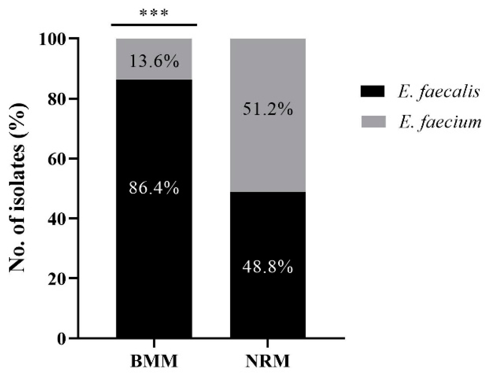
3.1. Identification of E. faecalis and E. faecium from BMM and NRM

In total, 81 and 41 Enterococcus spp. were isolated from BMM and NRM, respectively (Figure 1). According to the MALDI-TOF MS results, E. faecalis (70/81, 86.4%) was identified in BMM at an approximately six-fold higher rate than E. faecium (11/81, 13.6%) (p < 0.0001). In NRM, 21 isolates were identified as E. faecium (21/41, 51.2%), while the remaining isolates were identified as E. faecalis (20/41, 48.8%).

3.2. Detection of Virulence Genes

Among the 70 E. faecalis isolated from BMM samples, 85.7, 71.4, 54.3, and 30.0% harbored the gelE, asa1, esp, and cylA genes, respectively (Table 1). All 70 of the isolates identified as E. faecalis harbored at least one virulence gene and 20 of these isolates harbored all four virulence genes. Conversely, none of the virulence genes were detected in 10 of the 11 isolates identified as E. faecium in BMM, while only one isolate harbored both asa1 and gelE. Furthermore, in the case of NRM, none of the E. faecium harbored any of the virulence genes, whereas E. faecalis harbored asa1 (16/20, 80.0%), gelE (12/20, 60.0%), and esp (5/20, 25.0%) (Table 1). Notably, esp, gelE, and cylA were significantly more abundant in E. faecalis isolated from BMM than in those from NRM (p < 0.0001).

3.3. Biofilm, Gelatinase, and Hemolysin Production

The results of biofilm formation of the 122 Enterococcus spp. are shown in Figure 2A. Biofilm formation criteria were determined as follows: OD ≤ 0.052 = non-biofilm formation, 0.052 < OD ≤ 0.104 = weak biofilm formation, 0.104 < OD ≤ 0.208 = moderate biofilm formation, and OD > 0.208 = strong biofilm formation. All isolates formed a biofilm, with biofilm OD values ranging from 0.076 to 2.927 for BMM and 0.067 to 1.280 for NRM. Among the isolates from BMM, 53/81 (65.4%), 22/81 (27.2%), and 6/81 (7.4%) isolates formed strong, moderate, and weak biofilm, respectively. Among those isolated from NRM, 18/41 (43.9%), 12/41 (29.3%), and 11/41 (26.8%) isolates formed weak, moderate, and strong biofilms, respectively. The isolates from BMM demonstrated a significantly higher rate of strong biofilm formation than isolates from NRM (p < 0.0001).
Furthermore, according to Figure 2B, the isolates from BMM showed significantly higher positivity rates for gelatinase production than isolates from NRM, with 18/81 (22.2%) and 2/41 (4.9%), respectively (p < 0.05). In the case of hemolysin production, 2/81 (2.5%) of isolates from BMM were observed, whereas all isolates from NRM were negative.

3.4. Correlation between Virulence Genes and Corresponding Phenotype

The results of the phenotype of biofilm formation, gelatinase, and hemolysin in vitro and detection of related virulence genes by conventional PCR are presented in Table 2. Simultaneous virulence gene expression and strong biofilm formation were observed in isolates from BMM harboring gelE (43/81, 53.1%), asa1 (37/81, 45.7%), and esp (35/81, 43.2%), whereas simultaneous non-expression was observed in the isolates harboring esp (25/81, 30.9%), asa1 (14/81, 17.3%), and gelE (10/81, 12.3%). However, in the case of isolates from NRM, simultaneous expression of virulence genes and strong biofilm formation were observed in isolates harboring gelE (6/41, 14.6%), asa1 (5/41, 12.2%), and esp (5/41, 12.2%), whereas simultaneous non-expression was observed by the isolates with esp (30/41, 73.2%), gelE (24/41, 58.5%), and asa1 (19/41, 46.3%). Furthermore, the esp and gelE genes were significantly related to strong biofilm formation (p < 0.05).
Comparing the detection results of gelatinase and gelE gene of isolates from BMM, simultaneous expression was observed in 18 isolates (18/81, 22.2%), whereas simultaneous non-expression was observed in 20 isolates (20/81, 24.7%). In the case of isolates from NRM, simultaneous expression was demonstrated in 2 isolates (2/41, 4.9%), whereas simultaneous non-expression was demonstrated in 20 isolates (20/41, 48.8%). In addition, in the case of isolates from BMM, the cylA gene was positive for 21 isolates, with only 2 isolates showing β-hemolysis, whereas all isolates from NRM were negative for both of these. In the present study, all isolates from BMM and NRM positive for gelatinase and hemolysin activity were E. faecalis, and in the case of phenotypic expression, related genes (gelE and cylA) were also detected. Gelatinase production was significantly associated with the gelE gene, whereas hemolysin was not associated with the cylA gene (p < 0.05).

3.5. Antimicrobial Resistance of E. faecalis and E. faecium

The results of the antimicrobial resistance analysis using the disc diffusion method for E. faecalis and E. faecium isolates from BMM and NRM are shown in Table 3. Among the 81 isolates from BMM, the highest resistance rate was shown against TET (48/81, 59.3%), followed by ERY (22/81, 27.2%), C (17/81, 21.0%), RIF (15/81, 18.5%), DOX (4/81, 4.9%), CIP (1/81, 1.2%), and N (1/81, 1.2%). Among MDR isolates from BMM, the antimicrobial resistance patterns of E. faecalis were C-ERY-TET (5/13, 38.5%), C-DOX-ERY-TET (3/13, 23.1%), C-RIF-TET (2/13, 15.4%), C-ERY-RIF-TET (1/13, 7.7%), and ERY-RIF-TET (1/13, 7.7%). Interestingly, one E. faecium isolate from BMM demonstrated a DOX-ERY-RIF-TET (1/13, 7.7%) antimicrobial resistance pattern. However, E. faecalis and E. faecium isolates from NRM showed 14/41 (34.1%), 7/41 (17.1%), and 6/41 (14.6%) resistance to ERY, TET, and RIF, respectively. Interestingly, two MDR isolates from NRM showed strong biofilm-forming ability (OD > 0.208), and the antimicrobial resistance pattern was ERY-RIF-TET for the two isolates.

4. Discussion

A high SCC value (≥200,000 cells/mL) indicates an increase in leukocytes, reflecting an inflammatory reaction to mastitis, and is used as a general indicator of milk safety [20]. In the present study, E. faecalis and E. faecium were isolated from BMM with SCC values of 5–6 log cells/mL. Milk with an SCC value > 5 × 10⁵ cells/mL is unsuitable for human consumption in South Korea [20]. Although the intake of BMM is prohibited, Enterococcus spp. survive in herd bedding and housing, allowing transmission to humans through cross-contamination in milk bulk tanks, cheese vats, curd cutters, and to workers during milking [10,21,22]. Consistent with our study, the majority of Enterococcus spp. isolated from BMM and milk bulk tanks were identified as 91.13% and 90.24% E. faecalis, respectively [3,16]. Therefore, it might be effective and economical to intensively manage E. faecalis to reduce the damage caused by Enterococcus spp. to the dairy industry.
Since virulence genes of Enterococcus spp. can be transferred via plasmids, there is a risk that normal strains lacking genes for virulence factors may acquire these genes through conjugation [11]. Furthermore, the virulence factors of enterococci can enhance enterococcal infections and contribute to potential pathogenesis and disease severity in humans and other animals [11]. The gelE, which was the most abundant virulence gene in the current study, is involved in gelatinase production and can play an important role in the invasion and dissemination of Enterococcus spp. across the intestinal cell layer [14]. Furthermore, gelE has appeared more frequently in clinical isolates than in non-infectious strains [23]. Hemolysin (commonly called cytolysin and encoded by cylA) is an important factor in determining the lethality of endocarditis and both cytolysin and aggregation substance (encoded by asa1) was associated with lethality in 55% of animals when expressed [15]. In a previous study, gelE, esp, and cylA were reportedly present in 77.2, 68.4, and 54.5% of clinical Enterococcus spp. isolates, respectively [24]. In the present study, all virulence genes were more abundant in E. faecalis than in E. faecium; among these genes, gelE, esp, and cylA genes were significantly more abundant in BMM isolates than in NRM isolates, supporting the claim that virulence genes enhance enterococcal infections.
The genotype-phenotype discrepancies were observed for some isolates, and despite genetic evidence, a lack of phenotypic expression indicates the presence of silent genes [25]. Various environmental factors, such as ion concentration, temperature, and osmolarity of the medium, can downregulate genetic expression and this may have a negative effect on gene production [25]. However, silent genes may be activated by the balance of organisms in the intestinal flora, conditions in the gastrointestinal tract, and effects of bacterial synergism, as well as the presence and persistence of large numbers of viable enterococci in vivo [11]. The expression of cylA is greatly affected by the culture conditions and the hemolytic reaction was more common in clinical isolates than in food isolates [11,26]. Based on our findings, even if the phenotype is not expressed in vitro, it may occur in animals or humans, reinforcing the need to monitor the virulence genes of E. faecalis in BMM.
Strong biofilms protect microorganisms from extreme pH, osmolality, nutrient deficiencies, mechanical, and shear forces, as well as antimicrobial agents and host immune cells [27]. Similar to the results of Medeiros et al. (2014) the rate of strong biofilm formation of E. faecalis and E. faecium analyzed in the present study was significantly higher in isolates from BMM than in isolates from NRM [24]. Furthermore, many recent studies have investigated whether virulence genes affect biofilm formation [13,28,29,30]. The esp and gelE showed a significant association with strong biofilm formation in our study; in previous investigations, knockout mutants of these genes formed weak biofilm compared to the wild-type strains [13,31]. The gelatinase (encoded by gelE) modifies bacterial cell surface hydrophobicity to enhance biofilm formation through its ability to cleave substrates at hydrophobic residues [32]. Our study demonstrated that the presence of esp and gelE in isolates from BMM and NRM was significantly associated with strong biofilm formation.
Resistance to TET and ERY has reportedly increased in South Korea isolates from livestock in the last few decades, attributable to the use of these antimicrobial agents to treat clinical and subclinical mastitis [33,34]. Interestingly, C-resistant Enterococcus spp. generally show co-resistance to ERY, likely due to the adjacent location of ERY and C resistance genes in the same plasmid [35]. Although ERY is less marketed to the dairy industry than other antibiotics, ERY resistance likely stems from resistance to other members of the macrolides class, such as TYL, which is used to treat mastitis in South Korea [6,16]. Różańska et al. (2019) showed that among 426 Enterococcus spp. isolates, those resistant to ERY, C, and TYL showed similar resistance rates at 208 (48.83%), 191 (44.84%), and 181 (42.49%), respectively [3]. RIF is usually used in the treatment of MDR infections in combination with other antimicrobial agents to achieve a synergistic effect, which can limit treatment options for MDR pathogens [36]. Consistent with these facts, our results indicated high resistance rates to TET, ERY, C, and RIF, and 7 out of 15 MDR isolates included RIF resistance. The use of antibiotics in dairy cows presumably resulted in the detection of significantly more antibiotic-resistant Enterococcus spp. in BMM, as the predominance of antibiotic-resistant Enterococcus spp. results in bovine mastitis.
Considering its presence in the dairy industry, the serious human infection, and its virulence factors that can spread through cross-contamination, the findings of the current study support the importance of vigilant monitoring of E. faecalis in dairy products. Further studies using pulsed field gel electrophoresis or other subtyping tools, such as whole genome sequencing, are needed to confirm that the virulence genes and antibiotic resistance of isolates from raw milk are consistent with those found in farm environments and dairy products.

5. Conclusions

Our results suggest that isolates from BMM have higher positivity rates for virulence genes, exhibit stronger biofilm, gelatinase, and hemolysin formation, and have higher rates of antimicrobial resistance than those isolated from NRM. Furthermore, E. faecalis was a predominant species among Enterococcus spp. in BMM, showing high virulence characteristics. Taken together, controlling and monitoring E. faecalis harboring esp, gelE, and cylA genes could be the best intervention strategy for eliminating bovine mastitis, leading to improved public health.

Supplementary Materials

The following supporting information can be downloaded at: https://www.mdpi.com/article/10.3390/ani12111407/s1, Table S1: Polymerase chain reaction primers and product sizes for the detection of Enterococcus faecalis and Enterococcus faecium virulence genes.

Author Contributions

Conceptualization, H.-J.K. (Hyeon-Jin Kim) and H.-Y.Y.; Data curation, H.-J.K. (Hyeon-Jin Kim); Formal analysis, H.-J.K. (Hyeon-Jin Kim) and H.-Y.Y.; Funding acquisition, K.-H.S.; Investigation, H.-J.K. (Hyeon-Jin Kim); Methodology, H.-J.K. (Hyeon-Jin Kim); Project administration, K.-H.S.; Resource, H.-J.K. (Hye-Jeong Kang) and J.-S.M.; Supervision, K.-H.S.; Writing—original draft, H.-J.K. (Hyeon-Jin Kim) and H.-Y.Y.; Writing—review and editing, H.-J.K. (Hye-Jeong Kang), J.-S.M., Y.-S.J., K.-Y.S., and K.-H.S. All authors have read and agreed to the published version of the manuscript.

Funding

This paper was supported by the Konkuk University Researcher Fund in 2020. Additionally, this work was supported by the National Research Foundation of Korea (NRF) grant funded by the Korea government (MSIT) (2021R1H1A2092653).

Institutional Review Board Statement

Not applicable.

Informed Consent Statement

Not applicable.

Data Availability Statement

Not applicable.

Acknowledgments

The authors would like to thank Dongryeoul Bae for assistance with the techniques.

Conflicts of Interest

The authors declare no conflict of interest. The funders had no role in the design of the study; in the collection, analyses, or interpretation of data; in the writing of the manuscript, or in the decision to publish the results.

References

  1. Hogeveen, H.; Huijps, K.; Lam, T.J.G.M. Economic aspects of mastitis: New developments. N. Z. Vet. J. 2011, 59, 16–23. [Google Scholar] [CrossRef] [PubMed]
  2. Riekerink, R.G.O.; Barkema, H.W.; Veenstra, S.; Poole, D.E.; Dingwell, R.T.; Keefe, G.P. Prevalence of contagious mastitis pathogens in bulk tank milk in Prince Edward Island. Can. Vet. J. 2006, 47, 567. [Google Scholar]
  3. Różańska, H.; Lewtak-Piłat, A.; Kubajka, M.; Weiner, M. Occurrence of enterococci in mastitic cow’s milk and their antimicrobial resistance. J. Vet. Res. 2019, 63, 93–97. [Google Scholar] [CrossRef] [PubMed] [Green Version]
  4. Kiruthiga, A.; Padmavathy, K.; Shabana, P.; Naveenkumar, V.; Gnanadesikan, S.; Malaiyan, J. Improved detection of esp, hyl, asa1, gelE, cylA virulence genes among clinical isolates of Enterococci. BMC Res. Notes. 2020, 13, 170. [Google Scholar] [CrossRef] [PubMed] [Green Version]
  5. Franz, C.M.; Holzapfel, W.H.; Stiles, M.E. Enterococci at the crossroads of food safety? Int. J. Food Microbiol. 1999, 47, 1–24. [Google Scholar] [CrossRef]
  6. Nam, H.M.; Lim, S.K.; Kang, H.M.; Kim, J.M.; Moon, J.S.; Jang, K.C.; Jung, S.C. Prevalence and antimicrobial susceptibility of gram-negative bacteria isolated from bovine mastitis between 2003 and 2008 in Korea. Int. J. Dairy Sci. 2009, 92, 2020–2026. [Google Scholar] [CrossRef] [Green Version]
  7. Golob, M.; Pate, M.; Kušar, D.; Dermota, U.; Avberšek, J.; Papić, B.; Zdovc, I. Antimicrobial resistance and virulence genes in Enterococcus faecium and Enterococcus faecalis from humans and retail red meat. Biomed. Res. Int. 2019, 2019, 2815279. [Google Scholar] [CrossRef] [Green Version]
  8. Kang, I.B.; Kim, D.H.; Chon, J.W.; Seo, K.H. Effect of microbial control measures on farmstead cheesemaking and antimicrobial resistance of Staphylococcus aureus and Enterococcus spp. isolates. J. Food Saf. 2018, 38, e12432. [Google Scholar] [CrossRef]
  9. Murray, B.E.; Miller, W.R. Treatment of Enterococcal Infections; UpToDate: Waltham, MA, USA, 2018; Available online: https://www.uptodate.com/contents/treatment-of-enterococcal-infections (accessed on 31 May 2021).
  10. Kim, S.H.; Kim, D.H.; Lim, H.W.; Seo, K.H. High prevalence of non-faecalis and non-faecium Enterococcus spp. in farmstead cheesehouse and their applicability as hygiene indicators. LWT 2020, 126, 109271. [Google Scholar] [CrossRef]
  11. Eaton, T.J.; Gasson, M.J. Molecular screening of Enterococcus virulence determinants and potential for genetic exchange between food and medical isolates. Appl. Environ. Microbiol. 2001, 67, 1628–1635. [Google Scholar] [CrossRef] [Green Version]
  12. Mah, T.F. Biofilm-specific antibiotic resistance. Future Microbiol. 2012, 7, 1061–1072. [Google Scholar] [CrossRef] [PubMed] [Green Version]
  13. Mohamed, J.A.; Huang, D.B. Biofilm formation by enterococci. J. Med. Microbiol. 2007, 56, 1581–1588. [Google Scholar] [CrossRef] [PubMed] [Green Version]
  14. Zeng, J.; Teng, F.; Murray, B.E. Gelatinase is important for translocation of Enterococcus faecalis across polarized human enterocyte-like T84 cells. Infect. Immun. 2005, 73, 1606–1612. [Google Scholar] [CrossRef] [PubMed] [Green Version]
  15. Coburn, P.S.; Gilmore, M.S. The Enterococcus faecalis cytolysin: A novel toxin active against eukaryotic and prokaryotic cells. Cell. Microbiol. 2003, 5, 661–669. [Google Scholar] [CrossRef] [PubMed]
  16. Yoon, S.; Lee, Y.J. Molecular Characteristics of Enterococcus faecalis and Enterococcus faecium from Bulk Tank Milk in Korea. Animals 2021, 11, 661. [Google Scholar] [CrossRef] [PubMed]
  17. Vankerckhoven, V.; Van Autgaerden, T.; Vael, C.; Lammens, C.; Chapelle, S.; Rossi, R.; Goossens, H. Development of a multiplex PCR for the detection of asa1, gelE, cylA, esp, and hyl genes in enterococci and survey for virulence determinants among European hospital isolates of Enterococcus faecium. J. Clin. Microbiol. 2004, 42, 4473–4479. [Google Scholar] [CrossRef] [Green Version]
  18. Stepanović, S.; Vuković, D.; Hola, V.; Bonaventura, G.D.; Djukić, S.; Ćirković, I.; Ruzicka, F. Quantification of biofilm in microtiter plates: Overview of testing conditions and practical recommendations for assessment of biofilm production by staphylococci. Apmis 2007, 115, 891–899. [Google Scholar] [CrossRef]
  19. Clinical & Laboratory Standards Institute (CLSI). Performance Standards for Antimicrobial Susceptibility Testing, 30th ed.; CLSI Supplement M100-S3011; CLSI: Wayne, PA, USA, 2020. [Google Scholar]
  20. Yun, M.J.; Yoon, S.; Lee, Y.J. Monitoring and characteristics of major mastitis pathogens from bulk tank milk in Korea. Animals 2020, 10, 1562. [Google Scholar] [CrossRef]
  21. Lee, S.H.I.; Camargo, C.H.; Gonçalves, J.L.; Cruz, A.G.; Sartori, B.T.; Machado, M.B.; Oliveira, C.A.F. Characterization of Staphylococcus aureus isolates in milk and the milking environment from small-scale dairy farms of São Paulo, Brazil, using pulsed-field gel electrophoresis. J. Dairy Sci. 2012, 95, 7377–7383. [Google Scholar] [CrossRef] [Green Version]
  22. Cheng, W.N.; Han, S.G. Bovine mastitis: Risk factors, therapeutic strategies, and alternative treatments. Asian-Australas. J. Anim. Sci. 2020, 33, 1699. [Google Scholar] [CrossRef]
  23. Coque, T.M.; Patterson, J.E.; Steckelberg, J.M.; Murray, B.E. Incidence of hemolysin, gelatinase, and aggregation substance among enterococci isolated from patients with endocarditis and other infections and from feces of hospitalized and community-based persons. J. Infect. Dis. 1995, 171, 1223–1229. [Google Scholar] [CrossRef] [PubMed]
  24. Medeiros, A.W.; Pereira, R.I.; Oliveira, D.V.D.; Martins, P.D.; d’Azevedo, P.A.; Van der Sand, S.; Frazzon, A.P.G. Molecular detection of virulence factors among food and clinical Enterococcus faecalis strains in South Brazil. Braz. J. Microbiol. 2014, 45, 327–332. [Google Scholar] [CrossRef] [PubMed] [Green Version]
  25. Maasjost, J.; Lüschow, D.; Kleine, A.; Hafez, H.M.; Mühldorfer, K. Presence of virulence genes in Enterococcus species isolated from meat turkeys in Germany does not correlate with chicken embryo lethality. Biomed. Res. Int. 2019, 2019, 6147695. [Google Scholar] [CrossRef] [PubMed] [Green Version]
  26. Van Tyne, D.; Martin, M.J.; Gilmore, M.S. Structure, function, and biology of the Enterococcus faecalis cytolysin. Toxins 2013, 5, 895–911. [Google Scholar] [CrossRef] [Green Version]
  27. Sharma, D.; Misba, L.; Khan, A.U. Antibiotics versus biofilm: An emerging battleground in microbial communities. Antimicrob. Resist. Infect. Control. 2019, 8, 76. [Google Scholar] [CrossRef]
  28. Kafil, H.S.; Mobarez, A.M. Assessment of biofilm formation by enterococci isolates from urinary tract infections with different virulence profiles. J. King Saud Univ. Sci. 2015, 27, 312–317. [Google Scholar] [CrossRef] [Green Version]
  29. Cui, P.; Feng, L.; Zhang, L.; He, J.; An, T.; Fu, X.; Yang, X. Antimicrobial Resistance, Virulence Genes, and Biofilm Formation Capacity Among Enterococcus species From Yaks in Aba Tibetan Autonomous Prefecture, China. Front. Microbiol. 2020, 11, 1250. [Google Scholar] [CrossRef]
  30. Soares, R.O.; Fedi, A.C.; Reiter, K.C.; Caierão, J.; d’Azevedo, P.A. Correlation between biofilm formation and gelE, esp, and agg genes in Enterococcus spp. clinical isolates. Virulence 2014, 5, 634–637. [Google Scholar] [CrossRef] [Green Version]
  31. Heikens, E.; Bonten, M.J.; Willems, R.J. Enterococcal surface protein Esp is important for biofilm formation of Enterococcus faecium E1162. J. Bacteriol. Res. 2007, 189, 8233–8240. [Google Scholar] [CrossRef] [Green Version]
  32. Wang, L.; Dong, M.; Zheng, J.; Song, Q.; Yin, W.; Li, J.; Niu, W. Relationship of biofilm formation and gelE gene expression in Enterococcus faecalis recovered from root canals in patients requiring endodontic retreatment. J. Endod. 2011, 37, 631–636. [Google Scholar] [CrossRef]
  33. Kim, Y.B.; Seo, K.W.; Shim, J.B.; hyun Son, S.; Noh, E.B.; Lee, Y.J. Molecular characterization of antimicrobial-resistant Enterococcus faecalis and Enterococcus faecium isolated from layer parent stock. Poult. Sci. 2019, 98, 5892–5899. [Google Scholar] [CrossRef] [PubMed]
  34. Kim, H.J.; Koo, M. Occurrence, Antimicrobial Resistance and Molecular Diversity of Enterococcus faecium in Processed Pork Meat Products in Korea. Foods 2020, 9, 1283. [Google Scholar] [CrossRef] [PubMed]
  35. Wang, Y.; Lv, Y.; Cai, J.; Schwarz, S.; Cui, L.; Hu, Z.; Shen, J. A novel gene, optrA, that confers transferable resistance to oxazolidinones and phenicols and its presence in Enterococcus faecalis and Enterococcus faecium of human and animal origin. J. Antimicrob. Chemother. 2015, 70, 2182–2190. [Google Scholar] [CrossRef] [PubMed] [Green Version]
  36. Pankey, G.; Ashcraft, D.; Patel, N. In vitro synergy of daptomycin plus rifampin against Enterococcus faecium resistant to both linezolid and vancomycin. J. Antimicrob. Chemother. 2005, 49, 5166–5168. [Google Scholar] [CrossRef] [Green Version]
Figure 1. Comparison of the prevalence of Enterococcus faecalis and Enterococcus faecium isolates in bovine mastitis milk (BMM; n = 81) and bovine normal raw milk (NRM; n = 41). *** indicates significant difference in the prevalence rate of E. faecalis and E. faecium isolates in BMM (p < 0.0001; Pearson’s chi-square test). BMM: bovine mastitis milk, NRM: bovine normal raw milk.
Animals 12 01407 g001
Figure 2. Distribution of Enterococcus faecalis and Enterococcus faecium isolated from bovine mastitis milk (BMM) and bovine normal raw milk (NRM) according to their virulence factors. (A) Biofilm formation ability (OD values) and (B) gelatinase and hemolysin production. Biofilm formation criteria comprised non-biofilm formation (optical density (OD) ≤ 0.052), weak biofilm formation (0.052 < OD ≤ 0.104), moderate biofilm formation (0.104 < OD ≤ 0.208), and strong biofilm formation (OD > 0.208). BMM: bovine mastitis milk, NRM: bovine normal raw milk, W: weak, M: moderate, S: strong. *** indicates significant difference in strong biofilm formation between the BMM (S) and the NRM (S) group (p < 0.0001; Pearson’s chi-square test). * indicates significant difference in gelatinase production between the BMM and the NRM groups (p < 0.05; Pearson’s chi-square test).
Animals 12 01407 g002
Table 1. Distribution of virulence genes among Enterococcus faecalis isolates from bovine mastitis milk (BMM) and bovine normal raw milk (NRM).
Milk TypeNo. of IsolatesNo of Genes Encoding Virulence Factors (%)
esp ***asa1gelE ***cylA ***
BMM7038 (54.3%)50 (71.4%)60 (85.7%)21 (30.0%)
NRM205 (25.0%)16 (80.0%)12 (60.0%)0
*** indicates significant difference in virulence genes between bovine mastitis milk and normal raw milk (p < 0.0001; Pearson’s chi-square test). BMM: bovine mastitis milk, NRM: bovine normal raw milk.
Table 2. Correlation between virulence phenotype and genotype of Enterococcus faecalis and Enterococcus faecium isolated from bovine mastitis milk (BMM) and bovine normal raw milk (NRM).
Virulence FactorEncoding GenePhenotype and Genotype of Isolates from BMM and NRM
P+G+P−G+P+G−P−G−
BMMNRMBMMNRMBMMNRMBMMNRM
Strong biofilm formation
(=OD > 0.208)
esp *355301862530
asa137514111661419
gelE *4361861051024
GelatinasegelE *1824310002029
HemolysincylA20190006041
* indicates significant associations between virulence factors and related encoding genes (p < 0.05; Pearson’s chi-square test). BMM: bovine mastitis milk, NRM: bovine normal raw milk, P+: phenotypically expressed, P−: phenotypically not expressed, G+: encoding gene detected, G−: encoding gene not detected.
Table 3. Antimicrobial resistance of Enterococcus faecalis and Enterococcus faecium isolates from bovine mastitis milk (BMM) and bovine normal raw milk (NRM).
Antimicrobial
Agents
No (%) of Antimicrobial Resistance Isolates from BMM and NRM
BMMNRM
E. faecalis
(n = 70)
E. faecium
(n = 11)
Total
(n = 81)
E. faecalis
(n = 20)
E. faecium
(n = 21)
Total
(n = 41)
AMP000000
C16 (22.9%)1 (9.1%)17 (21.0%)000
CIP01 (9.1%)1 (1.2%)000
DOX3 (4.3%)1 (9.1%)4 (4.9%)000
TET45 (64.3%)3 (27.3%)48 (59.3%)4 (20%)3 (14.3%)7 (17.1%)
ERY19 (27.1%)3 (27.3%)22 (27.2%)1 (5%)13 (61.9%)14 (34.1%)
N01 (9.1%)1 (1.2%)000
RIF11 (15.7%)4 (36.4%)15 (18.5%)1 (5%)5 (23.8%)6 (14.6%)
VAN000000
MDR12 (17.1%)1 (9.1%)13 (16.0%)02 (9.5%)2 (4.9%)
BMM: bovine mastitis milk, NRM: bovine normal raw milk, AMP: ampicillin, C: chloramphenicol, CIP: ciprofloxacin, DOX: doxycycline, TET: tetracycline, ERY: erythromycin, N: nitrofurantoin, RIF: rifampicin, VAN: vancomycin, MDR: multidrug resistance.
Publisher’s Note: MDPI stays neutral with regard to jurisdictional claims in published maps and institutional affiliations.

