
Relationships between Body Condition Score (BCS), FAMACHA©-Score and Haematological Parameters in Alpacas (Vicugna pacos), and Llamas (Lama glama) Presented at the Veterinary Clinic

 , ,
, ,
Abstract
:Simple Summary
Abstract
1. Introduction
2. Material and Methods
2.1. Data Collection
2.2. Inclusion Criteria
2.3. Collected Parameters from the Animals
2.3.1. Basic Data about the Animal
2.3.2. Clinical Scores
BCS
FS
2.3.3. Haematological Parameters
PCV [L/L]
Haemoglobin (Hb) [g/L]
Total Leucocytes/White Blood Count (WBC) [G/L]
Lymphocytes, Segmented Neutrophils, Band Neutrophils, Eosinophils, Basophils, Monocytes, Normoblasts, all [%]
2.4. Statistical Analysis
3. Results
3.1. Population
- All alpacas (n = 259)
- All llamas (n = 41)
- Alpacas, male, cria (n = 26)
- Alpacas, male, adult (n = 87)
- Alpacas, female, cria (n = 40)
- Alpacas, female, adult (n = 106)
- Llamas, male, cria (n = 2)
- Llamas, male, adult (n = 21)
- Llamas, female, cria (n = 2)
- Llamas, female, adult (n = 16)
3.2. Clinical Parameters
3.2.1. BCS
3.2.2. FS
3.3. Haematological Parameters
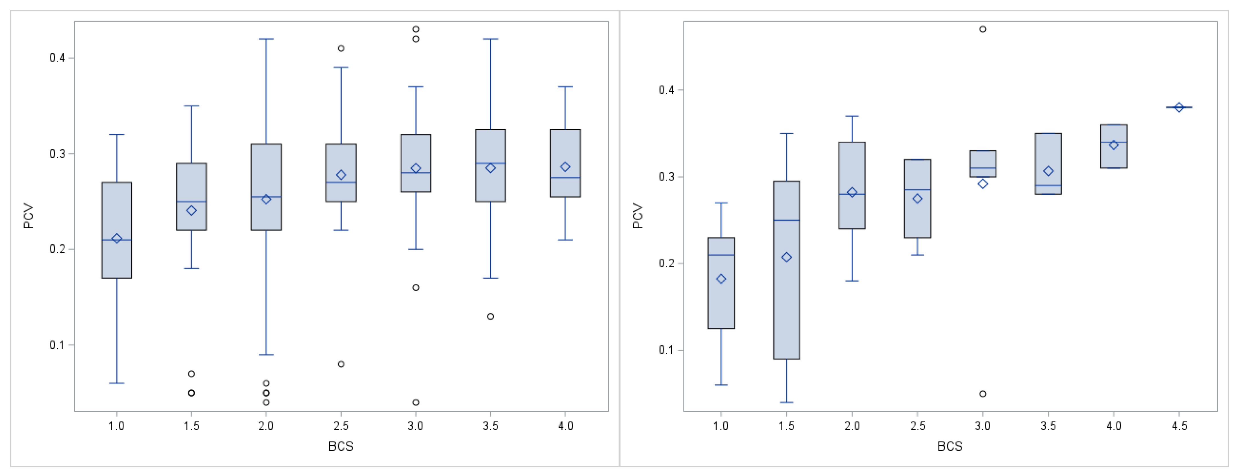
3.3.1. PCV
3.3.2. Hb
3.3.3. WBC
3.3.4. Lymphocytes
3.3.5. Neutrophils
3.3.6. Eosinophils
3.3.7. Basophils
3.3.8. Monocytes
3.3.9. Normoblasts
4. Discussion
5. Limitations
6. Conclusions
Supplementary Materials
Author Contributions
Funding
Institutional Review Board Statement
Data Availability Statement
Acknowledgments
Conflicts of Interest
References
- Neubert, S.; von Altrock, A.; Wendt, M.; Wagener, M.G. Llama and Alpaca Management in Germany-Results of an Online Survey among Owners on Farm Structure, Health Problems and Self-Reflection. Animals 2021, 11, 102. [Google Scholar] [CrossRef]
- Hengrave Burri, I.; Martig, J.; Sager, H.; Liesegang, A.; Meylan, M. South American Camelids in Switzerland. I. Population, Management and Health Problems. Schweiz. Arch. Für Tierheilkd. 2005, 147, 325–334. [Google Scholar] [CrossRef] [Green Version]
- Davis, R.; Keeble, E.; Wright, A.; Morgan, K. South American Camelids in the United Kingdom: Population Statistics, Mortality Rates and Causes of Death. Vet. Rec. 1998, 142, 162–166. [Google Scholar] [CrossRef]
- Van Saun, R.J. Nutritional Requirements and Assessing Nutritional Status in Camelids. Vet. Clin. North. Am. Food Anim. Pract. 2009, 25, 265–279. [Google Scholar] [CrossRef]
- Wagener, M.G.; Grimm, L.M.; Ganter, M. Anaemia in a Llama (Lama glama): Treatment, Regeneration and Differential Diagnoses. Vet. Rec. Case Rep. 2018, 6, e000638. [Google Scholar] [CrossRef]
- Jefferies, B. Body Condition Scoring and Its Use in Management. Tasman. J. Agric. 1961, 32, 19–21. [Google Scholar]
- Ferguson, J.D.; Galligan, D.T.; Thomsen, N. Principal Descriptors of Body Condition Score in Holstein Cows. J. Dairy Sci. 1994, 77, 2695–2703. [Google Scholar] [CrossRef]
- Edmonson, A.; Lean, I.; Weaver, L.; Farver, T.; Webster, G. A Body Condition Scoring Chart for Holstein Dairy Cows. J. Dairy Sci. 1989, 72, 68–78. [Google Scholar] [CrossRef]
- Johnson, L.W. Llama Nutrition. Vet. Clin. North Am. Food Anim. Pract. 1994, 10, 187–201. [Google Scholar] [CrossRef]
- Bath, G.F.; Malan, F.; Van Wyk, J. The “FAMACHA” Ovine Anaemia Guide to assist with the control of haemonchosis. In Proceedings of the 7th Annual Congress of the Livestock Health and Production Group of the South African Veterinary Association, Port Elizabeth, South Africa, 5–7 June 1996; p. 5. [Google Scholar]
- Storey, B.E.; Williamson, L.H.; Howell, S.B.; Terrill, T.H.; Berghaus, R.; Vidyashankar, A.N.; Kaplan, R.M. Validation of the FAMACHA© System in South American camelids. Vet. Parasitol. 2017, 243, 85–91. [Google Scholar] [CrossRef] [PubMed]
- Hilton, C.; Pugh, D.; Wright, J.; Waldridge, B.; Simpkins, S.; Heath, A. How To Determine and When to Use Body Weight Estimates and Condition Scores in Llamas. Vet. Med. 1998, 93, 1015–1018. [Google Scholar]
- Bromage, G. Feeding and Nutrition. In Llamas and Alpacas: A Guide to Management; The Crowood Press Ltd.: Marlborough, UK, 2006; pp. 34–45. [Google Scholar]
- Duncanson, G.R. Nutrition and Metabolic Diseases. In Veterinary Treatment of Llamas and Alpacas; CABI: Oxfordshire, UK, 2012; pp. 13–21. [Google Scholar]
- Wagener, M.G.; Ganter, M. Body Condition Scoring in South American Camelids. Der Prakt. Tierarzt 2020, 101, 684–696. [Google Scholar]
- Gauly, M. Fütterung. In Neuweltkameliden-Haltung, Zucht, Erkrankungen, 4th ed.; Thieme: Stuttgart, Germany, 2018; pp. 46–67. [Google Scholar]
- Van Saun, R.J. Nutritional Diseases of Llamas and Alpacas. Vet. Clin. North. Am. Food Anim. Pract. 2009, 25, 797–810. [Google Scholar] [CrossRef]
- Baumgartner, W.; Wittek, T.; Gauly, M. Allgemeiner Klinischer Untersuchungsgang. In Klinische Propädeutik der Haus- und Heimtiere, 9th ed.; Enke: Stuttgart, Germany, 2018; pp. 50–68. [Google Scholar]
- Proost, K.; Pardon, B.; Pollaris, E.; Flahou, T.; Vlaminck, L. Dental Disease in Alpacas. Part 2: Risk Factors Associated with Diastemata, Periodontitis, Occlusal Pulp Exposure, Wear Abnor-Malities, and Malpositioned Teeth. J. Vet. Intern. Med. 2020, 34, 1039–1046. [Google Scholar] [CrossRef]
- Frezzato, G.; Stelletta, C.; Murillo, C.E.P.; Simonato, G.; Cassini, R. Parasitological Survey To Address Major Risk Factors Threatening Alpacas in Andean Extensive Farms (Arequipa, Peru). J. Vet. Med Sci. 2020, 82, 1655–1661. [Google Scholar] [CrossRef]
- Edwards, E.E.; Garner, B.C.; Williamson, L.H.; Storey, B.E.; Sakamoto, K. Pathology of Haemonchus contortus in New World Camelids in the Southeastern United States: A Retrospective Review. J. Vet. Diagn. Investig. 2016, 28, 105–109. [Google Scholar] [CrossRef] [Green Version]
- Crosse, P.; Ayling, R.; Whitehead, C.; Szladovits, B.; English, K.; Bradley, D.; Solano-Gallego, L. First Detection of ’Candidatus Mycoplasma Haemolamae’infection in Alpacas in England. Vet. Rec. 2012, 171, 71–75. [Google Scholar] [CrossRef]
- Kaufmann, C.; Meli, M.; Hofmann-Lehmann, R.; Riond, B.; Zanolari, P. Epidemiology of’Candidatus Mycoplasma Haemolamae’infection in South American Camelids in Central Europe. J. Camelid Sci. 2011, 4, 23–29. [Google Scholar]
- Smith, B.B.; Pearson, E.G.; Timm, K.I. Third Compartment Ulcers in the Llama. Vet. Clin. North. Am. Food Anim. Pract. 1994, 10, 319–330. [Google Scholar] [CrossRef]
- Foster, A.; Bidewell, C.; Barnett, J.; Sayers, R. Haematology and Biochemistry in Alpacas and Llamas. In Practice 2009, 31, 276–281. [Google Scholar] [CrossRef]
- Andrews, A.; Cox, A. Suspected Nutritional Deficiency Causing Anaemia in Llamas (Lama glama). Vet. Rec. 1997, 140, 153–154. [Google Scholar] [CrossRef] [PubMed]
- Morin, D.; Garry, F.; Weiser, M.; Fettman, M.; Johnson, L. Hematologic Features of Iron Deficiency Anemia in Llamas. Vet. Pathol. 1992, 29, 400–404. [Google Scholar] [CrossRef] [PubMed]
- Luethy, D.; Stefanovski, D.; Salber, R.; Sweeney, R. Prediction of Packed Cell Volume after Whole Blood Transfusion in Small Ruminants and South American Camelids: 80 Cases (2006–2016). J. Vet. Intern. Med. 2017, 31, 1900–1904. [Google Scholar] [CrossRef] [PubMed] [Green Version]
- Van Wyk, J.A.; Bath, G.F. The FAMACHA System for Managing Haemonchosis in Sheep and Goats by Clinically Identifying Individual Animals for Treatment. Vet. Res. 2002, 33, 509–529. [Google Scholar] [CrossRef] [Green Version]
- Vilela, V.L.R.; Feitosa, T.F.; Linhares, E.F.; Athayde, A.C.R.; Molento, M.B.; Azevedo, S.S. FAMACHA© Method as an Auxiliary Strategy in the Control of Gastrointestinal Helminthiasis of Dairy Goats Under Semiarid Conditions of Northeastern Brazil. Vet. Parasitol. 2012, 190, 281–284. [Google Scholar] [CrossRef] [Green Version]
- Gauly, M.; Schackert, M.; Erhardt, G. Use of FAMACHA© Scoring System as a Diagnostic Aid for the Registration of Distinguishing Marks in the Breeding Program for Lambs Exposed to an Experimental Haemonchus contortus Infection. Dtsch. Tierarztl. Wochenschr. 2004, 111, 430–433. [Google Scholar]
- Koopmann, R.; Holst, C.; Epe, C. Experiences with the FAMACHA©-Eye-Colour-Chart for Identifying Sheep and Goats for Targeted Anthelmintic Treatment. Berl. Und Müncher Tierärztliche Wochenschr. 2006, 119, 436–442. [Google Scholar]
- Papadopoulos, E.; Gallidis, E.; Ptochos, S.; Fthenakis, G. Evaluation of the FAMACHA© System for Targeted Selective Anthelmintic Treatments for Potential Use in Small Ruminants in Greece. Small Rumin. Res. 2013, 110, 124–127. [Google Scholar] [CrossRef]
- Di Loria, A.; Veneziano, V.; Piantedosi, D.; Rinaldi, L.; Cortese, L.; Mezzino, L.; Cringoli, G.; Ciaramella, P. Evaluation of the FAMACHA System for Detecting the Severity of Anaemia in Sheep from Southern Italy. Vet. Parasitol. 2009, 161, 53–59. [Google Scholar] [CrossRef]
- Reynecke, D.P.; Van Wyk, J.A.; Gummow, B.; Dorny, P.; Boomker, J. Validation of the FAMACHA© Eye Colour Chart Using Sensitivity/Specificity Analysis on Two South African Sheep Farms. Vet. Parasitol. 2011, 177, 203–211. [Google Scholar] [CrossRef] [Green Version]
- Scheuerle, M.; Mahling, M.; Muntwyler, J.; Pfister, K. The Accuracy of the FAMACHA©-Method in Detecting Anaemia and Haemonchosis in Goat Flocks in Switzerland Under Field Conditions. Vet. Parasitol. 2010, 170, 71–77. [Google Scholar] [CrossRef]
- Kaplan, R.; Burke, J.; Terrill, T.; Miller, J.; Getz, W.; Mobini, S.; Valencia, E.; Williams, M.; Williamson, L.; Larsen, M. Validation of the FAMACHA© Eye Color Chart for Detecting Clinical Anemia in Sheep and Goats on Farms in the Southern United States. Vet. Parasitol. 2004, 123, 105–120. [Google Scholar] [CrossRef]
- Olah, S.; van Wyk, J.A.; Wall, R.; Morgan, E.R. FAMACHA©: A Potential Tool for Targeted Selective Treatment of Chronic Fasciolosis in Sheep. Vet. Parasitol. 2015, 212, 188–192. [Google Scholar] [CrossRef]
- Grace, D.; Himstedt, H.; Sidibe, I.; Randolph, T.; Clausen, P.-H. Comparing FAMACHA© Eye Color Chart and Hemoglobin Color Scale Tests for Detecting Anemia and Improving Treatment of Bovine Trypanosomosis in West Africa. Vet. Parasitol. 2007, 147, 26–39. [Google Scholar] [CrossRef] [PubMed]
- Viesselmann, L.C.; Videla, R.; Schaefer, J.; Chapman, A.; Wyrosdick, H.; Schaefer, D.M. Mycoplasma haemolamae and Intestinal Parasite Relationships With Erythrocyte Variables in Clinically Healthy Alpacas and Llamas. J. Vet. Intern. Med. 2019, 33, 2336–2342. [Google Scholar] [CrossRef] [PubMed]
- Cocquyt, C.M.; Van Amstel, S.; Cox, S.; Rohrbach, B.; Martín-Jiménez, T. Pharmacokinetics of Moxidectin in Alpacas Following Administration of an Oral or Subcutaneous Formulation. Res. Vet. Sci. 2016, 105, 160–164. [Google Scholar] [CrossRef] [PubMed]
- Galvan, N.; Middleton, J.R.; Nagy, D.W.; Schultz, L.G.; Schaeffer, J.W. Anthelmintic Resistance in a Herd of Alpacas (Vicugna Pacos). Can. Vet. J. 2012, 53, 1310. [Google Scholar] [PubMed]
- Viesselmann, L.C.; Videla, R.; Flatland, B. Verification of the Heska Element Point-of-Care Blood Gas Instrument for Use with Venous Blood From Alpacas and Llamas, With Determination of Reference Intervals. Vet. Clin. Pathol. 2018, 47, 435. [Google Scholar] [CrossRef]
- Gauly, M.; Maier, H.; Trah, M. Blood Collection, Haematological Values, Biochemical Parameters of New World Camelids. Tierärztliche Umsch. 1998, 53, 751–754. [Google Scholar]
- Wagener, M.G.; Grossmann, T.; Stöter, M.; Ganter, M. Hematological Diagnostics in Llamas and Alpacas. Der Prakt. Tierarzt 2018, 99, 481–493. [Google Scholar]
- Hengrave Burri, I.; Tschudi, P.; Martig, J.; Liesegang, A.; Meylan, M. South American Camelids in Switzerland. II. Reference Values for Blood Parameters. Schweiz. Arch. Für Tierheilkd. 2005, 147, 335–343. [Google Scholar] [CrossRef] [PubMed]
- Maia, D.; Rosalinski-Moraes, F.; Van Wyk, J.A.; Weber, S.; Sotomaior, C.S. Assessment of a Hands-On Method for FAMACHA© System Training. Vet. Parasitol. 2014, 200, 165–171. [Google Scholar] [CrossRef] [PubMed] [Green Version]
- Rafia, S.; Taghipour-Bazargani, T.; Khaki, Z.; Bokaie, S.; Tabrizi, S.S. Effect of Body Condition Score on Dynamics of Hemogram in Periparturient Holstein Cows. Comp. Clin. Pathol. 2012, 21, 933–943. [Google Scholar] [CrossRef]
- Torres-Chable, O.M.; García-Herrera, R.A.; González-Garduño, R.; Ojeda-Robertos, N.F.; Peralta-Torres, J.A.; Chay-Canul, A.J. Relationships Among Body Condition Score, FAMACHA© Score and Haematological Parameters in Pelibuey Ewes. Trop. Anim. Health Prod. 2020, 52, 3403–3408. [Google Scholar] [CrossRef]
- Munoz, A.; Riber, C.; Trigo, P.; Castejon, F. Hematology and Clinical Pathology Data in Chronically Starved Horses. J. Equine Vet. Sci. 2010, 30, 581–589. [Google Scholar] [CrossRef]
- Weiser, M.; Fettman, M.; Van Houten, D.; Johnson, L.; Garry, F. Characterization of Erythrocytic Indices and Serum Iron Values in Healthy Llamas. Am. J. Vet. Res. 1992, 53, 1776–1779. [Google Scholar]
- Hajduk, P. Haematological Reference Values for Alpacas. Aust. Vet. J. 1992, 69, 89–90. [Google Scholar] [CrossRef]
- Fowler, M.; Zinkl, J. Reference Ranges for Hematologic and Serum Biochemical Values in Llamas (Lama glama). Am. J. Vet. Res. 1989, 50, 2049–2053. [Google Scholar]
- Al-Izzi, S.; Abdouslam, O.; Al-Bassam, L.; Azwai, S. Haematological Parameters in Clinically Normal Llamas (Lama glama). Prax. Vet. 2004, 52, 225–232. [Google Scholar]
- Husakova, T.; Pavlata, L.; Pechova, A.; Tichy, L.; Hauptmanova, K. The Influence of Sex, Age and Season on the Haematological Profile of Alpacas (Vicugna pacos) in Central Europe. Vet. Med. 2015, 60, 407–414. [Google Scholar] [CrossRef] [Green Version]
- Goggs, R. Normoblasts: Not Always Normal. Vet. Rec. 2014, 175, 506–507. [Google Scholar] [CrossRef] [PubMed]
- Wagener, M.G.; Puff, C.; Stöter, M.; Schwennen, C.; Ganter, M. Regenerative Anaemia in Alpacas (Vicugna pacos) Can Lead to a Wrong Diagnosis of Leucocytosis. Vet. Rec. Case Rep. 2020, 8, e001257. [Google Scholar] [CrossRef]
- Newhall, D.A.; Oliver, R.; Lugthart, S. Anaemia: A Disease or Symptom. Neth. J. Med. 2020, 78, 104–110. [Google Scholar]


| Parameter | Alpaca vs. Llama | Juvenile vs. Adult | Anaemia vs. without Anaemia | |
|---|---|---|---|---|
| All | Alpacas | Alpacas | Llamas | |
| Bodyweight (kg) | *** | *** | *** | *** |
| BCS | n.s. | ** | *** | *** |
| FS | n.s. | * | ** | *** |
| PCV (L/L) | n.s. | n.s. | *** | *** |
| Hb (g/L) | n.s. | n.s. | *** | *** |
| WBC (G/L) | n.s. | n.s. | n.s. | n.s. |
| Lymphocytes (%) | ** | *** | n.s. | n.s. |
| Segmented neutrophils (%) | n.s. | ** | n.s. | n.s. |
| Band neutrophils (%) | n.s. | n.s. | * | n.s. |
| Eosinophils (%) | n.s. | ** | n.s. | ** |
| Basophils (%) | n.s. | n.s. | n.s. | n.s. |
| Monocytes (%) | n.s. | n.s. | * | n.s. |
| Normoblasts (%) | n.s. | n.s. | n.s. | * |
| Alpacas | BCS | FS | PCV | |||
|---|---|---|---|---|---|---|
| r = | r = | r = | ||||
| Bodyweight (kg) | 0.41 | *** | 0.03 | n.s. | 0.16 | * |
| BCS | −0.32 | *** | 0.29 | *** | ||
| FS | −0.32 | *** | −0.34 | *** | ||
| PCV (L/L) | 0.29 | *** | −0.34 | *** | ||
| Hb (g/L) | 0.28 | *** | −0.32 | *** | 0.94 | *** |
| WBC (G/L) | −0.08 | n.s. | −0.08 | n.s. | 0.00 | n.s. |
| Lymphoytes (%) | 0.09 | n.s. | −0.08 | n.s. | 0.12 | * |
| Segmented neutrophils (%) | −0.08 | n.s. | 0.04 | n.s. | −0.13 | * |
| Band neutrophils (%) | −0.12 | n.s. | 0.13 | n.s. | 0.08 | n.s. |
| Eosinophils (%) | 0.28 | *** | −0.11 | n.s. | −0.10 | n.s. |
| Basophils (%) | 0.11 | n.s. | −0.12 | n.s. | −0.13 | * |
| Monocytes (%) | 0.06 | n.s. | 0.03 | n.s. | 0.22 | *** |
| Normoblasts (%) | −0.09 | n.s. | 0.26 | *** | −0.20 | *** |
| Llamas | BCS | FS | PCV | |||
|---|---|---|---|---|---|---|
| r = | r = | r = | ||||
| Bodyweight (kg) | 0.66 | *** | −0.36 | * | 0.56 | *** |
| BCS | −0.55 | *** | 0.59 | *** | ||
| FS | −0.55 | *** | −0.73 | *** | ||
| PCV (L/L) | 0.59 | *** | −0.73 | *** | ||
| Hb (g/L) | 0.60 | *** | −0.72 | *** | 0.96 | *** |
| WBC (G/L) | −0.25 | n.s. | −0.06 | n.s. | 0.18 | n.s. |
| Lymphoytes (%) | 0.54 | *** | −0.17 | n.s. | 0.07 | n.s. |
| Segmented neutrophils (%) | −0.46 | ** | 0.25 | n.s. | −0.16 | n.s. |
| Band neutrophils (%) | −0.13 | n.s. | 0.22 | n.s. | −0.15 | n.s. |
| Eosinophils (%) | 0.61 | *** | −0.54 | *** | 0.50 | *** |
| Basophils (%) | 0.25 | n.s. | −0.04 | n.s. | 0.09 | n.s. |
| Monocytes (%) | −0.01 | n.s. | −0.05 | n.s. | 0.05 | n.s. |
| Normoblasts (%) | −0.30 | n.s. | 0.54 | *** | −0.53 | *** |
Publisher’s Note: MDPI stays neutral with regard to jurisdictional claims in published maps and institutional affiliations. |
© 2021 by the authors. Licensee MDPI, Basel, Switzerland. This article is an open access article distributed under the terms and conditions of the Creative Commons Attribution (CC BY) license (https://creativecommons.org/licenses/by/4.0/).
Share and Cite
Wagener, M.G.; Neubert, S.; Punsmann, T.M.; Wiegand, S.B.; Ganter, M. Relationships between Body Condition Score (BCS), FAMACHA©-Score and Haematological Parameters in Alpacas (Vicugna pacos), and Llamas (Lama glama) Presented at the Veterinary Clinic. Animals 2021, 11, 2517. https://doi.org/10.3390/ani11092517
Wagener MG, Neubert S, Punsmann TM, Wiegand SB, Ganter M. Relationships between Body Condition Score (BCS), FAMACHA©-Score and Haematological Parameters in Alpacas (Vicugna pacos), and Llamas (Lama glama) Presented at the Veterinary Clinic. Animals. 2021; 11(9):2517. https://doi.org/10.3390/ani11092517
Chicago/Turabian StyleWagener, Matthias Gerhard, Saskia Neubert, Teresa Maria Punsmann, Steffen B. Wiegand, and Martin Ganter. 2021. "Relationships between Body Condition Score (BCS), FAMACHA©-Score and Haematological Parameters in Alpacas (Vicugna pacos), and Llamas (Lama glama) Presented at the Veterinary Clinic" Animals 11, no. 9: 2517. https://doi.org/10.3390/ani11092517