Endometrial Status in Queens Evaluated by Histopathology Findings and Two Cytological Techniques: Low-Volume Uterine Lavage and Uterine Swabbing


Abstract
:Simple Summary
Abstract
1. Introduction
2. Materials and Methods
2.1. Animals
2.2. Sample Collection
2.3. Progesterone
2.4. Microbiology
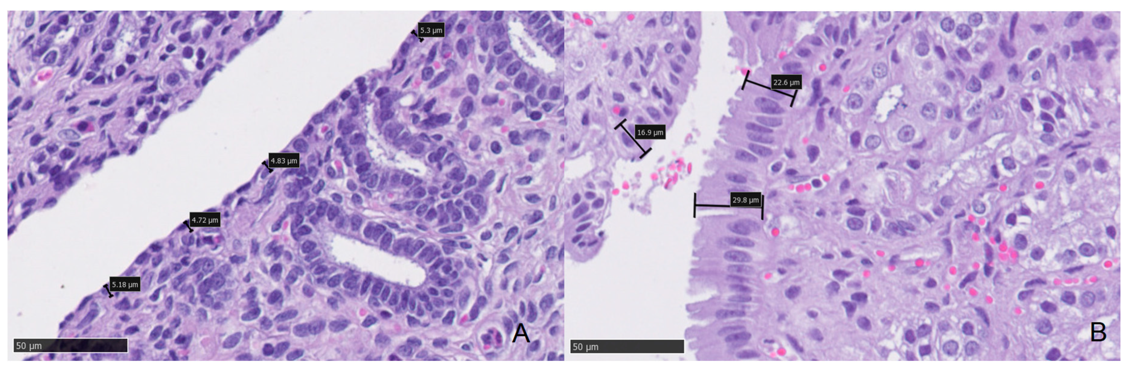
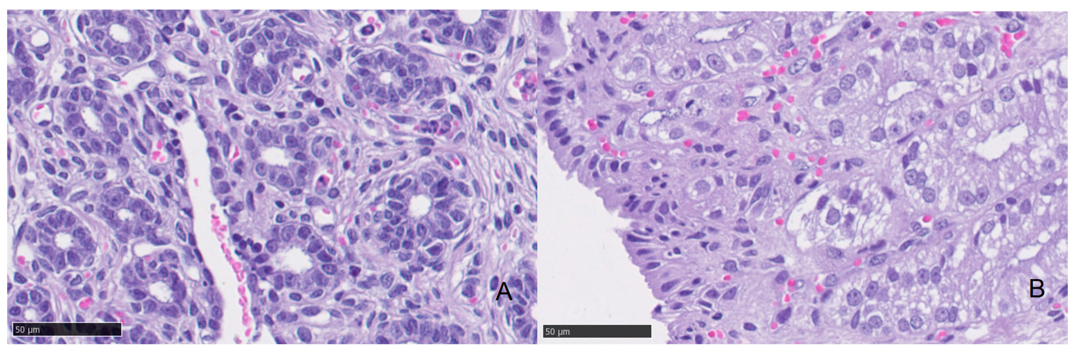
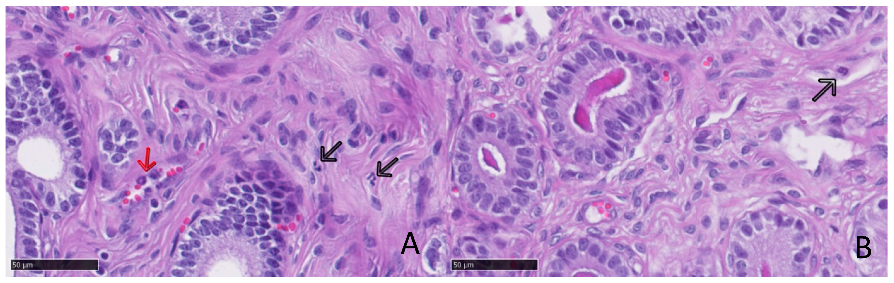
2.5. Histopathology
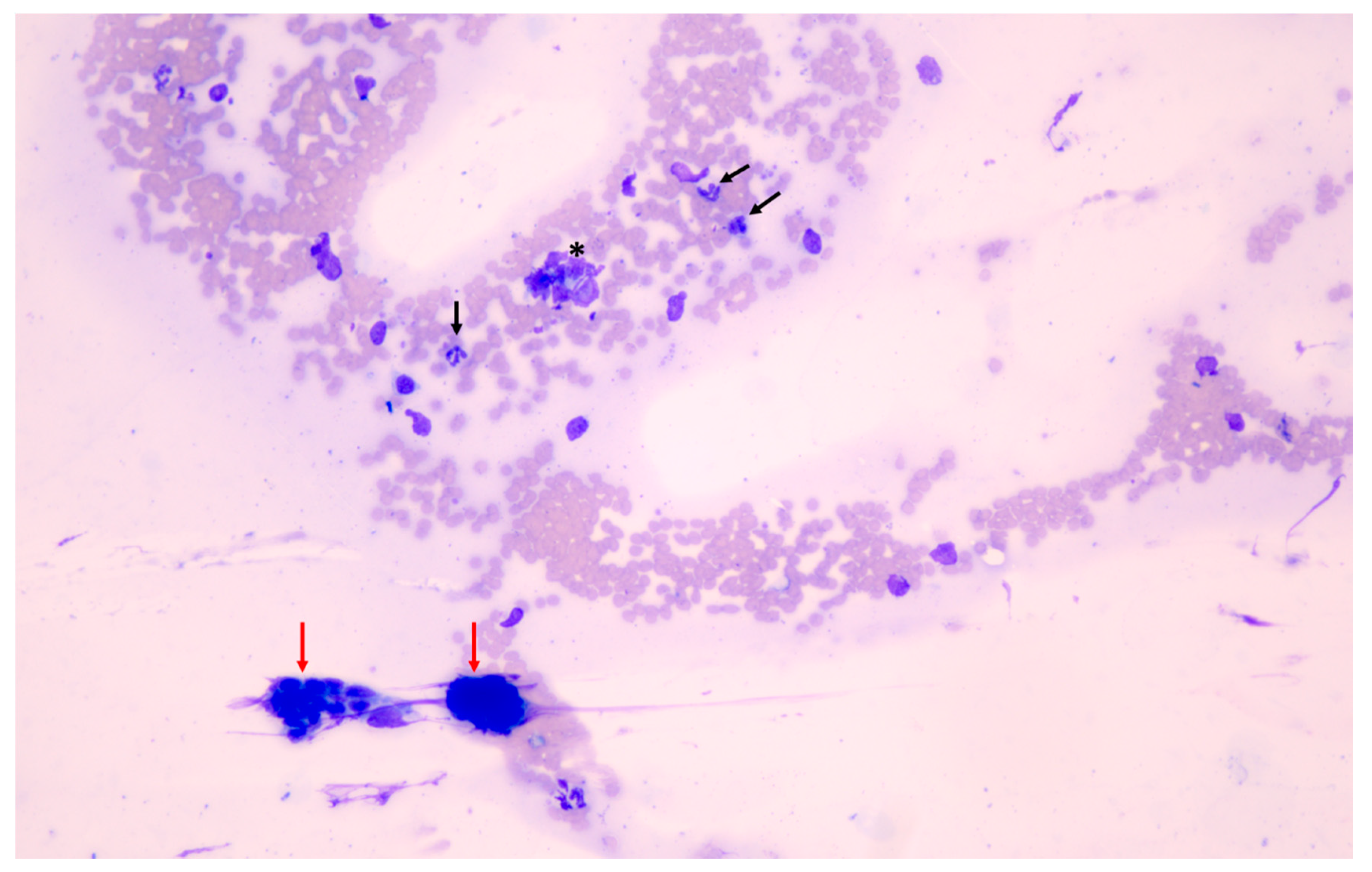
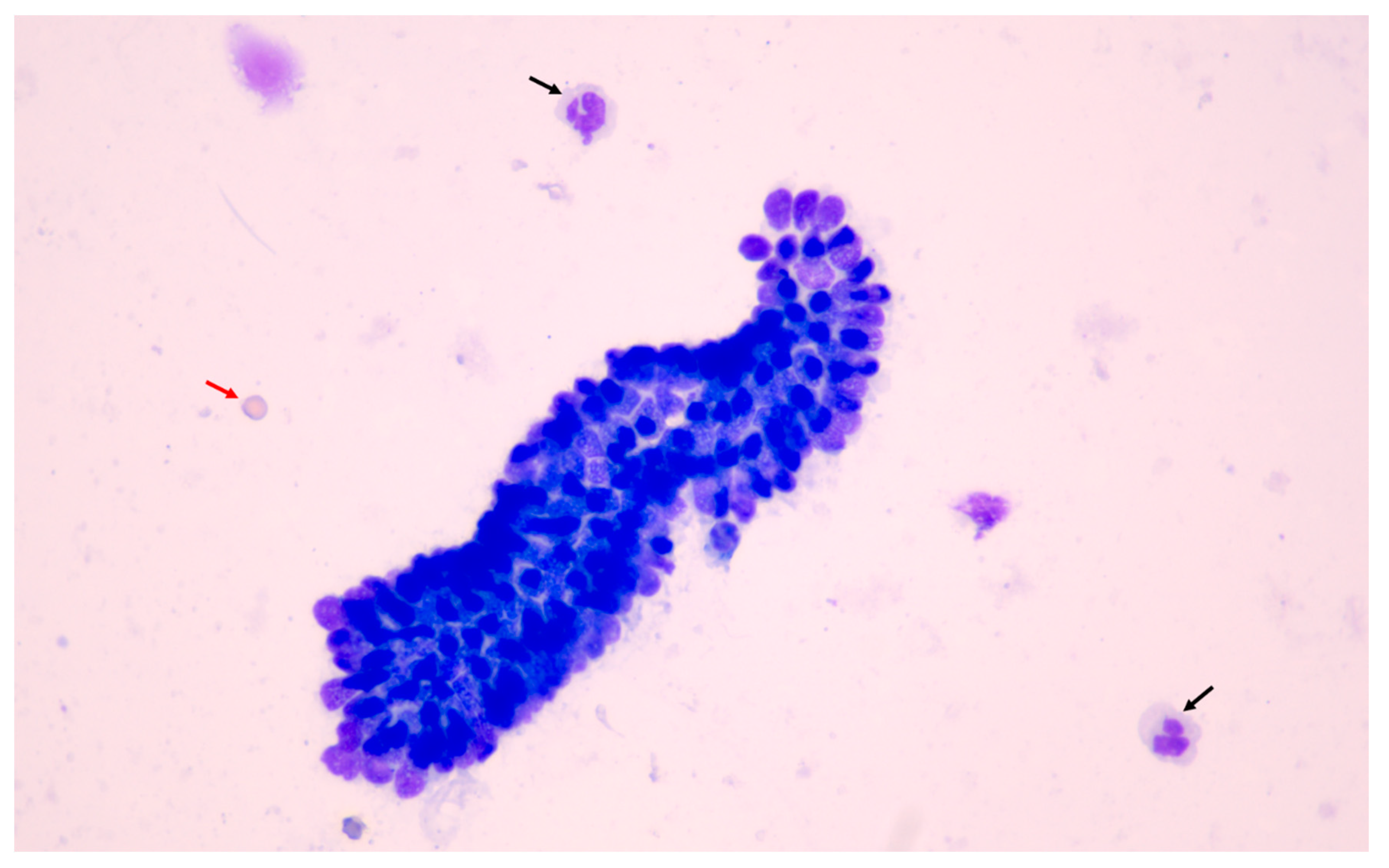
2.6. Cytology Samples
2.6.1. Smear Quality
2.6.2. Cellularity and Cell Morphology
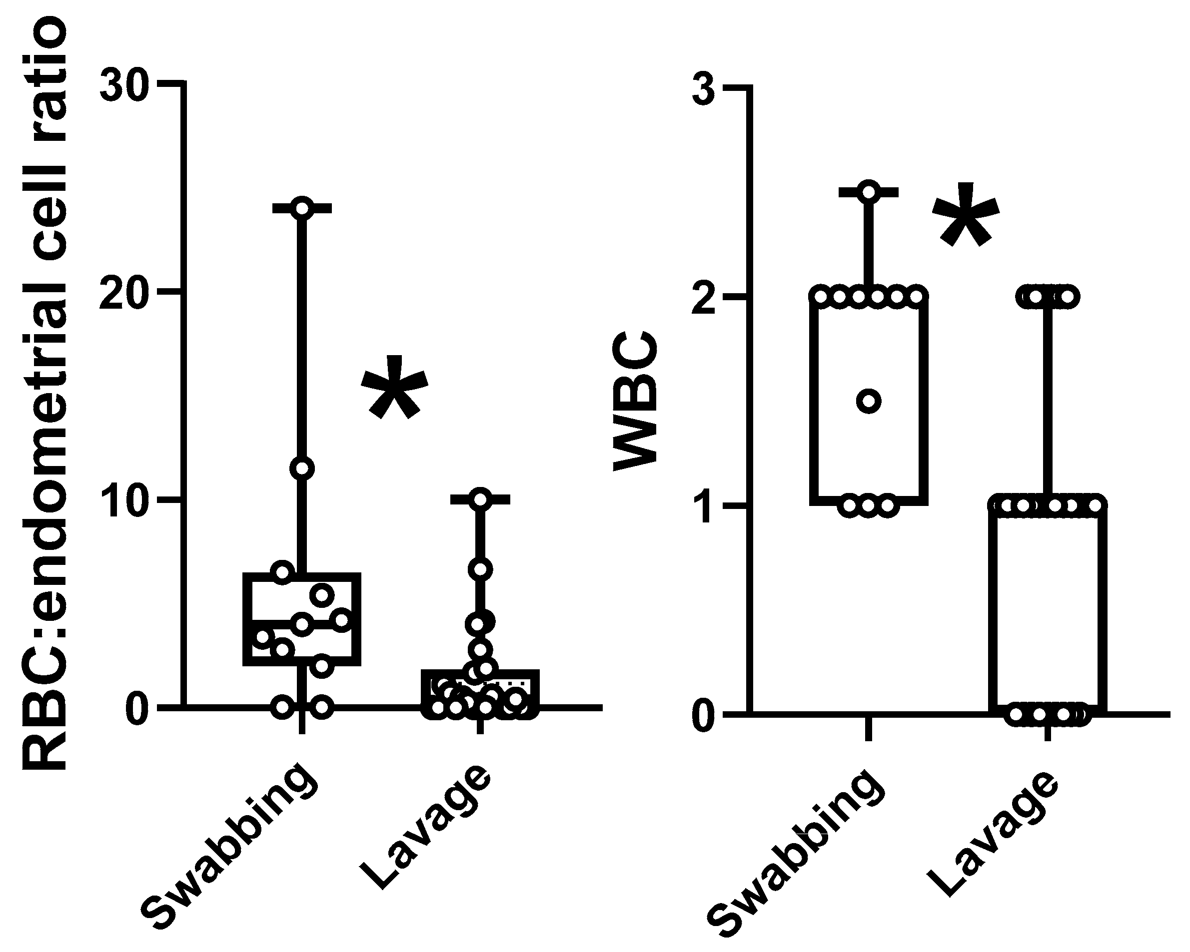
2.6.3. Cell Ratios
2.6.4. Inflammation
2.7. Statistical Analysis
3. Results
3.1. Progesterone
3.2. Microbiology
3.3. Histology
3.4. Cytology
4. Discussion
5. Conclusions
Author Contributions
Funding
Institutional Review Board Statement
Informed Consent Statement
Data Availability Statement
Conflicts of Interest
References
- Fontaine, E.; Levy, X.; Grellet, A.; Luc, A.; Bernex, F.; Boulouis, H.J.; Fontbonne, A. Diagnosis of Endometritis in the Bitch: A New Approach. Reprod. Dom. Anim. 2009, 44, 196–199. [Google Scholar] [CrossRef]
- LeBlanc, M.M.; Causey, R.C. Clinical and Subclinical Endometritis in the Mare: Both Threats to Fertility. Reprod. Domest. Anim. 2009, 44, 10–22. [Google Scholar] [CrossRef]
- Buczkowska, J.; Kozdrowski, R.; Nowak, M.; Ras, A.; Staroniewicz, Z.; Siemieniuch, M.J. Comparison of the biopsy and cytobrush techniques for diagnosis of subclinical endometritis in mares. Reprod. Biol. Endocrinol. 2014, 12, 27. [Google Scholar] [CrossRef] [PubMed] [Green Version]
- Kasimanickam, R.; Duffield, T.F.; Foster, R.A.; Gartley, C.J.; Leslie, K.E.; Walton, J.S.; Johnson, W.H. Endometrial cytology and ultrasonography for the detection of subclinical endometritis in postpartum dairy cows. Theriogenology 2004, 62, 9–23. [Google Scholar] [CrossRef] [PubMed]
- Wagener, K.; Gabler, C.; Drillich, M. A review of the ongoing discussion about definition, diagnosis and pathomechanism of subclinical endometritis in dairy cows. Theriogenology 2017, 94, 21–30. [Google Scholar] [CrossRef] [PubMed]
- Praderio, R.G.; García Mitacek, M.C.; Nuñez Favre, R.; Rearte, R.; De la Sota, R.L.; Stornelli, M.A. Uterine endometrial cytology, biopsy, bacteriology, and serum C-reactive protein in clinically healthy diestrus bitches. Theriogenology 2019, 131, 153–161. [Google Scholar] [CrossRef]
- Fuminori, K.; Takebayashi, A.; Ishida, M.; Nakamura, A.; Kitazawa, J.; Morimune, A.; Hirata, K.; Takahashi, A.; Tsuji, S.; Ono, T.; et al. Review: Chronic endometritis and its effect on reproduction. J. Obstet. Gynaecol. Res. 2019, 45, 951–960. [Google Scholar]
- Sheldon, I.M.; Price, S.B.; Cronin, J.; Gilbert, R.O.; Gadsby, J.E. Mechanisms of Infertility Associated with Clinical and Subclinical Endometritis in High Producing Dairy Cattle. Reprod. Domest. Anim. 2009, 44 (Suppl. S3), 1–9. [Google Scholar] [CrossRef]
- Cocchia, N.; Paciello, O.; Auletta, L.; Uccello, V.; Silvestro, V.; Mallardo, K.; Paraggio, G.; Pasolini, M.P. Comparison of the cytobrush, cottonswab, and low-volume uterine flush techniques to evaluate endometrial cytology for diagnosing endometritis in chronically infertile mares. Theriogenology 2012, 77, 89–98. [Google Scholar] [CrossRef]
- Sheldon, I.M.; Lewis, G.S.; LeBlanc, S.; Gilbert, R.O. Defining postpartum uterine disease in cattle. Theriogenology 2006, 65, 1516–1530. [Google Scholar] [CrossRef]
- Kasimanickam, R.; Duffield, T.F.; Foster, R.A.; Gartley, C.J.; Leslie, K.E.; Walton, J.S.; Walter, H.J. A comparison of the cytobrush and uterine lavage techniques to evaluate endometrial cytology in clinically normal postpartum dairy cows. Can. Vet. J. 2005, 46, 255–259. [Google Scholar]
- Földi, J.; Kulcsár, M.; Pécsi, A.; Huyghe, B.; de Sa, C.; Lohuis, J.A.; Cox, P.; Huszenicza, G. Bacterial complications of postpartum uterine involution in cattle. Anim. Reprod. Sci. 2006, 96, 265–281. [Google Scholar] [CrossRef]
- Axnér, E.; Agren, E.; Baverud, V.; Holst, B.S. Infertility in the cycling queen: Seven cases. J. Feline Med. Surg. 2005, 7, 59–63. [Google Scholar] [CrossRef]
- Fontbonne, A.; Prochowska, S.; Niewiadomska, Z. Infertility in purebred cats-A review of the potential causes. Theriogenology 2020, 158, 339–345. [Google Scholar] [CrossRef]
- Ley, W.B. Reproduçao em Éguas para Veterinários de Equinos; Roca: Sao Paulo, Brazil, 2006; p. 220. [Google Scholar]
- Troedsson, M.H.T. Equine Reproduction, 2nd ed.; Blackwell Publishing Ltd.: Ames, IA, USA, 2011; pp. 2608–2619. [Google Scholar]
- Barański, W.; Podhalicz-Dziegielewska, M.; Zduńczyk, S.; Janowski, T. The diagnosis and prevalence of subclinical endometritis in cows evaluated by different cytologic thresholds. Theriogenology 2012, 78, 1939–1947. [Google Scholar] [CrossRef]
- Nielsen, J.M. Endometritis in the mare: A diagnostic study comparing cultures from swab and biopsy. Theriogenology 2005, 64, 510–518. [Google Scholar] [CrossRef]
- Snider, T.A.; Sepoy, C.; Holyoak, G.R. Equine endometrial biopsy reviewed: Observation, interpretation, and application of histopathologic data. Theriogenology 2011, 75, 1567–1581. [Google Scholar] [CrossRef]
- Grifford, A.T.; Scarlett, J.M.; Schlafer, D.H. Histopathologic findings in uterine biopsy samples from subfertile bitches: 399 cases (1990–2005). J. Am. Vet. Med. Assoc. 2014, 244, 180–186. [Google Scholar] [CrossRef]
- Pascottini, O.B.; Hostens, M.; Dini, P.; Van Eetvelde, M.; Vercauteren, P.; Opsomer, G. Prevalence of cytological endometritis and effect on pregnancy outcomes at the time of insemination in nulliparous dairy heifers. J. Dairy Sci. 2016, 99, 9051–9056. [Google Scholar] [CrossRef] [Green Version]
- Reijnen, C.; Van der Putten, L.J.M.; Bulten, J.; Snijders, M.P.L.; Küsters-Vandevelde, H.V.N.; Sweegers, S.; Vos, M.C.; Van der Wurff, A.A.M.; Ligtenberg, M.J.L.; Massuger, L.F.A.G.; et al. Mutational analysis of cervical cytology improves diagnosis of endometrial cancer: A prospective multicentre cohort study. Int. J. Cancer 2020, 146, 2628–2635. [Google Scholar] [CrossRef]
- Paape, S.R.; Shille, V.M.; Seto, H.; Stabenfeldt, G.H. Luteal Activity in the Pseudopregnant Cat. Biol. Reprod. 1975, 13, 470–474. [Google Scholar] [CrossRef] [PubMed]
- Verhage, H.G.; Beamer, N.B.; Brenner, R.M. Plasma Levels of Estradiol and Progesterone in the Cat during Polyestrus, Pregnancy and Pseudopregnancy. Biol. Reprod. 1976, 14, 579–585. [Google Scholar] [CrossRef] [PubMed] [Green Version]
- Wildt, D.E.; Chan, S.Y.W.; Seager, S.W.J.; Chakraborty, P.K. Ovarian Activity, Circulating Hormones, and Sexual Behavior in the Cat. I. Relationships during the Coitus-Induced Luteal Phase and the Estrous Period without Mating. Biol. Reprod. 1981, 25, 15–28. [Google Scholar] [CrossRef] [Green Version]
- Schmidt, P.M.; Chakraborty, P.K.; Wildt, D.E. Ovarian Activity, Circulating Hormones and Sexual Behavior in the Cat. II. Relationships during Pregnancy, Parturition, Lactation and the Postpartum Estrus. Biol. Reprod. 1983, 28, 657–671. [Google Scholar] [CrossRef] [Green Version]
- Caballeros, J.E.; Camacho, C.; Cazales, N.; Estradé, M.J.; Fiala-Rechsteiner, S.; Jobim, M.I.M.; Mattos, R.C. Ultrastructural and histological characteristics of the equine endometrium at day 5 post ovulation. Theriogenology 2019, 132, 106–112. [Google Scholar] [CrossRef]
- Saltiel, A.; Gutierrez, A.; De Buen-llado, N.; Sosa, C. Cervico-endometrial cytology and physiological aspects of the post-partum mare. J. Reprod. Fertil. Suppl. 1987, 35, 305–309. [Google Scholar]
- Watts, J.; Wright, P.; Lee, C. Endometrial cytology of the normal bitch throughout the reproductive cycle. J. Small Anim. Pract. 1998, 39, 2–9. [Google Scholar] [CrossRef]
- Reilas, T.; Katila, T. Proteins and Enzymes in Uterine Lavage Fluid of Postpartum and Nonparturient Mares. Reprod. Domest. Anim. 2002, 37, 261–268. [Google Scholar] [CrossRef]
- Leroy, F.; Galand, P.; Chrétien, J. The mitotic action of ovarian hormones on the uterine and the vaginal epithelium during the oestrous cycle in the rat: A radioautographic study. J. Endocrinol. 1969, 45, 441–447. [Google Scholar] [CrossRef]
- Galand, P.; Leroy, F.; Chrétien, J. Effect of oestradiol on cell proliferation and histological changes in the uterus and vagina of mice. J. Endocrinol. 1971, 49, 243–252. [Google Scholar] [CrossRef]
- Martin, L.; Finn, C.A.; Trinder, G. Hypertrophy and hyperplasia in the mouse uterus after oestrogen treatment: An autoradiographic study. J. Endocrinol. 1973, 56, 133–144. [Google Scholar] [CrossRef] [Green Version]
- Conti, C.J.; Gimenez-Conti, I.B.; Zerbe, G.O.; Gerschenson, L.E. Differential Effects of Estradiol-17β and Progesterone on the Proliferation of Glandular and Luminal Cells of Rabbit Uterine Epithelium. Biol. Reprod. 1981, 24, 643–648. [Google Scholar] [CrossRef] [Green Version]
- Gerstenberg, C.; Allen, W.R.; Stewart, F. Cell proliferation patterns in the equine endometrium throughout the non-pregnant reproductive cycle. J. Reprod. Fertil. 1999, 116, 167–175. [Google Scholar] [CrossRef] [Green Version]
- Lai, M.D.; Lee, L.R.; Cheng, K.S.; Wing, L.Y.C. Expression of proliferating cell nuclear antigen in luminal epithelium during the growth and regression of rat uterus. J. Endocrinol. 2000, 166, 87–93. [Google Scholar] [CrossRef] [PubMed] [Green Version]
- Van Cruchten, S.; Van den Broeck, W.; D’haeseleer, M.; Simoens, P. Proliferation patterns in the canine endometrium during the estrous cycle. Theriogenology 2004, 62, 631–641. [Google Scholar] [CrossRef] [PubMed]
- Klein, C.; Troedsson, M.H. Transcriptional Profiling of Equine Conceptuses Reveals New Aspects of Embryo-Maternal Communication in the Horse. Biol. Reprod. 2011, 84, 872–885. [Google Scholar] [CrossRef] [Green Version]
- Chatdarong, K.; Rungsipipat, A.; Axner, E.; Linde Forsberg, C. Hysterographic appearance and uterine histology at different stages of the reproductive cycle and after progestagen treatment in the domestic cat. Theriogenology 2005, 64, 12–29. [Google Scholar] [CrossRef] [PubMed]
- Camozzato, G.C.; Martinez, M.N.; Bastos, H.B.A.; Fiala-Rechsteiner, S.; Meikle, A.; Jobim, M.I.M.; Gregory, R.M.; Mattos, R.C. Ultrastructural and histological characteristics of the endometrium during early embryo development in mares. Theriogenology 2019, 123, 1–10. [Google Scholar] [CrossRef]
- Walter, J.; Wehrend, A. Exfoliative endometrial cytology as a diagnostic aid in the gynaecological examination of broodmares. Pferdeheilkunde 2007, 39, 481–488. [Google Scholar] [CrossRef]
- Barlund, C.S.; Carruthers, T.D.; Waldner, C.L.; Palmer, C.W.A. A comparison of diagnostic techniques for postpartum endometritis in dairy cattle. Theriogenology 2008, 69, 714–723. [Google Scholar] [CrossRef]
- Zambelli, D.; Bini, C.; Cunto, M. Endoscopic Transcervical Catheterization in the Domestic Cat. Reprod. Domest. Anim. 2015, 50, 13–16. [Google Scholar] [CrossRef]


| Agar Media | Oxygen Conditions | Temperature |
|---|---|---|
| Blood | Aerobic | 37 °C |
| Anaerobic | 37 °C | |
| 5% CO2 | 37 °C | |
| MRS | Aerobic | 37 °C |
| Anaerobic | 37 °C | |
| 5% CO2 | 37 °C | |
| TSA | Aerobic | 37 °C |
| McConkey | Aerobic | 37 °C |
| Baird–Parker | Aerobic | 37 °C |
| Sabouraud | Aerobic | 28 °C |
| TSN | Anaerobic | 42 °C |
| Cytological Feature | Score | ||||
|---|---|---|---|---|---|
| 0 | 1 | 2 | 3 | 4 | |
| Mucus or Proteinaceous debris | Absent/clear background | Present but adequate for diagnosis | Abundant | Excessive, not adequate for diagnosis | - |
| Blood in Background (RBC/40×) | Absent (0–5) | Scattered (6–50) | Few (51–150) | Moderate (151–300) | Abundant (>300) |
| Cellularity | Absent | Scattered | Moderate | Abundant | - |
| Cell Preservation (% of broken cells) | Adequate (0) | Good (<25) | Fairly adequate (25–50) | Not useful for diagnosis (>50) | - |
| Epithelial Cells (number of cells) | Absent (0) | Scant (1–40) | Few (40–80) | Moderate (80–199) | Abundant (>200) |
| Individual Epithelial Cells | Absent | Scant | Few | Moderate | Abundant |
| Clusters Epithelial Cells (number of clusters) | Absent (0) | Scant (<10) | Few (10–40) | Moderate (40–80) | Abundant (>80) |
| Acinar Arrangement (HPF) | Absent (0) | Scattered <1/10 HPF | Moderate 1–3/10 HPF | Abundant >3/10 HPF | |
| Shape | Round | Low Columnar | Columnar | ||
| Pyknotic (%) | Absent (0) | Scant (<10) | Few (10–20) | Moderate (20–50) | Abundant (>50) |
| Cilia | Absent (0) | Scattered <1/10 HPF | Moderate 1–3/HPF | Abundant >3/10 HPF | |
| Vacuoles (%) | Absent (0) | Scattered (<20) | Moderate (20–60) Cells | Abundant (>60) Cells | |
| Nucleoli (%) | Absent (0) | Scattered <20 | Moderate 20–60 | Abundant >60 | |
| Mitosis (Number of mitoses) | Absent (0) | Scattered 0–1/10 HPF | Moderate 1–3/10 HPF | Abundant >3/10 HPF | |
| Serum Progesterone (ng/mL) | Uterine Lavage | Uterine Swabbing |
|---|---|---|
| Ovulated | 10.6 ± 1.3 (1.9–20.0) (n = 15) | 2.3 ± 0.5 (1.5–3.0) (n = 6) |
| Non-ovulated | 0.9 ± 0.1 (0.3–1.2) (n = 13) | 1.0 ± 0.1 (0.8–1.3) (n = 16) |
| Low Progesterone | High Progesterone | ||||||
|---|---|---|---|---|---|---|---|
| Median | Minimum | Maximum | Median | Minimum | Maximum | p | |
| Epithelial Height (cm) | 7.95 | 5.22 | 11.40 | 15.23 | 6.36 | 31.22 | 0.000 |
| Diameter 1 (cm) | 53.99 | 28.03 | 89.47 | 86.97 | 40.00 | 158.33 | 0.039 |
| Diameter 2 (cm) | 37.67 | 22.33 | 54.57 | 53.04 | 23.39 | 90.36 | 0.039 |
| Mean Diameter (cm) | 45.82 | 25.18 | 68.74 | 70.00 | 36.10 | 124.35 | 0.012 |
| Glandular Density (glands/400×) | 6.12 | 0.95 | 11.40 | 7.49 | 0.50 | 18.20 | 0.278 |
| Neutrophil (in 10 fields 400×) | 0.49 | 0.00 | 4.60 | 0.16 | 0.00 | 0.60 | 0.053 |
| Smear Quality | Technique | Score 0 | Score 1 | Score 2 | Score 3 | Score 4 | Comments |
|---|---|---|---|---|---|---|---|
| Mucus or Proteinaceous debris | Lavage | 28 (100%) | 0 (0%) | 0 (0%) | 0 (0%) | - | |
| Swabbing | 4 (18.18%) | 18 (81.81%) | 0 (0%) | 0 (0%) | - | ||
| Blood in Background (RBC) | Lavage | 9 (32.14%) | 8 (28.57%) | 4 (14.29%) | 4 (14.29%) | 3 (10.71%) | |
| Swabbing | 0 (0%) | 6 (27.27%) | 6 (27.27%) | 10 (45.45%) | 0 (0%) | ||
| Cellularity | Lavage | 6 (21.42%) | 9 (32.14%) | 8 (28.57%) | 5 (17.85%) | - | |
| Swabbing | 0 (0%) | 10 (45.45%) | 12 (54.54%) | 0 (0%) | - | ||
| Cell Preservation | Lavage | 8 (34.78%) | 6 (26.08%) | 4 (17.39%) | 5 (21.74%) | - | 5 No cells |
| Swabbing | 0 (0%) | 4 (18.18%) | 12 (54.54%) | 6 (27.27%) | - | ||
| Epithelial Cells | Lavage | 6 (21.42%) | 10 (35.71%) | 4 (14.29%) | 4 (14.29%) | 4 (14.29%) | |
| Swabbing | 0 (0%) | 12 (54.54%) | 6 (27.27%) | 4 (18.18%) | 0 (0%) | ||
| Individual Epithelial Cells | Lavage | 11 (39.29%) | 8 (28.57%) | 1 (3.57%) | 5 (17.86%) | 3 (10.71%) | |
| Swabbing | 0 (0%) | 10 (45.45%) | 10 (45.45%) | 2 (9.09%) | 0 (0%) | ||
| Clusters Epithelial Cells | Lavage | 7 (25%) | 6 (21.43%) | 7 (25%) | 2 (7.14%) | 6 (21.42%) | |
| Swabbing | 0 (0%) | 2 (9.09%) | 8 (36.36%) | 10 (45.45%) | 2 (9.09%) |
| Cell Characteristics | Technique | Score 0 | Score 1 | Score 2 | Score 3 | Score 4 | Comments |
|---|---|---|---|---|---|---|---|
| Acinar Arrangement | Lavage | 18 (81.81%) | 1 (4.55%) | 3 (13.64%) | 0 (0%) | - | 5 No cells/1 not evaluable |
| Swabbing | 16 (72.72%) | 4 (18.18%) | 2 (9.09%) | 0 (0%) | - | ||
| Shape | Lavage | 15 (68.18%) | 3 (13.64%) | 4 (18.18%) | - | - | 5 No cells/1 not evaluable |
| Swabbing | 22 (100%) | 0 (0%) | 0 (0%) | - | - | ||
| Pyknotic | Lavage | 13 (59.09%) | 9 (40.90%) | 0 (0%) | 0 (0%) | 0 (0%) | 5 No cells/1 not evaluable |
| Swabbing | 18 (81.81%) | 4 (18.18%) | 0 (0%) | 0 (0%) | 0 (0%) | ||
| Cilia | Lavage | 21 (95.45%) | 1 (4.54%) | 0 (0%) | 0 (0%) | - | 5 No cells/1 not evaluable |
| Swabbing | 22 (100%) | 0 (0%) | 0 (0%) | 0 (0%) | - | ||
| Vacuoles | Lavage | 16 (72.72%) | 3 (13.64%) | 3 (13.64%) | 0 (0%) | - | 5 No cells/1 not evaluable |
| Swabbing | 22 (100%) | 0 (0%) | 0 (0%) | 0 (0%) | - | ||
| Nucleoli | Lavage | 17 (77.27%) | 2 (9.09%) | 3 (13.64%) | 0 (0%) | - | 5 No cells/1 not evaluable |
| Swabbing | 20 (90.90%) | 1 (9.09%) | 1 (9.09%) | 0 (0%) | - | ||
| Mitosis | Lavage | 21 (95.45%) | 1 (4.54%) | 0 (0%) | 0 (0%) | - | 5 No cells/1 not evaluable |
| Swabbing | 22 (100%) | 0 (0%) | 0 (0%) | 0 (0%) | - |
| Category 0 | Category 1+ | Category 2+ | Category 3+ | |
|---|---|---|---|---|
| Swabbing | 2 (9%) | 8 (36%) | 11 (50%) | 1 (5%) |
| Lavage | 9 (32%) | 11 (39%) | 8 (29%) | 0 (0%) |
Publisher’s Note: MDPI stays neutral with regard to jurisdictional claims in published maps and institutional affiliations. |
© 2021 by the authors. Licensee MDPI, Basel, Switzerland. This article is an open access article distributed under the terms and conditions of the Creative Commons Attribution (CC BY) license (http://creativecommons.org/licenses/by/4.0/).
Share and Cite
Martí, A.; Serrano, A.; Pastor, J.; Rigau, T.; Petkevičiuté, U.; Calvo, M.À.; Arosemena, E.L.; Yuste, A.; Prandi, D.; Aguilar, A.; et al. Endometrial Status in Queens Evaluated by Histopathology Findings and Two Cytological Techniques: Low-Volume Uterine Lavage and Uterine Swabbing. Animals 2021, 11, 88. https://doi.org/10.3390/ani11010088
Martí A, Serrano A, Pastor J, Rigau T, Petkevičiuté U, Calvo MÀ, Arosemena EL, Yuste A, Prandi D, Aguilar A, et al. Endometrial Status in Queens Evaluated by Histopathology Findings and Two Cytological Techniques: Low-Volume Uterine Lavage and Uterine Swabbing. Animals. 2021; 11(1):88. https://doi.org/10.3390/ani11010088
Chicago/Turabian StyleMartí, Alba, Anna Serrano, Josep Pastor, Teresa Rigau, Ugné Petkevičiuté, Maria Àngels Calvo, Esteban Leonardo Arosemena, Aida Yuste, David Prandi, Adrià Aguilar, and et al. 2021. "Endometrial Status in Queens Evaluated by Histopathology Findings and Two Cytological Techniques: Low-Volume Uterine Lavage and Uterine Swabbing" Animals 11, no. 1: 88. https://doi.org/10.3390/ani11010088