Brief Report

The First Detection of Kudoa hexapunctata in Farmed Pacific Bluefin Tuna in South Korea, Thunnus orientalis (Temminck and Schlegel, 1844)

Department of Marine Biology and Aquaculture, College of Marine Science, Gyeongsang National University, Tongyeong 53064, Gyeongsangnam-do, Korea
*
Author to whom correspondence should be addressed.
Animals 2020, 10(9), 1705; https://doi.org/10.3390/ani10091705
Submission received: 5 September 2020 / Revised: 17 September 2020 / Accepted: 18 September 2020 / Published: 21 September 2020
(This article belongs to the Special Issue Fish Pathology)

Abstract

:

Simple Summary

In this study, we detected Kudoa hexapunctata in Pacific bluefin tuna (Thunnus orientalis) individuals that did not show any gross pathology lesions. Giemsa staining was used to identify clearer lesions than haematoxylin and eosin (H&E) staining that is used in general histological analysis. K. hexapunctata was separated through molecular biological methods, other than haematolocial and histological analysis. Individuals infected with K. hexapunctata showed relatively low haemoglobin (Hb) and haematocrit (Ht) values, and histological analysis revealed clear pseudocysts in the abdominal and dorsal muscles.

Abstract

The consumption of fish and shellfish worldwide is steadily increasing, and tuna is a particularly valuable fish species. However, infection caused by Kudoa spp. is causing problems in many fish including the Pacific bluefin tuna (Thunnus orientalis), and there is much controversy about the association of these infections with foodborne disease. In this study, using haematological and histological analyses of the blood and internal organs (liver, spleen, kidney, heart, stomach, intestine, gill, and muscle) of Pacific bluefin tuna cultured in South Korea, infection with Myxosporea was first identified, and molecular biological analysis was conducted. In this study, Kudoa hexapunctata was finally identified. The Pacific bluefin tunas analysed in this study did not show any gross pathology lesions, such as visible cysts and/or myoliquefaction, of infection with this species. The histological analytical results can provide guidelines for the identification of K. hexapunctata. In the case of K. hexapunctata-induced infection, unlike other countries, such as Japan, there have been no reports in South Korea, and this study is the first to detect K. hexapunctata infection in Pacific bluefin tuna cultured in South Korea. The correlation between K. hexapunctata and food poisoning is not yet clear, however, it is thought that continuous observation of its infection is necessary.

1. Introduction

Because fish and shellfish products have exhibited nutritive benefits and because of the increasing demand for healthy food, the consumption of fish and shellfish products is increasing annually [1,2]. Among fish, tuna has a very high commercial value in many countries [3]. However, parasitic diseases in fish can cause foodborne diseases in humans, according to the literature, which indicates that Kudoa septempunctata is a potential threat to human health [4,5,6]. Additionally of interest, are K. septempunctata in olive flounder (Paralichthys olivaceus), K. hexapunctata in tuna, and their possible implication in causing foodborne diseases [4,5,6]. Furthermore, Japanese patients exhibiting clinical diarrhoea had eaten tuna in which Kudoa hexapunctata was detected [7]. According to Suzuki et al. [7], K. hexapunctata is likely to be one of the causes of foodborne disease; however, there is still no clear evidence. In Japan, cases of patients with “unidentified foodborne disease” have increased recently, and it is emerging as a serious problem [8]. In olive flounder, Ohnishi et al. [9] said that K. septempunctata can invade human intestinal epithelial cells e.g., Caco-2 cells and exhibit sporoplasm invasion, causing severe damage. From this point of view, K. hexapunctata can be seen as one of the most important agents of parasitic disease in tuna, due to its possibility to cause a zoonotic parasitosis.
Kudoa spp. has several types of spores which can be divided into various types based on the shape of their spore valves [10]. The diagnostic methods involve mainly PCR (polymerase chain reaction) and real-time PCR methods using 18S rDNA or 28S rDNA [10]. However, currently, studies are focused on how Kudoa spp. affect the human body, and techniques for their detection, as it is difficult to determine the shape and distribution of Kudoa spp. [11].
Here, we demonstrate that the detection of K. hexapunctata in the cultured Pacific bluefin tuna of South Korea, is accompanied by histological and molecular biological results. In particular, histological analysis can be applied to samples without gross pathology lesions and can be used to monitor K. hexapunctata infection in individuals without visible cysts and myoliquefaction, which are typical gross pathology lesions of Kudoa spp. infection [12,13,14]. Therefore, this study aims to provide basic data so that various experimental methods can be applied to detect K. hexapunctata infection by combining the two above-mentioned assays with a molecular biological method and is the first report on K. hexapunctata infection in South Korean cultured Pacific bluefin tuna (Thunnus orientalis). Furthermore, through this study, we intend to provide a resource to suggest K. hexapunctata infection in South Korean farmed tuna and to analyse the correlation between the source of infection and food poisoning.

2. Materials and Methods

2.1. Sample Preparation

Five farmed Pacific bluefin tunas (Thunnus orientalis) were randomly sampled in December 2019 on Yokji Island for a health condition assessment. Samples of blood from the heart were taken from each sample, and the internal organs (liver, spleen, kidney, heart, stomach, intestine, gill, and muscle) were harvested. Additionally, for histological analysis, each organ was placed in 10% neutral-buffered formalin and for molecular biological analysis, each organ was stored at −80 °C until analysis.
Ethical approval: All experimental protocols followed the guidelines of the Institutional Animal Care and Use Committee of the Gyeongsang National University (approval number: 2020-0002).

2.2. Condition Factor

Following Barnham and Baxter [15] and Biswas et al. [16], the condition factor (CF) was calculated as follows: CF = 10 2 × W L 3 , where W = weight (g) and L = length (cm).
After calculation, CF values were compared Table 1 between each tuna, and the average condition of the tunas was calculated.

2.3. Morphological Analysis

For the morphological observation, abdominal and dorsal muscles (1 g) were sampled and homogenized using a 40 μm nylon cell strainer (Falcon, NY, USA). Staining was performed using Giemsa stain solution (Sigma-Aldrich, St. Louis, MI, USA) after the homogenates were suspended in 3 mL of PBS. For microscopic analysis, homogenates were smeared on a glass slide after being washed by distilled water.

2.4. Haematological Analysis

Each blood sample was collected in a heparin tube (BD, USA) for anticoagulation until arrival at the laboratory. The samples were centrifuged at 7000 RPM for 7 min, and the supernatants were used for haematological analysis by a DRI-CHEM 4000i instrument (Fujifilm, Tokyo, Japan). The following haematological parameters were analysed: alkaline phosphatase (ALP), blood urea nitrogen (BUN), calcium (Ca), glucose (GLU), glutamic oxaloacetic transaminase (GOT), glutamic pyruvic transaminase (GPT), lactate dehydrogenase (LDH), total cholesterol (TCHO), and total protein (TP). In the case of Ca and LDH, samples were diluted 2- and 10-fold in PBS for analysis. Haemoglobin (Hb) was measured using a haemoglobin colorimetric assay kit (Cayman Chemical, Ann Arbor, MI, USA) following the manufacturer’s protocol. Haematocrit (Ht) was calculated using heparinized capillary tubes (Paul Marienfeld Gmbh & Co. Kg, Lauda-Königshofen, Germany), and samples were placed in a haematocrit centrifuge machine (Scinco, Seoul, South Korea) at 12,000 RPM for, 5 min.

2.5. Histological Analysis

Dissection of internal organs (liver, spleen, kidney, heart, stomach, intestine, gill, and muscle) was followed by their fixation in 10% neutral-buffered formalin for 2 days. Then, small samples were taken from each organ, which were re-fixated in the same solution over 1 day before being gradually dehydrated with ethanolic solutions increasing from 70% to 100% ethanol. Samples were cleared further using xylene, after which, the samples were embedded in paraffin wax and, sectioned into slices with a 4 μm thickness. Finally, the sections were stained with haematoxylin-eosin (H&E) and Giemsa following routine protocols.

2.6. DNA Extraction, Polymerase Chain Reaction (PCR), and Sequence Analysis

Genomic DNA of tuna samples were extracted using an AccuPrep® genomic DNA extraction kit (Bioneer, Daejeon, South Korea) following the manufacturer’s guidelines. Homogenized muscle sample (10 mg) was used for polymerase chain reaction (PCR) to detect Kudoa spp. by histological data. All genomic DNA samples were stored at −80 °C until analysis. PCR was performed for amplifying 16S rRNA and 28S rRNA gene. Briefly, 10 μL of Exprime Taq Premix (GeNet Bio, Nonsan, South Korea), 7 μL of distilled water, 1 μL of genomic DNA, and 1 μL of each forward and reverse primer were mixed. Sequences of primers and PCR conditions are displayed in Table 2.
For sequencing, amplicons were extracted using a QIAquick® gel extraction kit (Qiagen, Hilden, Germany) following the manufacturer’s protocol. The purified PCR products were cloned into a pGEM® T-easy vector (Promega, Madison, Wisconsin, USA), and transformed into Escherichia coli JM109 according to a general protocol. After full propagation, plasmid DNA was extracted using a Hybrid-Q™ plasmid rapidprep kit (GeneAll®, Seoul, South Korea) and sequenced using a universal M13 primer set. Nucleotide sequence matching was performed using the basic local alignment search tool (BLAST) algorithm of the National Centre for Biotechnology Information (https://blast.ncbi.nlm.nih.gov/blast).

3. Results

3.1. Condition Factor and Haematological Data

Five condition factor (CF) values of Pacific bluefin tunas and haematological results are presented in Table 3. K. hexapunctata was detected in one tuna (number 5), which had only low haemoglobin (Hb) and haematocrit (Ht) levels of 10.9 g/dL and 34%, respectively, compared to those in the other tuna samples with an average of 15.02 g/dL (Hb) and 44.5% (Ht). However, the results have no statistical significance because of the number of samples.

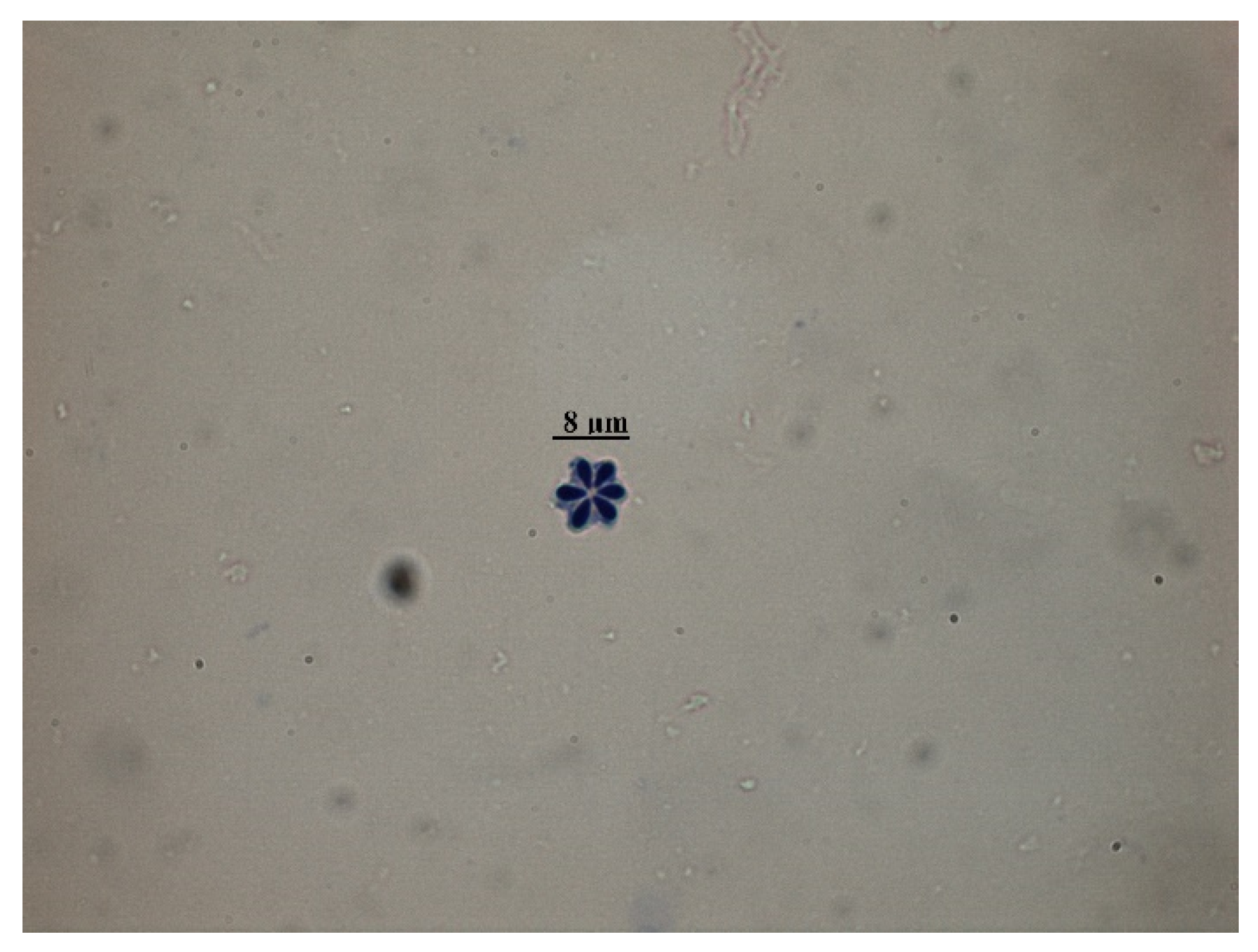
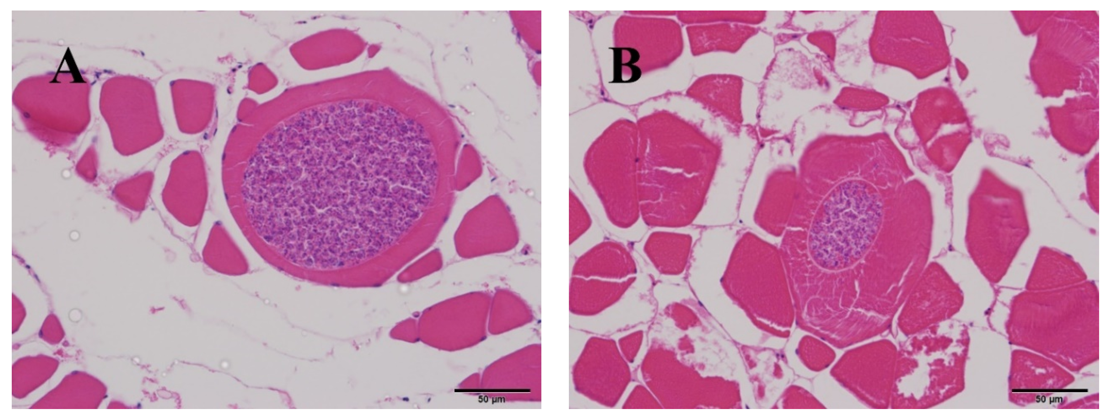
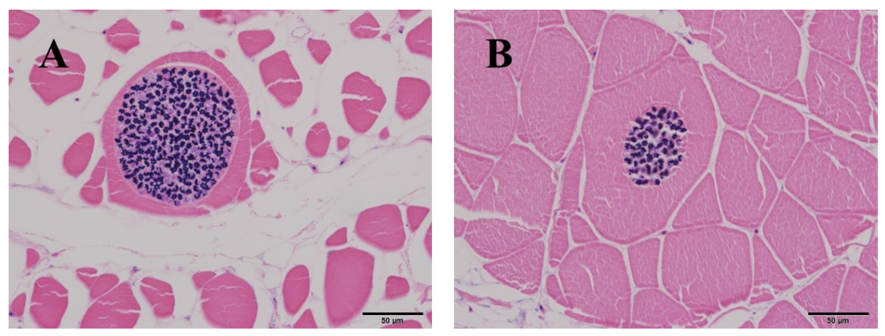
3.2. Morphological and Histological Analysis

The identified K. hexapunctata displayed six spores clearly (Figure 1). Histologically, the fifth sample only had pseudocysts in the abdominal and dorsal muscle (Figure 2 and Figure 3). By contrast, in the muscle of samples one to four, no pseudocysts indicating K. hexapunctata infection were observed in either the abdominal or dorsal sides. Similarly, liver, spleen, kidney, heart, stomach, intestine, and gill samples, including those of the fifth sample, did not display any evidence of K. hexapunctata-associated lesions.

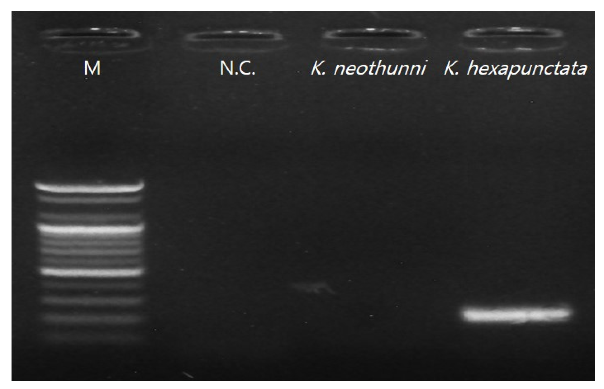
3.3. Polymerase Chain Reaction (PCR) and Sequence Analysis

Ribosomal DNA (rDNA) nucleotide sequencing was performed using two sets of primers to detect Kudoa spp. [6,17,19]. First, the pseudocysts were considered as kudoid parasites, and universal primers were used for the first isolation [17,19]; the primer set reported by Arai et al. [6] was used for secondary diagnosis, which distinguished species of Kudoa spp. from pseudocysts observed in histological data (Figure 2 and Figure 3). As a result, electrophoresis showed a target band size (197 bp) and sequences that ultimately were associated with K. hexapunctata (Figure 4 and Table 4).

4. Discussion

The Pacific bluefin tuna (Thunnus orientalis) used in this study did not shown any common gross pathology lesions, such as white or yellow cysts or myoliquefaction in the abdominal and/or dorsal muscle, that have been seen in olive flounder (Paralichthys olivaceus) infected by Kudoa septempunctata [20]. Nevertheless, we finally identified Kudoa hexapunctata using histological and molecular biological analyses. Although, it is generally known that K. hexapunctata does not cause myoliquefaction, further detailed studies are needed to investigate the correlation between the K. hexapunctata and myoliquefaction [6].
K. hexapunctata and Kudoa neothunni share many single nucleotide polymorphisms (SNPs) in the 28S rRNA gene [6]. In addition, Arai et al. [6] also detected K. neothunni in Pacific bluefin tuna, which is generally considered to be an infection of yellowfin tuna. Because of its host specificity, a molecular biological method was used to distinguish between K. hexapunctata and K. neothunni in this study. It is true that histological examination provides a guideline for definitive diagnosis; nevertheless, it can be seen that molecular biology methods have been included for accurate analysis.
In the present study, we investigated K. hexapunctata-infected tuna’s biomass and haematological data. It could be confirmed that the condition factor (CF), haemoglobin (Hb), and haematocrit (Ht) values of the fifth sample were relatively low, but the result was not statistically significant because the number of samples was small. Histologically, there were no specific pathological lesions such as necrosis or edema in other organs; nevertheless, further study on this part is needed. Moreover, on H&E staining, it was easy to identify myxosporea; however, the characteristic structure of K. hexapunctata was difficult to observe, and the spores of K. hexapunctata were much easier to observe through Giemsa staining.
There are no reports of South Korean tuna consumers complaining of food poisoning symptoms such as vomiting or diarrhoea; however, the causal relationship between K. hexapunctata and foodborne disease needs to be investigated in more depth by steadily checking the K. hexapunctata infection of tuna distributed in South Korea. Through this study, we confirmed that K. hexapunctata exists in cultured Pacific bluefin tuna in South Korea, and it is thought that there is a need to provide safer food resources to consumers through continuous surveillance of K. hexapunctata.

5. Conclusions

In conclusion, this is the first report of the detection of Kudoa hexapunctata from cultured Pacific bluefin tuna (Thunnus orientalis) in South Korea. The subjects used in this analysis had no visually identifiable gross pathology lesions such as visible cysts or myoliquefaction. The association between Kudoa spp. induced infection and food poisoning has been reported steadily in olive flounder (Paralichthys olivaceus) and Pacific bluefin tuna, but there is no clear evidence yet. Therefore, further studies need to test the potential pathogenicity in laboratory mammals for the purpose of speculating about possible foodborne disease.

Author Contributions

Conceptualization, G.K. and C.-I.P.; methodology, G.K.; formal analysis, G.K., D.-H.C., M.-J.H. and W.-S.W.; investigation, G.K.; resources, C.-I.P.; writing—original draft preparation, G.K.; writing—review and editing, K.-M.C. and M.-S.J.; supervision, C.-I.P.; project administration, C.-I.P.; funding acquisition, C.-I.P. All authors have read and agreed to the published version of the manuscript.

Funding

This research was funded by Ministry of Oceans and Fisheries, Korea (20180430).

Acknowledgments

This research was a part of the project titled, “Development of rapid and sensitive diagnostic methods for the quarantine of aquatic animals and their products”, funded by the Ministry of Oceans and Fisheries, Korea and the Marine Biotechnology Program of the Korea Institute of Marine Science and Technology promotion funded by the Ministry of Oceans and Fisheries (No. 20180430).

Conflicts of Interest

The authors declare no conflict of interest.

References

  1. Shamsi, S. Seafood-Borne Parasitic Diseases: A “One-Health” Approach Is Needed. Fishes 2019, 4, 9. [Google Scholar] [CrossRef] [Green Version]
  2. Kang, Y.M.; Kim, M.J.; Park, S.Y.; Heu, M.S.; Kim, J.S. Survey and Exposure Assessment of Biogenic Amines in Fish Species Commonly Consumed in Korea. J. Food Prot. 2019, 82, 151–158. [Google Scholar] [CrossRef] [PubMed]
  3. Vergara-Solana, F.J.; Araneda-Padilla, M.; Pardo, J.R.S.; Ortega-Garcia, S.; Seigo, J.C.; Ponce-Diaz, G. Growth and survival model of Pacific bluefin tuna (Thunnus orientalis) for capture-based aquaculture in Mexico. Aquac. Res. 2019, 50, 3549–3558. [Google Scholar] [CrossRef]
  4. Harada, T.; Kawai, T.; Sato, H.; Yokoyama, H.; Kumeda, Y. Development of a quantitative polymerase chain reaction assay for detection of Kudoa septempunctata in olive flounder (Paralichthys olivaceus). Int. J. Food Microbiol. 2012, 156, 161–167. [Google Scholar] [CrossRef] [PubMed]
  5. Kawai, T.; Sekizuka, T.; Yahata, Y.; Kuroda, M.; Kumeda, Y.; Iijima, Y.; Kamata, Y.; Sugita-Konishi, Y.; Ohnishi, T. Identification of Kudoa septempunctata as the Causative Agent of Novel Food Poisoning Outbreakes in Japan by Consumption of Paralichthys olivaceus in Raw Fish. Clin. Infect. Dis. 2012, 54, 1046–1052. [Google Scholar] [CrossRef] [PubMed] [Green Version]
  6. Arai, S.; Yoshinari, T.; Terajima, J.; Hara-Kudo, Y.; Ohnishi, T. Detection of Kudoa hexapunctata and Kudoa neothunni from retail raw tuna in Japan using a novel duplex polymerase chain reaction. Parasitol. Int. 2020, 75, 102048. [Google Scholar] [CrossRef] [PubMed]
  7. Suzuki, J.; Murata, R.; Yokoyama, H.; Sadamasu, K.; Kai, A. Detection rate of diarrhea-causing Kudoa hexapunctata in Pacific bluefin tuna Thunnus orientalis from Japanese waters. Int. J. Food Microbiol. 2015, 194, 1–6. [Google Scholar] [CrossRef] [PubMed]
  8. Kasai, A.; Tsuduki, H.; Jimenez, L.A.; Li, Y.C.; Tanaka, S.; Sato, H. Incidence of three Kudoa spp., K. neothunni, K. hexapunctata, and K. thunni (Myxosporea: Multivalvulida), in Thunnus tunas distributed in the western Pacific Ocean. Parasitol. Res. 2017, 116, 1137–1150. [Google Scholar] [CrossRef]
  9. Ohnishi, T.; Kikuchi, Y.; Furusawa, H.; Kamata, Y.; Sugita-Konishi, Y. Kudoa septempunctata Invasion Increases the Permeability of Human Intestinal Epithelial Monolayer. Foodborne Pathog. Dis. 2013, 10. [Google Scholar] [CrossRef]
  10. Yokoyama, H.; Suzuki, J.; Shirakashi, S. Kudoa hexapunctata n. sp. (Myxozoa: Multivalvulida) from the somatic muscle of Pacific bluefin tuna Thunnus orientalis and re-description of K. neothunni in yellowfin tuna T. albacares. Parasitol. Int. 2014, 63, 571–579. [Google Scholar] [CrossRef] [PubMed] [Green Version]
  11. Do, J.W.; Cho, M.; Jung, S.H.; Lee, N.S. A Study about Analysis Results for Kudoa septempunctata (Myxosporea: Multivalvulida) in Tissue at Olive Flounder, using PCR (polymerase chain reaction) and Histological Methods. Korean J. Environ. Biol. 2017, 35, 468–475. [Google Scholar] [CrossRef]
  12. Yokoyama, H.; Whipps, C.M.; Kent, M.L.; Mizuno, K.; Kawakami, H. Kudoa thyrsites from Japanese Flounder and Kudoa lateolabracis n. sp. from Chinese Sea Bass: Causative Myxozoans of Post-Mortem Myoliquefaction. Fish Pathol. 2004, 39, 79–85. [Google Scholar] [CrossRef]
  13. Matsukane, Y.; Sato, H.; Tanaka, S.; Kamata, Y.; Sugita-Konishi, Y. Kudoa septempunctata n. sp. (Myxosporea: Multivalvulida) from an aquacultured olive flounder (Paralichthys olivaceus) imported from Korea. Parasitol. Res. 2010, 107, 865–872. [Google Scholar] [CrossRef]
  14. Sugita-Konishi, Y.; Sato, H.; Ohnishi, T. Novel Foodborne Disease Associated with Consumption of Raw Fish, Olive Flounder (Paralichthys olivaceus). Food Saf. 2014, 2, 141–150. [Google Scholar] [CrossRef] [Green Version]
  15. Barnham, C.; Baxter, A. Fisheries Notes: Condition Factor, K, for Salmonid Fish; Department of Primary Industries: Orange, Australia, 1998; p. FN0005. [Google Scholar]
  16. Biswas, A.; Nakajima, M.; Nakao, T.; Takaoka, O.; Takii, K. Determination of suitable protein and lipid levels in diets for Pacific bluefin tuna, Thunnus orientalis at grow-out stage. Aquac. Sci. 2016, 64, 281–288. [Google Scholar]
  17. Barta, J.R.; Martin, D.S.; Liberator, P.A.; Dashkevicz, M.; Anderson, J.W.; Feighner, S.D.; Elbrecht, A.; Perkins-Barrow, A.; Jenkins, M.C.; Danforth, H.D.; et al. Phylogenetic Relationships among Eight Eimeria Species Infecting Domestic Fowl Inferred Using Complete Small Subunit Ribosomal DNA Sequences. J. Parasitol. 1997, 83, 262–271. [Google Scholar] [CrossRef]
  18. Fiala, I. The phylogeny of Myxosporea (Myxozoa) based on small subunit ribosomal RNA gene analysis. Int. J. Parasitol. 2016, 36, 1521–1534. [Google Scholar] [CrossRef]
  19. Bartosova, P.; Fiala, I.; Hypsa, V. Concatenated SSU and LSU rDNA data confirm the main evolutionary trends within myxosporeans (Myxozoa: Myxosporea) and provide an effective tool for their molecular phylogenetics. Mol. Phylogenet. Evol. 2009, 53, 81–93. [Google Scholar] [CrossRef] [PubMed]
  20. Grabner, D.S.; Yokoyama, H.; Shirakashi, S.; Kinami, R. Diagnostic PCR assays to detect and differentiate Kudoa septempunctata, K. thyrsites and K. lateolabracis (Myxozoa, Multivalvulida) in muscle tissue of olive flounder (Paralichthys olivaceus). Aquaculture 2012, 338, 36–40. [Google Scholar] [CrossRef]
Figure 1. Morphological observation of Kudoa hexapunctata by muscle biopsy of Pacific bluefin tuna (Thunnus orientalis) cultured in South Korea. Scale bar = 8 μm.
Animals 10 01705 g001
Figure 2. Histological analysis of abdominal and dorsal muscle in Pacific bluefin tuna (Thunnus orientalis) positively infected with Kudoa hexapunctata. H&E staining revealed: (A) abdominal muscle of the fifth sample; (B) abdominal muscle of the fifth sample. Scale bar = 50 μm.
Animals 10 01705 g002
Figure 3. Histological analysis of abdominal and dorsal muscle in Pacific bluefin tuna (Thunnus orientalis) positively infected with Kudoa hexapunctata. Giemsa staining revealed: (A) abdominal muscle of the fifth sample; (B) abdominal muscle of the fifth sample. Scale bar = 50 μm.
Animals 10 01705 g003
Figure 4. Electrophoresis results of secondary polymerase chain reaction (PCR) for distinguishing Kudoa neothunni from Kudoa hexapunctata (M = 2000 bp marker; N.C. = negative control).
Animals 10 01705 g004
Table 1. Standards of condition factor (CF) used in trout and salmonid fish (Barnham and Baxter, [15]).
Condition Factor (CF)Comments
1.60Excellent condition, trophy class fish.
1.40A good, well-proportioned fish
1.20A fair fish, acceptable to many anglers.
1.00A poor fish, long and thin.
0.80Extremely poor fish, resembling a barracuda, with a big head and narrow, thin body.
Table 2. Base sequence information and polymerase chain reaction (PCR) conditions of the primer set used in this study.
PrimerSequences (5′–3′)PCR ConditionsLength of Seq.Reference
ERIB1ACCTGGTTGATCCTGCCAG30 cycles of 95 °C for 1 min, 48 °C for 1 min and 72 °C for 2 min, followed by 10 min incubation at 72 °C-Barta et al. [17]
ERIB10CTTCCGCAGGTTCACCTACGG
MyxospecFTTCTGCCCTATCAACTWGTTG30 cycles of 95 °C for 1 min, 52 °C for 1 min and 72 °C for 2 min, followed by 10 min incubation at 72 °C769Ivan Fiala [18]
MyxospecRGGTTTCNCDGRGGGMCCAAC
K. hexFGCGACGGTTCAATCGTCAGIncubation for 2 min at 94 °C, 35 cycles of 30 s at 94 °C, 1 min at 68 °C, and a final extension for 5 min at 68 °C197Arai et al. [6]
K. hexRCACTGGTTGTTGAACTACCACACG
K. neoFGGCTACGTGCGGAAAAGTTGTA1045
K. neoRCGCGGATTCCAACCTATATCCAG
Table 3. Biomass, condition factor (CF) and haematological data of currently analysed Pacific bluefin tuna (Thunnus orientalis) 1 to 5.
Parameters12345
Weight (kg)17.417.814.510.413
Length (cm)103101989097
Condition factor (CF)1.591.731.541.431.42
ALP (U/L)170190382185367
BUN (mg/dL)2.22.131.82
Ca (mg/dL) 19.510.912.89.410.2
GLU (mg/dL)869697102102
GOT (U/L)453347563295354
GPT (U/L)42324016547
Haemoglobin (g/dL)16.2514.9914.3714.4710.90
Haematocrit (%)4145524034
LDH (U/L) 2503393497314287
TCHO (mg/dL)141141204204197
TP (g/dL)6.77.78.55.45.3
1 2-fold diluted in PBS for analysis; 2 10-fold diluted in PBS for analysis.
Table 4. Sequence data of first and secondary polymerase chain reaction (PCR) amplicon.
Kudoa hexapunctata from Cultured Pacific Bluefin Tuna (Thunnus orientalis) in South Korea
(1) 769 bp (Ivan Fiala [18]; Barta et al. [17])
5′-TTCTGCCCTATCAACTAGTTGGTGAGGTAGTGGCTCACCAAGGTTGTGACGGGTAACGGGGGATCAGGGTTCGATTCCGGAGAGGGAGCCTGAGAAACGGCTACCACATCTAAGGAAGGCAGCAGGCGCGCAAATTACCCAATCCAGACTTTGGGAGGTAGTGACGAGAAATACCGGAGTAGACCGTTAATTGGTTCACTATCGGAATGAACGTAATTTAATACCTTCGATGAGTAGCTACTGGAGGGCAAGTCTGGTGCCAGCAGCCGCGGTAATTCCAGCTCCAGTAGTGTATATCAAAATTGTTGCGGTTAAAACGCTCGTAGTTGGATTACAAAAGCTCTTTGGCGGTTAAATCAAGGTTTGATCGCTGTGGGGTTTTTTTATCGCGAGAGCCGCACGTGGGATTAAATTCTTGTGTGTGGTCACTTGCGAGGTGTGCCTTGAATAAAGCACAGTGCCCAAAGCAGGCGTAAGCTTGAATGTTATAGCATGGAACGATTATGTTAATCTTGTCGACTGTTGGTTGTTGGCAGTGGTCTCGATTAAAAGGGACATTTGAGGGCGTTAGTACTTGGTGGCGAGGGGTGAAATCCTTAGACCCATCAAAGACTAACTAATGCGAAAGCATTCGCCAAGAGTGTTTTCATTAATCAAGAACGAAAGTTGGAGGTTCGAAGACGATCAGATACCGTCCTAGTTCCATACAGTAAACTATGCCAACATGGGATTAGCCCGGTTTAATCCAGGTTGGTCCCTCAGAGAAACC-3′
(2) 197 bp (Arai et al. [6])
5′-CACTGGTTGTTGAACTACCACACGACTTTTCCGCACGTAGCCACAATGTGACCAGCACGAAACGCCGCACAGCACCAACCGACCATACATCACCAACGCTCCATCACATACATATCAGCACACGAGGCGGATCACCAGCCGCAACCACGAAACATTTGAGTGTTCCGTGACAGCCATGCTGACGATTGAACCGTCGC-3′

