Brief Report

Immune Responses of Asian Seabass Lates calcarifer to Dietary Glycyrrhiza uralensis

1
Tropical Aquaculture Research and Development Center, South China Sea Fisheries Research Institute, Chinese Academy of Fishery Sciences, Sanya 572018, China
2
Key Laboratory of South China Sea Fishery Resources Exploitation and Utilization, Ministry of Agriculture and Rural Affairs, Guangzhou 510300, China
3
Sanya Tropical Fisheries Research Institute, Sanya 572018, China
*
Author to whom correspondence should be addressed.
Animals 2020, 10(9), 1629; https://doi.org/10.3390/ani10091629
Submission received: 1 August 2020 / Revised: 26 August 2020 / Accepted: 9 September 2020 / Published: 11 September 2020

Abstract

:

Simple Summary

Due to the fact of their low toxicity, small side effects, and little residue, increasing attention has been paid to herbs as environmentally friendly immunostimulants. Results from the present study indicate that adding Glycyrrhiza uralensis to the feed can improve the growth and survival of Lates calcarifer and significantly promote the expression of immune-related genes in the liver and head kidney of Lates calcarifer. The optimum inclusion level of Glycyrrhiza uralensis should be 1–3%.

Abstract

To understand the impacts of dietary Glycyrrhiza uralensis on the immune responses of Lates calcarifer, the expression of immune-related genes including crp, c-3, c-4, mtor, mlst-8, eif4e, hsp-70, hsp-90, il-8il-8, il-10, tgfβ1, tnf, ifn-γ1, and mxf in L. calcarifer juveniles was evaluated in this study. Fish were fed experimental diets with G. uralensis levels of 0%, 1%, 3%, and 5% for 56 days. The results showed that dietary G. uralensis could improve the growth and survival of L. calcarifer and regulate the immune-related genes’ expression in L. calcarifer. Dietary G. uralensis significantly upregulated the expression level of crp, mtor, hsp-90, c-3, and c-4 genes in the liver of L. calcarifer, while hsp-70 gene expression was nearly downregulated. It did not upregulate the expression of elf4e and mlst-8 in the 1% and 3% inclusion groups, but it was the exact opposite in the 5% inclusion group. G. uralensis significantly affected the expression of il-8, il-10, tnf, ifn-γ1, mxf, and tgfβ1 in the head kidney of L. calcarifer. G. uralensis upregulated the expression of tnf and tgfβ1 consistently, but ifn-γ1 was at a low expression level. The expression of il-8 and il-10 was upregulated in the 1% group, while it was downregulated in the 5% group. The results from the present study indicate that dietary G. uralensis appeared to improve the immune function of L. calcarifer, and the optimum inclusion level should be between 1–3%.

1. Introduction

Asian seabass Lates calcarifer is widely distributed in the Indo–Pacific region. It is one of the most important marine aquaculture finfish in Australia and Asian countries, and the aquaculture of L. calcarifer increased to 76,842 tons in 2015 [1]. Because of its high nutritional value and rapid growth, L. calcarifer has become one of the main aquaculture species in southern China [2]. However, the outbreak of disease becomes a limiting factor in large-scale aquaculture of L. calcarifer. During aquaculture practices, high stocking density, grading, transporting, and environmental conditions may induce stress in fish, and this may cause disease outbreaks [3,4]. Diseases caused by bacteria, viruses or nutritional factors have been reported in farmed L. calcarifer such as scale drop syndrome (SDS) and megalocytiviral diseases [5,6]. Traditionally, antibiotics and chemotherapeutics administration are the major methods to treat the disease outbreaks, and these methods have a negative impact on the environment and fish. Recently, aquaculture specialists have made considerable progress in preventing and treating fish diseases through vaccines, probiotics, and immunostimulants [7,8,9].
Immunostimulants, which are feed additives derived from natural sources or synthetically, are capable of modulating the fish immune response and improving disease resistance. Therefore, people have begun to consider the immune stimulants used in the aquaculture industry [10,11]. Due to the low toxicity, small side effects, and little residue, increasing attention has been paid to herbs as environmentally friendly immunostimulants, and many studies have shown that herbs play a positive role in the prevention and treatment of fish diseases [11,12]. Glycyrrhiza uralensis, a leguminous flowering plant native to Asia, contains glycyrrhizic acid (GA) and other bioactive ingredients, and the extract of G. uralensis is widely used in China [13]. Evidence indicates that the extract of G. uralensis shows great potential as the immune stimulant in finfish aquaculture. Some reports have shown that feed supplemented with licorice extract or glycyrrhizic acid can improve the growth performance, immune response, stress resistance, and resistance to specified pathogens in fish species such as crucian (Carassius auratus), yellow croaker (Larimichthys crocea), and yellow catfish (Pelteobagrus fulvidraco) [14,15,16]. The addition of G. uralensis extract in the feed can promote the growth and lysozyme activity of yellow catfish and effectively reduce the mortality of fish infected with Flavobacterium columnare. Previous studies have explored the effects of plant or herbal, such as garlic, hirami lemon, and leaf meal, extracts as feed additives on the rearing performance of L. calcarifer, and positive effects in terms of growth and immunity have been observed (faster growth, higher disease resistance. and lower mortality rates) [4,9,10]. In this study, G. uralensis was used as the feed additive to test the immune responses of L. calcarifer. The expression of immune-related genes from fish liver and head kidney was evaluated, aiming to understand the immune responses of fish to immunostimulants and to provide more evidence for the availability of G. uralensis in fish feed.

2. Materials and Methods

2.1. Experimental Diets

The feed formulas were designed with reference to the nutritional requirements [17] of L. calcarifer (Table 1). Four types of feed were designed with the content of G. uralensis as the gradient (0%, 1%, 3%, and 5%). The basal feed without G. uralensis (0%) was used as the control group. All raw materials were crushed and extruded into feed with a diameter of 2.0 mm. Afterward, all the experimental feeds were stored at −20 °C until further usage.

2.2. Animals

L. calcarifer juveniles were produced by Tropical Aquaculture Research and Development Center, South China Sea Fisheries Research Institute, Chinese Academy of Fishery Sciences. The average weight of experimental fish ranged 14.58 ± 2.15 g, and the average length was 9.53 ± 1.86 cm.
This research was approved by the Animal Care and Use Committee of South China Sea Fisheries Research Institute, Chinese Academy of Fishery Sciences on 31 August 2018 (NO. 2018ZD01).

2.3. Experimental Design and Sampling

Before the experiment started, a total of 360 fish were randomly assigned to 12 tanks of 500 L (30 fish/tank) with a flow though system for a 14-day acclimation and fed with experimental diets. After acclimation, the feeding trail started and lasted 56 days. During the experimental period, fish feeding activity and death were observed and recorded, and the water quality was tested. The experiment was conducted in a circulation system and the water exchange rate was a 200% tank volume per day in each tank. The quality of water parameters was measured daily and maintained at a temperature of 27.5 ± 1.3 °C, a salinity of 32.5 ± 0.5‰, ammonia nitrogen < 0.01 mg/L, pH 7.5 ± 0.2, and nitrite nitrogen < 0.02 mg/L. The water quality parameters were maintained by adjusting the water exchange rate. Water was run through a mechanical filter and a biofilter and the water exchange occurred only when the water quality parameters exceeded the above ranges. The photoperiod was controlled at 14 h light and 10 h dark. Fish were fed ad libitum twice a day at 09:00 and 15:00. The feces and residual feeds were removed from the experimental tank daily by a siphon method. The residual feeds collected from each tank were dried and weighted.
At the end of the feeding trial, fish from each tank were counted for the final survival and were weighted to determine weight gain (WG). All the diets used in each tank were calculated to obtain feed intake (FI). Three fish from each tank were randomly collected after anaesthetizing with overdose of eugenol (7 mg/L eugenol, Shangchi Dental Material Co., Ltd., Changshu, China). Afterwards, fish were dissected on ice. Liver and kidney from each fish were stored in liquid nitrogen until further usage. Other fish growth performance indicators were calculated including specific growth rate (SGR), hepatosomatic index (HIS), and intraperitoneal fat ratio (IPF). The parameters were calculated as: WG = final body weight − initial body weight; FI = (feed consumed per tank/fish) / days, SGR = 100 × ((ln (final body weight) − ln (initial body weight)) /days; HIS = 100 × ((liver weight) / (whole-body weight)); IPF = 100 × ((intraperitoneal fat weight)/(whole-body weight)).

2.4. Gene Expression Analysis

The frozen tissue samples were homogenized in liquid nitrogen using a bioprep (Bioprep-24, Hangzhou Allsheng Instruments Co. Ltd., Hangzhou, China). The RNA extraction was performed according to the method described by Fu et al. [18]. The quantity of isolated RNA was determined by measuring their absorbance at 260 and 280 nm using an ND 5000 spectrophotometer (BioTeke Corporation, Beijing, China). Finally, the integrity of RNA was assessed using agarose gel (1%) electrophoresis. The cDNA was synthesized using the PrimeScript® RT Master Mix (Perfect Real Time, Takara Biomedical Technology (Dalian) Co., Ltd., Dalian, China). The synthesized cDNA samples were stored at −20 °C until further use. The 10 μL of reaction: 2 μL 5 × PrimeScript® RT Master Mix, RNA 500 ng, some Rnase Freed H2O (total volume was 10 μL). Reverse transcription reaction conditions: 37 °C, 30 min; 85 °C, 5 s.
The genes chosen for analysis by qPCR were selected from the L. calcarifer NCBI database (https://www.ncbi.nlm.nih.gov/). The Primer Premier 5 program (Premier Biosoft International, Palo Alto, CA, USA)was used for designing the primers of crp, c-3, c-4, mtor, mlst-8, eif4e, hsp-70, hsp-90, il-8, il-10, tgfβ1, tnf, ifn-γ1, mxf, and β-actin (Table 2). The direct and indirect effects of these gene transcription and translation products on fish immunity have been confirmed in numerous studies [19,20,21,22,23,24]. The qPCR was performed with the Real-Time qPCR analysis (Hangzhou Longgene Scientific Instrument Co., Ltd., Hangzhou, China) using SYBR Green (Tiangen Biotech Co., Ltd., Beijing, China) [18]. The 20 μL of reaction including 10 μL 2 × Real Universal PreMix, 0.6 μL 10 μM of each primer (10 μM), and 2 μL of diluted cDNA was initially denatured at 95 °C for 10 min and then amplified for 40 cycles (95 °C, 10 s, 58 °C, 20 s, and 72 °C 30 s). Each sample was subjected to qPCR for 3 times. At the end of each RT-qPCR cycle, the melting curve of the primer was analyzed to ensure that only specific products were obtained, and no primer dimer was formed. In addition, a negative control group without DNA a template was set to verify that the PCR process was not contaminated. The relative mRNA expression levels of the target genes were determined by the 2−ΔΔCt method and were normalized based on the level of housekeeping gene (β-actin). It has been verified that the reaction efficiency (E) was 90–105% and Pearson’s coefficients of determination (R2) > 0.98.

2.5. Statistical Analysis

The data were expressed as the mean ± standard deviation (SD). The software SPSS 19.0 (International Business Machines Corporation, Chicago, Illinois, USA) was used for statistical analysis, and one-way ANOVA and Least significant difference (LSD) test were used for inter-group comparison. The level of significance was set at p < 0.05. All data were tested for normality, homogeneity, and independence to satisfy the assumptions of ANOVA.

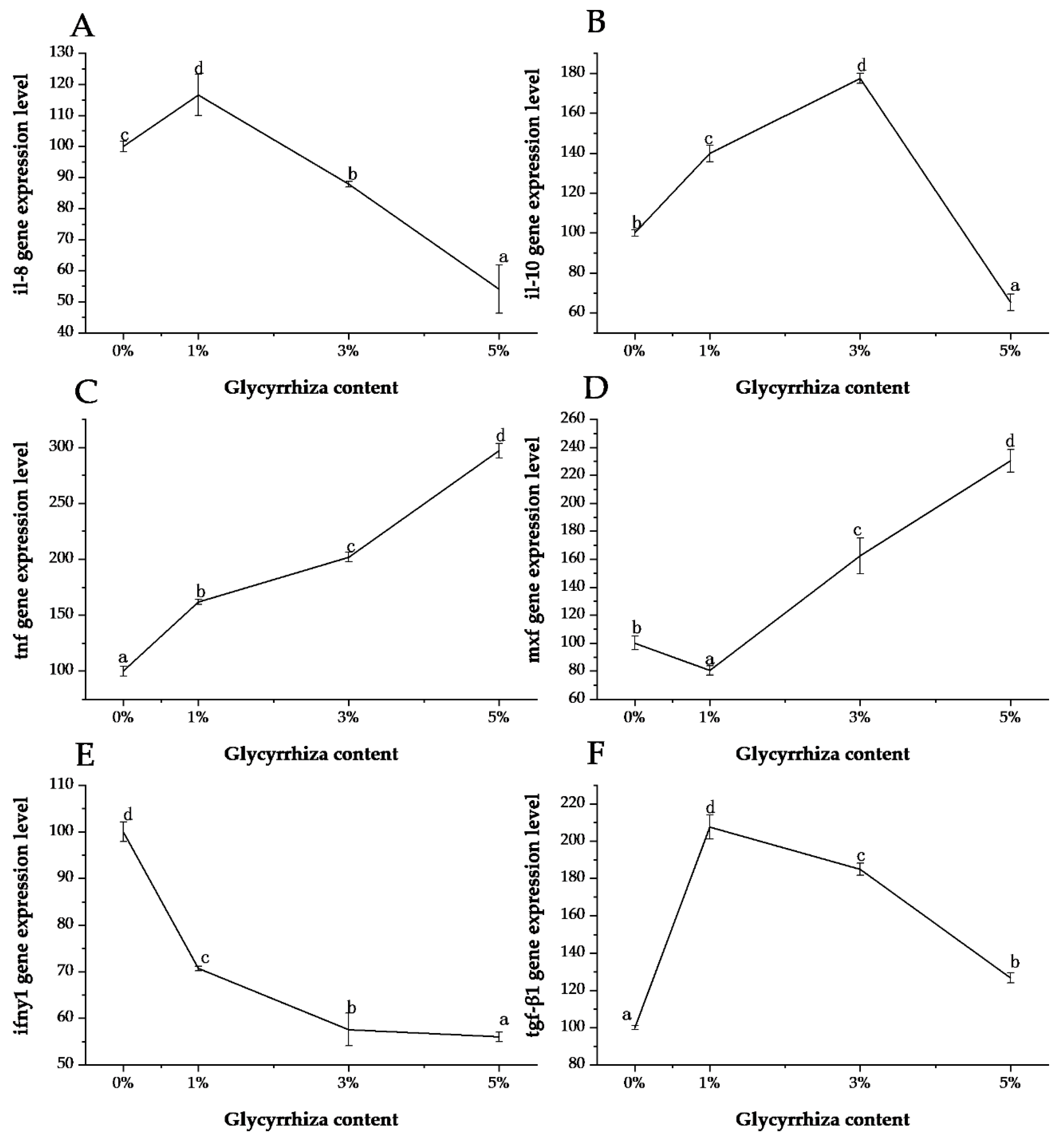
3. Results and Discussion

Compared with the control group, the weight gain rate and specific growth rate of each experimental group showed an upward trend, and both showed a significant increase at 5% of the experimental group (p < 0.05, Table 3). The survival rate increased with the increase of G. uralensis content. There was no significant difference in the feed intake and hepatosomatic index among all groups. Intraperitoneal fat ratio decreased with the increase of G. uralensis content, and there was a significant difference between the 5% group and other groups (p < 0.05).
Dietary G. uralensis significantly affected the expression of immune-related genes, such as crp eif4e mlst-8 mtor, hsp-70, hsp-90, c-3, and c-4, in the liver of L. calcarifer (p < 0.05, Figure 1). Compared with the control group, the relative expression level of the crp gene in the experimental group was significantly upregulated (p < 0.05), and the expression levels in the 3% and 5% groups were significantly higher than in the 1% group (p < 0.05). The expression levels between the 3% and 5% groups were not significantly different (p > 0.05). The eif4e gene expression levels in the 1% and 3% grade were significantly downregulated (p < 0.05) and was significantly upregulated in the 5% group (p < 0.05). The expression levels of the mlst-8 and mtor genes in the 1% and 3% groups were not significantly different from those in the control group (p > 0.05), but the relative expression levels in the 5% group were significantly upregulated (p < 0.05). G. uralensis had the opposite effect on genes hsp-70 and hsp-90. It almost completely inhibited the expression of the hsp-70 gene, but it had a significant upregulating effect on the hsp-90 gene. G. uralensis had the same effect on genes c-3 and c-4. The relative expression levels of c-3 and c-4 were not significantly different between the 1% group and the control group (p > 0.05) but were significantly upregulated in the other two groups (p < 0.05). Therefore, G. uralensis can promote the expression of most immune-related genes in the liver of L. calcarifer. Moreover, 5% G. uralensis can upregulate the expression of seven immune-related genes other than hsp-70.
G. uralensis significantly affected the expression of kidney immune-related genes including il-8, il-10, tnf, ifn-γ1, mxf, and tgfβ1 in L. calcarifer (p < 0.05, Figure 2). The content of 1% dietary G. uralensis could effectively upregulate the expression of il-8 in the kidney of L. calcarife, but as the proportion of G. uralensis increased, the expression level of il-8 was downregulated significantly (p < 0.05). The expression pattern of il-10 was almost the same as that of il-8; 1–3% dietary G. uralensis could upregulate its expression but in excess, its relative expression was significantly downregulated (p < 0.05). For tnf, its relative expression was significantly upregulated with the increase of G. uralensis content (p < 0.05). The expression of tnf was the exact opposite to the expression of ifn-γ1; G. uralensis appears to have a strong inhibitory effect on ifn-γ1. A small amount of G. uralensis inhibited the expression of the mxf gene, but the expression was significantly upregulated when its content increased (p < 0.05). Dietary G. uralensis significantly upregulated the expression level of tgfβ1 in fish (p < 0.05). The highest expression level of tgfβ1 was observed in fish fed with 1% G. uralensis group (p < 0.05).
As a traditional Chinese herb, G. uralensis is mainly composed of triterpenoid saponins, flavonoids, coumarins, alkaloids, polysaccharides, and amino acids [25]. Its pharmacological effects mainly include antitumor [26], antiarrhythmic [27], antispasmolysis [28], antitussive [29], anti-inflammatory [30], antiviral [31], and immunoregulatory [32]. In aquaculture applications, G. uralensis plays a certain role in promoting the immunity and antioxidant capacity of aquatic animals. The addition of fermented licorice to Epinephelus coioides feed can reduce the damage to liver tissue and enhance antioxidant capacity thus improving the survival rate of fish under nitrite stress [33]. G. uralensis significantly improves the anti-stress ability of Carassius auratus [34]. It also increases the host’s resistance to Aeromonas hydrophila [34,35].
In terms of growth, the results of this study show that the addition of G. uralensis to feed has an obvious promoting effect on the growth of L. calcarife juvenile. Similarly, in the white shrimp Litopenaeus vannamei, the specific growth rate of the feed group with glycyrrhizin is significantly higher than that of the control group [36]. Feeding G. uralensis diets significantly increased (p < 0.05) growth performance and antioxidant and immune response in yellow perch Perca flavescens [37]. Glycyrrhetinic acid has been shown to increase the activity of fish digestive enzymes and to increase the expression of tumor necrosis factor (tnf-α) and lipoprotein lipase (lpl) to promote lipolysis for energy, thereby saving more protein for deposition for increased growth performance [15]. In this study, it was also found that the increase of G. uralensis content in the feed reduced the intraperitoneal fat ratio of L. calcarife juveniles. This suggests that dietary supplementation of licorice may enhance protein deposition in juveniles by promoting lipolysis, which might result in increased growth performance. Furthermore, in commercial fish, intraperitoneal fat is usually removed along with the viscera as an inedible portion, and it is generally undesirable, so feeding G. uralensis diets improves the product quality of L. calcarife to some extent [38]. In addition, the survival rate of the juveniles in the experimental group was significantly increased, which was a direct reflection of the effect of licorice on fish immunity. This result is similar to previous reports in yellow croaker [15] and yellow catfish [16]. After nitrite stress, the survival rate of Epinephelus coioides supplemented with fermented G. uralensis is also significantly improved [33]. Meanwhile, dietary G. uralensis supplementation did not decrease but slightly increased the intake of feed in the juveniles. This indicates that G. uralensis is a feasible feed additive.
Reactive protein (crp) is a phylogenetically highly conserved plasma protein, with homologs in vertebrates and many invertebrates, that participates in the systemic response to inflammation [21]. The crp is capable of specifically binding to and modulating the function of mononuclear phagocytes [39]. In this study, the expression of crp gene in the liver of L. calcarifer was upregulated after fish intake of G. uralensis. The relative expression levels of crp gene were highest in the 3% and 5% groups. Eukaryotic translation initiation factor 4E (eif4e) plays a central role in the recognition of the 7-methylguanosine-containing cap structure of mRNA and the formation of initiation complexes during protein synthesis. The gene eif4e exists in both phosphorylated and non-phosphorylated forms, and the primary site of phosphorylation has been identified. Previous studies have suggested that eif4e phosphorylation facilitates its participation in protein synthesis [40]. Our study showed that a small amount of G. uralensis had a certain inhibitory effect on eif4e, but a significant promoting effect was observed when the dietary G. uralensis inclusion level was over 5%.
The mechanistic target of rapamycin (mtor) is the target of a molecule named rapamycin or sirolimus, which is a macrolide produced by Streptomyces hygroscopicus bacteria and that first gained attention because of its broad antiproliferative properties [41]. The mtor kinase nucleates two distinct protein complexes termed mtor-c1 and mtor-c2. The mtor-c1 responds to amino acids, stress, oxygen, energy, and growth factors and is acutely sensitive to rapamycin. The mtor-c2 responds to growth factors and regulates cell survival and metabolism as well as the cytoskeleton [42]. The mammalian lethal with SEC13 protein 8, is the binding protein of the target protein of rapamycin [43] and involved in both mtor-c1 and mtor-c2 [44]. In this study, the expression of mtor was upregulated in the 1% and 5% G. uralensis inclusion groups, and the highest expression level was observed in the 5% inclusion group. Similarly, the expression of mlst-8 was upregulated significantly in the 5% group. It may suggest that 5% G. uralensis could promote the expression of mtor and its binding protein gene mlst-8.
Heat shock proteins (hsp) belong to the family of highly conserved cellular proteins present in all organisms that have been examined [45]. Hsp-70 and hsp-90 are the two main proteins in the heat shock protein family [23]. Hsp-70 is known to assist the folding of nascent polypeptide chains, act as a molecular chaperone, and mediate the repair and degradation of altered or denatured proteins [46]. Hsp-90 is active in supporting various components of the cytoskeleton and steroid hormone receptors [47]. Our results showed that G. uralensis had a consistent inhibitory effect on the expression of hsp-70 gene in the liver of L. calcarifer, while it had the opposite effect on hsp-90, especially in the 5% inclusion group. Complement proteins c-3 and c-4 are also classified as acute phase reactants as their synthesis is upregulated during inflammation. In this study, low levels of G. uralensis did not affect the expression of c-3 and c-4 gene in the liver of L. calcarifer, while higher levels significantly upregulated their expression. This may suggest that higher dose G. uralensis can promote the expression of c-3 and c-4 effectively in L. calcarifer.
Interleukin-8 (il-8) is a chemokine that can activate neutrophils and has endogenous leukocyte chemokine and activation [48]. It is an important pleiotropic cytokine that mediates inflammatory responses and regulates the differentiation and proliferation of some immune cells. It mainly regulates the inflammatory response, which can not only inhibit mononuclear macrophages to release immune medium antigen presentation and cell phagocytosis [49]. In this study, the expression of il-8 gene in the kidney of L. calcarifer in 1% groups was significantly higher than the other groups, and the expression level was downregulated as the content of G. uralensis increased. However, the highest level of il-10 was found in the 3% group. Tumor necrosis factor (tnf), as a cytokine, it not only has cytotoxic effect on tumor cells, but also participates in a variety of pathophysiological processes such as antivirus, anti-infection, coagulation, fever and inflammation, shock, multi-organ failure and malignant fluid. Interferon (ifn) is a broad-spectrum antiviral glycoprotein secreted by recipient cells after viral infection of cells and the body or by nucleic acid bacterial endotoxin cytokinin. Ifn-γ1 is an important member of the ifn family, which also called Ⅱinterferon or immune interferon, mainly involved in inducing major histocompatibility antigen expression and immune regulation effect [50]. The results showed that the expression level of tnf was upregulated with the increase of content of G. uralensis, while the expression level of ifn-γ1 was the opposite.
Transforming growth factor beta (tgfβ) family is a kind of superfamily polypeptide which has the function of regulating cell growth and differentiation, and tgf-β1 is a member of this family [51]. In this study, we could clearly see that the relative expression level of the tgfβ1 gene in the kidney of L. calcarifer with G. uralensis was significantly higher than that of the control group. Myxovirus resistance (mx) is an antiviral protein that can be activated by ifn-I. Mx proteins belong to the dynamin superfamily and contain a tripartite guanosine triphosphate (GTP) binding domain which is essential for the antiviral activity [52,53]. In our study, adding 3–5% G. uralensis could significantly upregulate the expression of mxf gene.

4. Conclusions

In summary, dietary G. uralensis significantly improved growth performance and promoted the expression of immune-related genes in the liver and the kidney of L. calcarifer. Dietary G. uralensis can significantly upregulate the expression level of crp, mtor, hsp-90, c-3, and c-4 genes in fish liver, and significantly affected the expression of il-8, il-10, tnf, ifn-γ1, mxf, and tgfβ1 in fish kidney. Results from the present study indicated that dietary G. uralensis may improve the immune function of L. calcarifer, and the optimum inclusion level should be 1–3%. Adding G. uralensis to the feed will help to improve the growth, survival, and immunity of L. calcarife.

Author Contributions

Conceptualization, Z.M. and G.Y.; methodology, Z.M.; validation, R.Y. and Y.W.; formal analysis, Y.W.; investigation, M.H. and Y.W.; resources, W.Z.; writing—original draft preparation, R.Y., M.H., Z.F.; writing—review and editing, Z.M. and G.Y.; visualization, M.H. and Z.M.; supervision, Z.M. and G.Y.; project administration, Z.M.; funding acquisition, W.Z. All authors have read and agreed to the published version of the manuscript.

Funding

This study was supported by Guangxi Innovation Driven Development Special Fund Project (grant no. Guike AA18242031), Central Public-Interest Scientific Institution Basal Research Fund (CAFS NO. 2020XT03, 2020XT0301,2020TD55), Central Public-Interest Scientific Institution Basal Research Fund, South China Sea Fisheries Research Institute (grant no. CAFS-2018ZD01).

Conflicts of Interest

The authors declare no conflict of interest.

References

  1. FAO. Cultured Aquatic Species Information Programme. Lates calcarifer (Block, 1790). 2019. Available online: http://www.fao.org/fishery/culturedspecies/Lates_calcarifer/en (accessed on 10 July 2020).
  2. Ou, Y. Study on artificial breeding of Lates calcarifer (Bloch). Ocean Fish. 2008, 12, 30–32. [Google Scholar]
  3. Lim, K.C.; Yusoff, F.M.; Shariff, M.; Kamarudin, M.S.; Nagao, N. Dietary supplementation of astaxanthin enhances hemato-biochemistry and innate immunity of Asian seabass, Lates calcarifer (Bloch, 1790). Aquaculture 2019, 512, 11. [Google Scholar] [CrossRef]
  4. Talpur, A.D.; Ikhwanuddin, M. Dietary effects of garlic (Allium sativum) on haemato-immunological parameters, survival, growth, and disease resistance against Vibrio harveyi infection in Asian sea bass, Lates calcarifer (Bloch). Aquaculture 2012, 364, 6–12. [Google Scholar] [CrossRef]
  5. Dong, H.T.; Jitrakorn, S.; Kayansamruaj, P.; Pirarat, N.; Rodkhum, C.; Rattanarojpong, T.; Senapin, S.; Saksmerprome, V. Infectious spleen and kidney necrosis disease (ISKND) outbreaks in farmed barramundi (Lates calcarifer) in Vietnam. Fish Shellfish Immunol. 2017, 68, 65–73. [Google Scholar] [CrossRef]
  6. Dong, H.T.; Taengphu, S.; Sangsuriya, P.; Charoensapsri, W.; Phiwsaiya, K.; Sornwatana, T.; Khunrae, P.; Rattanarojpong, T.; Senapin, S. Recovery of Vibrio harveyi from scale drop and muscle necrosis disease in farmed barramundi, Lates calcarifer in Vietnam. Aquaculture 2017, 473, 89–96. [Google Scholar] [CrossRef]
  7. Syed Raffic Ali, S.; Ambasankar, K.; Praveena, P.E.; Nandakumar, S.; Saiyad Musthafa, M. Effect of dietary prebiotic inulin on histology, immuno-haematological and biochemical parameters of Asian seabass (Lates calcarifer). Aquac. Res. 2018, 49, 2732–2740. [Google Scholar] [CrossRef]
  8. Ali, S.S.R.; Ambasankar, K.; Praveena, P.E.; Nandakumar, S.; Syamadayal, J. Effect of dietary fructooligosaccharide supplementation on growth, body composition, hematological and immunological parameters of Asian seabass (Lates calcarifer). Aquac. Int. 2017, 25, 837–848. [Google Scholar]
  9. Shiu, Y.L.; Lin, H.L.; Chi, C.C.; Yeh, S.P.; Liu, C.H. Effects of hirami lemon, Citrus depressa Hayata, leaf meal in diets on the immune response and disease resistance of juvenile barramundi, Lates calcarifer (block), against Aeromonas hydrophila. Fish Shellfish Immunol. 2016, 55, 332–338. [Google Scholar] [CrossRef]
  10. Devakumar, C.; Chinnasamy, A. Dietary administration of natural immunostimulants on growth performance, haematological, biochemical parameters and disease resistance of Asian Sea bass Lates calcarifer (Bloch, 1790). Aquac. Res. 2017, 48, 1131–1145. [Google Scholar] [CrossRef]
  11. Wang, W.; Sun, J.; Liu, C.; Xue, Z. Application of immunostimulants in aquaculture: Current knowledge and future perspectives. Aquac. Res. 2017, 48, 1–23. [Google Scholar] [CrossRef]
  12. Harikrishnan, R.; Balasundaram, C.; Heo, M.S. Impact of plant products on innate and adaptive immune system of cultured finfish and shellfish. Aquaculture 2011, 317, 1–15. [Google Scholar] [CrossRef]
  13. Wang, X.; Zhang, H.; Chen, L.; Shan, L.; Fan, G.; Gao, X. Liquorice, a unique “guide drug” of traditional Chinese medicine: A review of its role in drug interactions. J. Ethnopharmacol. 2013, 150, 781–790. [Google Scholar] [CrossRef] [PubMed]
  14. Wen, W. The immunoregulative effects of liquorice extract on crucian. Acta Hydrobiol. Sinica 2007, 31, 655–660. [Google Scholar]
  15. Wang, Q.; Shen, J.; Yan, Z.; Xiang, X.; Mu, R.; Zhu, P.; Yao, Y.; Zhu, F.; Chen, K.; Chi, S.; et al. Dietary Glycyrrhiza uralensis extracts supplementation elevated growth performance, immune responses and disease resistance against Flavobacterium columnare in yellow catfish (Pelteobagrus fulvidraco). Fish Shellfish Immunol. 2020, 97, 153–164. [Google Scholar] [CrossRef] [PubMed]
  16. Xu, H.; Ai, Q.; Mai, K.; Xu, W.; Wang, J.; Zuo, R. Effects of dietary supplementation of glycyrrhizic acid on growth performance, survival, innate immune response and parasite resistance in juvenile large yellow croaker, Larimichthys crocea (Richardson). Aquac. Res. 2015, 46, 86–94. [Google Scholar] [CrossRef]
  17. Zhenhua, M. Culture Biology and Processing of Lates calarifer; China Agricultural Press: Beijing, China, 2019. [Google Scholar]
  18. Fu, Z.; Yang, R.; Chen, X.; Qin, J.G.; Gu, Z.; Ma, Z. Dietary non-protein energy source regulates antioxidant status and immune response of barramundi (Lates calcarifer). Fish Shellfish Immunol. 2019, 95, 697–704. [Google Scholar] [CrossRef] [PubMed]
  19. Bird, S.; Zou, J.; Secombes, C.J. Advances in fish cytokine biology give clues to the evolution of a complex network. Curr. Pharm. Des. 2006, 12, 3051–3069. [Google Scholar] [CrossRef]
  20. Secombes, C.J.; Wang, T.; Hong, S.; Peddie, S.; Crampe, M.; Laing, K.J.; Cunningham, C.; Zou, J. Cytokines and innate immunity of fish. Dev. Comp. Immunol. 2001, 25, 713–723. [Google Scholar] [CrossRef]
  21. Black, S.; Kushner, I.; Samols, D. C-reactive protein. J. Biol. Chem. 2004, 279, 48487–48490. [Google Scholar] [CrossRef] [Green Version]
  22. Watts, M.; Munday, B.L.; Burke, C.M. Immune responses of teleost fish. Australian Vet. J. 2001, 79, 570–574. [Google Scholar] [CrossRef]
  23. Basu, N.; Todgham, A.E.; Ackerman, P.A.; Bibeau, M.R.; Nakano, K.; Schulte, P.M.; Iwama, G.K. Heat shock protein genes and their functional significance in fish. Gene 2002, 295, 173–183. [Google Scholar] [CrossRef]
  24. Laplante, M.; Sabatini, D.M. mTOR Signaling in Growth Control and Disease. Cell 2012, 149, 274–293. [Google Scholar] [CrossRef] [Green Version]
  25. Wang, Q.; Ren, H.; Cao, X. Research and Utilization Statue of Licorice. Chin. Agric. Sci. Bull. 2011, 27, 290–295. [Google Scholar]
  26. Kobayashi, M.; Fujita, K.; Katakura, T.; Utsunomiya, T.; Pollard, R.B.; Suzuki, F. Inhibitory effect of glycyrrhizin on experimental pulmonary metastasis in mice inoculated with b16 melanoma. Anticancer Res. 2002, 22, 4053–4058. [Google Scholar]
  27. Zhang, M.; Shen, Y. Advances in studies on the cardioprotectiio of glycyrrhizic acid compound and flavones. Drugs Clin. 2012, 27, 429–434. [Google Scholar]
  28. Wang, F.; Su, Y. Pharmacological effect and clinical Application of lradix Glycyrrhizae. Lishizhen Med. Mater. Med. Res. 2002, 13, 303–304. [Google Scholar]
  29. Zhu, Y.L.; Xie, Q.M.; Chen, J.Q.; Zhang, S.J. Inhibition of flavone from Glycyrrhiza uralensis on capsaicin-induced cough reflex in guinea pig. Chin. Tradit. Herb. Drugs 2006, 37, 1048–1051. [Google Scholar]
  30. Huang, Q.; Ma, Z. Pharmacological research progress of glycyrrhizin acid. Drug Eval. Res. 2011, 34, 384–387. [Google Scholar]
  31. Liu, Q. An overview of the chemical constituents and pharmacological effects of licorice. Chin. Med. Mod. Distance Educ. China 2011, 9, 84. [Google Scholar]
  32. Zhang, M.; Shen, Y. Advances in study on glycyrrhizic acid and its derivatives in anti-inflammatory and antiallergy. Drugs Clin. 2011, 26, 359–364. [Google Scholar]
  33. Weng, Q.; Li, Z.; Lu, K.; Wang, L.; Zhang, C.; Song, K. Effects of fermented licorice root under the stress of nitrite on blood indexes and antioxidant capacity of rhizoid grouper. Feed Res. 2019, 42, 24–27. [Google Scholar]
  34. Wang, W.B.; Fang, P.; Lin, X.T.; Xia, L.; Qi, C.B.; Wang, J.G.; Sun, J. Effect of Liquorice Extracts on the Resistance of Carassius auratus to Stress and Pathogen Infection. Freshw. Fish. 2007, 270, 3–6. [Google Scholar]
  35. Chen, C.; Chen, X.; Chen, C. Effect of feeding glycyrrhizine on the resistance of Chinese soft-shelled turtle (Peiodiscus sinesis) against Aromonas hydrophila. J. Huazhong Agric. Univ. 2000, 19, 577–580. [Google Scholar]
  36. Chen, X.; Mai, K.; Zhang, W.; Wang, X.; Ai, Q.; Xu, W.; Liufu, Z.; Ma, H.; Tan, B. Effects of Dietary Glycyrrhizin on Growth and Nonspecific Immunity of White Shrimp, Litopenaeus vannamei. J. World Aquac. Soc. 2010, 41, 665–674. [Google Scholar] [CrossRef]
  37. Elabd, H.; Wang, H.P.; Shaheen, A.; Yao, H.; Abbass, A. Feeding Glycyrrhiza glabra (liquorice) and Astragalus membranaceus (AM) alters innate immune and physiological responses in yellow perch (Perca flavescens). Fish Shellfish Immunol. 2016, 54, 374–384. [Google Scholar] [CrossRef] [Green Version]
  38. Helland, S.J.; Grisdale-Helland, B. Growth, feed utilization and body composition of juvenile Atlantic halibut (Hippoglossus hippoglossus) fed diets differing in the ratio between the macronutrients. Aquaculture 1998, 166, 49–56. [Google Scholar] [CrossRef]
  39. Ballou, S.P.; Lozanski, G. Induction of inflammatory cytokine release from cultured human monocytes by C-reactive protein. Cytokine 1992, 4, 361–368. [Google Scholar] [CrossRef]
  40. Minich, W.B.; Balasta, M.L.; Goss, D.J.; Rhoads, R.E. Chromatographic resolution of in vivo phosphorylated and nonphosphorylated eukaryotic translation initiation factor eIF-4E: Increased cap affinity of the phosphorylated form. Proc. Natl. Acad. Sci. USA 1994, 91, 7668–7672. [Google Scholar] [CrossRef] [PubMed] [Green Version]
  41. Cafferkey, R.; Young, P.R.; McLaughlin, M.M.; Bergsma, D.J.; Koltin, Y.; Sathe, G.M.; Faucette, L.; Eng, W.K.; Johnson, R.K.; Livi, G.P. Dominant missense mutations in a novel yeast protein related to mammalian phosphatidylinositol 3-kinase and VPS34 abrogate rapamycin cytotoxicity. Mol. Cell. Biol. 1993, 13, 6012–6023. [Google Scholar] [CrossRef] [Green Version]
  42. Ma, X.J.M.; Blenis, J. Molecular mechanisms of mtor-mediated translational control. Nat. Rev. Mol. Cell Biol. 2009, 10, 307–318. [Google Scholar] [CrossRef] [PubMed]
  43. Loewith, R.; Jacinto, E.; Wullschleger, S.; Lorberg, A.; Crespo, J.L.; Bonenfant, D.; Oppliger, W.; Jenoe, P.; Hall, M.N. Two TOR complexes, only one of which is rapamycin sensitive, have distinct roles in cell growth control. Mol. Cell 2002, 10, 457–468. [Google Scholar] [CrossRef]
  44. Jacinto, E.; Loewith, R.; Schmidt, A.; Lin, S.; Rüegg, M.A.; Hall, A.; Hall, M.N. Mammalian TOR complex 2 controls the actin cytoskeleton and is rapamycin insensitive. Nat. Cell Biol. 2004, 6, 1122–1128. [Google Scholar] [CrossRef] [PubMed]
  45. Morimoto, R.I.; Georgopoulos, C.J.S. Heat Shock. In Book Reviews: Stress Proteins in Biology and Medicine; Cold Spring Harbor Laboratory: New York, NY, USA, 1990; Volume 249, pp. 572–573. [Google Scholar]
  46. Kiang, J.G.; Tsokos, G.C. Heat shock protein 70 kDa: Molecular biology, biochemistry, and physiology. Pharmacol. Therap. 1998, 80, 183–201. [Google Scholar] [CrossRef]
  47. Young, J.C.; Moarefi, I.; Hartl, F.U. Hsp90: A specialized but essential protein-folding tool. J. Cell Biol. 2001, 154, 267–273. [Google Scholar] [CrossRef]
  48. Laing, K.J.; Secombes, C.J. Chemokines. Dev. Comp. Immunol. 2004, 28, 443–460. [Google Scholar] [CrossRef] [PubMed]
  49. Bogdan, C.; Vodovotz, Y.; Nathan, C. Macrophage deactivation by interleukin 10. J. Exp. Med. 1991, 174, 1549–1555. [Google Scholar] [CrossRef] [PubMed] [Green Version]
  50. Ai, Q.U.; Wang, C.R. Interferons and Their Current Progress. J. Xuzhou Norm. Univ. 2002, 20, 57–60. [Google Scholar]
  51. Franzen, P.; Heldin, C.H.; Miyazono, K. The GS domain of the transforming growth factor-beta type I receptor is important in signal transduction. Biochem. Biophys. Res. Commun. 1995, 207, 682–689. [Google Scholar] [CrossRef]
  52. Staeheli, P. Interferon-induced proteins and the antiviral state. Adv. Virus Res. 1990, 38, 147–200. [Google Scholar]
  53. Pitossi, F.; Blank, A.; Schröder, A.; Schwarz, A.; Hüssi, P.; Schwemmle, M.; Pavlovic, J.; Staeheli, P. A functional GTP-binding motif is necessary for antiviral activity of Mx proteins. J. Virol. 1993, 67, 6726–6732. [Google Scholar] [CrossRef] [Green Version]
Figure 1. Relative expression of Glycyrrhiza uralensis on immune-related genes in the liver of L. calcarifer. (A) crp; (B) eif4e; (C) mlst-8; (D) mtor; (E) hsp-70; (F) hsp-90; (G) c-3; (H) c-4; Different superscript letters indicate significant differences among grades (p < 0.05) and the letters go from a to d, indicating that the level of expression increases. Error bars represent the standard error.
Animals 10 01629 g001
Figure 2. Relative expression of Glycyrrhiza uralensis on immune-related genes in the head kidney of L. calcarifer. (A) il-8; (B) il-10; (C) tnf; (D) mxf; (E) ifn-γ1; (F) tgf-β1; Different superscript letters indicate significant differences among grades (p < 0.05) and the letters go from a to d, indicating that the level of expression increases. Error bars represent theß standard error.
Animals 10 01629 g002
Table 1. Feed formula and ingredient list.
Ingredients0% Control Group1% Test Group3% Test Group5% Test Group
Fish Meal50505050
Flour23222018
Soybean Meal12.912.912.912.9
Vitamin Premix0.50.50.50.5
Mineral Premix0.50.50.50.5
Fish Oil13131313
Glycyrrhiza Meal0135
Choline Chloride0.10.10.10.1
Dry Ingredients
Crude Protein41.4441.3141.0640.81
Crude Lipid17.5317.5117.4617.41
Crude Ash9.269.229.139.05
Total Energy20.2820.1219.7919.46
Notes: (1) vitamin premix (mg or IU·kg−1): vitamin A 900,000 IU, vitamin D 250,000 IU, vitamin K3 60 IU, vitamin E 50 IU, vitamin B1 320 mg, vitamin B2 1090 mg, vitamin B5 2000 mg, vitamin B6 500 mg, vitamin B12 116 mg, vitamin C 5000 mg, niacin 40 mg, folic acid 5 mg, calcium pantothenate 20 mg, phaseomannite 150 mg, biotin 0.2 mg; (2) mineral premix (g·100 g−1): MgSO4·7H2O 3.0, KCl 0.7, KI 0.015, ZnSO4·7H2O 0.14, MnSO4·4H2O 0.03, CuCl2 0.05, CoCl·6H2O 0.005, FeSO4·7H2O 0.15, KH2PO4·H2O 45.0, CaCl2 28.0. The dietary energy was calculated as protein: 23.64 MJ·kg−1, lipid: 39.54 MJ·kg−1, carbohydrate: 17.15 MJ·kg−1.
Table 2. Primer of the immune-related genes in L. calcarifer used in qPCR.
SampleGene AbbreviationPrimer Sequence (5′–3′)Amplicon Size (bp)Accession No.
Head Kidneyil-8F: TCTGACTGTTCCTGAGGCTATC92XM_018695863
R: GACGTCCAATGGGCTTTCT
il-10F: TGCTGCCGTTTTGTGGAG194XM_018686737
R: ACCGTGCTCAGGTAAAAGTCC
tgfβ1F: TACCTCGCTTCCCGTTTC105XM_018665504
R: CTGCTCATCCTCAGTCCCTC
tnfF: AAGGACTCCGCTGAGAAAAC241XM_018699809
R: TGAACGATGCCTGGCTGTA
ifn-γ1F: TACCAGGAGCAGGACAAGC134NM_001360734
R: TCGTCAGGCAGCGAACTT
mxfF: GGTGGACAAAGGCACAGAA215AY821518
R: GTTTAGGAACGGTGGCATG
LivercrpF: ACCGAACTGAAGACCACGAT106HQ652974
R: TGGGGCACCTCAAACAAA
c-3F: AAATGCTGCCATCGTTCC175XM_018679796
R: CCAGTGACCTTCAGACCAAA
c-4F: CGAGGTTGAACGAAAAGGAC97XM_018688206
R: CACAGCAAGCAAAGCCACT
mtorF: GTTTCTTCCGCTCCATTTC110XM_018675222
R: CAGGGCTTCATTCACTTCA
mlst-8F: TGATTCAACACTATTAGCCACA212XM_018687802
R: TTTCCACGCACCACAGG
eif4eF: TGACGACTACAGCGATGAT183XM_018697729
R: GTGTCTGCGTGGGATTG
hsp-70F: CTGGAGTCCTACGCTTTCAA204HQ646109
R: CTTGCTGATGATGGGGTTAC
hsp-90F: ACGATGATGAGCAGTATGCC201XM018661637
R: CAAACAGGGTGATGGGGTA
Head Kidney and Liver β-actinF: AACCAAACGCCCAACAACT112XM_018667666
R: ATAACTGAAGCCATGCCAATG
Note: C-reactive protein (crp), complement c-3 (c-3), complement c-4 (c-4), mechanistic target of rapamycin (mtor), mammalian lethal with SEC13 protein 8 (mlst-8), eukaryotic translation initiation factor 4E (eif4e), heat shock cognate 70 kDa protein (hsp-70), heat shock cognate 90 kDa protein (hsp-90), interleukin-8 (il-8), interleukin-10 (il-10), transforming growth factor beta-1 (tgfβ1), tumor necrosis factor (tnf), interferon gamma 1 (ifn-γ1), and myxovirus resistance factor (mxf) genes.
Table 3. Effects of different levels of Glycyrrhiza uralensis in feed on the growth performance of L. calcarifer.
Productivity IndexExperimental Diets
0%1%3%5%
WG (g fish−1)10.74 ± 0.35 a14.91 ± 0.06 b13.91 ± 3.34 b16.33 ± 2.47 b
SGR (% d−1)1.04 ± 0.05 a1.31 ± 0.08 ab1.23 ± 0.21 ab1.37 ± 0.21 b
Survival (%)73.33 ± 16.67 a79.25 ± 11.15 ab98.89 ± 1.92 b95.56 ± 5.09 b
FI (g fish−1·d−1)0.76 ± 0.060.86 ± 0.060.84 ± 0.080.90 ± 0.09
HIS (%)2.32 ± 0.282.12 ± 0.312.49 ± 0.431.86 ± 0.32
IPF (%)5.76 ± 1.42 b5.51 ± 0.50 b4.09 ± 1.35 b2.90 ± 1.01 a
Note: WG, weight gain; SGR, specific growth rate; FI, feed intake; HIS, hepatosomatic index; IPF, intraperitoneal fat ratio. Different superscript letters indicate significant differences among grades (p < 0.05) and the letters go from a to b, indicating that the value increases.

