Article

Transcriptomic Analysis of Testicular Gene Expression in Normal and Cryptorchid Horses

1
College of Animal Science and Technology, Henan University of Animal Husbandry and Economy, Zhengzhou 450046, China
2
College of Animal Science and Technology, Northwest A&F University, Yangling 712100, China
3
College of Animal Science and Technology, Shihezi University, Shihezi 832003, China
4
Henan Genetic Protection Engineering Research Center for Livestock and Poultry, Zhengzhou 450046, China
*
Author to whom correspondence should be addressed.
Animals 2020, 10(1), 102; https://doi.org/10.3390/ani10010102
Submission received: 19 November 2019 / Revised: 30 December 2019 / Accepted: 6 January 2020 / Published: 8 January 2020
(This article belongs to the Special Issue Horse Breeding and Genetics)

Abstract

:

Simple Summary

Cryptorchidism is a common congenital malformation that results in impaired fertility in horses. The high abdominal temperature and the effects of this disease lead to differences in gene expression between retained testes and descended testes (DTs). Here, we focus on the genetic effects of cryptorchidism. All the differentially expressed genes (DEGs) between undescended testes (UDTs) and DTs were analyzed in this study. A total of 84 DEGs were associated with functions related to sperm development and male reproductive performance. Our study has provided fundamental transcriptomic data for future studies on equine testes and cryptorchidism.

Abstract

Testes produce sperm, and investigations into gene expression in the testes will enhance the understanding of the roles of testicular genes in male reproduction. Cryptorchidism, the failure of one or both testes to descend into the scrotal sac, is a common congenital malformation in horses. The major clinical consequence of this abnormality is impaired fertility. The aim of this study was to analyze the expression patterns of testicular genes and to identify the differentially expressed genes (DEGs) in testes between cryptorchid and normal horses. In this study, the gene expression patterns in equine testes and the DEGs between mature descended testes (DTs) and undescended testes (UDTs) were identified by RNA-seq and validated by real-time qPCR. Our results provide comprehensive transcriptomic data on equine testes. The transcriptomic analysis revealed 11 affected genes that were downregulated in UDTs, possibly as a result of the higher temperature in the abdomen than in the scrotal sac. These 11 genes have previously been associated with male reproduction, and their downregulation might explain the impaired fertility of cryptorchid horses. Two homozygous missense mutations detected in horses with cryptorchidism were absent in normal horses and were listed as potential pathogenic mutations; these mutations should be verified in the future.

1. Introduction

Cryptorchidism, or impaired testicular descent, is the failure of one or both testes to descend into the scrotal sac and is a common congenital malformation in horses. The major clinical consequence of this abnormality is impaired fertility [1] and a significantly increased risk of testicular malignancy due to the negative effects of elevated temperatures on the seminiferous tubules [2,3]. Undescended testes (UDTs) carry the risk of malignant transformation. The interstitial cells of retained testes do not appear to be as sensitive to heat as the cells of the seminiferous epithelium. Thus, while bilateral cryptorchid stallions often exhibit normal secondary sex characteristics, including libido, due to the testosterone (T) produced by interstitial cells, they may not produce viable spermatozoa [1]. Understanding the genetic impact of cryptorchidism on horse testes will provide new insights into the clinical symptoms caused by cryptorchidism. Therefore, we assessed the effects of cryptorchidism on gene expression levels and investigated the genetic factors that lead to impaired stallion fertility.
Testicular descent from the abdomen into the scrotum occurs in two distinct phases: The transabdominal phase and the inguinoscrotal phase [4,5,6]. Failure of this process causes UDTs or cryptorchidism due to impaired development of the gubernaculum [7,8]. Recent studies have revealed that major genetic regulators of testicular descent include the Leydig cell-derived hormones insulin-like factor 3 (INSL3) and its receptor relaxin family peptide 2 (RXFP2) in humans and mice [7,8,9,10]. INSL3 is a member of the insulin hormone superfamily expressed in the developing testes [11] and is involved in the transabdominal phase of testicular descent [12]. Mutation of INSL3 results in bilateral intraabdominal cryptorchidism in humans and mice [7,8]. Furthermore, polymorphisms in the androgen receptor (AR), TGFBR3, and Hoxa11 genes and in genetic loci coding for cytoskeleton-associated proteins have been recognized as contributing risk factors for cryptorchidism [13,14,15,16,17]. A chance observation of high incidences of cryptorchidism in humans and animals led to the decision to study impaired testicular descent. Until now, no causal genetic mutation has been found to be the genetic cause for horse cryptorchidism.
To our knowledge, there have been very few studies on the association between cryptorchidism and testicular gene expression levels, and the pathogenesis leading to equine cryptorchidism remains unclear. Herein, we report a complete dataset detailing the testicular transcriptomes of Chinese horses obtained using next-generation sequencing (NGS). NGS technologies, including transcriptome sequencing (RNA-seq), are transcriptomic profiling technologies for mapping and quantifying transcriptomes [18,19,20,21,22,23]. Aberrant transcription and differentially expressed genes (DEGs) between descended testis (DT) tissues and UDT tissues were detected; the findings enhance understanding of the effects of the abdominal environment on gene expression. Furthermore, mutations found in cryptorchid horses but not in normal horses might enable an explanation of equine cryptorchidism in more detail.

2. Materials and Methods

2.1. Ethics Statement

The protocols involving animals used in this study were approved by the Faculty Animal Policy and Welfare Committee of Northwest A&F University (FAPWC-NWAFU, protocol number NWAFAC1008).

2.2. Horse Testicular Tissue Collection

Testicular tissue was obtained from Guanzhong horses (n = 5) and Chakouyi horses (n = 3), of which two individuals (GU4 and CKY2) had unilateral cryptorchidism. Two retained testes and eight normal testes were collected by a professional veterinarian during castration. Guanzhong and Chakouyi are two Chinese horse breeds, the first a crossbred, and the second a native population. The sample information and sampling locations are displayed in Table S1, and the average age of all the samples was 2.0700 ± 1.0593. The testes of GU4 individuals are shown in Figure S1. All the tissue samples were immediately frozen in liquid nitrogen until RNA extraction.

2.3. RNA Extraction and Quality Analysis

Total RNA was extracted from the horse testicular tissue using TRIzol Reagent (Invitrogen, Carlsbad, CA, USA) according to the manufacturer’s instructions. RNA degradation and contamination were monitored on 1% agarose gels. The total RNA quantity and purity were checked using a NanoPhotometer® spectrophotometer (Implen, Westlake Village, CA, USA). The RNA concentration was measured using a Qubit® RNA Assay Kit and a Qubit® 2.0 Fluorometer (Life Technologies, Carlsbad, CA, USA). The RNA integrity was assessed using an RNA Nano 6000 Assay Kit and an Agilent Bioanalyzer 2100 system (Agilent Technologies, Santa Clara, CA, USA) with a threshold RNA integrity number (RIN) >7.0.

2.4. Library Construction and Sequencing

A total of 1.5 μg of RNA per sample were used as input material for RNA library preparation. Briefly, mRNA was purified from total RNA using poly-T oligo-attached magnetic beads (Invitrogen, Carlsbad, CA, USA). Following purification, the mRNA was fragmented into small pieces using divalent cations at an elevated temperature. Then, the cleaved RNA fragments were reverse transcribed to create the final cDNA library, which was generated using an NEBNext® UltraTM RNA Library Prep Kit for Illumina® (NEB, Ipswich, MA, USA) according to the manufacturer’s recommendations, and index codes were added to associate the sequences with each sample. To select cDNA fragments of the right length, the library fragments were purified with an AMPure XP system (Beckman Coulter, Beverly, CA, USA). Then, the index-coded samples were clustered on a cBot Cluster Generation System using a HiSeq 4000 PE Cluster Kit (Illumina, San Diego, CA, USA) according to the manufacturer’s instructions. After cluster generation, the prepared libraries were sequenced on an Illumina HiSeq 4000 platform, and 150 bp paired-end reads were generated.

2.5. Gene Expression Analysis

The equine genome EquCab2.0 (GCF_000002305.2) and the Y chromosome gene sequences from [24] were utilized as the reference genome and sequences for read mapping using TopHat, and the unique mapped reads were further analyzed. The read count was estimated with htseq-count. The expression levels of the assembled transcripts were normalized, and the DEGs were identified based on their size factors (SFs), relative log expression (RLE), and trimmed means of M-values (TMMs) with the DESeq2 R package (1.8.1) (https://bioconductor.org/packages/release/bioc/vignettes/DESeq2/inst/doc/DESeq2.html). The fold changes (in log2 scale) and the corresponding p-values and padj values relative to the significance thresholds were estimated for the DEGs according to the normalized gene expression levels. Based on the expression levels, genes with a padj <0.05 and a |log2-fold change| >1 were considered differentially expressed in this study. The functional associations of the DEGs were analyzed, and the protein functions were predicted with the Database for Annotation, Visualization, and Integrated Discovery (DAVID) 6.8 (https://david.ncifcrf.gov/). All searches were conducted with a minimum false discovery rate (FDR) of <0.05 as the threshold.

2.6. Real-Time qPCR Verification of the mRNA Sequencing Results

Total RNA was reverse transcribed into cDNA by PrimeScript RT Enzyme in a reaction including gDNA Eraser (RR047, TaKaRa, Dalian, China). Each 10 μL of real-time qPCR included 5 μL of SYBR Green Real-time PCR Master Mix, 0.8 μL of cDNA, and 0.4 μL of each primer. The PCR conditions consisted of 1 cycle at 94 °C for 30 s followed by 40 cycles at 95 °C for 5 s, 61–67 °C for 30 s, and 65 °C for 5 s on a Bio-Rad CFX96 instrument; fluorescence was acquired at 95 °C in the single mode. The relative expression levels were determined using the 2−ΔΔCt method with GAPDH as the control.

3. Results

3.1. Overview of the Sequenced RNAs from Horse Testes

This study used RNA-seq to compare the gene expression of normal testes and retained testes. The RNA-seq generated a total of 73.15 million raw reads, with an average of 7.31 million reads, and a total of 70.80 million clean reads, with an average of 7.08 million (range: 5.98 to 8.88 million) clean reads for each sample (Table 1). The read alignment showed that 87.53% of the reads (61,997,273 reads) mapped to the equine genome (EcuCab2) on average. Of the mapped reads, 84.45% mapped to unique positions, and 1.41% mapped to multiple positions (Table 1). Only the uniquely mapped reads were considered in subsequent analyses.
We conducted a principal component analysis (PCA) on the 10 samples and found that the two UDTs (CKY2b and GU4b), the five normal DTs of Guanzhong horses (GU1, GU2, GU3, GU4a, and GU5), and the three normal DTs of Chakouyi horses (CKY1, CKY2a, and CKY3) tended to form clusters. The testes were classified as normal DTs (PC1 < 10) or UDTs (PC1 > 10) based on PC1 (Figure 1). The current study included three comparisons: A comparison of the gene expression and DEGs between Guanzhong and Chakouyi horses (group 1), a comparison of the gene expression and DEGs between the DTs and UDTs of cryptorchids (group 2), and a comparison of the gene expression and DEGs between the DTs of normal horses and the UDTs of cryptorchid horses (group 3).

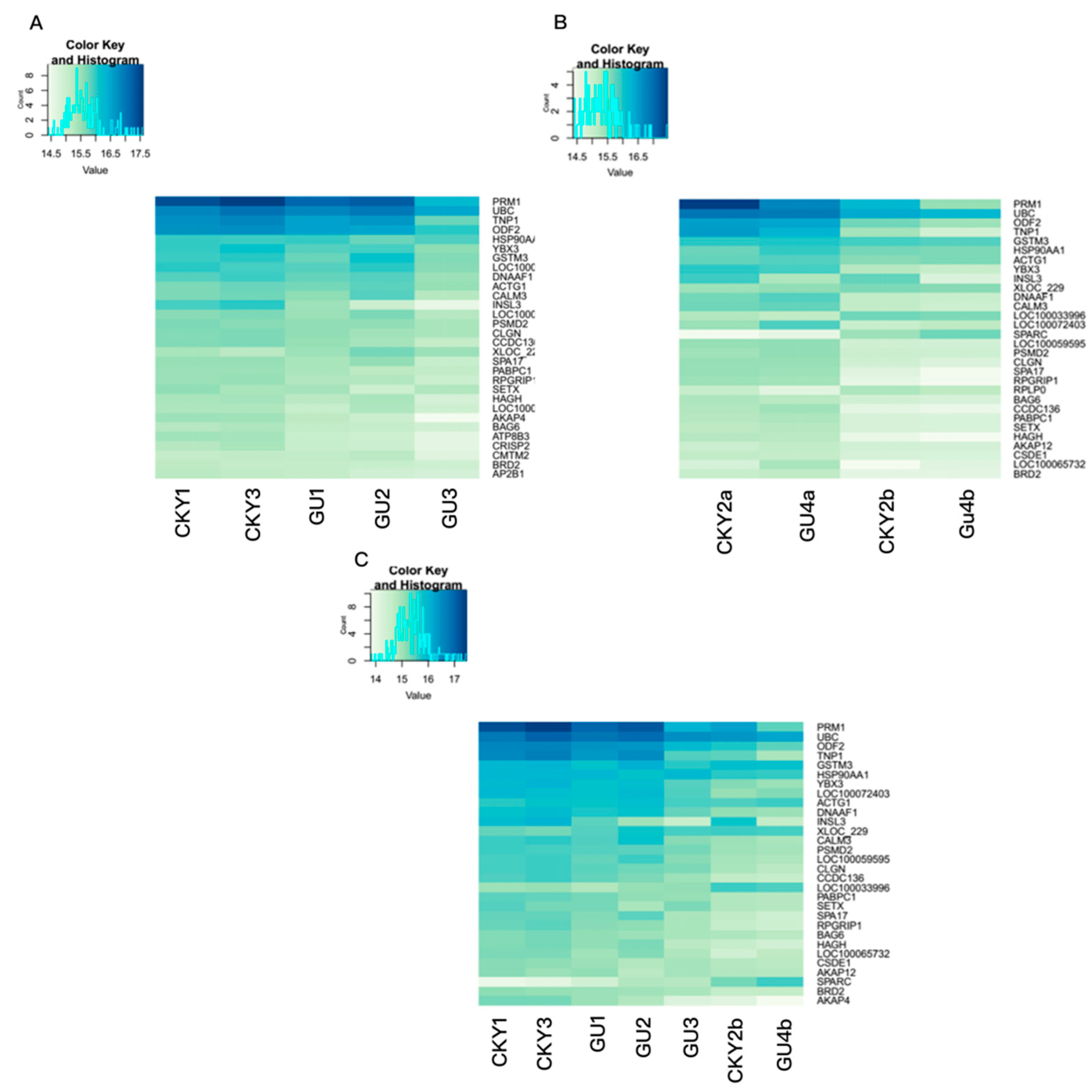
3.2. Top Genes Expressed in Horse Testicular Tissue

The top 30 genes expressed in the testicular tissue in the three comparisons are shown in Figure 2. Strikingly, eight genes, PRM1, UBC, TNP1, ODF2, HSP90AA1, YBX3, GSTM3, and ACTG1, were included among the top 10 genes in all three comparisons, suggesting that these eight genes are highly expressed in horse testes. As shown in Figure 2B,C, compared to normal DTs, UDTs exhibited downregulation of some genes, including TNP1, DNAAF1, CALM3, CLGN, SPA17, and RPGRIP1; these results reveal the existence of different expression patterns between normal and retained testes.

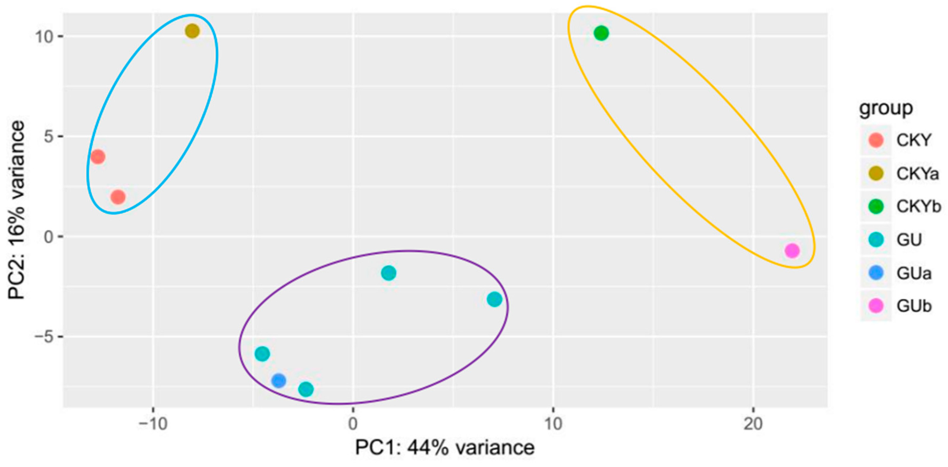
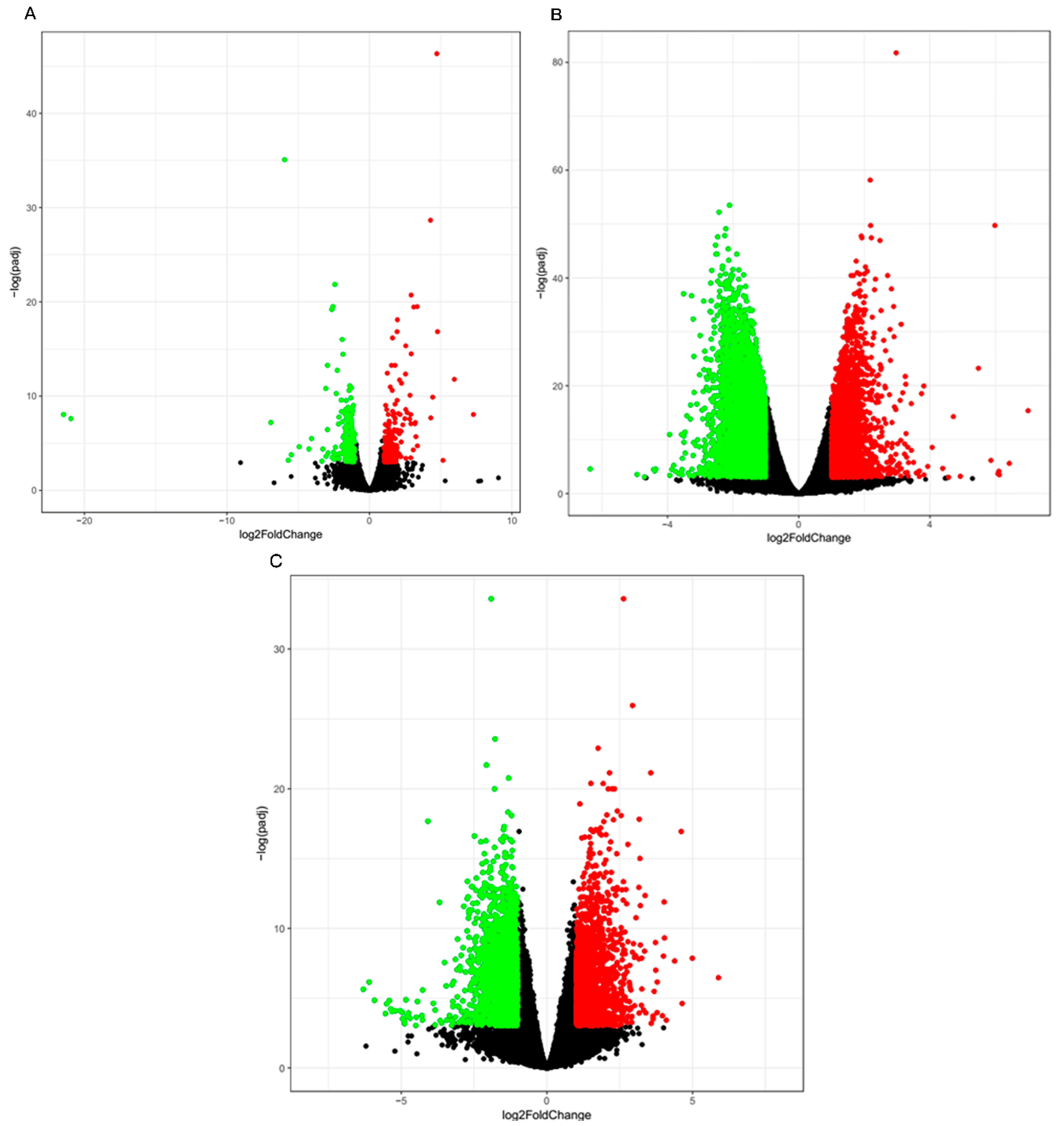
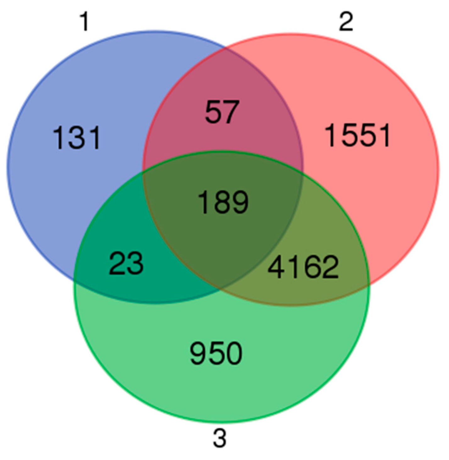
3.3. Differential Gene Expression Analysis

The RNA-seq technique enabled analysis of the differential expression profiles via analysis of the transcript abundance with a high sensitivity. When a p < 0.05 and a |log2 fold change| ≥1 were used as cutoffs, a total of 400, 5959, and 5324 DEGs, respectively, were identified in the three comparisons defined above. A total of 191 DEGs were downregulated in the Guanzhong horses compared to the Chakouyi horses while 219 DEGs were upregulated (Figure 3A). Sets of 3341 and 2618 transcripts were downregulated and upregulated in the CKY2b and GU4b testes, respectively, compared to the CKY2a and GU4a testes (Figure 3B). A total of 3049 genes were downregulated in UDTs (CKY2b and GU4b) compared to normal horse testes (CKY1, CKY3, GU1, GU2, and GU3) while 2275 genes were upregulated in these tissues (Figure 3C). Many genes showed differential expression between DTs and UDTs, as shown in Figure 3B,C, indicating that the expression of these genes might be associated with the reproductive performance of cryptorchids. The top 10 genes that were upregulated and downregulated in the three groups are shown in Tables S3–S5.

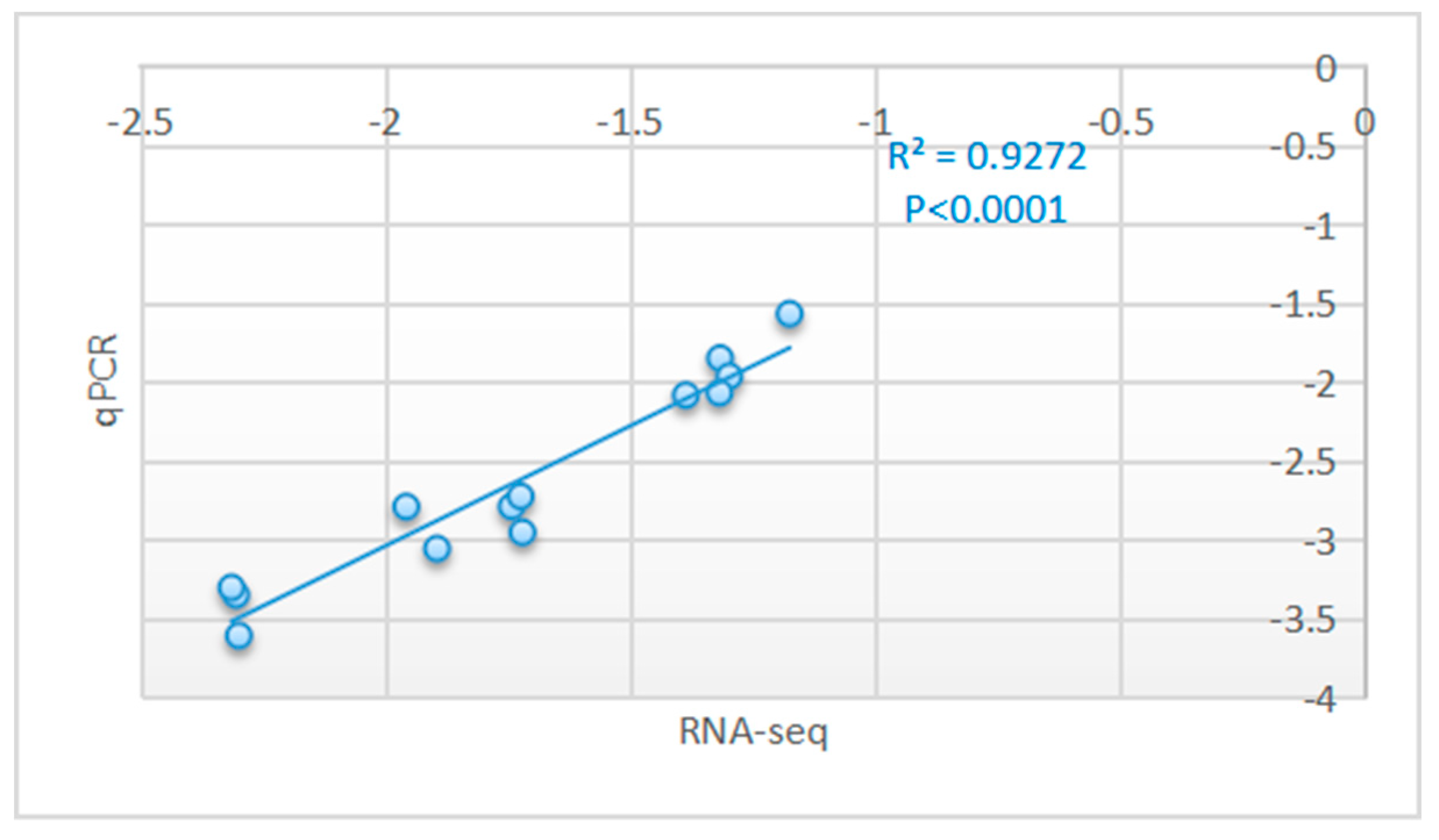
3.4. Real-Time qPCR Validation of the RNA-Seq Results

To validate the gene expression results obtained by sequencing, the expression levels of 13 DEGs, namely, ATP1A4, ROPN1, NME8, CATSPER3, CATSPER1, AKAP4, MNS1, CABS1, LRGUK, TSGA10, PRM1, CAPZA3, and ENKUR, were analyzed through qPCR. Detailed information on the primers for the 13 genes is given in Table S6. The log2 fold change values determined by RNA-seq and the log2 values determined by qPCR through the 2−ΔΔCt method with normalization to GAPDH for the comparisons of these 13 genes are shown in the electronic Supplementary Materials in Table S7. The expression patterns of these 13 genes were consistent with the RNA-seq results according to a Pearson correlation coefficient of 0.9272 and p < 0.0001 (Figure 4); quantification of the differential expression levels of the genes indicated significant findings and validated the repeatability and reproducibility of the gene expression data in this study.

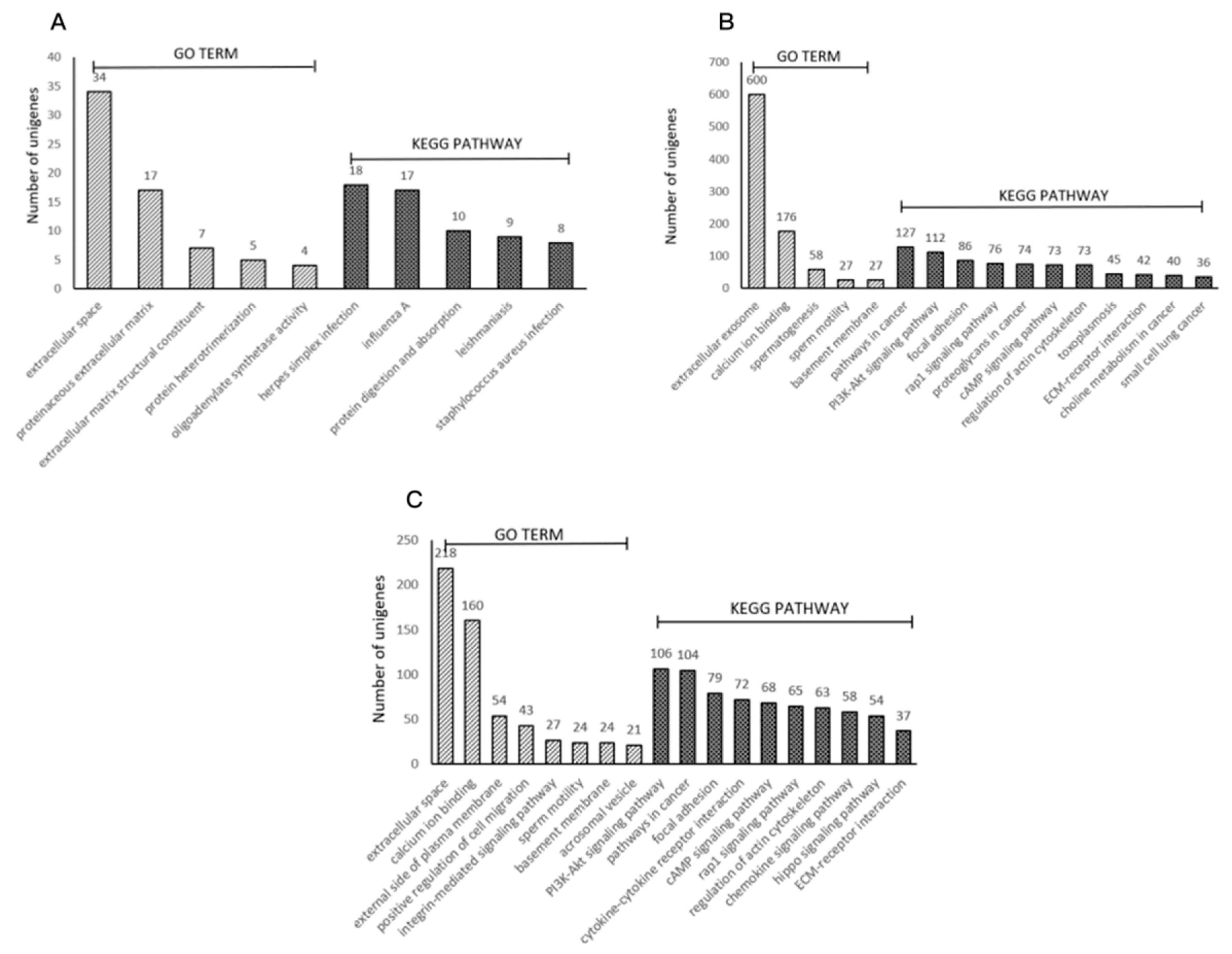
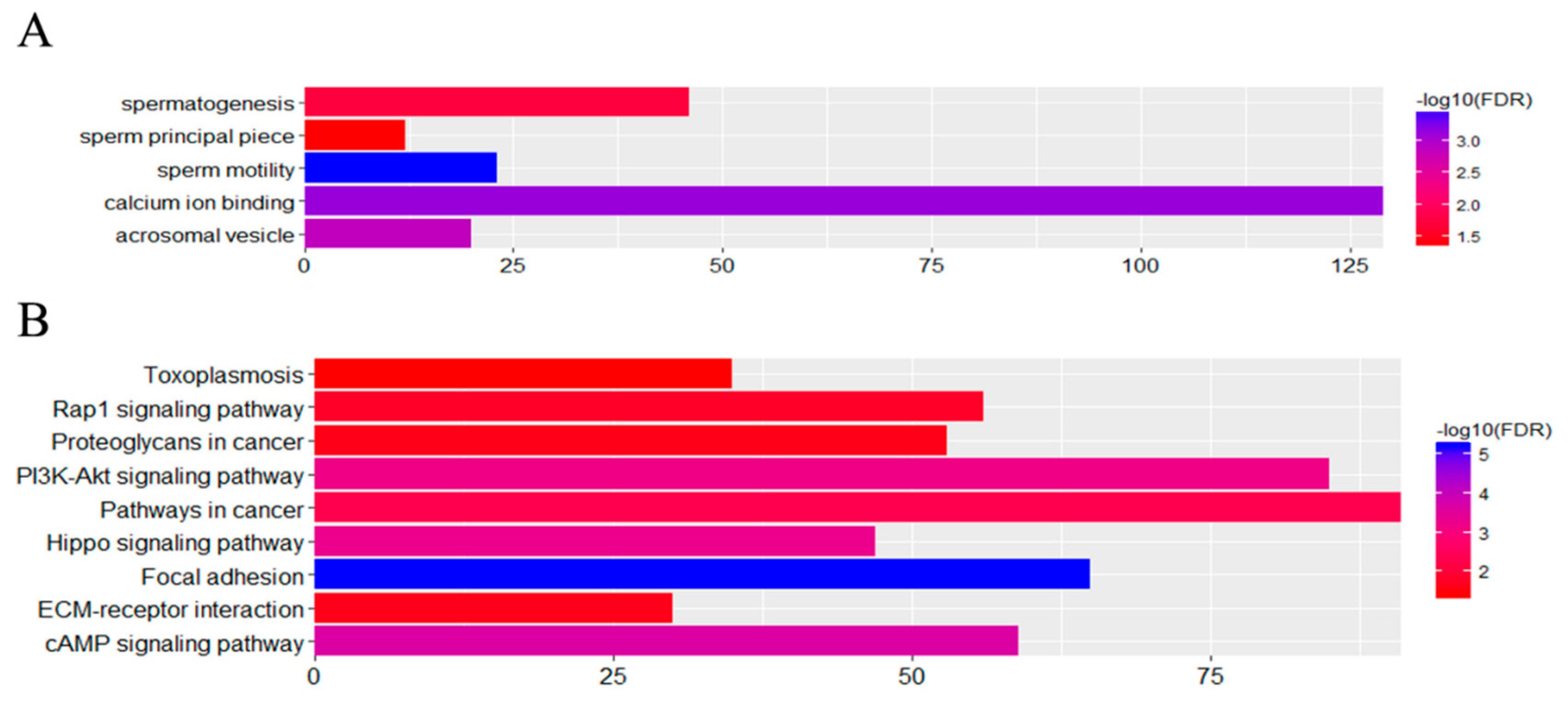
3.5. Functional Associations of the DEGs

Various genes cooperate with each other to exercise their biological functions. Accordingly, we performed Kyoto Encyclopedia of Genes and Genomes (KEGG) analysis and Gene Ontology (GO) analysis for terms in the biological process, cellular component, and molecular function categories to further elucidate the biological functions of the DEGs. The DEGs were significantly enriched for 5, 5, and 8 GO terms and 5, 11, and 10 KEGG pathways that met the criterion of an FDR < 0.05 in the three comparison groups (Figure 5). The most significantly enriched GO terms were the extracellular space term (GO: 0005615) with 34 associated genes, the extracellular exosome term (GO: 0070062) with 600 associated genes, and the extracellular space term (GO: 0005615) with 218 associated genes (Figure 5). In particular, the DEGs enriched for the spermatogenesis, sperm motility, and acrosomal vesicle terms are associated with sperm structure and function. The KEGG pathway terms showing the highest levels of significance were the herpes simplex infection term (ecb05168) with 18 enriched DEGs, the pathways in cancer term (ecb05200) with 127 enriched DEGs, and the PI3K-Akt signaling pathway term (ecb04151) with 106 enriched DEGs (Figure 5).

3.6. Effects of Cryptorchidism on Gene Expression

Our investigations focused on determining the effects of cryptorchidism on testicular gene expression levels in horses. An examination of the overlap of the three groups revealed that 4162 DEGs were shared by groups 2 and 3 when the DEGs between breeds were excluded (Figure 6). These DEGs were assigned to five GO terms and eight KEGG pathways with the criterion of an FDR < 0.05 (Figure 7). It is worth noting that within the GO cellular component category, the acrosomal vesicle (GO: 0001669) and sperm principal piece (GO: 0097228) terms were enriched while within the biological process category, the spermatogenesis (GO: 0007283) and sperm motility (GO: 0030317) terms were enriched (Figure 7). There were 84 DEGs associated with these functions that might influence sperm development and male reproductive performance. Thus, our results support the hypothesis that high abdominal temperature or other factors associated with cryptorchidism impact sperm production and relevant gene expression levels.

3.7. Single-Nucleotide Polymorphisms (SNPs) May Cause Cryptorchidism in Horses

Our investigations identified 1,032,196 SNPs in total, of which 146,610 were located in exonic regions. There were 48,083 missense mutations and 77,143 synonymous mutations. Herein, 46 SNPs, including 2 homozygous mutations and 44 heterozygous mutations, were detected in horses with unilateral cryptorchidism but not in normal horses. These variations occurred within 29 genes and are listed in Table 2.

4. Discussion

In this study, we compared gene expression levels between UDTs and DTs to determine the genetic effects of cryptorchidism in horses. The statistical power of the analysis of the present research was not so high, because only two samples, belonging to two individuals of Guanzhong and Chakouyi horse breeds, showing unilateral cryptorchidism, were available for the study. This is due to the rarity of the cryptorchidic condition being found in horses. A total of 84 DEGs were associated with functions that might influence sperm development and male reproductive performance; among them, 11 genes were downregulated and were associated with more than two functional terms in our study. These genes have been previously reported to be associated with the fibrous sheaths (FSs) of spermatozoa, nuclear morphology maintenance during meiotic prophase, sperm construction, and male infertility (Table S7). In particular, three of them, CATSPERD, CATSPER1, and CATSPER3, are sperm ion channel proteins involved in sperm hyperactivated motility, which facilitates sperm penetration through the zona pellucida [25,26,27,28]. The expression analysis in the current study, showed that CatSper1–4 was expressed in horse testes, consistent with previous research [25,29,30]. Furthermore, we found lower expression levels of CatSper1–4 in UDTs than in normal mature DTs. Other studies carried out on ejaculated sperm have found that CatSper transcript levels are positively correlated with sperm motility [31,32]; this association could be the result of essential roles of sperm CatSper transcripts in sperm functions, such as motility, capacitation, and the acrosomal reaction [31,33]. Therefore, cryptorchidism leads to reduced expression levels of the CatSper1–4 genes in UDTs and impaired fertility in horses.
The gene expression levels of AKAP3 and AKAP4 were significantly lower in UDTs than in DTs in this study. AKAP3 and AKAP4 are the most abundant structural protein components of the sperm FS [34]. It is generally accepted that the FS provides mechanical support and flexibility to the sperm flagellum and provides a scaffold for signaling enzymes and glycolysis that is necessary for hyperactivated motility during capacitation [34,35,36,37]. No research on the relationship between AKAP gene expression and cryptorchidism has been reported. Based on this study, AKAP3 and AKAP4 are thought to be downregulated in retained testes as a result of the abdominal environment.
ROPN1 has also been localized to the FS and binds to AKAP3 and AKAP4 [34,38]. ROPN1L and ROPN1 compensate for each other in the absence of the opposing protein, possibly to maintain AKAP3 incorporation in the FS. We found that the ROPN1 and ROPN1L genes both showed significantly lower expression in UDTs than in DTs, indicating that retained testicles have higher frequencies of sperm with dysplasia or do not produce sperm at all. SPAG6 deficiency causes sperm motility defects and morphological abnormalities [39]. The SPAG6 gene plays an important role in maintaining flagellar structure and function in mammals [39]. Therefore, cryptorchidism might significantly downregulate the expression of the ROPN1, ROPN1L, and SPAG6 genes.
SNP calling was used to clarify the genetic pathogenesis of equine cryptorchidism. A large number of SNPs were screened; however, only 46 missense mutations were detected in horses with unilateral cryptorchidism but not in normal horses. Unfortunately, none of them were consistent with the causal mutations reported previously. Among the 46 missense SNPs, two were homozygous, located within the PRICKLE3 and PPP1R42 genes, although no relationship was shown between cryptorchidism and these two genes. The PRICKLE3 gene has been found to regulate ciliogenesis and cilia growth in gastrocoel roof plates [40]. PPP1R42 contains a protein phosphatase-1-binding site and translocates from the apical nucleus to the centrosome at the base of the flagellum during spermiogenesis [41]. Therefore, the homozygous SNPs within the PRICKLE3 and PPP1R42 genes are considered to be related to horse anorchism, although further verification is needed.

5. Conclusions

In conclusion, DEGs between normal DTs and UDTs were identified, and many genes were up- or downregulated in UDTs as a result of cryptorchidism. The decreased expression of 11 genes is considered to be related to the impaired fertility of horses with cryptorchidism. In addition, two homozygous SNPs in the PRICKLE3 and PPP1R42 genes are proposed to cause cryptorchidism in horses. A limitation of the present study is the low sample size, since only two individuals showing unilateral cryptorchidism were available for analysis, but this condition is rare in horses. An increased sample size for analysis is aimed for in further research to corroborate the results of the present study.

Supplementary Materials

The following are available online at https://www.mdpi.com/2076-2615/10/1/102/s1. Additional supporting information may be found in the online version of this article. Table S1: Information on the horse testicular samples. Table S2: Top 10 unigenes that were upregulated or downregulated in the testes in Guanzhong horses compared with Chakouyi horses. Table S3: Top 10 unigenes with higher or lower expression in UDTs than in DTs. Table S4: Top 10 unigenes that were upregulated or downregulated in horses with UDTs compared with normal horses with DTs. Table S5: Information on the primers used in qPCR. Table S6: Log2 |fold change| values determined by RNA-seq and log2 values determined by qPCR through the 2−ΔΔCt method for 13 verified genes. Table S7: Functions of 11 genes affected by increased temperature. Figure S1: Testes of GU4 horses. (A): Normal DT; (B): UDT.

Author Contributions

Conceptualization: H.H. and C.L.; methodology: Y.G.; validation: H.H. and C.L.; formal analysis: Q.C.; investigation: H.H., H.D.; resources: W.L. and C.L.; data curation: H.H. and Q.C.; writing—original draft preparation: H.H.; writing—review and editing: C.L., and R.D.; visualization: Y.G.; supervision: C.L.; project administration: H.H. and C.L.; funding acquisition: H.H., J.L. and C.L. All authors have read and agreed to the published version of the manuscript.

Funding

This work was supported by the National Natural Science Foundation of China (81270439 and 31601915) and the PhD Start-up Fund of Henan University of Animal Husbandry and Economy (906/24030042 and 906/24030103).

Conflicts of Interest

The authors declare no conflict of interest.

Data Availability

The full set of raw data from this study has been deposited in the National Center for Biotechnology Information’s Sequence Read Archive (SRA) and is accessible through the SRA accession number SRR8303651-830365160.

References

  1. Eik-Nes, K.B. Secretion of testosterone by the eutopio and the cryptorchid testes in the same dog. Can. J. Physiol. Pharmacol. 1966, 144, 629–633. [Google Scholar] [CrossRef]
  2. Kogan, S.J. Fertility in cryptorchidism. Eur. J. Pediatrics 1987, 146 (Suppl. 2), S21–S24. [Google Scholar] [CrossRef] [PubMed]
  3. Benson, R.C.; Beard, C.M.; Kelalis, P.P.; Kurland, L.T. Malignant potential of the cryptorchid testis. Mayo Clin. Proc. 1991, 66, 372–378. [Google Scholar] [CrossRef]
  4. Hutson, J.M.; Baker, M.; Terada, M.; Zhou, B.; Paxton, G. Hormonal control of testicular descent and the cause of cryptorchidism. Reprod. Fertil. Dev. 1994, 6, 151–156. [Google Scholar] [CrossRef] [PubMed]
  5. Hutson, J.M. A biphasic model for the hormonal control of testicular descent. Lancet 1985, 326, 419–421. [Google Scholar] [CrossRef]
  6. Satokata, I.; Benson, G.; Maas, R. Sexually dimorphic sterility phenotypes in Hoxa10-deficient mice. Nature 1995, 374, 460–463. [Google Scholar] [CrossRef]
  7. Zimmermann, S.; Steding, G.; Emmen, J.M.; Brinkmann, A.O.; Nayernia, K.; Holstein, A.F.; Engel, W.; Adham, I.M. Targeted disruption of the INSL3 gene causes bilateral cryptorchidism. Mol. Endocrinol. 1999, 13, 681–691. [Google Scholar] [CrossRef]
  8. Nef, S.; Parada, L.F. Cryptorchidism in mice mutant for INSL3. Nat. Genet. 1999, 22, 295–299. [Google Scholar] [CrossRef]
  9. Overbeek, P.A.; Gorlov, I.P.; Sutherland, R.W.; Houston, J.B.; Harrison, W.R.; Boettger-Tong, H.L.; Bishop, C.E.; Agoulnik, A.I. A transgenic insertion causing cryptorchidism in mice. Genesis 2001, 30, 26–35. [Google Scholar] [CrossRef]
  10. El Houate, B.; Rouba, H.; Sibai, H.; Barakat, A.; Chafik, A.; Chadli el, B.; Imken, L.; Bogatcheva, N.V.; Feng, S.; Agoulnik, A.I.; et al. Novel mutations involving the INSL3 gene associated with cryptorchidism. J. Urol. 2007, 177, 1947–1951. [Google Scholar] [CrossRef]
  11. Zimmermann, S.; Schottler, P.; Engel, W.; Adham, I.M. Mouse Leydig insulin-like (Ley I-L) gene: Structure and expression during testis and ovary development. Mol. Endocrinol. 1997, 13, 681–691. [Google Scholar] [CrossRef] [PubMed]
  12. Foresta, C.; Zuccarello, D.; Garolla, A.; Ferlin, A. Role of hormones, genes and environment in human cryptorchidism. Endocr. Rev. 2008, 29, 560–580. [Google Scholar] [CrossRef] [PubMed] [Green Version]
  13. Lu, P.; Wang, Y.; Wang, F.; Huang, J.; Zeng, Y.; He, D.; Huang, H.; Cheng, Z. Genetic analysis of HOXA11 gene in Chinese patients with cryptorchidism. Andrologia 2017, 50, e12790. [Google Scholar] [CrossRef]
  14. Barthold, J.S.; Wang, Y.; Kolon, T.F.; Kollin, C.; Nordenskjöld, A.; Olivant Fisher, A.; Figueroa, T.E.; BaniHani, A.H.; Hagerty, J.A.; Gonzaléz, R.; et al. Pathway analysis supports association of nonsyndromic cryptorchidism with genetic loci linked to cytoskeleton dependent functions. Hum. Reprod. 2015, 30, 2439–2451. [Google Scholar] [CrossRef] [PubMed] [Green Version]
  15. Barthold, J.S.; Wang, Y.; Kolon, T.F.; Kollin, C.; Nordenskjöld, A.; Olivant Fisher, A.; Figueroa, T.E.; BaniHani, A.H.; Hagerty, J.A.; Gonzalez, R.; et al. Phenotype specific association of the TGFBR3 locus with nonsyndromic cryptorchidism. J. Urol. 2015, 193, 1637–1645. [Google Scholar] [CrossRef] [PubMed] [Green Version]
  16. Ferlin, A.; Garolla, A.; Bettella, A.; Bartoloni, L.; Vinanzi, C.; Roverato, A.; Foresta, C. Androgen receptor gene CAG and GGC repeat lengths in cryptorchidism. Eur. J. Endocrinol. 2005, 152, 419–425. [Google Scholar] [CrossRef] [PubMed]
  17. Nagraj, S.; Seah, G.J.; Farmer, P.J.; Davies, B.; Southwell, B.; Lewis, A.G.; Hutson, J.M. The development and anatomy of the gubernaculum in Hoxa11 knockout mice. J. Pediatric Surg. 2011, 46, 387–392. [Google Scholar] [CrossRef]
  18. Hitzemann, R.; Bottomly, D.; Darakjian, P.; Walter, N.; Iancu, O.; Searles, R.; Wilmot, B.; McWeeney, S. Genes, behavior and next-generation RNA sequencing. Genes Brain Behav. 2013, 12, 1–12. [Google Scholar] [CrossRef]
  19. Marston, D.A.; McElhinney, L.M.; Ellis, R.J.; Horton, D.L.; Wise, E.L.; Leech, S.L.; David Lamballerie, X.; Fooks, A.R. Next generation sequencing of viral RNA genomes. BMC Genom. 2013, 14, 444. [Google Scholar] [CrossRef] [Green Version]
  20. Zhou, Y.; Sun, J.; Li, C.; Wang, Y.; Li, L.; Cai, H.; Lan, X.; Lei, C.; Zhao, X.; Chen, H. Characterization of Transcriptional Complexity during Adipose Tissue Development in Bovines of Different Ages and Sexes. PLoS ONE 2014, 9, e101261. [Google Scholar] [CrossRef]
  21. Sultan, M.; Schulz, M.H.; Richard, H.; Magen, A.; Klingenhoff, A.; Scherf, M.; Seifert, M.; Borodina, T.; Soldatov, A.; Parkhomchuk, D.; et al. A global view of gene activity and alternative splicing by deep sequencing of the human transcriptome. Science 2008, 321, 956–960. [Google Scholar] [CrossRef] [PubMed] [Green Version]
  22. Richard, H.; Schulz, M.H.; Sultan, M.; Nürnberger, A.; Schrinner, S.; Balzereit, D.; Dagand, E.; Rasche, A.; Lehrach, H.; Vingron, M.; et al. Prediction of alternative isoforms from exon expression levels in RNA-Seq experiments. Nucleic Acids Res. 2010, 38, e112. [Google Scholar] [CrossRef] [PubMed] [Green Version]
  23. Mortazavi, A.; Williams, B.A.; McCue, K.; Schaeffer, L.; Wold, B. Mapping and quantifying mammalian transcriptomes by RNA-Seq. Nat. Methods 2008, 5, 621–628. [Google Scholar] [CrossRef]
  24. Paria, N.; Raudsepp, T.; Pearks Wilkerson, A.J.; O’Brien, P.C.; Ferguson-Smith, M.A.; Love, C.C.; Arnold, C.; Rakestraw, P.; Murphy, W.J.; Chowdhary, B.P. A gene catalogue of the euchromatic male-specific region of the horse Y chromosome: Comparison with human and other mammals. PLoS ONE 2011, 6, e21374. [Google Scholar] [CrossRef] [PubMed]
  25. Ren, D.; Navarro, B.; Perez, G.; Jackson, A.C.; Hsu, S.; Shi, Q.; Tilly, J.L.; Clapham, D.E. A sperm ion channel required for sperm motility and male fertility. Nature 2001, 413, 603–609. [Google Scholar] [CrossRef]
  26. Qi, H.; Moran, M.M.; Navarro, B.; Chong, J.A.; Krapivinsky, G.; Krapivinsky, L.; Kirichok, Y.; Ramsey, I.S.; Quill, T.A.; Clapham, D.E. All four CatSper ion channel proteins are required for male fertility and sperm cell hyperactivated motility. Proc. Natl. Acad. Sci. USA 2007, 104, 1219–1223. [Google Scholar] [CrossRef] [Green Version]
  27. Quill, T.A.; Sugden, S.A.; Rossi, K.L.; Doolittle, L.K.; Hammer, R.E.; Garbers, D.L. Hyperactivated sperm motility driven by CatSper2 is required for fertilization. Proc. Natl. Acad. Sci. USA 2003, 100, 14869–14874. [Google Scholar] [CrossRef] [Green Version]
  28. Xia, J.; Reigada, D.; Mitchell, C.H.; Ren, D. CATSPER channel-mediated Ca2+ entry into mouse sperm triggers a tail-to-head propagation. Biol. Reprod. 2007, 77, 551–559. [Google Scholar] [CrossRef] [Green Version]
  29. Quill, T.A.; Ren, D.; Clapham, D.E.; Garbers, D.L. A voltage-gated ion channel expressed specifically in spermatozoa. Proc. Natl. Acad. Sci. USA 2001, 98, 12527–12531. [Google Scholar] [CrossRef] [Green Version]
  30. Song, C.; Gao, B.; Wu, H.; Xie, Y.; Wang, X.; Li, B.; Chen, G.; Mao, J. Molecular cloning, spatial and temporal expression analysis of CatSper genes in the Chinese Meishan pigs. Reprod. Biol. Endocrinol. 2011, 9, 1–12. [Google Scholar] [CrossRef] [Green Version]
  31. Li, H.G.; Ding, X.F.; Liao, A.H.; Kong, X.B.; Xiong, C.L. Expression of CatSper family transcripts in the mouse testis during post-natal development and human ejaculated spermatozoa: Relationship to sperm motility. Mol. Hum. Reprod. 2007, 13, 299–306. [Google Scholar] [CrossRef] [PubMed] [Green Version]
  32. Lambard, S.; Galeraud-Denis, I.; Martin, G.; Levy, R.; Chocat, A.; Carreau, S. Analysis and significance of mRNA in human ejaculated sperm from normozoospermic donors: Relationship to sperm motility and capacitation. Mol. Hum. Reprod. 2004, 10, 535–541. [Google Scholar] [CrossRef] [PubMed] [Green Version]
  33. Gur, Y.; Breitbart, H. Mammalian sperm translate nuclear-encoded proteins by mitochondrial-type ribosomes. Genes Dev. 2006, 20, 411–416. [Google Scholar] [CrossRef] [PubMed] [Green Version]
  34. Eddy, E.M.; Toshimori, K.; O’Brien, D.A. Fibrous sheath of mammalian spermatozoa. Microsc. Res. Tech. 2003, 61, 103–115. [Google Scholar] [CrossRef] [PubMed]
  35. Krisfalusi, M.; Miki, K.; Magyar, P.L.; O’Brien, D.A. Multiple glycolytic enzymes are tightly bound to the fibrous sheath of mouse spermatozoa. Biol. Reprod. 2006, 75, 270–278. [Google Scholar] [CrossRef] [PubMed] [Green Version]
  36. Miki, K.; Qu, W.; Goulding, E.H.; Willis, W.D.; Bunch, D.O.; Strader, L.F.; Perreault, S.D.; Eddy, E.M.; O’Brien, D.A. Glyceraldehyde 3-phosphate dehydrogenase-S, a sperm-specific glycolytic enzyme, is required for sperm motility and male fertility. Proc. Natl. Acad. Sci. USA 2004, 101, 16501–16506. [Google Scholar] [CrossRef] [Green Version]
  37. Miki, K.; Willis, W.D.; Brown, P.R.; Goulding, E.H.; Fulcher, K.D.; Eddy, E.M. Targeted disruption of the Akap4 gene causes defects in sperm flagellum and motility. Dev. Biol. 2002, 248, 331–342. [Google Scholar] [CrossRef] [Green Version]
  38. Brown, P.R.; Miki, K.; Harper, D.B.; Eddy, E.M. A-kinase anchoring protein 4 binding proteins in the fibrous sheath of the sperm flagellum. Biol. Reprod. 2003, 68, 2241–2248. [Google Scholar] [CrossRef] [Green Version]
  39. Teves, M.E.; Sears, P.R.; Li, W.; Zhang, Z.; Tang, W.; van Reesema, L.; Costanzo, R.M.; Davis, C.W.; Knowles, M.R.; Strauss, J.F., 3rd; et al. Sperm-Associated Antigen 6 (SPAG6) Deficiency and Defects in Ciliogenesis and Cilia Function: Polarity, Density, and Beat. PLoS ONE 2014, 9, e107271. [Google Scholar] [CrossRef] [Green Version]
  40. Chu, C.W.; Ossipova, O.; Ioannou, A.; Sokol, S.Y. Prickle3 synergizes with Wtip to regulate basal body organization and cilia growth. Sci. Rep. 2016, 6, 24104. [Google Scholar] [CrossRef] [Green Version]
  41. Devaul, N.; Wang, R.; Sperry, A.O. PPP1R42, a PP1 binding protein, regulates centrosome dynamics in ARPE-19 cells. Biol. Cell 2013, 105, 359–371. [Google Scholar] [CrossRef] [PubMed] [Green Version]
Figure 1. PCA of horse testicular tissues. CKYa represents CKY2a; CKY represents CKY1 and CKY3; GU represents GU1, GU2, GU3, and GU5; GUa represents GU4a; CKY2b represents CKY2b; and GUb represents GU4b.
Animals 10 00102 g001
Figure 2. Top 30 genes expressed in horse testicular tissue. (A) Gene expression in Guanzhong and Chakouyi horses. (B) Gene expression in DTs and UDTs of cryptorchids. (C) Gene expression in DTs of normal horses and UDTs of cryptorchids.
Animals 10 00102 g002
Figure 3. Volcano plot displaying the DEGs in horse testicular tissue. Red dots: upregulated genes. Green dots: downregulated genes. (A) DEGs between Guanzhong and Chakouyi horses. (B) DEGs between DTs and UDTs of cryptorchids. (C) DEGs between DTs of normal horses and UDTs of cryptorchids.
Animals 10 00102 g003
Figure 4. Correlations in the mRNA expression levels of 13 randomly selected DEGs in equine testicular tissues using RNA-seq and qPCR. The x- and y-axes show the log2 ratios of mRNA levels as measured by RNA-seq and qPCR, respectively.
Animals 10 00102 g004
Figure 5. Distributions of the GO and KEGG categories assigned to the DEGs in horse testicular tissues. The x-axis indicates the GO and KEGG groups, and the y-axis indicates the number of genes associated with the group. (A) Functional characterization of the DEGs between Guanzhong and Chakouyi horses. (B) Functional characterization of the DEGs between DTs and UDTs of cryptorchids. (C) Functional characterization of the DEGs between DTs of normal horses and UDTs of cryptorchids. The number of genes is shown on top of each bar.
Animals 10 00102 g005
Figure 6. Venn diagram of the unigenes distributed in three groups. Group 1: DEGs between Guanzhong and Chakouyi horses; group 2: DEGs between DTs and UDTs of cryptorchids; group 3: DEGs between DTs of normal horses and UDTs of cryptorchids.
Animals 10 00102 g006
Figure 7. Distribution of the GO and KEGG categories assigned to the DEGs in groups 2 and 3. (A) GO terms assigned to 4162 DEGs. (B) Eight KEGG pathways assigned to 4162 DEGs.
Animals 10 00102 g007
Table 1. Summary of the transcripts in horse testicular tissues.
SampleRaw ReadsClean ReadsClean BasesTotal Mapped ReadsMultiple Mapped ReadsUniquely Mapped Reads
CKY161,721,59259,762,1588,700,950,79252,179,635 (87.31%)813,963 (1.36%)50,385,369 (84.31%)
CKY2a77,719,68675,527,31210,936,704,23366,175,695 (87.62%)1,074,061 (1.42%)63,821,947 (84.50%)
CKY2b72,480,27270,588,36010,234,970,74462,176,229 (88.08%)1,002,706 (1.42%)60,005,651 (85.01%)
CKY392,420,91488,846,73612,869,802,57378,285,911 (88.11%)1,305,326 (1.47%)75,421,193 (84.89%)
GU174,459,30072,055,25410,494,742,85963,316,272 (87.87%)994,823 (1.38%)61,130,797 (84.84%)
GU273,099,62670,758,15410,296,493,98461,319,858 (86.66%)1,017,616 (1.44%)59,085,846 (83.50%)
GU379,108,43076,613,63211,153,734,66367,734,267 (88.41%)1,113,341 (1.45%)65,306,502 (85.24%)
GU4a73,056,30870,657,31810,241,905,46260,878,044 (86.16%)980,165 (1.39%)58,722,122 (83.11%)
GU4b65,345,24863,299,0929,212,135,31455,446,209 (87.59%)869,078 (1.37%)53,551,478 (84.60%)
GU562,041,33859,931,8948,714,823,98652,460,612 (87.53%)831,832 (1.39%)50,626,486 (84.47%)
average73,145,27170,803,99110,285,626,46161,997,273 (87.53%)1,000,291 (1.41%)59,805,739 (84.45%)
Table 2. Candidate SNPs associated with cryptorchidism.
GeneNCBI Accession NumberExonNucleotideProteinGenotype
Normal HorseCryptorchid Horse
LOC102147809XM_005602655.2exon 1c.A14Gp.E5GAAAG
ZNF37AXM_005602647.2exon 5c.T450Ap.H150QTTTA
DDX60LXM_014737782.1exon 22c.G2747Ap.R916QGGGA
FNIP2XM_005607793.2exon 13c.C2612Tp.T871MGGGA
c.C2231Tp.T744IGGGA
C3H16orf46XM_001501937.3exon 3c.G62Cp.S21TCCCG
GK2NM_001256933.1exon 1c.A230Gp.E77GAAAG
BMP2KXM_014738530.1exon 10c.C1109Tp.T370MGGGA
PNPLA8XM_005609080.2exon 10c.C2110Tp.P704SGGGA
ALDH9A1XM_001492799.4exon 1c.C125Tp.A42VCCCT
LOC100058290XM_001495326.4exon 7c.C479Gp.A160GGGGC
GPSM2XM_001493568.5exon 14c.G2006Ap.S669NCCCT
TMEM198XM_001494150.4exon 1c.G242Tp.R81LGGGT
LOC102148548XM_014741362.1exon 3c.G2650Ap.D884NCCCT
c.G2627Cp.R876TCCCG
c.G2068Ap.V690MCCCT
c.A1679Tp.E560VTTTA
c.A1648Gp.M550VTTTC
c.C581Tp.P194LGGGA
c.A437Tp.Q146LTTTA
c.A369Tp.L123FTTTA
c.T362Cp.M121TAAAG
c.A354Cp.E118DTTTG
c.G278Ap.S93NCCCT
PPP1R42XM_005613086.2exon 8c.A799Gp.I267VAAGG
TMEM68NM_001309262.1exon 1c.A70Gp.I24VAAAG
HACE1XM_014734298.1exon 20c.A2257Gp.M753VTTTC
FBF1XM_005597144.2exon 9c.C614Tp.P205LCCCT
ABCA9XM_001916991.3exon 8c.T974Cp.V325ATTTC
HIRIP3XM_014730026.1exon 4c.A958Gp.K320ETTTC
ANKRD31XM_014730406.1exon 11c.G1451Tp.R484LGGGT
exon 15c.A2804Gp.Y935CAAAG
exon 19c.T3918Ap.H1306QTTTA
c.C3922Tp.R1308WCCCT
RMDN2XM_005600050.2exon 2c.A13Gp.T5ATTTC
VWA8XM_001493030.5exon 41c.G5224Ap.A1742TGGGA
CCDC168XM_014732045.1exon 3c.A16781Gp.E5594GTTTC
c.C14732Gp.P4911RGGGC
c.A10753Tp.N3585YTTTA
c.G10363Ap.E3455KCCCT
POM121L2XM_001495084.5exon 1c.A397Cp.T133PTTTG
SKIV2LXM_001492580.3exon 9c.C833Tp.P278LCCCT
XPO5XM_005603953.2exon 19c.A1996Gp.I666VTTTC
MMP9NM_001111302.1exon 9c.A1414Gp.T472AAAAG
PSD3XM_001488201.5exon 2c.G144Ap.M48ICCCT
PRICKLE3XM_001495412.5exon 8c.G1222Tp.A408SCCAA

