
Methicillin-Resistant Staphylococcus epidermidis Lineages in the Nasal and Skin Microbiota of Patients Planned for Arthroplasty Surgery

 , , , and
Abstract
:1. Introduction
2. Materials and Methods
2.1. Culture, DNA Extraction, and Illumina Sequencing
2.2. Phylogenetic Analysis
2.3. Identification of Genes and Gene Variants Associated with Antimicrobial Resistance
2.4. Identification of STs
2.5. Identification of Genetic Traits in S. epidermidis Associated with PJIs
2.6. Statistics
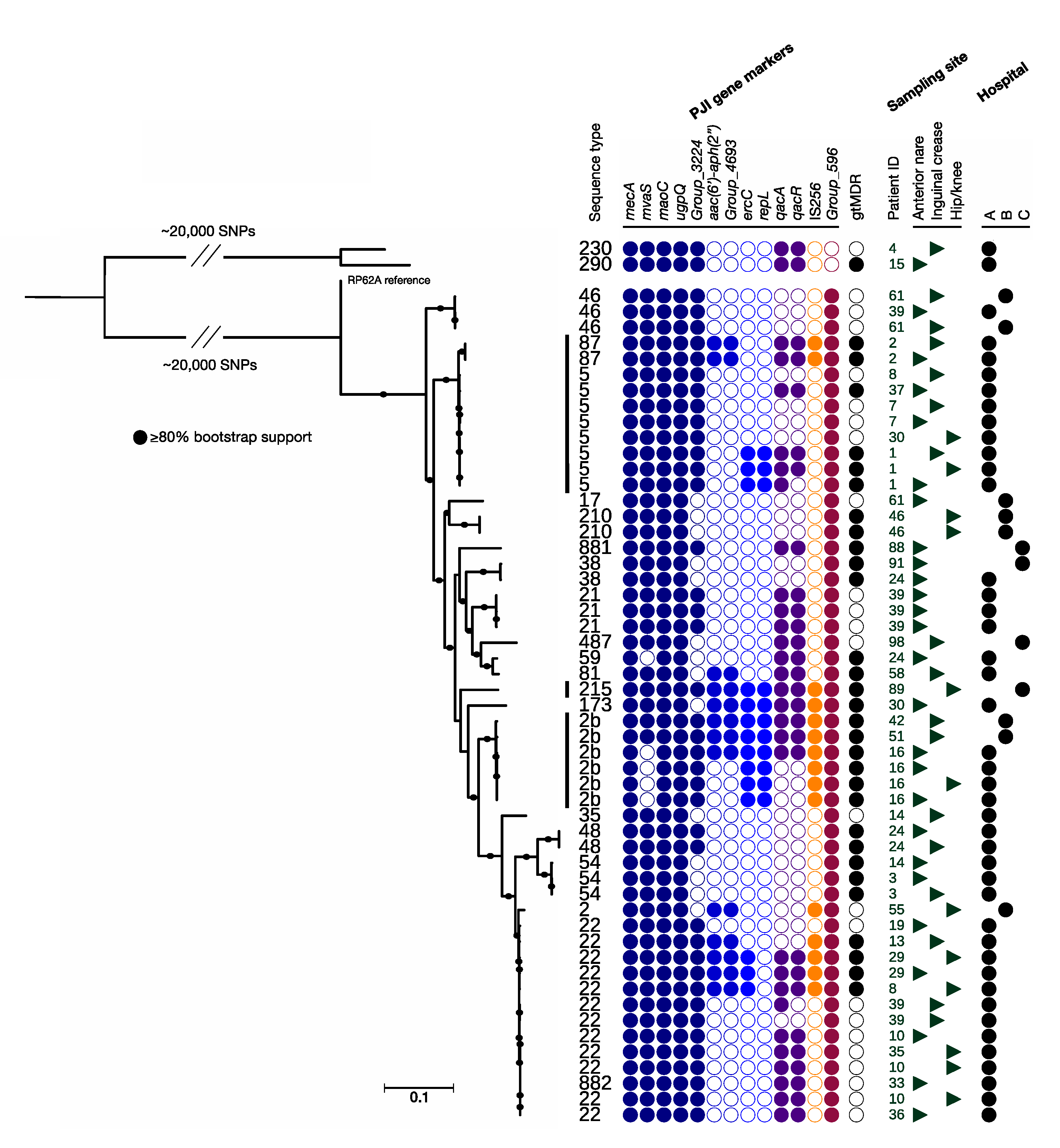
3. Results
4. Discussion
5. Conclusions
Supplementary Materials
Author Contributions
Funding
Institutional Review Board Statement
Informed Consent Statement
Data Availability Statement
Conflicts of Interest
References
- Stacy, A.; Belkaid, Y. Microbial guardians of skin health. Science 2019, 363, 227–228. [Google Scholar] [CrossRef] [PubMed]
- Becker, K.; Heilmann, C.; Peters, G. Coagulase-negative staphylococci. Clin. Microbiol. Rev. 2014, 27, 870–926. [Google Scholar] [CrossRef] [PubMed] [Green Version]
- Zimmerli, W. Clinical presentation and treatment of orthopaedic implant-associated infection. J. Intern. Med. 2014, 276, 111–119. [Google Scholar] [CrossRef] [PubMed]
- Cahill, J.L.; Shadbolt, B.; Scarvell, J.M.; Smith, P.N. Quality of life after infection in total joint replacement. J. Orthop. Surg. 2008, 16, 58–65. [Google Scholar] [CrossRef] [PubMed]
- Zmistowski, B.; Karam, J.A.; Durinka, J.B.; Casper, D.S.; Parvizi, J. Periprosthetic joint infection increases the risk of one-year mortality. J. Bone Jt. Surg. Am. 2013, 95, 2177–2184. [Google Scholar] [CrossRef] [Green Version]
- Gundtoft, P.H.; Pedersen, A.B.; Varnum, C.; Overgaard, S. Increased Mortality After Prosthetic Joint Infection in Primary THA. Clin. Orthop. Relat. Res. 2017, 475, 2623–2631. [Google Scholar] [CrossRef] [Green Version]
- Kapadia, B.H.; Berg, R.A.; Daley, J.A.; Fritz, J.; Bhave, A.; Mont, M.A. Periprosthetic joint infection. Lancet 2016, 387, 386–394. [Google Scholar] [CrossRef]
- Inacio, M.C.S.; Graves, S.E.; Pratt, N.L.; Roughead, E.E.; Nemes, S. Increase in Total Joint Arthroplasty Projected from 2014 to 2046 in Australia: A Conservative Local Model with International Implications. Clin. Orthop. Relat. Res. 2017, 475, 2130–2137. [Google Scholar] [CrossRef] [Green Version]
- Nemes, S.; Rolfson, O.; W-Dahl, A.; Garellick, G.; Sundberg, M.; Karrholm, J.; Robertsson, O. Historical view and future demand for knee arthroplasty in Sweden. Acta Orthop. 2015, 86, 426–431. [Google Scholar] [CrossRef] [Green Version]
- Kurtz, S.M.; Lau, E.C.; Son, M.S.; Chang, E.T.; Zimmerli, W.; Parvizi, J. Are We Winning or Losing the Battle with Periprosthetic Joint Infection: Trends in Periprosthetic Joint Infection and Mortality Risk for the Medicare Population. J. Arthroplast. 2018, 33, 3238–3245. [Google Scholar] [CrossRef]
- Nemes, S.; Gordon, M.; Rogmark, C.; Rolfson, O. Projections of total hip replacement in Sweden from 2013 to 2030. Acta Orthop. 2014, 85, 238–243. [Google Scholar] [CrossRef] [PubMed] [Green Version]
- Stefansdottir, A.; Johansson, D.; Knutson, K.; Lidgren, L.; Robertsson, O. Microbiology of the infected knee arthroplasty: Report from the Swedish Knee Arthroplasty Register on 426 surgically revised cases. Scand. J. Infect. Dis. 2009, 41, 831–840. [Google Scholar] [CrossRef] [PubMed]
- Lindgren, V.; Gordon, M.; Wretenberg, P.; Karrholm, J.; Garellick, G. Deep infection after total hip replacement: A method for national incidence surveillance. Infect. Control Hosp. Epidemiol. Off. J. Soc. Hosp. Epidemiol. Am. 2014, 35, 1491–1496. [Google Scholar] [CrossRef] [PubMed]
- Tande, A.J.; Patel, R. Prosthetic Joint Infection. Clin. Microbiol. Rev. 2014, 27, 302–345. [Google Scholar] [CrossRef] [PubMed] [Green Version]
- Benito, N.; Mur, I.; Ribera, A.; Soriano, A.; Rodriguez-Pardo, D.; Sorli, L.; Cobo, J.; Fernandez-Sampedro, M.; Del Toro, M.D.; Guio, L.; et al. The Different Microbial Etiology of Prosthetic Joint Infections according to Route of Acquisition and Time after Prosthesis Implantation, Including the Role of Multidrug-Resistant Organisms. J. Clin. Med. 2019, 8, 673. [Google Scholar] [CrossRef] [PubMed] [Green Version]
- Peel, T.N.; Cole, N.C.; Dylla, B.L.; Patel, R. Matrix-assisted laser desorption ionization time of flight mass spectrometry and diagnostic testing for prosthetic joint infection in the clinical microbiology laboratory. Diagn Microbiol. Infect. Dis. 2014. [Google Scholar] [CrossRef]
- Lourtet-Hascoet, J.; Felice, M.P.; Bicart-See, A.; Bouige, A.; Giordano, G.; Bonnet, E. Species and antimicrobial susceptibility testing of coagulase-negative staphylococci in periprosthetic joint infections. Epidemiol. Infect. 2018, 146, 1771–1776. [Google Scholar] [CrossRef]
- Flurin, L.; Greenwood-Quaintance, K.E.; Patel, R. Microbiology of polymicrobial prosthetic joint infection. Diagn Microbiol. Infect. Dis. 2019. [Google Scholar] [CrossRef]
- Cizmic, Z.; Feng, J.E.; Huang, R.; Iorio, R.; Komnos, G.; Kunutsor, S.K.; Metwaly, R.G.; Saleh, U.H.; Sheth, N.; Sloan, M. Hip and Knee Section, Prevention, Host Related: Proceedings of International Consensus on Orthopedic Infections. J. Arthroplast. 2019, 34, S255–S270. [Google Scholar] [CrossRef]
- Abouljoud, M.M.; Alvand, A.; Boscainos, P.; Chen, A.F.; Garcia, G.A.; Gehrke, T.; Granger, J.; Kheir, M.; Kinov, P.; Malo, M.; et al. Hip and Knee Section, Prevention, Operating Room Environment: Proceedings of International Consensus on Orthopedic Infections. J. Arthroplast. 2019, 34, S293–S300. [Google Scholar] [CrossRef]
- Aboltins, C.A.; Berdal, J.E.; Casas, F.; Corona, P.S.; Cuellar, D.; Ferrari, M.C.; Hendershot, E.; Huang, W.; Kuo, F.C.; Malkani, A.; et al. Hip and Knee Section, Prevention, Antimicrobials (Systemic): Proceedings of International Consensus on Orthopedic Infections. J. Arthroplast. 2019, 34, S279–S288. [Google Scholar] [CrossRef] [PubMed]
- Hellmark, B.; Söderquist, B.; Unemo, M.; Nilsdotter-Augustinsson, Å. Comparison of Staphylococcus epidermidis isolated from prosthetic joint infections and commensal isolates in regard to antibiotic susceptibility, agr type, biofilm production, and epidemiology. Int. J. Med. Microbiol. 2013, 303, 32–39. [Google Scholar] [CrossRef] [PubMed]
- Both, A.; Huang, J.; Lausmann, C.; Wisselberg, S.; Buettner, H.; Zahar, A.; Alawi, M.; Luoto, L.; Kendoff, D.; Rohde, H. Inter- and Intra-clonal Diversity in S. epidermidis from Prosthetic Joint Infections. In Proceedings of the 29th ECCMID, Amsterdam, The Netherlands, 13–16 April 2019. [Google Scholar]
- Kyung Wee, S.; Leboreiro, C.; Aydin, A.; Warren, S.; Mchugh, T.D.; Sharma, H. The Molecular Epidemiology of Staphylococcus Epidermidis Prosthetic Joint Infection in a UK Specialist Orthopaedic Hospital. In Proceedings of the 29th ECCMID, Amsterdam, The Netherlands, 13–16 April 2019. [Google Scholar]
- Sanchez Morillo, A.; Espinal, P.; Benito, N.; Rivera, A.; Gutierrez, C.; Pere, B.; Mirelis, B.; Coll, P.; Navarro, F. Staphylococcus epidermidis in prosthetic joint infections: Epidemiology and pathogenicity factors. In Proceedings of the 28th ECCMID, Madrid, Spain, 21–24 April 2018. [Google Scholar]
- Månsson, E.; Bech Johannesen, T.; Nilsdotter-Augustinsson, Å.; Söderquist, B.; Stegger, M. Comparative genomics of Staphylococcus epidermidis from prosthetic joint infections and nares highlights genetic traits associated to antimicrobial resistance, not virulence. Microb. Genom. 2020, 000504. [Google Scholar] [CrossRef]
- Hischebeth, G.T.; Randau, T.M.; Ploeger, M.M.; Friedrich, M.J.; Kaup, E.; Jacobs, C.; Molitor, E.; Hoerauf, A.; Gravius, S.; Wimmer, M.D. Staphylococcus aureus versus Staphylococcus epidermidis in periprosthetic joint infection—Outcome analysis of methicillin-resistant versus methicillin-susceptible strains. Diagn Microbiol. Infect. Dis. 2019, 93, 125–130. [Google Scholar] [CrossRef] [PubMed]
- Post, V.; Harris, L.G.; Morgenstern, M.; Mageiros, L.; Hitchings, M.D.; Meric, G.; Pascoe, B.; Sheppard, S.K.; Richards, R.G.; Moriarty, T.F. Comparative Genomics Study of Staphylococcus epidermidis Isolates from Orthopedic-Device-Related Infections Correlated with Patient Outcome. J. Clin. Microbiol. 2017, 55, 3089–3103. [Google Scholar] [CrossRef] [Green Version]
- Oh, J.; Byrd, A.L.; Deming, C.; Conlan, S.; Program, N.C.S.; Kong, H.H.; Segre, J.A. Biogeography and individuality shape function in the human skin metagenome. Nature 2014, 514, 59–64. [Google Scholar] [CrossRef] [Green Version]
- Sanzen, L.; Walder, M. Antibiotic resistance of coagulase-negative staphylococci in an orthopaedic department. J. Hosp. Infect. 1988, 12, 103–108. [Google Scholar] [CrossRef]
- Thore, M.; Kühn, I.; Löfdahl, S.; Burman, L.G. Drug-resistant coagulase-negative skin staphylococci: Evaluation of four marker systems and epidemiology in an orthopaedic ward. Epidemiol. Infect. 1990, 105, 95–105. [Google Scholar] [CrossRef] [Green Version]
- Stefánsdóttir, A.; Johansson, Å.; Lidgren, L.; Wagner, P.; W-Dahl, A. Bacterial colonization and resistance patterns in 133 patients undergoing a primary hip- or knee replacement in Southern Sweden. Acta Orthop. 2013, 84, 87–91. [Google Scholar] [CrossRef] [Green Version]
- James, P.J.; Butcher, I.A.; Gardner, E.R.; Hamblen, D.L. Methicillin-resistant Staphylococcus epidermidis in infection of hip arthroplasties. J. Bone Jt. Surg. Br. 1994, 76, 725–727. [Google Scholar] [CrossRef]
- Stegger, M.; Andersen, P.S.; Kearns, A.; Pichon, B.; Holmes, M.A.; Edwards, G.; Laurent, F.; Teale, C.; Skov, R.; Larsen, A.R. Rapid detection, differentiation and typing of methicillin-resistant Staphylococcus aureus harbouring either mecA or the new mecA homologue mecA(LGA251). Clin. Microbiol. Infect. 2012, 18, 395–400. [Google Scholar] [CrossRef] [PubMed] [Green Version]
- Sahl, J.W.; Lemmer, D.; Travis, J.; Schupp, J.M.; Gillece, J.D.; Aziz, M.; Driebe, E.M.; Drees, K.P.; Hicks, N.D.; Williamson, C.H.D.; et al. NASP: An accurate, rapid method for the identification of SNPs in WGS datasets that supports flexible input and output formats. Microb. Genom. 2016, 2, e000074. [Google Scholar] [CrossRef] [PubMed]
- Li, H.; Durbin, R. Fast and accurate short read alignment with Burrows-Wheeler transform. Bioinformatics 2009, 25, 1754–1760. [Google Scholar] [CrossRef] [PubMed] [Green Version]
- McKenna, A.; Hanna, M.; Banks, E.; Sivachenko, A.; Cibulskis, K.; Kernytsky, A.; Garimella, K.; Altshuler, D.; Gabriel, S.; Daly, M.; et al. The Genome Analysis Toolkit: A MapReduce framework for analyzing next-generation DNA sequencing data. Genome Res. 2010, 20, 1297–1303. [Google Scholar] [CrossRef] [PubMed] [Green Version]
- Stamatakis, A. RAxML version 8: A tool for phylogenetic analysis and post-analysis of large phylogenies. Bioinformatics 2014, 30, 1312–1313. [Google Scholar] [CrossRef]
- Roach, D.J.; Burton, J.N.; Lee, C.; Stackhouse, B.; Butler-Wu, S.M.; Cookson, B.T.; Shendure, J.; Salipante, S.J. A Year of Infection in the Intensive Care Unit: Prospective Whole Genome Sequencing of Bacterial Clinical Isolates Reveals Cryptic Transmissions and Novel Microbiota. PLoS Genet. 2015, 11, e1005413. [Google Scholar] [CrossRef]
- Salipante, S.J.; SenGupta, D.J.; Cummings, L.A.; Land, T.A.; Hoogestraat, D.R.; Cookson, B.T. Application of whole-genome sequencing for bacterial strain typing in molecular epidemiology. J. Clin. Microbiol. 2015, 53, 1072–1079. [Google Scholar] [CrossRef] [Green Version]
- Bankevich, A.; Nurk, S.; Antipov, D.; Gurevich, A.A.; Dvorkin, M.; Kulikov, A.S.; Lesin, V.M.; Nikolenko, S.I.; Pham, S.; Prjibelski, A.D.; et al. SPAdes: A new genome assembly algorithm and its applications to single-cell sequencing. J. Comput. Biol. 2012, 19, 455–477. [Google Scholar] [CrossRef] [Green Version]
- Seemann, T. Prokka: Rapid prokaryotic genome annotation. Bioinformatics 2014, 30, 2068–2069. [Google Scholar] [CrossRef]
- Zankari, E.; Hasman, H.; Cosentino, S.; Vestergaard, M.; Rasmussen, S.; Lund, O.; Aarestrup, F.M.; Larsen, M.V. Identification of acquired antimicrobial resistance genes. J. Antimicrob. Chemother. 2012, 67, 2640–2644. [Google Scholar] [CrossRef]
- Castanheira, M.; Watters, A.A.; Mendes, R.E.; Farrell, D.J.; Jones, R.N. Occurrence and molecular characterization of fusidic acid resistance mechanisms among Staphylococcus spp. from European countries (2008). J. Antimicrob. Chemother. 2010, 65, 1353–1358. [Google Scholar] [CrossRef] [PubMed] [Green Version]
- Zolfo, M.; Tett, A.; Jousson, O.; Donati, C.; Segata, N. MetaMLST: Multi-locus strain-level bacterial typing from metagenomic samples. Nucleic Acids Res. 2017, 45, e7. [Google Scholar] [CrossRef] [PubMed] [Green Version]
- Hellmark, B.; Unemo, M.; Nilsdotter-Augustinsson, å.; Söderquist, B. Antibiotic susceptibility among Staphylococcus epidermidis isolated from prosthetic joint infections with special focus on rifampicin and variability of the rpoB gene. Clin. Microbiol. Infect. 2009, 15, 238–244. [Google Scholar] [CrossRef] [PubMed] [Green Version]
- Yamada, M.; Yoshida, J.; Hatou, S.; Yoshida, T.; Minagawa, Y. Mutations in the quinolone resistance determining region in Staphylococcus epidermidis recovered from conjunctiva and their association with susceptibility to various fluoroquinolones. Br. J. Ophthalmol. 2008, 92, 848–851. [Google Scholar] [CrossRef] [PubMed] [Green Version]
- Trong, H.N.; Prunier, A.L.; Leclercq, R. Hypermutable and fluoroquinolone-resistant clinical isolates of Staphylococcus aureus. Antimicrob. Agents Chemother. 2005, 49, 2098–2101. [Google Scholar] [CrossRef] [PubMed] [Green Version]
- Dale, G.E.; Broger, C.; D’Arcy, A.; Hartman, P.G.; DeHoogt, R.; Jolidon, S.; Kompis, I.; Labhardt, A.M.; Langen, H.; Locher, H.; et al. A single amino acid substitution in Staphylococcus aureus dihydrofolate reductase determines trimethoprim resistance. J. Mol. Biol. 1997, 266, 23–30. [Google Scholar] [CrossRef] [Green Version]
- Jolley, K.A.; Bray, J.E.; Maiden, M.C.J. Open-access bacterial population genomics: BIGSdb software, the PubMLST.org website and their applications. Wellcome Open Res. 2018, 3, 124. [Google Scholar] [CrossRef]
- Bradley, P.; Gordon, N.C.; Walker, T.M.; Dunn, L.; Heys, S.; Huang, B.; Earle, S.; Pankhurst, L.J.; Anson, L.; de Cesare, M.; et al. Rapid antibiotic-resistance predictions from genome sequence data for Staphylococcus aureus and Mycobacterium tuberculosis. Nat. Commun. 2015, 6, 10063. [Google Scholar] [CrossRef] [Green Version]
- Kock, R.; Friedrich, A.; On Behalf of The Original Author Group, C. Systematic literature analysis and review of targeted preventive measures to limit healthcare-associated infections by meticillin-resistant Staphylococcus aureus. Euro Surveill 2014, 19, 20860. [Google Scholar] [CrossRef] [Green Version]
- Mohanty, S.S.; Kay, P.R. Infection in total joint replacements. Why we screen MRSA when MRSE is the problem? J. Bone Jt. Surg. Br. 2004, 86, 266–268. [Google Scholar] [CrossRef] [Green Version]
- European Centre for Disease Prevention and Control. Multidrug-Resistant Staphylococcus Epidermidis—8 November 2018; ECDC: Stockholm, Sweden, 2018. [Google Scholar]
- Muhlhofer, H.M.L.; Deiss, L.; Mayer-Kuckuk, P.; Pohlig, F.; Harrasser, N.; Lenze, U.; Gollwitzer, H.; Suren, C.; Prodinger, P.; R, V.O.N.E.-R.; et al. Increased Resistance of Skin Flora to Antimicrobial Prophylaxis in Patients Undergoing Hip Revision Arthroplasty. Vivo 2017, 31, 673–676. [Google Scholar] [CrossRef] [Green Version]
- Atkins, G.J.; Alberdi, M.T.; Beswick, A.; Blaha, J.D.; Bingham, J.; Cashman, J.; Chen, A.F.; Cooper, A.M.; Cotacio, G.L.; Fraguas, T.; et al. General Assembly, Prevention, Surgical Site Preparation: Proceedings of International Consensus on Orthopedic Infections. J. Arthroplast. 2019, 34, S85–S92. [Google Scholar] [CrossRef] [PubMed]
- Addetia, A.; Greninger, A.L.; Adler, A.; Yuan, S.; Makhsous, N.; Qin, X.; Zerr, D.M. A novel qacA allele results in an elevated chlorhexidine gluconate minimum inhibitory concentration in cutaneous Staphylococcus epidermidis isolates. Antimicrob Agents Chemother. 2019, 6, e02607-18. [Google Scholar] [CrossRef] [Green Version]
- Kärrholm, J.; Mohaddes, M.; Odin, D.; Vinblad, J.; Rogmark, C.; Rolfson, O. Swedish Hip Arthroplasty Register Annual Report 2017; Swedish Hip Arhroplasty Registry: Göteborg, Sweden, 2018. [Google Scholar]
- Engesaeter, L.B.; Espehaug, B.; Lie, S.A.; Furnes, O.; Havelin, L.I. Does cement increase the risk of infection in primary total hip arthroplasty? Revision rates in 56,275 cemented and uncemented primary THAs followed for 0-16 years in the Norwegian Arthroplasty Register. Acta Orthop. 2006, 77, 351–358. [Google Scholar] [CrossRef] [PubMed] [Green Version]
- Otto, M. Staphylococcus epidermidis—The ‘accidental’ pathogen. Nat. Rev. Microbiol. 2009, 7, 555–567. [Google Scholar] [CrossRef] [Green Version]
- Tornero, E.; Garcia-Ramiro, S.; Martinez-Pastor, J.C.; Bori, G.; Bosch, J.; Morata, L.; Sala, M.; Basora, M.; Mensa, J.; Soriano, A. Prophylaxis with teicoplanin and cefuroxime reduces the rate of prosthetic joint infection after primary arthroplasty. Antimicrob. Agents Chemother. 2015, 59, 831–837. [Google Scholar] [CrossRef] [Green Version]


| STs of MRSE Strains per Sample | |||||||
|---|---|---|---|---|---|---|---|
| Patient | Age | Sex | Hospital | Nasal Mucosa | Inguinal Crease | Hip/Knee | SNV Distance between Samples 1 |
| 1 | 84 | M | A | 5 | 5 | 5 | 6–40 |
| 2 | 83 | M | A | 87 | 87 | − | 2 |
| 3 | 80 | F | A | 54 | 54 | − | 12 |
| 4 | 79 | F | A | − | 230 | − | |
| 7 | 76 | F | A | 5 | 5 | − | 1 |
| 8 | 75 | M | A | − | 5 | 22 | 3917 |
| 10 | 75 | F | A | 22 | − | 22, 22 | 13–20 |
| 13 | 73 | F | A | − | 22 | − | |
| 14 | 71 | M | A | 54 | 35 | − | 3917 |
| 15 | 71 | F | A | 290 | − | − | |
| 16 | 70 | F | A | 2, 2, 2 | − | 2 | 9–10 |
| 19 | 61 | M | A | 22 | − | − | |
| 24 | 54 | F | A | 48, 38, 59 | 48 | − | 3–8466 |
| 29 | 43 | F | A | 22 | − | 22 | 19 |
| 30 | 73 | F | A | 173 | − | 5 | 5842 |
| 33 | 78 | F | A | 882 | − | − | |
| 35 | 73 | F | A | − | − | 22 | |
| 36 | 60 | F | A | 22 | − | − | |
| 37 | 83 | F | A | 5 | − | − | |
| 39 | 74 | F | A | 21, 21, 21, 46 | 22, 22 | − | 5641–6381 |
| 42 | 77 | M | B | − | 2 | − | |
| 46 | 70 | M | B | − | − | 210, 210 | |
| 51 | 61 | F | B | − | 2 | − | |
| 55 | 60 | M | B | − | − | 2 | |
| 58 | 81 | M | B | − | 81 | − | |
| 61 | 57 | F | B | 17 | 46, 46 | − | 5411–5414 |
| 88 | 46 | M | C | 881 | − | − | |
| 89 | 64 | M | C | − | − | 215 | |
| 91 | 49 | M | C | 38 | − | − | |
| 98 | 53 | F | C | − | 487 | − | |
| Antimicrobial Agent | Nasal Mucosa (n = 19) | Inguinal Crease (n = 15) | Hip/Knee (n = 10) | Total | |
|---|---|---|---|---|---|
| Aminoglycosides | 6 (32%) | 5 (33%) | 5 (50%) | 16 (36%) | n.s |
| Fluoroquinolones | 7 (37%) | 6 (40%) | 5 (50%) | 18 (41%) | n.s |
| Fusidic acid | 6 (32%) | 2 (13%) | 1 (10%) | 9 (20%) | n.s |
| MLS 1 | 12 (63%) | 9 (60%) | 7 (70%) | 28 (63%) | n.s |
| Rifampicin | 0 | 0 | 0 | 0 | |
| TMP/SMX 2 | 9 (63%) | 5 (33%) | 4 (40%) | 18 (41%) | n.s |
| Multidrug-resistant | 12 (63%) | 8 (53%) | 6 (60%) | 26 (59%) | n.s |
Publisher’s Note: MDPI stays neutral with regard to jurisdictional claims in published maps and institutional affiliations. |
© 2021 by the authors. Licensee MDPI, Basel, Switzerland. This article is an open access article distributed under the terms and conditions of the Creative Commons Attribution (CC BY) license (http://creativecommons.org/licenses/by/4.0/).
Share and Cite
Månsson, E.; Tevell, S.; Nilsdotter-Augustinsson, Å.; Johannesen, T.B.; Sundqvist, M.; Stegger, M.; Söderquist, B. Methicillin-Resistant Staphylococcus epidermidis Lineages in the Nasal and Skin Microbiota of Patients Planned for Arthroplasty Surgery. Microorganisms 2021, 9, 265. https://doi.org/10.3390/microorganisms9020265
Månsson E, Tevell S, Nilsdotter-Augustinsson Å, Johannesen TB, Sundqvist M, Stegger M, Söderquist B. Methicillin-Resistant Staphylococcus epidermidis Lineages in the Nasal and Skin Microbiota of Patients Planned for Arthroplasty Surgery. Microorganisms. 2021; 9(2):265. https://doi.org/10.3390/microorganisms9020265
Chicago/Turabian StyleMånsson, Emeli, Staffan Tevell, Åsa Nilsdotter-Augustinsson, Thor Bech Johannesen, Martin Sundqvist, Marc Stegger, and Bo Söderquist. 2021. "Methicillin-Resistant Staphylococcus epidermidis Lineages in the Nasal and Skin Microbiota of Patients Planned for Arthroplasty Surgery" Microorganisms 9, no. 2: 265. https://doi.org/10.3390/microorganisms9020265