Article

Resistome, Mobilome and Virulome Analysis of Shewanella algae and Vibrio spp. Strains Isolated in Italian Aquaculture Centers

1
Department of Diagnostics and Public Health, University of Verona, Strada Le Grazie 8, 37134 Verona, Italy
2
Department of Neurosciences, Biomedicine and Movement Sciences, University of Verona, Strada Le Grazie 8, 37134 Verona, Italy
*
Author to whom correspondence should be addressed.
Microorganisms 2020, 8(4), 572; https://doi.org/10.3390/microorganisms8040572
Submission received: 3 March 2020 / Revised: 2 April 2020 / Accepted: 13 April 2020 / Published: 15 April 2020
(This article belongs to the Section Environmental Microbiology)

Abstract

:
Antimicrobial resistance is a major public health concern restricted not only to healthcare settings but also to veterinary and environmental ones. In this study, we analyzed, by whole genome sequencing (WGS) the resistome, mobilome and virulome of 12 multidrug-resistant (MDR) marine strains belonging to Shewanellaceae and Vibrionaceae families collected at aquaculture centers in Italy. The results evidenced the presence of several resistance mechanisms including enzyme and efflux pump systems conferring resistance to beta-lactams, quinolones, tetracyclines, macrolides, polymyxins, chloramphenicol, fosfomycin, erythromycin, detergents and heavy metals. Mobilome analysis did not find circular elements but class I integrons, integrative and conjugative element (ICE) associated modules, prophages and different insertion sequence (IS) family transposases. These mobile genetic elements (MGEs) are usually present in other aquatic bacteria but also in Enterobacteriaceae suggesting their transferability among autochthonous and allochthonous bacteria of the resilient microbiota. Regarding the presence of virulence factors, hemolytic activity was detected both in the Shewanella algae and in Vibrio spp. strains. To conclude, these data indicate the role as a reservoir of resistance and virulence genes in the environment of the aquatic microbiota present in the examined Italian fish farms that potentially might be transferred to bacteria of medical interest.

Graphical Abstract

1. Introduction

Shewanellae are non-fermentative Gram-negative, motile rods that inhabit aquatic and sedimentary environments. Cases of human infections involving Shewanella are rare but may include skin and soft tissue infections, septicemia, hepatobiliary disease, otitis media and pneumonia. These infections are described in immunocompromised patients with renal failure, neutropenia, hepatobiliary disease, diabetes or those involved in trauma accidents [1,2,3,4,5]. Usually, the antibiotic therapy adopted includes beta-lactams, aminoglycosides and quinolones. These bacteria are generally susceptible to third and fourth generation cephalosporins, carbapenems, beta-lactamase inhibitor combinations, aminoglycosides, chloramphenicol, erythromycin, aztreonam and quinolones [2,5,6]. However, resistance to these drugs is increasing due to the presence in their chromosome of class D beta-lactamase encoding genes (blaOXA) conferring resistance to carbapenems, class C beta-lactamases (blaAmpC) which decrease the susceptibility to cephalosporins and qnr genes responsible for resistance to quinolones [4,7,8,9,10,11,12,13,14]. Furthermore, resistance to colistin, currently a last-resort antibiotic in human medicine, has been reported as well, due to the presence of the chromosomal eptA gene encoding for phosphoethanolamine transferase [12,15,16]. Regarding virulence-associated factors, hemolytic activity has been reported [12]. Also vibrios are included in the marine microbial community and they are able to cause severe or mild gastroenteritis in humans [17,18]. Most of the species belonging to the genus Vibrio are harmless to humans except for the “big four” representing by V. cholerae, V. parahaemolyticus, V. vulnificus and V. alginolyticus [19]. All of them are waterborne and foodborne pathogens. V. cholerae, V. parahaemolyticus but also other environmental vibrios may contribute to the spread of resistance through mobile genetic elements (MGEs) [20]. V. anguillarum represents the causative agent of a deadly haemorrhagic septicaemic disease affecting various marine and fresh or brackish water fish, bivalves and crustaceans [21]. It harbors a genome containing several MGEs such as plasmids, superintegrons and insertion sequences (ISs) carrying cargo genes, particularly resistance and virulence genes [21]. Tetracyclines, aminoglycosides, third generation cephalosporins and fluoroquinolones are generally used as antibiotic therapy in human vibriosis outbreaks [13,22] but, several cases of multidrug-resistant (MDR) vibrios are reported in literature even to the last-resort antibiotics used in human medicine such as carbapenems [22,23,24,25,26,27].
Another mechanism of resistance that can be detected, using whole genome sequencing (WGS), is the presence of multidrug efflux pump systems. They represent the first line of defense against antibiotics as these pumps decrease the intracellular level of drugs while the bacterial cell activates other levels of protection such as the production of enzymes [28]. Bacterial MDR efflux pumps are classified into five different structural families: the adenosine triphosphate (ATP)-binding cassette (ABC) superfamily [29], the multidrug and toxic compound extrusion (MATE) family [30], the major facilitator superfamily (MFS) [31], the small multidrug resistance (SMR) family [32], and the resistance/nodulation/division (RND) superfamily [33]. Their mechanism of action relies on exporting the drug out of the cell and are very common in Gram negative bacteria included Shewanellaceae and Vibrionaceae strains. As an example, the detection of mexF gene in Shewanella is required for resistance to antibiotics such as chloramphenicol and tetracycline [34]. Integrative and conjugative elements (ICEs), belonging in particular to SXT/R391 family and carrying genes involved in antimicrobial resistance (AMR), virulence and heavy metal resistance, are the main mobile elements found in these aquatic bacteria which are able to contribute to the spread of these genes in the environment [35,36,37]. Moreover, also class I integron and other transposable elements (insertion sequences) have been detected in these two genera [21,38,39,40].
Next-generation sequencing (NGS) is becoming a powerful tool in microbiology facilitating the detection of AMR and virulence factors encoding genes as well as those associated to their genetic mechanisms [41]. In particular, WGS has already numerous applications in AMR ranging from the development of novel antibiotics, surveillance systems both in human and veterinary medicine, the study of the evolution of resistance in real-time under a variety of conditions to the development of diagnostic tests [42]. In this work, we used this technology to investigate the resistome, mobilome and virulome of 12 MDR S. algae and Vibrio spp. strains isolated in several Italian aquaculture centers in order to study the resistance mechanisms involved, their contribution as antimicrobial resistance gene (ARG) reservoir and the possible outcomes in public health.

2. Materials and Methods

2.1. Strain Identification, Phenotypical and Biochemical Analysis

Twelve MDR marine bacteria belonging to a collection of Shewanellaceae and Vibrionaceae strains isolated in fish farms located along the coast of the Adriatic Sea, in Italy [43], were selected for the WGS analysis. The selection was based on their resistance (antimicrobial susceptibility testing) and biochemical profiles (positiveness to Blue Carba Test). Growth on ChromID® extended spectrum beta-lactamase (ESBL) medium (Biomérieux, Marcy l’Étoile, France) was tested in order to detect ESBL-producing strains. The Blue Carba Test (BCT) [44] was used to detect the presence of carbapenemases through imipenem hydrolysis. Carbapenemase activity was revealed when the test and the negative-control solutions were green versus blue, respectively. Non-carbapenemase producers remained blue or green on both solutions. Bacterial growth was carried out overnight on trypticase soy agar (TSA; Oxoid Ltd., Basingstoke, Hampshire, England) supplemented with NaCl 1% at 37 °C for shewanellae whereas the growth was sustained on TCBS (Oxoid Ltd., Basingstoke, Hampshire, England) at 37 °C for V. parahaemolyticus VPE116 and at room temperature for V. anguillarum 28AD. Species identification was confirmed using BLAST analysis of the predicted 16S rRNA gene sequence.

2.2. Antimicrobial Susceptibility Testing

Antimicrobial susceptibility testing was performed using disk diffusion method in Mueller Hinton (MH) agar plates (Bio-Rad, Cressier Switzerland) according to EUCAST guidelines. The antibiotics used in this study were at following concentrations: amoxicillin (25 µg/mL), amoxicillin+clavulanic acid (30 µg/mL), ticarcillin (75 µg/mL), ticarcillin+clavulanic acid (85 µg/mL), piperacillin (75 µg/mL), piperacillin+tazobactam (85 µg/mL), ceftazidime (10 µg/mL), cefepime (30 µg/mL), temocillin (30 µg/mL), cefoxitin (30 µg/mL), cefotaxime (30 µg/mL), cefepime (30 µg/mL), cephalothin (30 µg/mL), aztreonam (30 µg/mL), imipenem (10 µg/mL), ertapenem (10 µg/mL), meropenem (10 µg/mL), trimethoprim+sulfamethoxazole (25 µg/mL), colistin (50 µg/mL), chloramphenicol (30 µg/mL), sulfonamides (200 µg/mL), fosfomycin (200 µg/mL), tobramycin (10 µg/mL), kanamycin (30 µg/mL), gentamicin (15 µg/mL), amikacin (30 µg/mL), nalidixic acid (30 µg/mL), ciprofloxacin (5 µg/mL), tetracycline (30 µg/mL), and tigecycline (15 µg/mL).

2.3. Genomic DNA Extraction and Genome Sequencing

Genomic DNA (gDNA) was extracted using the CTAB method and 1 µg of gDNA was used by Illumina TruSeq DNA PCR-free kit (Illumina, Milan, Italy) for library preparation. WGS was performed by Illumina NextSeq® 500 platform (Illumina, San Diego, CA, USA). It enabled the sequencing of 150 bp paired-end DNA reads using an average sequencing coverage of 400X. Reads underwent a quality control that included an inspection of the overall sequencing quality (FastQC software; https://www.bioinformatics.babraham.ac.uk/projects/fastqc/), trimming of the low-quality sequences and removal of the adapter sequences using Scythe v0.991 (https://github.com/ucdavis-bioinformatics/scythe) and Sickle v1.33 softwares (https://codeload.github.com/najoshi/sickle/tar.gz/v1.33). Then reads were assembled into several genome contigs by SPAdes v3.10.1 assembler [45]. FASTQ reads from all sequences and strains were submitted to the European Nucleotide Archive (https://www.ebi.ac.uk/ena) under BioProject number PRJEB36298.

2.4. Genome Annotation

Genome annotation was carried out using Prodigal v2.6, a tool provided by PROKKA v1.12 software [46]. Antimicrobial resistance genes were detected using several databases such as ResFinder, the Comprehensive Antibiotic Resistance Database (CARD), ABRicate and Antibiotic Resistance Gene ANNOTation (ARG-ANNOT) [47,48,49]. Plasmids and other mobile genetic elements such as integrons and transposons were investigated using INTEGRALL [50] and ISFinder databases [51], whereas VR profile v2.0 and ICEBerg v2.0 were used for ICE detection [52]. Finally the PHASTER web server was used for the detection of phages [53] whereas Recycler tool [54] for plasmids. Virulence factors were detected using gene annotation and prediction performed by Prodigal.

3. Results

3.1. Bacterial Strain Characterization, Phenotypical and Biochemical Results

All the marine strains were identified at species level by WGS analysis blasting the predicted 16S rRNA gene. The 12 strains, under study, were identified as S. algae (n = 10), V. anguillarum (n = 1) and V. parahaemolyticus (n = 1) as reported in Table 1.
The BCT resulted positive for every S. algae isolate. Carbapenemase activity was revealed for the 10 S. algae strains whereas a doubtful result was obtained for V. anguillarum 28AD. The strain VPE116 resulted negative (blue versus blue) and it was used as a negative control (non-carbapenemase producing strain). Moreover, strain growth was inhibited on ChromID® ESBL medium indicating the susceptibility to third generation cephalosporins and the absence of ESBLs.

3.2. Antimicrobial Susceptibility Testing

Antimicrobial resistance profiles were obtained by standard antimicrobial susceptibility testing performed according to EUCAST guidelines. As regards the S. algae isolates, resistance towards β-lactams (including imipenem), colistin, sulfonamides and fosfomycin was reported (Table 1).
Only the strains 57CP and 38LV were resistant also to tetracycline and tigecycline. V. anguillarum 28AD was resistant to the same antibiotics detected in all the S. algae strains including temocillin, amikacin, ticarcillin alone and combined with clavulanic acid as well. V. parahaemolyticus VPE116 showed resistance to aminoglycosides (tobramycin, kanamycin, amikacin and gentamicin), colistin and sulfonamides but susceptible to imipenem, cephalothin and cefotaxime.

3.3. Genome Features

Genome sizes, total number of the generated reads, contigs, predicted genes, proteins, hypothetical proteins, RNA encoding genes and %GC contents are reported in Table S1 (Supplementary Material) for each isolates. A k-mer value of 97 was selected to obtain the best genome assembly for all the strains.

3.4. Resistome Analysis

The resistome analysis evidenced resistance to several antibiotic classes including beta-lactams, quinolones, tetracyclines, macrolides, polymyxins, chloramphenicol and heavy metals.
Resistance to beta-lactams was mainly due to the presence in S. algae strains of blaOXA-55-like, blaAmpC and mexB-OprM genes which conferred resistance to carbapenems, cephalosporins and penicillins. A blaAmpC encoding gene was detected in V. anguillarum 28AD whereas mexA-OprM and blaCARB-19 were found in V. parahaemolyticus VPE116 (Table 2).
These chromosome-encoded beta-lactamases are regulated by different transcriptional regulators. In particular, in all S. algae strains the blaOXA-55-like expression is regulated by a member of LysR family transcriptional regulator as already reported in literature [55]. Concerning blaAmpC, it is located near cat (chloramphenicol acetyltransferase), HTH-type transcriptional regulator DmlR, czcA (cobalt-zinc-cadmium resistance protein), OprM (Outer membrane protein OprM) and pqiAB (paraquat-inducible protein AB) genes (Figure 1).
AmpC induction is mediated by beta-lactams and regulated by AmpD, AmpE, AmpR, a LysR-type regulator and AmpG, a series of genes not always found in our sequences. This is probably due to the difficulties encountered in short read sequencing. As regards the blaAmpC detected in V. anguillarum 28AD isolate, it is regulated by the same system described above but the cat gene, although not involved in the beta-lactamase regulation, is not present near the blaAmpC gene. Only its activator AmpR has been detected. By contrast, strain VPE116 harbored the blaCARB-19 gene which was located near the cusAB genes involved in silver, copper and fosfomycin resistance.
Concerning the resistance to quinolones, QnrVC6, an integron-mediated quinolone resistance protein, was found only in V. anguillarum 28AD isolate, while QnrA7, a plasmid-mediated quinolone resistance (PMQR) protein found in S. algae, was present only in some of the shewanellae strains here studied (Table 2). The isolates 353M, 219VB, 38LV, 57CP and 60CP, instead, resulted positive for the presence of the qnrA3 gene encoding a quinolone-resistant protein reported also in Escherichia coli and Salmonella enteritidis [56,57]. Mutations on gyrase A and ParC (topoisomerase IV) genes were found in quinolone resistance determining regions (QRDRs). However, the most important mutations, Ser83 and Asp87, were conserved in all the strains in gyrA but not in parC protein sequences where a Ser83Pro was detected. Conversely, the gyrB and parE protein sequences did not show mutations of particular interest in QRDRs.
Several efflux pump families were detected in our S. algae and Vibrio spp. strains (Table 2).
Quinolone resistance can be mediated by efflux mechanisms using efflux pumps belonging to the MATE and MFS families. In particular, MdtK, EmrAB-TolC and MepAB were present in all the analyzed strains, whereas NorM, Bmr3, MdtH and MfpA were found only in the vibrio isolates (Table 2). Moreover, the regulation of the EmrAB-TolC system was described by the presence of an HTH-type transcriptional regulator DmlR (belonging to LysR family transcriptional regulator) which could regulate the EmrAB expression. The DmlR regulator is followed by an acetyltransferase belonging to the GNAT family N-acetyltransferase, which could contribute to quinolone resistance in the S. algae isolates.
A list of an important multidrug resistance efflux pump system belonging to RND family conferring resistance to a variety of molecules was identified for each strain analyzed and reported in Table 2. Among them, AcrAB-TolC, AcrE-TolC, MdtAC-TolC, EmrD (MFS family) and MdtN were found in all the S. algae strains. EmrYK-TolC, a member of the MFS family, was found only in some S. algae (144bCP, 178CP, 146bCP, 83CP, 57CP, 60CP) and in the two vibrios. In addition, V. parahaemolyticus VPE116 harbored the mdtG (MFS family) and mdlB (ABC family) genes contributing to the generation of a resistant phenotype against fluoroquinolones and fosfomycin, respectively.
As concerns pumps involved in tetracycline resistance, tet34, tet35 as well as tetR, their regulator, were detected only in the two vibrios. Finally, an ABC-type tripartite multidrug efflux pump, MacAB-TolC, was responsible for macrolide antibiotic resistance. Also MdtE-TolC, a RND-type efflux pump, contributed to macrolide and beta-lactam resistance. The first was detected in all the studied strains while the second one was found in the vibrio genomes but not in the S. algae ones (Table 2).
Phosphoethanolamine transferase (eptA), a chromosomal encoding gene, was detected in both the two genera causing a resistant phenotype towards polymyxins. Moreover, cat and mdtL genes, involved in the chloramphenicol resistance, were found.
Resistance to heavy metal was also observed (Table S2, Supplementary Material). Some of the genes were found in all the 12 analyzed strains, namely all the genes involved in resistance to arsenic, copper, molybdenum and the czcD (cobalt-zinc-cadmium resistance protein), corC and mgtE genes involved in magnesium resistance. Others were typical of vibrios such as czcR, zur, znuA, znuB, znuC (cobalt, zinc and cadmium resistance) or of S. algae, chrA for chromium, corA for magnesium and nikR for nickel resistance.

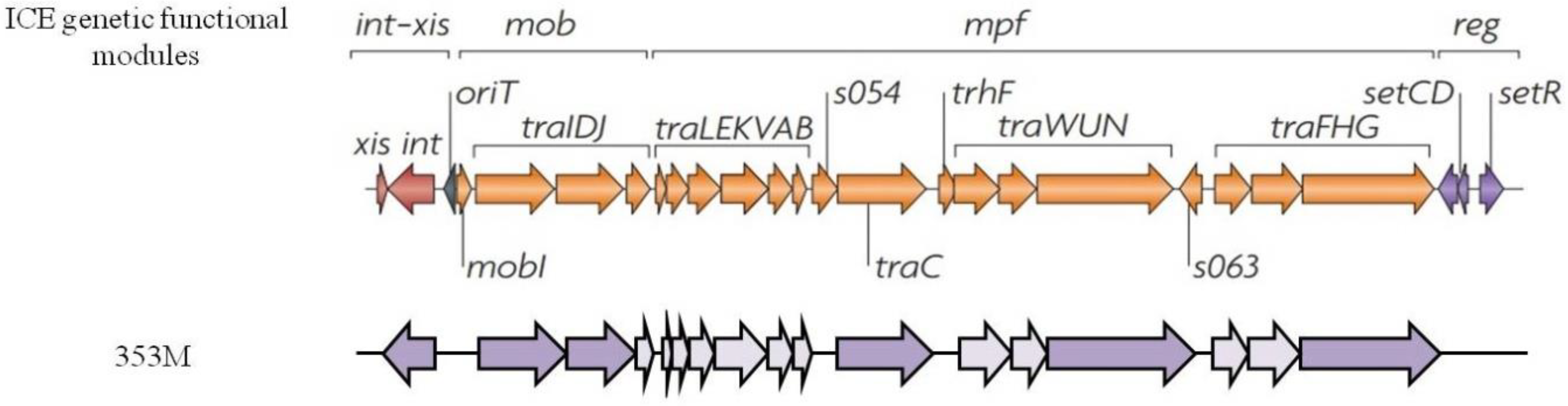
3.5. Mobilome Analysis

The MGE analysis has revealed the absence of circular elements such as plasmids. Anyway, ICEs, class I integrons, ISs and bacteriophages were detected. Of interest was the presence in 353M, a strain isolated in the open sea, of several genetic elements associated to int-xis (integration and excision module), mob (DNA mobilization and processing module) and mpf (mating-pair formation module) machineries that are typical of ICEs. The isolate 353M carried the following genes: traID, traC, traN, traG, a tyrosine-type XerD recombinase (integrase), a site-specific integrase and several other conjugal transfer proteins predicted as hypothetical proteins (Figure 2) but containing protein domains involved in the conjugative process as confirmed by BLAST analysis.
In particular, the Tra amino acid sequences showed high similiarity (99–100%) to Salmonella enterica ICE transfer apparatus (TraC, TraG, TraI, TraN) whereas TraD revealed 61.71% of similarity to the same protein detected in Pseudoalteromonas sp. GutCa3 (WP_101217907). Concerning the integration module, the site-specific integrase sequence protein was 89.09% similar to that found in S. putrefaciens (WP_086903394.1) whereas the XerD recombinase showed low similarity (41.03%) to the tyrosine-type recombinase/integrase found in the marine bacterium Halofilum ochraceum (WP_070988554.1). Furthermore, several genes involved in the partitioning such as ParAB, replication (RepA, DNA polymerase III, DNA topoisomerase III, DNA binding proteins, IncW-like replication protein), recombination (RecF, SbcC) and several other genetic elements associated to type II, III and IV secretion systems were revealed. In addition, in 8 out of 10 S. algae strains, we found a 200 kbp genetic element containing metabolism, virulence factor, antibiotic and heavy metal resistance encoding genes. Most of these resistance genes were efflux pumps. They were also flanked by integration and recombination genes such as integrases, endonucleases, transposases and phage-associated elements. They seem to be associated to ICE elements considering their large size and the presence of the genes mentioned above. To confirm this, we have also found in the surrounding of these elements a tRNAPhe encoding gene as a possible insertion site in the host chromosome. Moreover, an integration host factor (ihf) gene was found to flank this region with the alfa subunit near to the tRNAPhe gene and the beta subunit at the opposite end of this genetic element. Conversely, these elements were absent in the vibrios under study.
Class I integron elements were detected in all the S. algae and in V. anguillarum 28AD but not in V. parahaemolyticus VPE116 in which only an IntI was found. IntI was also detected in 5 out of 10 S. algae strains. Regarding ISs, several families already described in Enterobacteriaceae and in other aquatic bacteria such as Aeromonas salmonicida, V. vulnificus, S. loihica, S. oneidensis, S. baltica, V. anguillarum, V. parahaemolyticus, V. splendidus and S. putrefaciens were present in the S. algae or Vibrio spp. isolates (Table 3).
The presence of bacteriophages and their related elements was investigated in this study as well. As reported in Table 3, several prophages were detected also if incomplete. Interestingly, the presence of phages associated to other aquatic bacteria such as Aeromonas phage phiO18P (NC_009542) and Shewanella sp. phage 1/44 (NC_025463) was shown in the S. algae strains. Moreover, phages derived from Enterobacteriaceae family members such as Escherichia phage D108 (NC_013594), Enterobacteria phage phi92 (NC_023693) and Enterobacterial phage mEp213 (NC_019720) were detected in the S. algae and in V. parahaemolyticus VPE116 isolates. Vibrio phage VP882 (NC_009016) and bacteriophage phi 1.45 were found only in V. parahaemolyticus VPE116 whereas the strain 38LV did not harbor any phages. In addition to these elements, the inovirus Gp2 encoding gene was found in 6 out of 12 strains (144bCP, 146bCP, 178CP, 38LV, 83CP, 28AD). This protein is associated to a genus of viruses able to infect Gram-negative and Gram-positive bacteria. In addition, a defense system from foreign DNA, the CRISPR-Cas system, was detected in 3 out of 10 S. algae strains (219VB, 38LV, 82CP) and in V. parahaemolyticus VPE116 isolate.

3.6. Virulome Analysis

Virulence factor encoding genes were investigated both in the S. algae and Vibrio spp. isolates (Table S2, Supplementary Material). Hemolytic activity is predominant in these strains. In particular, hlyD, hlyD family secretion protein and hemolysin III were all found in the S. algae strains as well as a complex machinery for the secretion of these factors such as type I, II, III, IV and VI secretion systems. Regarding V. anguillarum 28AD, a thermolabile hemolysin, hemolysin III, hemagglutinin, the repeats-in-toxin subunit A (rtxA) and type I, II, III, IV and VI secretion systems were found. In addition to these factors, V. parahaemolyticus VPE116 harbored hemolysin D and the thermostable hemolysin delta-VPH as well.

3.7. Bacteriocins

Furthermore, some bacteriocin encoding genes such as lodA, L-lysine 6-oxidase, lodB, a putative FAD-dependent oxidoreductase and cvpA, a colicin V production protein were detected in all the S. algae strains. In V. anguillarum 28AD, the expressing hydrolase activity gene vabF was detected.

4. Discussion

Antibiotics are widely used in human medicine, food producing animals and agricultural activities to prevent and treat infections. In the aquaculture sector, a lack of legislation has led to an indiscriminate use of antibiotics which may spread in the aquatic environment and sediment promoting antimicrobial resistance in the aquatic microbiota [43,59]. Therefore, these bacteria represent a potential reservoir of ARGs [60] and a risk to public health [61]. The classes of antibiotics used both for therapeutic and prophylaxis, on the basis of legal permissions in Italian aquaculture, include beta-lactams (amoxicillin, ampicillin), tetracyclines (clortetracycline, tetracycline, oxytetracycline), amphenicols (florfenicol, thiamphenicol), macrolides (erythromycin), sulfonamides (all, included trimethoprim+sulfonamide) and quinolones (oxolinic acid, flumequin, enrofloxacin) [62,63]. A review of the scientific literature on AMR in Italian aquaculture has evidenced a common resistance to beta-lactams, tetracycline, erythromycin and trimethoprim, and a general susceptibility to chloramphenicol, tobramycin and flumequin [64]. Of interest, multiple antibiotic resistance profiles have been reported for Aeromonas and Vibrio [65,66,67,68].
In our study we report the results obtained concerning the resistome, mobilome and virulome analysis of 12 MDR marine bacterial strains by WGS, providing new insights on the genes involved in the AMR mechanisms.
The presence of class C and class D beta-lactamases is already reported for S. algae but few data are available for V. anguillarum and V. parahaemolyticus. In this study, we detected a chromosome-encoded AmpC in the V. anguillarum 28AD isolate, and a class A beta-lactamase (CARB-19) in V. parahaemolyticus VPE116 conferring resistance to penicillins and cephalosporins (first- and second-generation), and to amino- and carboxy-penicillins, respectively. Interestingly, the presence of pqiAB genes in our S. algae strains near the blaAmpC gene and of cusAB genes near the blaCARB-19 in V. parahaemolyticus VPE116 may suggest the important role of heavy metal resistance-encoding genes in inducing the beta-lactam resistant phenotype in these marine bacteria.
Fluoroquinolones are frequently used in aquaculture. Some pentapeptide proteins belonging to the Qnr group were detected in the analyzed strains. Although these genes are generally located in plasmids (PMQR, except for QnrVC-like proteins), they were here detected in the bacterial chromosome both in Shewanella and Vibrio genera as already reported by Poirel and coauthors [69]. In fact, the origin of qnrA-like determinants was identified as being the chromosome of S. algae [8]. Then, their association to plasmids has allowed their spread in other bacterial species. It is noted that PMQR determinants generally confer only low-level quinolone resistance that alone does not exceed the clinical breakpoint but they can favor the selection of additional resistance mechanisms [70].
Of interest is the presence of some S. algae strains which carry the QnrA7 and belong to a group of strains isolated from the same aquaculture center while the strains harboring the QnrA3 protein have been obtained in other different sites and fish farms. Moreover, the qnrVC6 gene, usually found in V. cholerae, was here detected in V. anguillarum 28AD. This is not really surprising, since qnrVC-like genes are currently spreading in other Vibrio species and Gram negatives such as Pseudomonas aeruginosa, Pseudomonas putida and Citrobacter freundii [71,72]. Interestingly, although the Qnr-like genes should provide resistance to quinolones, the phenotypes of all our strains are susceptible to ciprofloxacin and nalidixic acid, the two fluoroquinolones tested for.
Conversely, all the S. algae strains provided a resistant phenotype to fosfomycin, but the WGS analysis failed to detect the genes involved. This is probably due to the fact that they are still unknown and not reported on databases. Considering the data reported in literature, very high MICs were observed by Torri and coworkers [73] in clinical S. algae isolates during an Italian survey on shewanella-associated infections. This is probably an intrinsic resistance present in S. algae strains whose gene may be chromosomal-encoded considering a few presence of plasmids in these strains.
Regarding tetracycline resistance, only vibrios harbored tet34- or/and tet35-encoding genes. In particular, the tet34 gene has been found in some Gram-negative genera (Pseudomonas, Serratia and Vibrio) and is unique to environmental bacteria [74]. Surprisingly, two S. algae strains, 38LV and 57CP, were resistant to tigecycline, a recent commercialized antibiotic used against MDR pathogens to treat hospital-acquired pneumonia, ventilator-associated pneumonia and other infections [75]. Tigecycline is scarcely investigated in aquaculture because it is mainly used in human medicine. The mechanism of resistance is still unknown, probably due to the presence of efflux pumps widely detected by WGS.
Globally, the data obtained for the resistome study seem to be in line with that reported for Italian aquaculture [64,65,66,67,68].
The mobilome analysis has been particularly difficult to perform, because of the Illumina technology which uses a mating pair and short read sequencing. Despite some difficulties, we identified integrative and conjugal elements in the 353M strain isolated in the open sea. This is an interesting isolate considering its scarce relation to aquaculture strains but potentially able to behave as an ARG reservoir in the same environment facilitating the spread of these elements and of their genes carried on to other marine bacteria or human pathogens. Furthermore, the presence of large size elements of ICE associated in S. algae strains allow us to highlight the contribution of these elements in aquaculture that, to the best of our knowledge, has never been investigated. The role of the detected bacteriophages has yet to be clarified. However, their involvement in the horizontal gene transfer of ARGs that flank these elements during the excision process, packing these bacterial ARGs in the mature virion particle and disseminating them in the genome of a new host cell, is not to be excluded. A scheme of how ARGs transfer occurs among bacteria is described in Figure 3.
Interestingly, both S. algae and V. parahaemolyticus VPE116 strains were shown to be prone to infection by bacteriophages belonging to different genera of marine and human pathogenic bacteria. Other transposable elements such as IS family transposases have been investigated. They are intracellular MGEs which need to be integrated in other mobile elements such as plasmids, bacteriophages or ICEs. Most of these ISs did not carry passenger genes such as transcription regulators, methyltransferases and antibiotic resistance, only the isolate 38LV carried a MFS transporter and an AraC family transcriptional regulator downstream the IS481 family transposase, whereas the isolates 82CP and 353M carried a LysR and an AraC family transcriptional regulators downstream the transposase genes, respectively.

5. Conclusions

In conclusion, NGS technology is a useful means of detecting ARGs, MGEs and virulence factors although its reliability is dependent upon the availability of updated databases. Moreover, the genotype analysis sometimes does not correspond to the phenotype as in our case for some classes of antibiotics (fosfomycin and quinolones). Thus, the results obtained from this technology can act as a potential warning about the resistance of a microorganism. However, although transcriptome or proteome analysis is more adequate in detecting the real presence of the products generated by the predicted ARGs, WGS can help in analyzing the surrounding of the ARGs, the regulators involved during the transcription and possible mobilization of the MGEs. Here, we only analyzed the presence of these elements suggesting their possible involvement in the final MDR phenotype of our strains. Further studies should be performed to confirm how the MGEs act in the aquatic microbial community and if the genetic transfer occurs but this is not the scope of the current work. Among the data obtained, particularly interesting are those deriving from the mobilome analysis of these three species (S. algae, V. parahaemolyticus and V. anguillarum). The presence of several bacteriophages, large ICEs and ISs of different families has allowed us to decipher the players that might have a crucial role in spreading ARGs and virulence factors in the aquatic environment.

Supplementary Materials

The following are available online at https://www.mdpi.com/2076-2607/8/4/572/s1: Word Document: Supplementary Material. Table S1: Genome features of every isolates, Table S2: List of heavy metal and other antimicrobial compound resistance genes found in the genomes of S. algae and Vibrio spp. Strains.

Author Contributions

Conceptualization, V.Z., G.M. and M.M.L.; software, L.V. and G.M.; validation, C.P.; formal analysis, L.V.; investigation, V.Z. and C.P.; resources, G.M. and M.M.L.; data curation, V.Z., L.V. and G.M.; writing—original draft preparation, V.Z. and M.M.L.; writing—review and editing, V.Z. and M.M.L.; visualization, V.Z.; supervision, G.M. and M.M.L.; project administration, M.M.L.; funding acquisition, M.M.L. All authors have read and agreed to the published version of the manuscript.

Funding

This work was supported by the Italian Ministry of Education, University and Research and by Project CARIVERONA 2017—B31G17000640005 entitled “Diffusione della resistenza agli antibiotici carbapenemici: ricerca dei serbatoi ambientali”.

Conflicts of Interest

The authors declare no conflict of interest. The funders had no role in the design of the study; in the collection, analyses, or interpretation of data; in the writing of the manuscript; or in the decision to publish the results.

References

  1. Jacob-Kokura, S.; Chan, C.Y.; Kaplan, L. Bacteremia and empyema caused by Shewanella algae in a trauma patient. Ann. Pharmacother. 2014, 48, 128–136. [Google Scholar] [CrossRef] [PubMed]
  2. Janda, J.M.; Abbott, S.L. The genus Shewanella: From the briny depths below to human pathogen. Crit. Rev. Microbiol. 2014, 40, 293–312. [Google Scholar] [CrossRef] [PubMed]
  3. Muñoz-Gallego, I.; Chaves, F.; Orellana, M.A. Epidemiological and clinical characteristics of Shewanella spp. infections in a tertiary hospital in Madrid. Infect. Dis. (Lond.) 2016, 48, 760–762. [Google Scholar] [CrossRef]
  4. Jousset, A.B.; Dabos, L.; Bonnin, R.A.; Girlich, D.; Potron, A.; Cabanel, N.; Dortet, L.; Glaser, P.; Naas, T. CTX-M-15-producing Shewanella species clinical isolate expressing OXA-535, a chromosome-encoded OXA-48 variant, putative progenitor of the plasmid-encoded OXA-436. Antimicrob. Agents Chemother. 2017, 62, e018791-17. [Google Scholar] [CrossRef] [PubMed] [Green Version]
  5. Yousfi, K.; Bekal, S.; Usongo, V.; Touati, A. Current trends of human infections and antibiotic resistance of the genus Shewanella. Eur. J. Clin. Microbiol. Infect. Dis. 2017, 36, 1353–1362. [Google Scholar] [CrossRef] [PubMed]
  6. Satomi, M. The Family Shewanellaceae. In The Prokaryotes, 4th ed.; Rosenberg, E., DeLong, E.F., Lory, S., Stackebrandt, E., Thompson, F., Eds.; Springer: Berlin/Heidelberg, Germany, 2014. [Google Scholar]
  7. Poirel, L.; Héritier, C.; Nordmann, P. Chromosome-encoded Ambler class D-lactamase of Shewanella oneidensis as a progenitor of carbapenem-hydrolyzing oxacillinase. Antimicrob. Agents Chemother. 2004, 48, 348–351. [Google Scholar] [CrossRef] [PubMed] [Green Version]
  8. Poirel, L.; Rodriguez-Martinez, J.M.; Mammeri, H.; Liard, A.; Nordmann, P. Origin of plasmid-mediated quinolone resistance determinant QnrA. Antimicrob. Agents Chemother. 2005, 49, 3523–3525. [Google Scholar] [CrossRef] [Green Version]
  9. Kim, D.M.; Kang, C.I.; Lee, C.S.; Kim, H.B.; Kim, E.C.; Kim, N.J.; Oh, M.D.; Choe, K.W. Treatment failure due to emergence of resistance to carbapenem during therapy for Shewanella algae bacteremia. J. Clin. Microbiol. 2006, 44, 1172–1174. [Google Scholar] [CrossRef] [Green Version]
  10. Lascols, C.; Podglajen, I.; Verdet, C.; Gauthier, V.; Gutmann, L.; Soussy, C.J.; Collatz, E.; Cambau, E. A plasmid-borne Shewanella algae gene, qnrA3, and its possible transfer In Vivo between Kluyvera ascorbata and Klebsiella pneumonia. J. Bacteriol. 2008, 190, 5217–5223. [Google Scholar] [CrossRef] [Green Version]
  11. Potron, A.; Poirel, L.; Nordmann, P. Origin of OXA-181, an emerging carbapenem-hydrolyzing oxacillinase, as a chromosomal gene in Shewanella xiamenensis. Antimicrob. Agents Chemother. 2011, 55, 4405–4407. [Google Scholar] [CrossRef] [Green Version]
  12. Cimmino, T.; Olaitan, A.O.; Rolain, J.M. Whole genome sequence to decipher the resistome of Shewanella algae, a multidrug-resistant bacterium responsible for pneumonia, Marseille, France. Expert Rev. Anti Infect. Ther. 2016, 14, 269–275. [Google Scholar] [CrossRef]
  13. Ceccarelli, D.; van Essen-Zandbergen, A.; Veldman, K.T.; Tafro, N.; Haenen, O.; Mevius, D.J. Chromosome-based blaOXA-48-like variants in Shewanella species isolates from food-producing animals, fish, and the aquatic environment. Antimicrob. Agents Chemother. 2017, 61, e010131-16. [Google Scholar] [CrossRef] [PubMed] [Green Version]
  14. Huang, Y.H.; Tang, Y.Y.; Cheng, J.F.; Wu, Z.Y.; Mao, Y.C.; Liu, P.Y. Genome analysis of multidrug-resistant Shewanella algae isolated from human soft tissue sample. Front. Pharmacol. 2018, 9, 419. [Google Scholar] [CrossRef]
  15. Holt, H.M.; Søgaard, P.; Gahrn-Hansen, B. Ear infections with Shewanella alga: A bacteriologic, clinical and epidemiologic study of 67 cases. Clin. Microbiol. Infect. 1997, 3, 329–334. [Google Scholar] [CrossRef] [PubMed] [Green Version]
  16. Telke, A.A.; Rolain, J.M. Functional genomics to discover antibiotic resistance genes: The paradigm of resistance to colistin mediated by ethanolamine phosphotransferase in Shewanella algae MARS 14. Int. J. Antimicrob. Agents 2015, 46, 648–652. [Google Scholar] [CrossRef] [PubMed]
  17. Jacobs-Slifka, K.M.; Newton, A.E.; Mahon, B.E. Vibrio alginolyticus infections in the USA, 1988–2012. Epidemiol. Infect. 2017, 145, 1491–1499. [Google Scholar] [CrossRef] [Green Version]
  18. Ahmed, H.A.; El Bayomi, R.M.; Hussein, M.A.; Khedr, M.H.E.; Abo Remela, E.M.; El-Ashram, A.M.M. Molecular characterization, antibiotic resistance pattern and biofilm formation of Vibrio parahaemolyticus and V. cholerae isolated from crustaceans and humans. Int. J. Food Microbiol. 2018, 274, 31–37. [Google Scholar] [CrossRef]
  19. Baker-Austin, C.; Trinanes, J.; Gonzalez-Escalona, N.; Martinez-Urtaza, J. Non-cholera vibrios: The microbial barometer of climate change. Trends Microbiol. 2017, 25, 76–84. [Google Scholar] [CrossRef] [Green Version]
  20. Ceccarelli, D.; Salvia, A.M.; Sami, J.; Cappuccinelli, P.; Colombo, M.M. New cluster of plasmid-located class 1 integrons in Vibrio cholerae O1 and a dfrA15 cassette-containing integron in Vibrio parahaemolyticus isolated in Angola. Antimicrob. Agents Chemother. 2006, 50, 2493–2499. [Google Scholar] [CrossRef] [Green Version]
  21. Holm, K.O.; Bækkedal, C.; Söderberg, J.J.; Haugen, P. Complete genome sequences of seven Vibrio anguillarum strains as derived from PacBio sequencing. Genome Biol. Evol. 2018, 10, 1127–1131. [Google Scholar] [CrossRef]
  22. Bier, N.; Schwartz, K.; Guerra, B.; Strauch, E. Survey on antimicrobial resistance patterns in Vibrio vulnificus and Vibrio cholerae non-O1/non-O139 in Germany reveals carbapenemase-producing Vibrio cholerae in coastal waters. Front. Microbiol. 2015, 6, 1179. [Google Scholar] [CrossRef] [PubMed]
  23. Aberkane, S.; Compain, F.; Barraud, O.; Ouédraogo, A.S.; Bouzinbi, N.; Vittecoq, M.; Jean-Pierre, H.; Decré, D. Non-O1/non-O139 Vibrio cholerae avian isolate from France cocarrying the blaVIM-1 and blaVIM-4 genes. Antimicrob. Agents Chemother. 2015, 59, 6594–6596. [Google Scholar] [CrossRef] [PubMed] [Green Version]
  24. Chowdhury, G.; Pazhani, G.P.; Sarkar, A.; Rajendran, K.; Mukhopadhyay, A.K.; Bhattacharya, M.K.; Ghosh, A.; Ramamurthy, T. Carbapenem resistance in clonally distinct clinical strains of Vibrio fluvialis isolated from diarrheal samples. Emerg. Infect. Dis. 2016, 22, 1754–1761. [Google Scholar] [CrossRef] [PubMed] [Green Version]
  25. Mangat, C.S.; Boyd, D.; Janecko, N.; Martz, S.L.; Desruisseau, A.; Carpenter, M.; Reid-Smith, R.J.; Mulvey, M.R. Characterization of VCC-1, a novel Ambler class A carbapenemase from Vibrio cholerae isolated from imported retail shrimp sold in Canada. Antimicrob. Agents Chemother. 2016, 60, 1819–1825. [Google Scholar] [CrossRef] [PubMed] [Green Version]
  26. Lee, L.H.; Ab Mutalib, N.S.; Law, J.W.; Wong, S.H.; Letchumanan, V. Discovery on antibiotic resistance patterns of Vibrio parahaemolyticus in Selangor reveals carbapenemase producing Vibrio parahaemolyticus in marine and freshwater fish. Front. Microbiol. 2018, 9, 2513. [Google Scholar] [CrossRef] [PubMed]
  27. Di, D.Y.W.; Shin, H.; Han, D.; Unno, T.; Hur, H.G. High genetic diversity of Vibrio parahaemolyticus isolated from tidal water and mud of southern coast of South Korea. FEMS Microbiol. Ecol. 2019, 95, fiz022. [Google Scholar] [CrossRef]
  28. Rahman, T.; Yarnall, B.; Doyle, D.A. Efflux drug transporters at the forefront of antimicrobial resistance. Eur. Biophys. J. 2017, 46, 647–653. [Google Scholar] [CrossRef]
  29. Lubelski, J.; Konings, W.N.; Driessen, A.J. Distribution and physiology of ABC-type transporters contributing to multidrug resistance in bacteria. Microbiol. Mol. Biol. Rev. 2007, 71, 463–476. [Google Scholar] [CrossRef] [Green Version]
  30. Kuroda, T.; Tsuchiya, T. Multidrug efflux transporters in the MATE family. Biochimica Biophysica Acta 2009, 1794, 763–768. [Google Scholar] [CrossRef]
  31. Pao, S.S.; Paulsen, I.T.; Saier, M.H., Jr. Major facilitator superfamily. Microbiol. Mol. Biol. Rev. 1998, 62, 1–34. [Google Scholar] [CrossRef] [Green Version]
  32. Chung, Y.J.; Saier, M.H., Jr. SMR-type multidrug resistance pumps. Curr. Opin. Drug Discov. Dev. 2001, 4, 237–245. [Google Scholar]
  33. Nikaido, H. Structure and mechanism of RND-type multidrug efflux pumps. Adv. Enzymol. Relat. Areas Mol. Biol. 2011, 77, 1–60. [Google Scholar]
  34. Groh, J.L.; Luo, Q.; Ballard, J.D.; Krumholz, L.R. Genes that inhance the ecological fitness of Shewanella oneidensis MR-1 in sediments reveal the value of antibiotic resistance. Appl. Environ. Microbiol. 2007, 73, 492–498. [Google Scholar] [CrossRef] [PubMed] [Green Version]
  35. Pembroke, J.T.; Piterina, A.V.C. A novel ICE in the genome of Shewanella putrefaciens W3-18-1: Comparison with the SXT/R391 ICE-like elements. FEMS Microbiol. Lett. 2006, 264, 80–88. [Google Scholar] [CrossRef] [Green Version]
  36. Carraro, N.; Burrus, V. Biology of Three ICE Families: SXT/R391, ICEBs1, and ICESt1/ICESt3. Microbiol. Spectr. 2014, 2, 1–20. [Google Scholar] [CrossRef] [PubMed]
  37. Fang, Y.; Wang, Y.; Li, Z.; Liu, Z.; Li, X.; Diao, B.; Kan, B.; Wang, D. Distribution and genetic characteristics of SXT/R391 Integrative Conjugative Elements in Shewanella spp. from China. Front. Microbiol. 2018, 9, 920. [Google Scholar] [CrossRef] [PubMed] [Green Version]
  38. Kamruzzaman, M.; Nischibuchi, M. Detection and characterization of a functional insertion sequence, ISVpa2, in Vibrio parahaemolyticus. Gene 2008, 409, 92–99. [Google Scholar] [CrossRef] [PubMed] [Green Version]
  39. Jiang, Y.; Yao, L.; Li, F.; Tan, Z.; Zhai, Y.; Wang, L. Chacterization of antimicrobial resistance of Vibrio parahaemolyticus from cultured sea cucumbers (Apostichopus japonicas). Lett. Appl. Microbiol. 2014, 59, 147–154. [Google Scholar] [CrossRef]
  40. Almuzara, M.; Montaña, S.; Lazzaro, T.; Uong, S.; Parmeciano Di Noto, G.; Traglia, G.; Bakai, R.; Centrón, D.; Iriarte, A.; Quiroga, C.; et al. Genetic analysis of a PER-2-producing Shewanella sp. strain harbouring a variety of mobile genetic elements and antibiotic resistance determinants. J. Glob. Antimicrob. Resist. 2017, 11, 81–86. [Google Scholar] [CrossRef] [Green Version]
  41. Rossen, J.W.A.; Friedrich, A.W.; Moran-Gilad, J.; ESCMID Study Group for Genomic and Molecular Diagnostics (ESGMD). Practical issues in implementing whole-genome-sequencing in routine diagnostic microbiology. Clin. Microbiol. Infect. 2018, 24, 355–360. [Google Scholar] [CrossRef] [Green Version]
  42. Köser, C.U.; Ellington, M.J.; Peacock, S.J. Whole-genome sequencing to control antimicrobial resistance. Trends Genet. 2014, 30, 401–407. [Google Scholar] [CrossRef] [PubMed] [Green Version]
  43. Labella, A.; Gennari, M.; Ghidini, V.; Trento, I.; Manfrin, A.; Borrego, J.J.; Lleo, M.M. High incidence of antibiotic multi-resistant bacteria in coastal areas dedicated to fish farming. Mar. Pollut. Bull. 2013, 70, 197–203. [Google Scholar] [CrossRef] [PubMed]
  44. Pires, J.; Novais, A.; Peixe, L. Blue-carba, an easy biochemical test for detection of diverse carbapenemase producers directly from bacterial cultures. J. Clin. Microbiol. 2013, 51, 4281–4283. [Google Scholar] [CrossRef] [PubMed] [Green Version]
  45. Bankevich, A.; Nurk, S.; Antipov, D.; Gurevich, A.A.; Dvorkin, M.; Kulikov, A.S.; Lesin, V.M.; Nikolenko, S.I.; Pham, S.; Prjibelski, A.D.; et al. SPAdes: A new genome assembly algorithm and its applications to single-cell sequencing. J. Comput. Biol. 2012, 19, 455–477. [Google Scholar] [CrossRef] [PubMed] [Green Version]
  46. Seemann, T. Prokka: Rapid prokaryotic genome annotation. Bioinformatics 2014, 30, 2068–2069. [Google Scholar] [CrossRef] [PubMed]
  47. Gupta, S.K.; Padmanabhan, B.R.; Diene, S.M.; Lopez-Rojas, R.; Kempf, M.; Landraud, L.; Rolain, J.M. ARG-ANNOT, a new bioinformatic tool to discover antibiotic resistance genes in bacterial genomes. Antimicrob. Agents Chemother. 2014, 58, 212–220. [Google Scholar]
  48. Kleinheinz, K.A.; Joensen, K.G.; Larsen, M.V. Applying the ResFinder and VirulenceFinder web-services for easy identification of acquired antibiotic resistance and E. coli virulence genes in bacteriophage and prophage nucleotide sequences. Bacteriophage 2014, 4, e27943. [Google Scholar] [CrossRef] [Green Version]
  49. Jia, B.; Raphenya, A.R.; Alcock, B.; Waglechner, N.; Guo, P.; Tsang, K.K.; Lago, B.A.; Dave, B.M.; Pereira, S.; Sharma, A.N.; et al. CARD 2017: Expansion and model-centric curation of the comprehensive antibiotic resistance database. Nucleic Acids Res. 2017, 45, D566–D573. [Google Scholar] [CrossRef]
  50. Moura, A.; Soares, M.; Pereira, C.; Leitão, N.; Henriques, I.; Correia, A. INTEGRALL: A database and search engine for integrons, integrases and gene cassettes. Bioinformatics 2009, 25, 1096–1098. [Google Scholar] [CrossRef] [Green Version]
  51. Siguier, P.; Perochon, J.; Lestrade, L.; Mahillon, J.; Chandler, M. ISfinder: The reference centre for bacterial insertion sequences. Nucleic Acids Res. 2006, 34, D323–D326. [Google Scholar] [CrossRef] [Green Version]
  52. Liu, M.; Li, X.; Xie, Y.; Bi, D.; Sun, J.; Li, J. ICEberg 2.0: An updated database of bacterial integrative and conjugative elements. Nucleic Acids Res. 2019, 47, D660–D665. [Google Scholar] [CrossRef] [PubMed]
  53. Arndt, D.; Grant, J.R.; Marcu, A.; Sajed, T.; Pon, A.; Liang, Y.; Wishart, D.S. PHASTER: A better, faster version of the PHAST phage search tool. Nucleic Acids Res. 2016, 44, W16–W21. [Google Scholar] [CrossRef] [PubMed] [Green Version]
  54. Rozov, R.; Brown Kav, A.; Bogumil, D.; Shterzer, N.; Halperin, E.; Mizrahi, I.; Shamir, R. Recycler: An algorithm for detecting plasmids from de novo assembly graphs. Bioinformatics 2017, 33, 475–482. [Google Scholar]
  55. Héritier, C.; Poirel, L.; Nordmann, P. Genetic and biochemical characterization of a chromosome-encoded carbapenem-hydrolyzing Ambler class D beta-lactamase from Shewanella algae. Antimicrob. Agents Chemother. 2004, 48, 1670–1675. [Google Scholar] [CrossRef] [PubMed] [Green Version]
  56. Poole, K. Efflux pumps as antimicrobial resistance mechanisms. Ann. Med. 2007, 39, 162–176. [Google Scholar] [CrossRef] [PubMed]
  57. Anes, J.; McCusker, M.P.; Fanning, S.; Martins, M. The ins and outs of RND efflux pumps in Escherichia coli. Front. Microbiol. 2015, 6, 587. [Google Scholar] [CrossRef] [PubMed] [Green Version]
  58. Wozniak, A.F.; Waldor, M.K. Integrative and conjugative elements: Mosaic mobile genetic elements inabling dynamic lateral gene flow. Nat. Rev. Microbiol. 2010, 8, 552–563. [Google Scholar] [CrossRef]
  59. Teuber, M. Veterinary use and antibiotic resistance. Curr. Opin. Microbiol. 2001, 4, 493–499. [Google Scholar] [CrossRef]
  60. Hexing, W.; Lingshuang, R.; Xin, Y.; Jing, H.; Yue, C.; Gengsheng, H.; Qingwu, J. Antibiotic residues in meat, milk and aquatic products in Shanghai and human exposure assessment. Food Control 2017, 80, 217–225. [Google Scholar]
  61. Sorum, H. Antimicrobial Drug Resistance in Fish Pathogens. In Antimicrobial Resistance in Bacteria of Animal Origin; Aarestrup, F.M., Ed.; ASM Press: Washington, DC, USA, 2006; pp. 213–238. [Google Scholar]
  62. OIE. Report of the Meeting of the OIE ad hoc Group on Antimicrobial Resistance; Annex 9; OIE: Paris, France, 2018. [Google Scholar]
  63. Regulation, H. The Council of the European Communities. Council Regulation (ECC) No 2377/90 Laying down a Community Procedure for the Establishment of Maximum Residue Limits of Veterinary Medicine Products in Foodstuffs of Animal Origins; Annex III. Off. J. Eur. Commun. 1990, 224, 1–8. [Google Scholar]
  64. Scarano, C.; Spanu, C.; Ziino, G.; Pedonese, F.; Dalmasso, A.; Spanu, V.; Virdis, S.; De Santis, E.P. Antibiotic resistance of Vibrio species isolated from Sparus aurata reared in Italian mariculture. New Microbiol. 2014, 37, 329–337. [Google Scholar] [PubMed]
  65. Ottaviani, D.; Bacchiocchi, I.; Masini, L.; Leoni, F.; Carraturo, A.; Giammarioli, M.; Sbaraglia, G. Antimicrobial susceptibility of potentially pathogenic halophilic vibrios isolated from seafood. Int. J. Antimicrob. Agents 2001, 18, 135–140. [Google Scholar] [CrossRef]
  66. Laganà, P.; Caruso, G.; Minutoli, E.; Zaccone, R.; Santi, D. Susceptibility to antibiotics of Vibrio spp. and Photobacterium damsela ssp. piscicida strains isolated from Italian aquaculture farms. New Microbiol. 2011, 34, 53–63. [Google Scholar] [PubMed]
  67. Ottaviani, D.; Leoni, F.; Talevi, G.; Masini, L.; Santarelli, S.; Rocchegiani, E.; Susini, F.; Montagna, C.; Monno, R.; D’Annibale, L.; et al. Extensive investigation of antimicrobial resistance in Vibrio parahaemolyticus from shellfish and clinical sources, Italy. Int. J. Antimicrob. Agents 2013, 42, 187–193. [Google Scholar] [CrossRef] [PubMed]
  68. Scarano, C.; Piras, F.; Virdis, S.; Ziino, G.; Nuvoloni, R.; Dalmasso, A.; De Santis, E.P.L.; Spanu, C. Antibiotic resistance of Aeromonas spp. strains isolated from Sparus aurata reared in Italian mariculture farms. Int. J. Food Microbiol. 2018, 284, 91–97. [Google Scholar] [CrossRef]
  69. Poirel, L.; Cattoir, V.; Nordmann, P. Plasmid-mediated quinolone resistance; interactions between human, animal, and environmental ecologies. Front. Microbiol. 2012, 2, 3–24. [Google Scholar] [CrossRef] [Green Version]
  70. Correia, S.; Poeta, P.; Hébraud, M.; Capelo, J.L.; Igrejas, G. Mechanisms of quinolone action and resistance: Where do we stand? J. Med. Microb. 2017, 66, 551–559. [Google Scholar] [CrossRef]
  71. Bado, I.; Papa-Ezdra, R.; Cordeiro, N.; Outeda, M.; Caiata, L.; García-Fulgueiras, V.; Seija, V.; Vignoli, R. Detection of qnrVC6, within a new genetic context, in an NDM-1-producing Citrobacter freundii clinical isolate from Uruguay. J. Glob. Antimicrob. Resist. 2018, 14, 95–98. [Google Scholar] [CrossRef]
  72. Liu, J.; Yang, L.; Chen, D.; Peters, B.M.; Li, L.; Li, B.; Xu, Z.; Shirtliff, M.E. Complete sequence of pBM413, a novel multidrug resistance megaplasmid carrying qnrVC6 and blaIMP-45 from Pseudomonas aeruginosa. Int. J. Antimicrob. Agents 2018, 51, 145–150. [Google Scholar] [CrossRef]
  73. Torri, A.; Bertini, S.; Schiavone, P.; Congestrì, F.; Matteucci, M.; Sparacino, M.; Testa, G.; Pedna, M.F.; Sambri, V. Shewanella algae infection in Italy: Report of 3 years’ evaluation along the coast of the northern Adriatic Sea. New Microb. New Infect. 2018, 23, 39–43. [Google Scholar] [CrossRef]
  74. Miranda, C.D.; Kehrenberg, C.; Ulep, C.; Schwarz, S.; Roberts, M.C. Diversity of tetracycline resistance genes in bacteria from Chilean salmon farms. Antimicrob. Agents Chemother. 2003, 47, 883–888. [Google Scholar] [CrossRef] [PubMed] [Green Version]
  75. Grossman, T.H. Tetracycline antibiotics and resistance. Cold Spring Harb. Perspect. Med. 2016, 6, a025387. [Google Scholar] [CrossRef] [PubMed] [Green Version]
  76. The Biology Notes. Available online: https://thebiologynotes.com/carbapenem-resistant-gram-negative-bacteria/ (accessed on 15 January 2020).
Figure 1. Genetic surrounding of the blaAmpC gene found in the S. algae strains. The black arrow represents the blaAmpC gene, the grey one the transcriptional regulator, whereas the white ones the other genes found in this structure.
Microorganisms 08 00572 g001
Figure 2. Description of the integrative and conjugative element (ICE) detected in the isolate 353M. At the top of the picture the genetic functional modules present in ICEs are reported [58]; at the bottom the encoding genes found in the strain 353M (dark violet) belonging to xis/int, mob and mpf modules and the other conjugal proteins (light violet) predicted as hypothetical but containing protein domains involved in conjugation and transfer at BLAST analysis are shown.
Microorganisms 08 00572 g002
Figure 3. Scheme of the mechanisms of antimicrobial resistance genes (ARGs) transfer that are usually involved in bacterial cells: transduction, conjugation and transformation [76]. These mechanisms can occur also in the aquatic environment among autochthonous bacteria (aquatic bacterial species) and human pathogenic species that are temporally present in the aquatic environment. In the transduction process (A), a mature bacteriophage can move bacterial (virulence, antimicrobial resistance (AR), metabolism) genes to a new host cell. These genes can also be captured in a plasmid and spread by conjugation. In the mechanism of conjugation (B), a plasmid containing integrons, ISs, or transposons carrying ARGs can be transferred by ssDNA intermediate from a donor to a recipient cell. Also ICEs can use this mechanism followed by integration in the bacterial chromosome. During the transformation (C), the bacterial lysis can release DNA encoding for ARGs in the aquatic environment. This exogenous DNA can be captured from natural competent bacteria and integrated in their chromosomes.
Microorganisms 08 00572 g003
Table 1. List of multidrug-resistant (MDR) marine isolates under study. Sampling dates, sources, species identification and antimicrobial resistance profiles are reported for each strain.
StrainSampling DateSource16S rRNA Gene IdentificationAntimicrobial Resistance Profile
353M10/08/2010Water
Open sea
Shewanella algaeCEF, FOX, IPM, AMX, CS, SUL, FOS
178CP13/06/2011Water
Veneto II station
Shewanella algaeCEF, FOX, IPM, AMX, CS, SUL, FOS
146bCP13/06/2011Water
Veneto II station
Shewanella algaeCEF, FOX, IPM, AMX, CS, SUL, FOS
144bCP13/06/2011Water
Veneto II station
Shewanella algaeCEF, FOX, IPM, AMX, CS, SUL, FOS
219VB30/09/2010Water
Veneto I station
Shewanella algaeCEF, FOX, IPM, AMX, CS, SUL, FOS
82CP15/04/2011Water
Veneto II station
Shewanella algaeCEF, FOX, IPM, CS, SUL, FOS
38LV22/07/2011Water
Varano lake
Shewanella algaeCEF, FOX, IPM, AMX, CS, SUL, FOS, TET, TGC
57CP15/04/2011Water
Veneto II station
Shewanella algaeCEF, FOX, IPM, CS, SUL, FOS, TET, TGC
60CP15/04/2011Water
Veneto II station
Shewanella algaeCEF, FOX, IPM, AMX, CS, SUL, FOS
83CP15/04/2011Water
Veneto II station
Shewanella algaeCEF, FOX, IPM, AMX, CS, SUL, FOS
28AD01/02/2007European seabassVibrio anguillarumCEF, FOX, AMX, TIC, AMC, TCC, TEM, CS, SUL, AKN
VPE11626/06/2007Water
Caleri lagoon
Vibrio parahaemolyticusAMX, TIC, TMN, KMN, AKN, GMI, CS, SUL
AMC, amoxicillin-clavulanic acid; AMX, amoxicillin; AKN, amikacin; CEF, cephalothin; CS, colistin; CTX, cefotaxime; FOS, fosfomycin; FOX, cefoxitin; GMI, gentamicin; IPM, imipenem; KMN, kanamycin; SUL, sulfonamides; TCC, ticarcillin-clavulanic acid; TEM, temocillin; TET, tetracycline; TGC, tigecycline; TIC, ticarcillin; TMN, tobramycin.
Table 2. List of resistance genes found in S. algae and Vibrio spp. grouped on the basis of their mechanism and antimicrobial target.
AntimicrobialPentapeptide ProteinEnzymeEfflux SystemPump FamilyStrain
Beta-lactams OXA-55-like All S. algae
AmpC All S. algae, V. anguillarum 28AD
MexAB-OprMRNDAll S. algae, V. parahaemolyticus VPE116
FluoroquinolonesQnrA3 353M, 219VB, 38LV, 57CP, 60CP
QnrA7 144bCP, 178CP, 146bCP, 82CP, 83CP
QnrVC6 V. anguillarum 28AD
MdtKMATEAll S. algae, V. anguillarum 28AD
EmrAB-TolCMFSAll S. algae, V. parahaemolyticus VPE116
MepAB All S. algae, V. anguillarum 28AD
NorMMATEV. anguillarum 28AD, V. parahaemolyticus VPE116
Bmr3MFSV. anguillarum 28AD
MdtHMFSV. anguillarum 28AD
MfpA V. anguillarum 28AD
Multiple substrates AcrAB-TolCRNDAll S. algae, V. parahaemolyticus VPE116
AcrEF-TolCRNDAll S. algae, V. parahaemolyticus VPE116
MdtABC-TolCRNDAll strains
EmrDMFSAll strains
EmrYK-TolCMFSAll strains excepting for 353M, 219VB, 82CP, 38LV
MdtNRNDAll strains
DrrAABCAll strains excepting for V. anguillarum 28AD
StpMFSAll S. algae
BcrMFSAll S. algae
MdtGMFSV. parahaemolyticus VPE116
MdlBABCV. parahaemolyticus VPE116
Tetracyclines TetRMFSV. parahaemolyticus VPE116
Tet34MFSV. anguillarum 28AD, V. parahaemolyticus VPE116
Tet35MFSV. parahaemolyticus VPE116
Macrolides MacAB-TolCABCAll strains
MdtE-TolCRNDV. anguillarum 28AD, V. parahaemolyticus VPE116
Polymyxins EptA All strains
Chloramphenicol MdtLMFSAll strains
Cat All strains excepting for V. anguillarum 28AD
ABC, ATP binding cassette; MATE, multidrug and toxic compound extrusion; MFS, major facilitator system; RND, resistance-nodulation-cell division.
Table 3. Summary of mobile genetic elements found in S. algae and Vibrio spp. genomes.
StrainInsertion Sequence (IS) FamilyIS Family SubgroupIntegron/IntegraseBacteriophage
353MIS630, ISL3, IS3
IS630
IS3
IS4
ISSpu8
ISSlo2
ISVvu3
IntI
Class I integron
Prophage-1 incomplete
219VBIS200/IS605, IS630, ISL3
IS481
IS110, IS3
IS630
IS4
ISSpu18
ISSpu8
ISVvu3
Class I integronAeromonas phage phiO18P (NC_009542)
144BCPISL3
IS481
IS630/ISSpu8
IS4
IS3, IS630, IS110 IS200/IS605
IS110, IS4
ISSpu18
ISVvu3
ISSod
ISSba
Class I integronEscherichia phage D108 (NC_013594)
Shewanella sp. phage 1/44 (NC_025463)
Prophage-1 and Prophage-2 incomplete
178CPISL3
IS481
IS630/ISSpu8
IS4
IS3, IS630, IS110 IS200/IS605
IS110, IS4
ISSpu18
ISVvu3
ISSod
ISSba
Class I integronEscherichia phage D108 (NC_013594)
Shewanella sp. phage 1/44 (NC_025463)
Prophage-1 and Prophage-2 incomplete
82CPIS630, ISL3, IS481/ISSpu18, IS110, IS3
IS630
ISSod16IntI
Class I integron
Prophage-1
Aeromonas phage phiO18P (NC_009542)
146BCPISL3
IS481
IS630/ISSpu8
IS4
IS3, IS630, IS110 IS200/IS605
IS110, IS4
ISSpu18
ISVvu3
ISSod
ISSba
Class I integronEscherichia phage D108 (NC_013594)
Shewanella sp. phage 1/44 (NC_025463)
Prophage-1 and Prophage-2 incomplete
83CPIS200/IS605, ISL3, IS110
IS630
IS630
IS4
IS3
ISSpu8
ISVvu3
ISSba6
ISSlo2
IntI
Class I integron
Prophage-1
Aeromonas phage phiO18P (NC_009542)
38LVISL3
IS481
IS110
IS630
ISSpu18
ISSod16
IntI
Class I integron
-
57CPIS200/IS605, ISL3, IS110 IS3
IS630
IS630
IS4
ISSpu8
ISSod16
ISVvu3
ISSba6
IntI
Class I integron
Enterobacteria phage phi92 (NC_023693)
incomplete
60CPIS200/IS605, ISL3, IS110
IS3
IS630
ISSlo2
ISSod16
Class I integron2 Unknown Prophages incomplete
28ADIS200/IS605, IS5/IS1182, ISAs1
IS30
ISSpu13
ISVa6
IntI
Class I integron
2 Unknown Prophages incomplete
VPE116IS5/IS1182
IS5
IS5
IS3
ISSpu14
ISVpa3
ISVisp3
IntIEnterobacterial phage mEp213 (NC_019720)
Vibrio phage VP882 (NC_009016)

