Article

Role of SrtA in Pathogenicity of Staphylococcus lugdunensis

1
Institute of Medical Microbiology, University Hospital Münster, 48149 Münster, Germany
2
Friedrich Loeffler-Institute of Medical Microbiology, University Medicine Greifswald, 17475 Greifswald, Germany
*
Author to whom correspondence should be addressed.
Microorganisms 2020, 8(12), 1975; https://doi.org/10.3390/microorganisms8121975
Submission received: 6 November 2020 / Revised: 4 December 2020 / Accepted: 9 December 2020 / Published: 11 December 2020
(This article belongs to the Special Issue Staphylococcal Infections (Host and Pathogenic Factors))

Abstract

:
Among coagulase-negative staphylococci (CoNS), Staphylococcus lugdunensis has a special position as causative agent of aggressive courses of infectious endocarditis (IE) more reminiscent of IEs caused by Staphylococcus aureus than those by CoNS. To initiate colonization and invasion, bacterial cell surface proteins are required; however, only little is known about adhesion of S. lugdunensis to biotic surfaces. Cell surface proteins containing the LPXTG anchor motif are covalently attached to the cell wall by sortases. Here, we report the functionality of Staphylococcus lugdunensis sortase A (SrtA) to link LPXTG substrates to the cell wall. To determine the role of SrtA dependent surface proteins in biofilm formation and binding eukaryotic cells, we generated SrtA-deficient mutants (ΔsrtA). These mutants formed a smaller amount of biofilm and bound less to immobilized fibronectin, fibrinogen, and vitronectin. Furthermore, SrtA absence affected the gene expression of two different adhesins on transcription level. Surprisingly, we found no decreased adherence and invasion in human cell lines, probably caused by the upregulation of further adhesins in ΔsrtA mutant strains. In conclusion, the functionality of S. lugdunensis SrtA in anchoring LPXTG substrates to the cell wall let us define it as the pathogen’s housekeeping sortase.

1. Introduction

As known for Staphylococcus aureus and several coagulase-negative staphylococci (CoNS), Staphylococcus lugdunensis is also part of the human microbiota colonizing miscellaneous skin surface habitats [1,2,3,4]. Infections due to this opportunistic pathogen resemble those caused by S. aureus rather than “classical” CoNS infections [5]. In particular, aggressive and highly destructive courses of native and prosthetic valve infectious endocarditis (IE) have been reported [6]. Despite its clinical impact, only a few factors contributing to the pathogenicity of S. lugdunensis have been described, including a fibrinogen binding protein (Fbl), a von Willebrand-factor binding protein (vWbl), and a multifunctional autolysin (AtlL) [7,8,9,10,11].
To initiate invasive infections, staphylococcal cells irrespective of the species must adhere to cells of the host tissue or to the extracellular matrix (ECM) [12]. This complex multifactorial process is mediated by strong interactions of cell wall-anchored (CWA) proteins with host structures. In S. aureus, four distinct categories of CWA proteins have been identified, and the microbial surface components recognizing adhesive matrix molecules (MSCRAMMs) were elucidated as the largest group [13,14]. CWA proteins are characterized by a sorting signal containing a carboxy-terminal LPXTG anchor motif [15]. In S. aureus and many other Gram-positive species, the LPXTG-sorting signal is cleaved by the membrane-bound transpeptidase sortase. Sortase A (SrtA) of S. aureus is a 206 amino acid peptide that catalyzes a transpeptidation process [16,17,18,19] and is considered the housekeeping sortase [20]. The cleavage between the threonine and glycine residues is followed by an amide linkage between the carboxyl group of threonine and the amino group of a pentaglycine cross-bridge of the cell membrane-attached peptidoglycan [16,19]. Mutants lacking the srtA gene are defective in the cell wall anchoring of LPXTG proteins [16,17,21]. Thus, cells are unable to anchor cell surface proteins of S. aureus important for the adherence to eukaryotic cell structures. In consequence, ΔsrtA mutants were attenuated to establish infections as shown in a murine model [22]. In addition to SrtA, a further S. aureus sortase isoform, sortase B (SrtB), has been described [23]. More recently, a SrtA-deficient mutant of S. lugdunensis was significantly less virulent than the parental strain in a rat IE model [24]. Therefore, the use of SrtA inhibitors might represent a promising anti-virulence therapy strategy to disrupt the anchoring of bacterial surface proteins, which are critical for the pathogen´s adherence to the host.
Here, we report the functionality of S. lugdunensis SrtA to anchor LPXTG substrates to the cell wall and defined it as the pathogen´s housekeeping sortase. Generating srtA-deficient mutants, we determined the role of S. lugdunensis SrtA-dependent surface proteins in biofilm formation and invasion of eukaryotic cells. Finally, we confirmed the influence of a functional SrtA on the gene expression of further LPXTG proteins.

2. Materials and Methods

2.1. Bacterial Strains and Culture Media

Lysogeny broth (LB) or agar were used for cultivation of Escherichia coli and staphylococci were cultivated in tryptic soy broth (TSB) or agar (TSA) (Difco, Detroit, Detroit, MI, USA), brain heart infusion (BHI) broth or agar (Merck, Darmstadt, Darmstadt, Germany) and Mueller Hinton (MH) broth or agar (Mast, Merseyside, Bootle, UK). Antibiotics were added to MH agar in appropriate amounts (ampicillin, 100 µg/mL, Sigma; erythromycin, 10 µg/mL Serva; and chloramphenicol, 10 µg/mL, Serva, Heidelberg, Germany) for selection of resistance in E. coli or S. lugdunensis. All bacterial strains and plasmids used in this study are presented in Table 1.

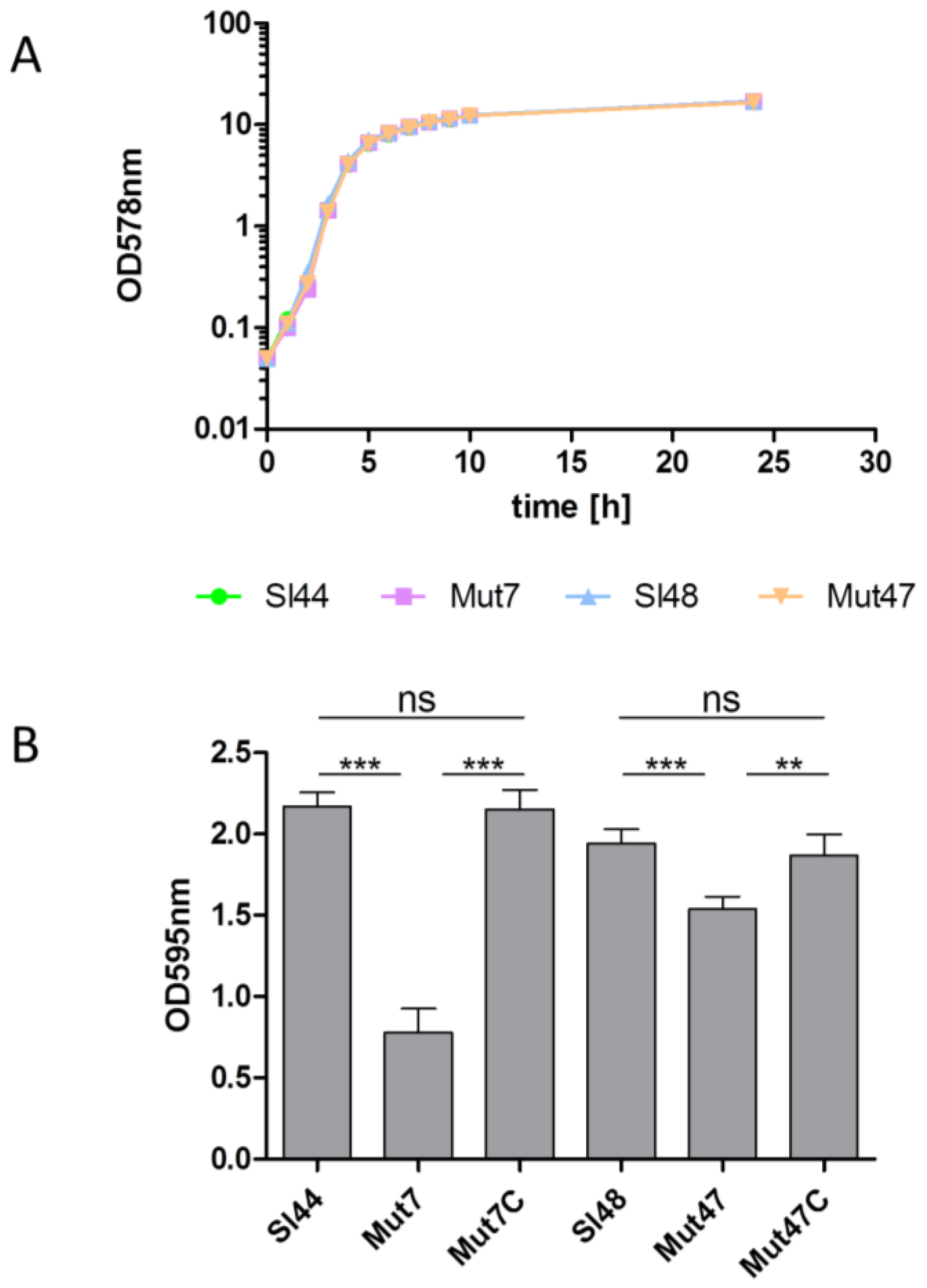
2.2. Growth Characteristics

Bacteria were grown in BHI overnight followed by dilution of the culture in 100 mL fresh BHI in 500 mL flask to an optical density 578 nm (OD578) of 0.05. Flasks were incubated at 160 rpm at 37 °C and the OD578 were determined hourly for a period of 10 h followed by final sampling after 24 h to establish growth curves. For growth experiments in little volumes, bacteria were grown in 5 mL BHI in a glass tube for 18 h at 37 °C with shaking at 160 rpm. Next day, OD578 were measured against un-inoculated BHI after vortex of growth.

2.3. Characterization of Agglutination

The Pastorex Staph Plus (Sanofi Diagnostic Pasteur, S.A., Marnes la Coquette, France), a rapid agglutination test for the simultaneous detection of the S. aureus fibrinogen affinity antigen (clumping factor), protein A, and capsular polysaccharides were used to differentiate wild type and mutant strains.

2.4. Biofilm Formation

Bacteria were grown in 5 mL BHI, TSB, BHI + 0.5% glucose or TSB + 0.5% glucose in glass test tubes for 8 h at 37 °C with shaking at 160 rpm. Afterwards, the cells were diluted 1:100 with the same type of fresh medium and 100 µL were added to wells in quadruplicate in a 96 well microtiterplate. Plates were incubated at 37 °C without shaking overnight. Next day bacteria were removed, plates were washed with PBS, and the biofilm bacteria mix were fixed with ice cold methanol for 10 min at −20 °C. After washing once with PBS, the adhered biofilm bacteria were stained with crystal violet for 10 min at room temperature. Excess stain was removed by washing 3x with PBS. The adhered bacteria were brought into solution by the addition of 100 µL of 35% acetic acid to each well. Finally, the plates were read at OD595 in the iMark Microplate Reader (Bio Rad).

2.5. DNA Manipulations and Transformations

Staphylococcal cells were lysed with recombinant lysostaphin (20 U/mL, Applied Micro, New York, NY, USA). Genomic DNA isolated by using QIAamp DNA Mini Blood Kit (Qiagen, Hilden, Germany) and plasmid DNA were prepared using the Qiagen Plasmid Mini kit (Qiagen). DNA fragments were isolated from agarose gels using the QIAEX II Gel extraction kit (Qiagen).

2.6. Construction of a srtA-Deficient Mutant

The method for the generation of a srtA lacking mutant was same as described before [7]. In brief, a primer set 1 (SrtA1FH, and SrtA1RE,) and primer set 2 (SrtA2FE, and SrtAl2RB,) were used to amplify PCR products of 809 and 793 bp, respectively, from genomic DNA of S. lugdunensis strain Sl48. The PCR products were ligated into the shuttle vector pBT9 and the ligation mixes were transformed into E. coli TG1 cells followed by incubation on LB plates containing ampicillin. A clone containing srtA gene as an insert was designated as pBTsrtA. The restriction analysis and sequence data of clone pBTsrtA confirmed srtA gene as an insert. A 1,479 bp ermB fragment was PCR-amplified using primers ermbF, and ermbR from the plasmid pEC4 containing the staphylococcal transposon Tn551 in a ClaI restriction site [28]. The ermB primers were designed from NCBI accession # AF239773. The ermB cassette was restricted with EcoRI and ligated into the EcoRI-restricted pBTsrtA. The freshly prepared E. coli TG1 cells were transformed with the ligation mix and cultivated on ampicillin- and erythromycin-containing LB plates. Clones with the plasmid conferring resistance to both antibiotics were designated as pBTsrtAE and were selected for further work. For the construction of the srtA allelic replacement mutant, protoplasts of S. lugdunensis strains Sl44 and Sl48 transformed with pBTsrtAE as described by Palma et al. [29]. Chloramphenicol-sensitive and erythromycin-resistant colonies were detected by replica plating protocol onto plates containing chloramphenicol or erythromycin at 37 °C overnight. Clones that were sensitive for chloramphenicol and resistant to erythromycin were designated Mut7 (wild-type strain Sl44) or Mut47 (Wild-type strain Sl48) and were selected for further analyses. Correct insertion of the ermB was confirmed by using srtA1FH, srtA2RB, ermBF and ermBR primers.

2.7. The Complementation of a srtA-Deficient Mutant

A PCR product of srtA gene including the ribosomal binding site was PCR amplified using primer set (SrtA1FB and SrtA2RH) from genomic DNA of S. lugdunensis Sl48. The PCR product was restricted with HindIII and BamHI and ligated into the pCU1 plasmid. Freshly prepared E. coli TG1 cells were transformed with ligation mix. A plasmid containing srtA as an insert was designated pCUsrtA. The isolated plasmid pCUsrtA transformed by protoplast method into Mut7 and Mut47. Transformants were grown on TSA plates containing 10 µg of chloramphenicol and 10 µg of erythromycin per mL. Clones expressing SrtA were designated as Mut7C and Mut47C.

2.8. Cell Protein Preparations

Briefly, bacteria were grown overnight in BHI at 37 °C with shaking (160 rpm) and cells were harvested by centrifugation and washed twice with phosphate buffered saline. Then the cell surface proteins from PBS washed bacterial cells pellets were extracted by following methods: In the first method, bacterial cell surface proteins were generated by the LiCl method as described earlier [30]. In a second method we used the hydroxylamine hydrochloride method as described by Ton-That et al. [17]. In brief, bacteria were grown in two tubes containing 5 mL BHI overnight at 37 °C and 160 rpm. After centrifugation of the cells (4000 rpm 4 °C for 10 min), 1.5 mL of 50 mM Tris HCl pH 7.0 was added to the control pellet. To the other pellet we added 1.5 mL 100 mM hydroxylamine HCl in 50 mM Tris HCl pH 7.0. Both sets were stirred at 600 rpm at 37 °C in a heating block for 1 h. After centrifugation of the cells (13,000 rpm 4 °C for 10 min, the proteins in both supernatants were precipitated by addition of TCA to a final concentration of 10%. After centrifugation of the precipitated proteins (13,000 rpm 4 °C for 10 min), 200 µL of a 2× standard Laemmli SDS Page Sample buffer were added to the pellets and heated at 95 °C for 3 min. Samples were ready to load on SDS page gel. Whole cell lysates were prepared by the suspensions of cell pellets solved in 50 mM Tris/HCl pH 8.0 containing 15 mM NaCl. Lysostaphin (20 U/mL) and protease inhibitor cocktail (Sigma) were added and the mixture was incubated at 37 °C for 30 min. Then DNase was added to break DNA and, after centrifugation, liquid supernatants were used as whole cell lysate. For the generation of cell wall, cytoplasmic, and membrane proteins, S. lugdunensis were grown overnight in BHI at 37 °C with shaking (160 rpm) and cells were harvested by centrifugation and washed twice with phosphate buffered saline, and adjusted to an optical density at 600 nm (OD600) of 20. A 1-mL portion of the bacterial suspension was pelleted and resuspended in 200 μL of digestion buffer (50 mM Tris-HCl, 20 mM MgCl2, 30% [wt/vol] raffinose; pH 7.5) containing complete mini-EDTA-free protease inhibitors (Roche). Cell wall proteins were solubilized by digestion with lysostaphin (500 μg/mL) at 37 °C for 30 min. Protoplasts were harvested by centrifugation (5000× g, 15 min) and the supernatant was retained as the cell wall fraction. Protoplast pellets were washed once in digestion buffer, sedimented (5000× g, 15 min), and resuspended in ice-cold lysis buffer (50 mM Tris-HCl [pH 7.5]) containing protease inhibitors and DNase (80 μg/mL). Protoplasts were lysed on ice by vortexing. Complete lysis was confirmed by phase-contrast microscopy. The membrane fractions were obtained by centrifugation at 18,500× g for 1 h at 4 °C. The pellets (membrane fraction) were washed once and resuspended in ice-cold lysis buffer. Cell wall fractions and protoplast suspensions were centrifuged under the same conditions and the pellets were resuspended in 200 μL of lysis buffer. The liquid supernatant from protoplast suspension retained as cytosolic fraction.

2.9. SDS-PAGE and Ligand Overlay Analysis

The prepared cell surface proteins were separated in SDS-PAGE mini gel approaches. For Western ligand blot analysis, fibronectin (Fn) (Chemicon, Temecula, CA, USA), fibrinogen (Fg) (Calbiochem, San Diego, CA, USA), collagen type I (Cn) (Sigma; Sigma product #7774) or vitronectin (Vn) were purified by the method of Yatohgo et al. [28] and labeled with biotin (Roche, Mannheim, Germany). The cell surface proteins separated by SDS-PAGE were transferred electrophoretically (Trans-blot SD, Bio-Rad, Munich, Germany) onto nitrocellulose membranes (Schleicher and Schüll, Dassel, Germany) and were blocked with 3% BSA (bovine serum albumin fraction V, Sigma). The nitrocellulose membranes with blotted proteins were exposed to biotinylated ligands, treated with avidin alkaline phosphatase and subsequently bands were revealed using NBT (Nitrotetrazolium Blue chloride) and BCIP (5-Brom-4-chlor-3-indoxylphosphat) as recommended by the manufacturer´s protocol (Bio-Rad).

2.10. Expression of Recombinant Sortase-A

The srtA gene was PCR amplified from genomic DNA of S. lugdunensis using the primer set srtAF and srtAR. The srtA gene was ligated into the pQE30 expression vector and the vector was transformed into E. coli TG1 cells. E. coli TG1 cells were cultivated in LB medium plus 100 µg/mL ampicillin at 37 °C and 150 rpm and IPTG (Isopropyl-β-D-thiogalactopyranosid, Sigma) 1 mM per mL was added at OD578 0.5 to induce the expression of SrtA. E. coli cells were lysed by lysozyme (1 mg/mL lysis buffer) and SrtA was purified in a single step under native conditions using Ni-nitrilotriacetic acid (NINTA) resin column according to the manufacturer’s recommendation (Qiagen).

2.11. Polyclonal Antibodies

Polyclonal antibodies against the recombinant SrtA were raised commercially (Genosphere Biotechnologies, Paris, France) in two rabbits by applying standard procedures of 70 days with 4 immunizations. The IgG fraction from the crude antiserum was obtained on protein-A column (Pierce, Rockford, IL, USA). In IgG preparations, naturally occurring anti-staphylococcal antibodies were removed by mixing the sera with 10 volumes of LiCl cell surface extracts from strain Mut47, which does not produce the SrtA, and immune-complexes were partially removed by centrifugation at 14,000 rpm at 4 °C for 15 min. For control Western immunoblotting, cell proteins of the wild type were separated on a SDS-Page, transferred onto a nitrocellulose membrane (Schleicher and Schuell, Dassel, Germany), and probed with anti-SrtA sera. Bound rabbit immunoglobulin G was detected with alkaline phosphatase-conjugated goat anti-rabbit immunoglobulins (Dako Diagnostika, Hamburg, Germany) with NBT/BCIP color reaction (Bio-Rad).

2.12. ELISA Adherence assays

The employed ELISA adherence assays of staphylococci to ECM proteins and host cells were done as described earlier [7].

2.13. Cell Culture and Flow Cytometric Invasion Assay

For flow cytometric invasion assay, three types of host cells were used (Table 1). The human endothelial cell line, Ea.hy926 [31], was grown in DMEM (Sigma-Aldrich) supplemented with 10% fetal calf serum (FCS).The fibroblasts (human fetal lung cells) were cultivated as described earlier [31,32]. Another epithelial cell line, the human bladder carcinoma cell line 5637 (ATCC HTB-9™), which secretes several functionally active cytokines, was also used in this study and maintained as described by Quentmeier et al. [33]. Fluorescein-5-isothiocyanate (FITC)-labeling of bacterial cells was performed as described elsewhere [34,35] and labeled cells were used within 24 h. The flow cytometric invasion assay was performed as described previously with minor modifications, such as the addition of propidium iodide just before samples were analyzed on a FacsCALIBUR™ (BD Bioscience) [36]. Results were normalized according to the mean fluorescence intensity of the respective bacterial preparation, as determined by flow cytometry. The invasiveness of the laboratory S. aureus strain Cowan I was set as 100% and the results are shown as means ± SEM of three independent experiments performed in duplicates.

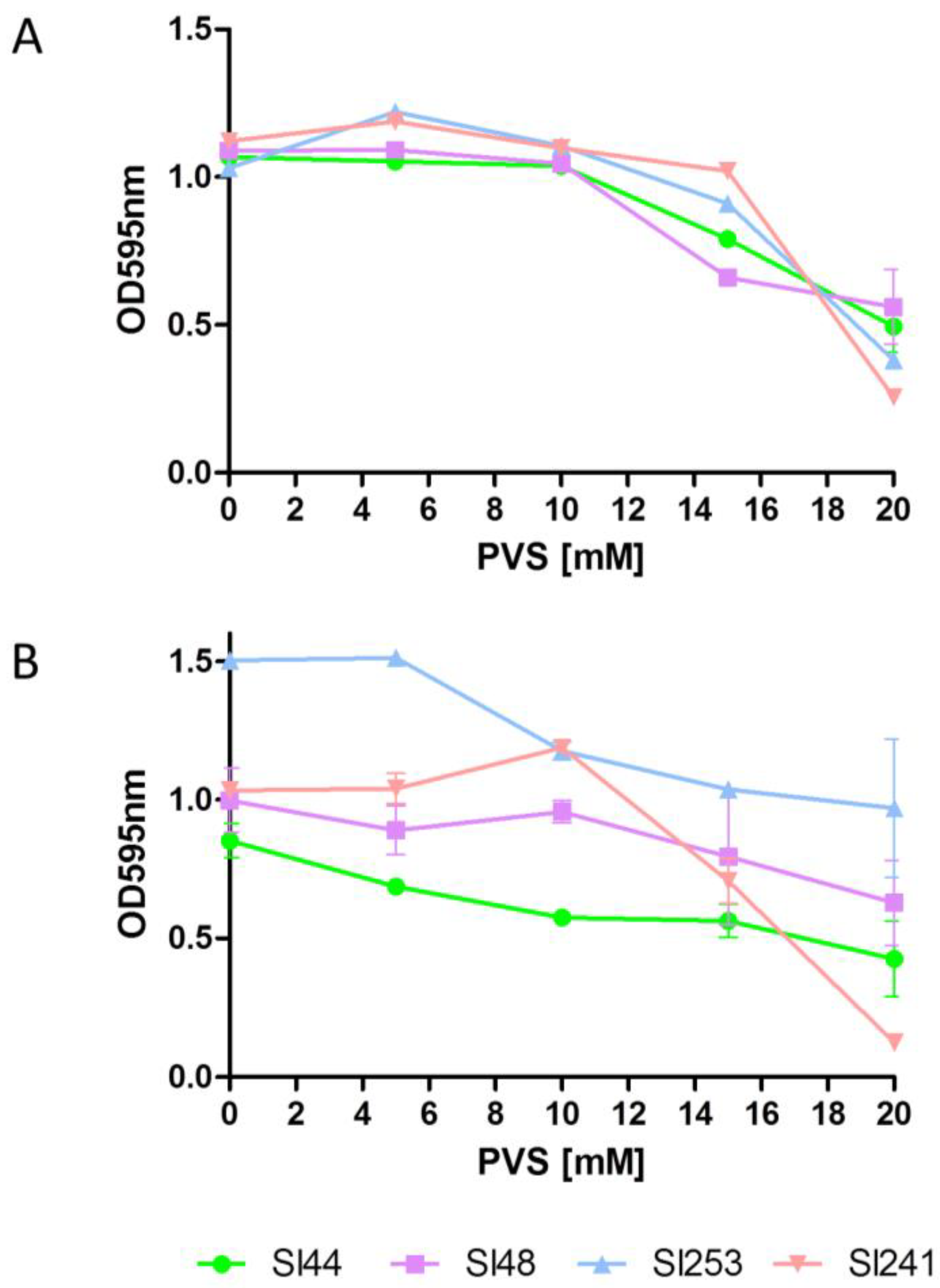
2.14. Sortase Inhibitor PVS

5 mL BHI in a glass tube was inoculate with S. lugdunensis and grown overnight at 37 °C with 160 rpm. Next day, cultures were diluted to an OD595 of 0.01. 180 µL of diluted culture was mixed with 20 µL of 10× concentrated phenyl vinyl sulfone (PVS) in a microtiter plate well. Plates were incubated at 37 °C for 18 h. Growth was determined in a Biorad reader at absorbance 595 nm. The lowest concentration that inhibited the cell growth was considered to be the MIC. For determination of effects of PVS on binding of bacteria to Fn and Fg, microtiter plate wells were coated with Fn and Fg separately. Overnight cultures were diluted to an OD595 of 0.01. 180 µL of diluted culture were mixed with 20 µL of concentrated phenyl vinyl sulfone (PVS) to the respective concentrations in a microtiter plate well. Plates were incubated at room temperature for 1 h. Adherence was determined in a Biorad reader at absorbance of 595 nm.

2.15. Hydroxylaminolysis of LPXTG Peptide

Reactions were performed in 260 μL volume containing 50 mM Hepes buffer pH 7.5, 5 mM CaCl2, and 10 μM LPETG fluorescent labeled peptides (DABCYL-LPETG-EDANS). The mixture was heated at 95 °C for 5 min. Then 10 µL of staphylococcal LiCl-extracts or 10 μM recombinant sortase-A, 5 µM of the sortase inhibitor p-hydroxymecuribenzoic acid (pHMB), or 10 mM DTT were added. Reactions were incubated for 1 h at 37 °C and then analyzed fluorometrically at 350 nm for excitations and 495 nm for recordings. Experiments were performed in triplicates.

2.16. Real-Time Reverse-Transcription PCR (qtRT-PCR)

For RNA isolation, S. lugdunensis was grown in BHI for 18 h at 37 °C with 160 rpm. After bacterial RNA isolation, real-time amplification and transcripts quantification was done as described earlier [37]. Primer sequences are given in Table 2.

2.17. Statistical Analysis

Using GraphPad Instat3, the statistical significance of the results was analyzed by ANOVA in combination with Bonferroni´s post test (compare all pairs of column or compare selected pairs of column) or Dunnett´s post test (compare all columns vs. control). Differences with p values ≤ 0.05 were considered as significant and are indicated with asterisks: * (p ≤ 0.05), ** (p ≤ 0.01), and *** (p ≤ 0.001).

3. Results

3.1. Sortase A-Dependent Proteins

A comparative bioinformatic analysis of publicly available S. lugdunensis genomes identified the presence of only 11 sortase-A dependent proteins (Table 3) [39]. All 11 MSCRAMMs were found to be highly conserved in the S. lugdunensis strains as genomic data from strain HKU09-01, M23590, VCU139 and N920143 revealed [39]. However, there exist only minor differences in the number of repeats within the stalk regions that in turns affect the length of mature proteins [39].

3.2. Alignment of Sortase A Sequences

In silico analyses identified homologs of S. aureus srtA in published genomes of S. lugdunensis. A nucleotide NCBI BLAST search identified SLGD_00472 (strain HKU09-01), srtA (strain N920143 and M23590), and SEVCU139_1681 (strain VCU139) as homologs of staphylococcal srtA. A ClustalW alignment of amino acid sequences was performed for pairwise comparison of SrtA between the four S. lugdunensis strains (N920143, HKU09-01, M23590, VCU139) and S. aureus SrtA. The SrtA sequences of four strains of S. lugdunensis (N920143, HKU09-01, M23590, VCU139) showed an intra-alignment score of 98–100%. The identity of SrtA sequences from four S. lugdunensis strains to S. aureus Newman was between 76–77% and revealed significant similarities. We identified conserved amino acid residues in the region corresponding to the calcium binding cleft of the S. aureus SrtA, such as the three glutamate residues Glu105, Glu108 and Glu166 as well as the aspartate residue Asp112, which is also highly conserved in the SrtA sequences of the S. lugdunensis strains (Figure 1) [40].

3.3. Generation of Sortase-A Mutants

To generate knockout mutants of srtA, we used the homologous recombination method leading to an insertion of ermB cassette into the srtA genes of S. lugdunensis strains Sl48 and Sl44 (Figure 2A). The srtA deficient mutants Mut7 and Mut47 were analyzed by PCR amplification using primers that anneal to ermB or to sequences flanking the srtA gene. The ermB gene could be amplified from the chromosomal DNA of the srtA deficient mutants Mut7 and Mut47, but not from the isogenic parent strains Sl48 and Sl44 respectively. Amplifications with primers specific for sequences flanking srtA revealed the insertion of ermB into the srtA genes of strains Mut7 and Mut47. The final double cross-over srtA::ermB allelic replacement in the ΔsrtA mutant was confirmed by PCR amplification. Figure 2 shows the map of srtA as well as the positions of the primer pairs and the expected sizes of the amplified PCR products. The expected PCR product size with primers SrtA1F and SrtA2R was 2.2 Kb for the wild type and is 3.1 kb for the mutant. The observed sizes for the increased PCR products could only be generated from the ΔsrtA mutants with primers SrtA1F and SrtA2R, but not with DNA of the wild type strains. In addition, amplifications of the ermB cassette were only successful with the ΔsrtA mutant, but not with wild type strain DNAs using primers Ery-FE and Ery-RE which confirmed the allelic replacement of the wild type srtA gene by srtA::ermB (Figure 2). Results are exemplary shown for the Sl48 parental and Mut47 mutant strain.

3.4. Characterisation of the Surface Proteins by SDS Page and Western Blot Experiments

First, we verified the absence of the sortase A protein in mutant strains and confirmed their complementation with plasmid coded srtA. Specific polyclonal antibodies rose in rabbits against recombinant sortase-A lacking the N-terminal signal peptide, revealed sortase-A as a polypeptide of 26 kDa in staphylococcal extracts. Immunoblotting with anti-SrtA antibodies failed to detect the SrtA in the whole cell extract from mutant strains, but complementation of the mutant strain with plasmid pCUSrtA encoded sortase-A restored the appearance of the sortase-A (Figure 3A). As a control, the transformation of the mutant strain with empty vector DNA did not affect the expression of the SrtA (data not shown). Together, these results confirmed the successful depletion of the srtA gene from the genome of S. lugdunensis. The SrtA is described as a membrane bound protein. Therefore, an attempt was made to localize SrtA within S. lugdunensis cells. For this purpose, S. lugdunensis cultivated in BHI broth medium was fractionated into the extracellular fraction, cell wall fraction, cytosol fraction and membrane digest. SrtA was detected in the membrane digest (Figure 3B). Strongest signal was observed for the membrane fractions. Only weak signals were found for the other protein fractions. It shows that SrtA of S. lugdunensis is also a membrane bound protein.

3.5. The Surface Proteomes of the Wild-Type and Mutant Strains Differ in the Absence of the SrtA

The cell surface protein expression patterns were determined by extracting the whole cells in 0.1 M hydroxylamine hydrochloride. In Coomassie Blue-stained SDS-PAGEs, the wild type and the mutant strains showed similar but non-identical patterns of proteins. In general, gels revealed less protein concentrations and protein bands for the mutants strains compared to the wild type isolates. In all strains, lower molecular mass proteins were detected in the both wild type and mutant strains, but higher molecular mass proteins were dominant in the wild type strains (Figure 3C). The results were confirmed by Western immunoblots probed with a mixture of antibodies raised in rabbits against formaldehyde fixed whole cells of S. lugdunensis. This experiment revealed a lack of bands for several proteins of the cell surface protein extracts of the ΔsrtA mutant strains, although these bands were present in wild type strains (Figure 3D).

3.6. Determination of Growth and Biofilm Formation of the ΔsrtA Mutants

The parental strains of S. lugdunensis Sl44/Sl48 and ΔsrtA mutants Mut7/Mut47 showed nearly the same growth rate for 24 h when cultivated in 100 mL of BHI in 500 mL flask (Figure 4A). Furthermore, in little cultivation volumes of only 5 mL in 14 mL glass tubes, no difference in growth could be observed (data not shown). Next, we determined the biofilm formation of all strains. S. lugdunensis wild type strains and the ΔsrtA isogenic mutants were grown in BHI, TSB, and were supplemented with or without additional 0.5% (w/v) glucose. The addition of 0.5% glucose to TSB resulted in a strong biofilm formation but it was not observed in BHI with glucose. Mut7 produced 70% less biofilm in TSB plus glucose compared to the wild type strain Sl44. Mut47 showed only a decrease of around 25% biofilm formation capability in TSB plus glucose compared to the parental strain Sl48. In the complemented mutants Mut47C and Mut47C, the biofilm formation was completely restored to the levels of the wild-type strains Sl44 and Sl48 (Figure 4B).

3.7. Recombinant SrtA As Well As Cell Extracts Catalyzes Hydroxylaminolysis

To verify whether our recombinant SrtA as well as native SrtA of cell extracts still had catalytic activities, we incubated them with LPXTG fluorescent peptides (DABCYL-LPETG-EDANS) and CaCl2. An increase in fluorescence is observed when cleavage of DABCYL-LPETG-EDANS peptide separates the fluorophore from Dabcyl which in turn confirmed the well described catalytic mechanism of the sortase. First, we tested the catalytic activity of the recombinant sortase SrtA from S. lugdunensis in this experimental setup. We observed an increase in fluorescence caused by SrtA which confirmed the cleavage of the DABCYL-LPETG-EDANS peptide (Figure 5A). As hydroxylaminolysis of LPXTG peptides depends on the sulfhydryl of the sortase, an addition of the known sortase inhibitor pHMB at least strongly abolished enzymatic activity of SrtA. However, the enzymatic activity was restored when DTT was added. We next tested cell lysates containing the native SrtA of the wild type strain Sl48, the mutant Mut47, the complemented mutant Mut47C and compare them with the recombinant SrtA in one experiment (Figure 5B). We observed an increase in fluorescence intensity when cell extracts of Sl48, Mut47C and the recombinant SrtA were incubated with the LPXTG peptide in presence of CaCl2. As expected, only low activity was measured for cell extracts of the mutant strain Mut47 and the control (Figure 5B). Comparable results were found in cell extracts of the Sl44 wild type, the SrtA deficient mutant Mut7, and the Mut7C complemented mutant strains.

3.8. Agglutination Test and Adherence of ΔsrtA Mutants to Immobilized Fibronectin (Fn), Fibrinogen (Fg), and Vitronectin (Vn)

Clumping factor (ClfA) of S. aureus is a SrtA substrate and constitutes an important virulence factor. It binds to the C-terminus of the γ-chain of fibrinogen and allowed the adhesion to different eukaryotic cell types. Besides, the ClfA is used as an antigen for rapid diagnostic identification based on latex-agglutination test systems. S. lugdunensis also possesses a clumping factor (Fbl) with an amino acid identity of 58% to ClfA of S. aureus. Hence, we applied the Pastorex Staph Plus (Sanofi Diagnostic Pasteur, S.A., Marnes la Coquette, France) rapid agglutination test for simultaneous detection of the fibrinogen affinity antigen (clumping factor) combined with the detection of protein A and capsular polysaccharides of S. aureus. The WT strains showed positive agglutination reactions, but the ΔsrtA strains did not reveal any agglutination (Figure 6A). It suggested the absence of Fbl in mutant strains, but we verified this result and measured the adherence of the ΔsrtA mutants to different ECM proteins via ELISA assays. All strains were investigated for binding to immobilized Fn, Fg, and Vn. Compared to wild-type strain Sl44, the adherence of the ΔsrtA mutant Mut7 was significantly decreased to immobilized Fg, Fn, and Vn (Figure 6B). The binding of complemented mutant Mut7C was indistinguishable to the parent level binding capacity. For the mutant strain Mut47, the results differed a little bit. We observed only a strong decrease in binding Fg, but the binding to Fn- and Vn-coated surfaces of Mut47 decreased less and not to a significant extent. However, binding of complemented mutant Mut47C to Fg, Fn and Vn was restored to the same extent as the wild type strain Sl48 (Figure 6B).

3.9. Sortase A Inhibition Resulted in Decreased Biofilm Formation and Binding to Fg and Fn

The function of sortase A could be blocked by different classes of sortase A inhibitors like berberine chloride (BBCl), phenyl vinyl sulfone (PVS) and pHMB as shown before in Section 3.7 [41]. In our study, we used the well-known SrtA blocking reagent PVS. Our experiments with PVS showed MIC’s of about 8-12 mM for strains Sl48, Sl44 and the corresponding ΔsrtA mutants (Figure 7).
The sortase A inhibition by PVS appeared to be highly effective because at 8 mM concentration we observed a reduction in biofilm formation of strains Sl48 and Sl44, but also for the corresponding ΔsrtA mutants. We further determined the effect of PVS on the binding capability of S. lugdunensis to Fn- and Fg-coated surfaces (Figure 8). A PVS treatment decreased the ability of S. lugdunensis to adhere to Fn- and Fg-coated surfaces. PVS at a concentration of 12 mM abolishes the binding to Fn of the parental strains Sl48 and Sl44 and the ΔsrtA mutants Mut47 and Mut7. The effective PVS concentration to stop binding to Fg is about 16–20 mM (Figure 8).

3.10. Hydroxylamine HCl Treatment Decrease Binding to Immobilized Fg and Fn

A hydroxylamine treatment causes a formation of the C-terminal threonine hydroxamate of surface proteins, which are thereby released into the culture medium (17). Therefore, a hydroxylamine exposure of S. lugdunensis should result in a decreased adherence to Fg, Fn or other eukaryotic proteins. Here, we incubated different wild type strains (Sl44, Sl48, Sl253 and Sl241) with different concentrations of hydroxalamine HCL and found that 20 mM hydroxylamine HCl tendentially reduced the ability to adhere to Fn (Figure 9A). The reduction of Fg adherence was much lower than that observed for adherence to Fn (Figure 9B). In addition, we observed strain specific differences in binding Fg and Fn.

3.11. Adherence and Invasion to Eucaryotic Cell Lines

LPXTG motif cell surface proteins are important for the adherence of staphylococci to eukaryotic cells. As shown above, ΔsrtA mutants showed decreased adherence to different ECM proteins. Therefore we tested the adherence of the wild type, mutant, and complemented strains to three different eukaryotic cell lines. Surprisingly, the ΔsrtA mutant Mut47 showed only slight reduced adherence to confluent epithelial cells, fibroblast, and 5637 cells. However, another ΔsrtA mutant Mut7 bound more to the above mention three cell lines. However, these differences were not statistically significant (Figure 10).
Next, we analyzed the invasion to the same eukaryotic cell lines. In experiments with FITC-labeled wild type and mutant strains, we observed a significant increase in invasion of both mutant strains to 5637 cells, Ea.hy926, and fibroblast cells (Figure 11). In addition, we observed that the Mut7 strain reached same invasion levels as the other mutant Mut47, but the wild type Sl44 revealed a comparable low capability to invade these cell lines. However, complemented strains showed the same invasion behavior as the respective wild type strains.

3.12. SrtA Influence the Gene Expression of Further Adhesins

As shown for ΔsrtA mutants of other species [42], we finally measured the influence of a srtA gene deletion on the gene expression of two further LPXTG proteins. Using real-time PCR, expression of specific mRNA for the LPXTG-bearing adhesin proteins Fbl and vWbF were compared in wild type strains Sl44, Sl48 and their respective ΔsrtA mutants Mut7 and Mut47. The RT-PCR analyses of fbl and vwbF genes showed a modest up-regulation of about 2 fold in both ΔsrtA mutant backgrounds (Table 4), verifying results observed for sortase mutants of other species [42].

4. Discussion

The LPXTG-anchored proteins are covalently anchored to the cell surface of many bacteria and play key roles as virulence factors for the establishment of bacterial infections [43]. The executive enzyme, the sortase, is essential for the functional assembly of cell surface virulence factors and, hence, important for the pathogenesis of staphylococci and in particular S. aureus [22,44]. Mutations in the srtA gene result in non-anchoring of 19 surface proteins to the cell envelope [23]. Consequently, S. aureus sortase mutants are defective in assembling surface proteins and are highly attenuated in the pathogenesis shown in animal infection studies [23]. However, only a few reports are available on the properties of S. lugdunensis in terms of interaction with ECM molecules or host tissues, as well as on its possession of the LPXTG-anchored proteins and secretable expanded repertoire adhesive molecules (SERAMs). S. lugdunensis strains were shown to bind to varying extents to collagen, fibronectin, vitronectin, laminin, fibrinogen, thrombospondin, plasminogen, and human IgG [45]. A comparative bioinformatic analysis of publicly available S. lugdunensis genomes identified the presence of 11 sortase A dependent proteins (Table 3) [39]. In the published genome sequence of S. lugdunensis strain N920143, Heilbronner et al. (2011) identified fbl, vWbF, isdB, isdJ and seven more genes coding for proteins with a LPXTG motif [39]. In the present study, we report the characterization of S. lugdunensis housekeeping SrtA and its role in LPXTG motif proteins’ cell wall anchoring. To address the role of SrtA to adhere to different ECMs and cell lines, stable and defined ΔsrtA mutants of S. lugdunensis were generated by allelic replacement as described before and respective complemented mutant strains were generated to exclude any polar effects [30].
The SrtA from four S. lugdunensis strains (N920143, HKU09-01, M23590, and VCU139) showed identity of 76-77% to the SrtA of S. aureus strain Newman, and a very high identity of 98-100% among themselves. Specific residues identified in the region corresponding to the described calcium binding cleft in the S. aureus SrtA were also highly conserved in the amino acid sequence of the S. lugdunensis SrtA. In our study, the recombinant SrtA as well as whole cell extracts of S. lugdunensis could catalyze the hydrolysis of a fluorescent quenched polypeptide carrying the LPXTG motif, which is characteristic of sortases of other species (Figure 5) [16,23]. In addition, it was observed that the S. lugdunensis SrtA activity also required calcium ions for hydroxyaminolysis of LPXTG peptide, as reported previously for SrtA of S. aureus [40]. Therefore, we propose a quite similar catalytic mechanism as shown for SrtA from S. aureus [16,17]. These assumption was approved by further phenotypic analyzes showing an essential role for SrtA in anchoring LPXTG proteins, as ΔsrtA mutants differ in their cell surface proteome from that of the wild type strains, showing the absence of several proteins in the ΔsrtA mutants (Figure 3). It’s therefore not surprising that we found a reduced biofilm formation and a reduced adherence to Fn, Fg, and Vn in the ΔsrtA mutants (Figure 4B and Figure 6). Similar results were shown for S. aureus as the inhibition of SrtA activity caused loss of binding to Fn, Fg, and IgG and reduced biofilm formation [46,47]. Furthermore, these results are consistent with our observations that the ΔsrtA mutants showed no agglutination in the Pastorex test.
The presence of an active sortase enzyme in S. aureus was found essential for the adherence to host eukaryotic cell [46]. In a previous study, the ΔsrtA mutants of S. aureus and Lactococcus salivarius showed significant reductions in adhesion to different epithelial cell lines compared to the WT strain [48,49]. Here in this report, the ΔsrtA mutant Mut47 showed only slightly reduced adherence to confluent epithelial cells, fibroblast, and 5637 cells, whereas another ΔsrtA mutant Mut7 bound to above mention cell lines (Figure 10). Obviously, the differences were not statistically significant, which was surprising because formerly we observed significant lower adherences to Fn, Fg, and Vn. As shown previously, the autolysin AtlL of S. lugdunensis was identified as a major adhesin being crucially involved in the internalization process to cells [47]. Here, we showed a fairly strong imbalance in the proteome of the cell surface compartment in the mutant strains. These probably make AtlL more available to the cell surface, which might compensate for the loss of other LPXTG adhesins in case of Mut47 and eventually enhance, to a limited extent, the binding of Mut7 to eukaryotic cells. On the other hand, we cannot exclude the absence of the respective LPXTG counterpart proteins on the cell surfaces of the eukaryotic cell lines under our experimental conditions. This rather would lead to indistinguishable results between the adherence of wild types and ΔsrtA mutant to cell lines, similar to what we have observed here.
We further observed that SrtA is not only involved in the covalent binding of LPXTG motif proteins to cell wall, but also influenced the transcriptional regulation of at least two adhesin genes fbl and vWbF. RT-PCR results showed that expression of fbl and vWbI genes were upregulated at least 2 fold in the ΔsrtA mutants relative to their wild type strains (Table 4). This is in agreement with the previous finding that mutations in the srtA gene result in the upregulated expression of cell surface proteins in an oral Streptococcus strain [42]. Nobbs et al. (2007) reported a significant upregulation of the expression of adhesin genes like sspA/B, cshA/B, and fbpA in ΔsrtA deficient mutants relative to their wild type strains of S. gordonii [50]. Moreover, Hall et al. (2019) recently described a quality control mechanism that monitors the processing of LPXTG adhesins by SrtA via measuring the left C-terminal cleaved LPXTG proteins (C-peps) which stayed membrane located and are recognized by a previously uncharacterized intramembrane two-component system (TCS) [51]. Prevention of C-peps generation de-repressed this TCS and resulted in increased expression of further adhesins compensating for the loss of LXPTG adhesins [51]. Since TCS is conserved in streptococci but not in staphylococci, the same C-pep-driven regulatory circuit is unlikely in S. lugdunensis, but similar regulation processes cannot be ruled out and might be an explanation for the upregulation of fbl and vWbF. Thus, the comparable high adherence to host cells of ΔsrtA mutants might also be a result of the upregulation of other adhesins in the mutant strains and is object of future research. However, detailed in vitro infection studies are necessary to understand the impact of the single LPXTG proteins of S. lugdunensis to the respective adherence mechanisms.
A sudden inhibition of the SrtA activity could disrupt the pathogenesis of bacterial infections [22]. The surface-protein anchoring is sensitive to sulfhydryl-modifying reagents like methanethiosulfonates, berberine chloride, (Z)-3-(2,5-dimethoxyphenyl)-2-(4-methoxyphenyl)acrylonitrile or PVS under in vitro conditions [17,42,52]. Here, the SrtA inhibitor PVS reduced the biofilm formation and also reduced the adherence to Fg and Fn (Figure 7 and Figure 8). Further, a hydroxylamine HCl treatment, releasing LPXTG proteins from the cell wall, was found to greatly reduce the adherence of four wild type strains of S. lugdunensis to immobilized Fg and Fn (Figure 9). This clearly showed the important role of the LPXTG proteins in S. lugdunensis for the binding to some ECM proteins, and confirmed the data observed for S. aureus [23,53]. The inhibition of the sortase activity may provide a novel approach for the treatment of infections with staphylococci and a development of such inhibitors could complement the current dependence on antibiotics. Therefore, SrtA is an attractive target to attenuate virulence and hamper infections. In addition, the ΔsrtA mutant of S. lugdunensis showed significant defects in virulence including reduced bacteremia, reduced bacterial spreading to the kidneys and reduced size/density of endocardial vegetations [24]. This highlights the importance of LPXTG-anchored proteins in the pathogenesis of this germ. These and our results suggest that several surface proteins probably act in concert to promote the adhesion to different host structures and enable a survival in the host [24]. Sortase A mutants of S. aureus displayed strongly reduced virulence in various infection models including experimental sepsis, highlighting the impact of properly displayed surface proteins during infection [54,55]. Therefore, the ΔsrtA mutants were recently discussed as vaccine candidates due to the attenuated but still immune-stimulating phenotype [56].
In conclusions, SrtA was found as the housekeeping sortase of S. lugdunensis like in other Gram-positive bacteria and in particular in S. aureus. Disrupting the presentation of surface proteins by gene deletion or blocking the activity of SrtA via different SrtA inhibitors could therefore effectively cause a reduction in binding to ECM proteins and could probably disrupt the pathogenesis of bacterial infections and promote bacterial clearance by the host. Although some strain-specific differences may exist, SrtA could be an attractive target to attenuate the virulence of S. lugdunensis.

Author Contributions

Conceptualization, M.H. and K.B.; methodology, M.H.; software, M.H.; validation, M.H., C.K. and K.B.; formal analysis, M.H.; investigation, M.H.; resources, M.H.; data curation, M.H.; writing—original draft preparation, M.H., C.K.; writing—review and editing, C.K., K.B.; visualization, M.H., C.K.; supervision, K.B.; project administration, M.H.; funding acquisition, K.B. All authors have read and agreed to the published version of the manuscript.

Funding

This study was funded in part from the IZKF (Be2/023/11).

Acknowledgments

The paper is dedicated to Georg Peters (1951–2018).

Conflicts of Interest

The authors declare no conflict of interest.

References

  1. Becker, K.; Heilmann, C.; Peters, G. Coagulase-negative staphylococci. Clin. Microbiol. Rev. 2014, 27, 870–926. [Google Scholar] [CrossRef] [Green Version]
  2. Sundqvist, M.; Bieber, L.; Smyth, R.; Kahlmeter, G. Detection and identification of Staphylococcus lugdunensis are not hampered by use of defibrinated horse blood in blood agar plates. J. Clin. Microbiol 2010, 48, 1987–1988. [Google Scholar] [CrossRef] [Green Version]
  3. van der Mee-Marquet, N.; Achard, A.; Mereghetti, L.; Danton, A.; Minier, M.; Quentin, R. Staphylococcus lugdunensis infections: High frequency of inguinal area carriage. J. Clin. Microbiol. 2003, 41, 1404–1409. [Google Scholar] [CrossRef] [Green Version]
  4. Kaspar, U.; Kriegeskorte, A.; Schubert, T.; Peters, G.; Rudack, C.; Pieper, D.H.; Wos-Oxley, M.; Becker, K. The culturome of the human nose habitats reveals individual bacterial fingerprint patterns. Environ. Microbiol. 2016, 18, 2130–2142. [Google Scholar] [CrossRef]
  5. Frank, K.L.; Del Pozo, J.L.; Patel, R. From clinical microbiology to infection pathogenesis: How daring to be different works for Staphylococcus lugdunensis. Clin. Microbiol. Rev. 2008, 21, 111–133. [Google Scholar] [CrossRef] [Green Version]
  6. Anguera, I.; Del, R.A.; Miro, J.M.; Matinez-Lacasa, X.; Marco, F.; Guma, J.R.; Quaglio, G.; Claramonte, X.; Moreno, A.; Mestres, C.A.; et al. Staphylococcus lugdunensis infective endocarditis: Description of 10 cases and analysis of native valve, prosthetic valve, and pacemaker lead endocarditis clinical profiles. Heart 2005, 91, e10. [Google Scholar] [CrossRef] [Green Version]
  7. Hussain, M.; Steinbacher, T.; Peters, G.; Heilmann, C.; Becker, K. The adhesive properties of the Staphylococcus lugdunensis multifunctional autolysin AtlL and its role in biofilm formation and internalization. Int. J. Med. Microbiol. 2015, 305, 129–139. [Google Scholar] [CrossRef]
  8. Marlinghaus, L.; Becker, K.; Korte, M.; Neumann, S.; Gatermann, S.G.; Szabados, F. Construction and characterization of three knockout mutants of the fbl gene in Staphylococcus lugdunensis. APMIS 2012, 120, 108–116. [Google Scholar] [CrossRef]
  9. Nilsson, M.; Bjerketorp, J.; Wiebensjo, A.; Ljungh, A.; Frykberg, L.; Guss, B. A von Willebrand factor-binding protein from Staphylococcus lugdunensis. FEMS Microbiol. Lett. 2004, 234, 155–161. [Google Scholar] [CrossRef]
  10. Nilsson, M.; Bjerketorp, J.; Guss, B.; Frykberg, L. A fibrinogen-binding protein of Staphylococcus lugdunensis. FEMS Microbiol. Lett. 2004, 241, 87–93. [Google Scholar] [CrossRef] [Green Version]
  11. Donvito, B.; Etienne, J.; Denoroy, L.; Greenland, T.; Benito, Y.; Vandenesch, F. Synergistic hemolytic activity of Staphylococcus lugdunensis is mediated by three peptides encoded by a non-agr genetic locus. Infect. Immun. 1997, 65, 95–100. [Google Scholar] [CrossRef] [Green Version]
  12. Heilmann, C. Adhesion mechanisms of staphylococci. Adv. Exp. Med. Biol. 2011, 715, 105–123. [Google Scholar] [CrossRef]
  13. Foster, T.J.; Geoghegan, J.A.; Ganesh, V.K.; Hook, M. Adhesion, invasion and evasion: The many functions of the surface proteins of Staphylococcus aureus. Nat. Rev. Microbiol. 2014, 12, 49–62. [Google Scholar] [CrossRef] [Green Version]
  14. Patti, J.M.; Höök, M. Microbial adhesins recognizing extracellular matrix macromolecules. Curr. Opin. Cell. Biol. 1994, 6, 752–758. [Google Scholar] [CrossRef]
  15. Navarre, W.W.; Schneewind, O. Proteolytic cleavage and cell wall anchoring at the LPXTG motif of surface proteins in gram-positive bacteria. Mol. Microbiol. 1994, 14, 115–121. [Google Scholar] [CrossRef]
  16. Mazmanian, S.K.; Liu, G.; Ton-That, H.; Schneewind, O. Staphylococcus aureus sortase, an enzyme that anchors surface proteins to the cell wall. Science 1999, 285, 760–763. [Google Scholar] [CrossRef]
  17. Ton-That, H.; Liu, G.; Mazmanian, S.K.; Faull, K.F.; Schneewind, O. Purification and characterization of sortase, the transpeptidase that cleaves surface proteins of Staphylococcus aureus at the LPXTG motif. Proc. Natl. Acad. Sci. USA 1999, 96, 12424–12429. [Google Scholar] [CrossRef] [Green Version]
  18. Perry, A.M.; Ton-That, H.; Mazmanian, S.K.; Schneewind, O. Anchoring of surface proteins to the cell wall of Staphylococcus aureus. III. Lipid II is an in vivo peptidoglycan substrate for sortase-catalyzed surface protein anchoring. J. Biol. Chem. 2002, 277, 16241–16248. [Google Scholar] [CrossRef] [Green Version]
  19. Ruzin, A.; Severin, A.; Ritacco, F.; Tabei, K.; Singh, G.; Bradford, P.A.; Siegel, M.M.; Projan, S.J.; Shlaes, D.M. Further evidence that a cell wall precursor [C(55)-MurNAc-(peptide)-GlcNAc] serves as an acceptor in a sorting reaction. J. Bacteriol. 2002, 184, 2141–2147. [Google Scholar] [CrossRef] [Green Version]
  20. Spirig, T.; Weiner, E.M.; Clubb, R.T. Sortase enzymes in Gram-positive bacteria. Mol. Microbiol. 2011, 82, 1044–1059. [Google Scholar] [CrossRef]
  21. Oshida, T.; Sugai, M.; Komatsuzawa, H.; Hong, Y.M.; Suginaka, H.; Tomasz, A. A Staphylococcus aureus autolysin that has an N-acetylmuramoyl-L-alanine amidase domain and an endo-beta-N-acetylglucosaminidase domain: Cloning, sequence analysis, and characterization. Proc. Natl. Acad. Sci. USA 1995, 92, 285–289. [Google Scholar] [CrossRef] [Green Version]
  22. Mazmanian, S.K.; Liu, G.; Jensen, E.R.; Lenoy, E.; Schneewind, O. Staphylococcus aureus sortase mutants defective in the display of surface proteins and in the pathogenesis of animal infections. Proc. Natl. Acad. Sci. USA 2000, 97, 5510–5515. [Google Scholar] [CrossRef] [Green Version]
  23. Mazmanian, S.K.; Ton-That, H.; Su, K.; Schneewind, O. An iron-regulated sortase anchors a class of surface protein during Staphylococcus aureus pathogenesis. Proc. Natl. Acad. Sci. USA 2002, 99, 2293–2298. [Google Scholar] [CrossRef] [Green Version]
  24. Heilbronner, S.; Hanses, F.; Monk, I.R.; Speziale, P.; Foster, T.J. Sortase A promotes virulence in experimental Staphylococcus lugdunensis endocarditis. Microbiology 2013, 159, 2141–2152. [Google Scholar] [CrossRef] [Green Version]
  25. Edgell, C.J.; McDonald, C.C.; Graham, J.B. Permanent cell line expressing human factor VIII-related antigen established by hybridization. Proc. Natl. Acad. Sci. USA 1983, 80, 3734–3737. [Google Scholar] [CrossRef] [Green Version]
  26. Giard, D.J.; Aaronson, S.A.; Todaro, G.J.; Arnstein, P.; Kersey, J.H.; Dosik, H.; Parks, W.P. In vitro cultivation of human tumors: Establishment of cell lines derived from a series of solid tumors. J. Natl. Cancer Inst. 1973, 51, 1417–1423. [Google Scholar] [CrossRef]
  27. Fogh, J.; Wright, W.C.; Loveless, J.D. Absence of HeLa cell contamination in 169 cell lines derived from human tumors. J. Natl. Cancer Inst. 1977, 58, 209–214. [Google Scholar] [CrossRef]
  28. Khan, S.A.; Novick, R.P. Terminal nucleotide sequences of Tn551, a transposon specifying erythromycin resistance in Staphylococcus aureus: Homology with Tn3. Plasmid 1980, 4, 148–154. [Google Scholar] [CrossRef]
  29. Palma, M.; Nozohoor, S.; Schennings, T.; Heimdahl, A.; Flock, J.I. Lack of the extracellular 19-kilodalton fibrinogen-binding protein from Staphylococcus aureus decreases virulence in experimental wound infection. Infect. Immun. 1996, 64, 5284–5289. [Google Scholar] [CrossRef] [Green Version]
  30. Hussain, M.; Becker, K.; von Eiff, C.; Schrenzel, J.; Peters, G.; Herrmann, M. Identification and characterization of a novel 38.5-kilodalton cell surface protein of Staphylococcus aureus with extended-spectrum binding activity for extracellular matrix and plasma proteins. J. Bacteriol. 2001, 183, 6778–6786. [Google Scholar] [CrossRef] [Green Version]
  31. Tuchscherr, L.; Löffler, B.; Buzzola, F.R.; Sordelli, D.O. Staphylococcus aureus adaptation to the host and persistence: Role of loss of capsular polysaccharide expression. Future Microbiol. 2010, 5, 1823–1832. [Google Scholar] [CrossRef]
  32. Hussain, M.; Haggar, A.; Heilmann, C.; Peters, G.; Flock, J.I.; Herrmann, M. Insertional inactivation of Eap in Staphylococcus aureus strain Newman confers reduced staphylococcal binding to fibroblasts. Infect. Immun. 2002, 70, 2933–2940. [Google Scholar] [CrossRef] [Green Version]
  33. Quentmeier, H.; Zaborski, M.; Drexler, H.G. The human bladder carcinoma cell line 5637 constitutively secretes functional cytokines. Leuk. Res. 1997, 21, 343–350. [Google Scholar] [CrossRef]
  34. Sinha, B.; Francois, P.; Que, Y.A.; Hussain, M.; Heilmann, C.; Moreillon, P.; Lew, D.; Krause, K.H.; Peters, G.; Herrmann, M. Heterologously expressed Staphylococcus aureus fibronectin-binding proteins are sufficient for invasion of host cells. Infect. Immun. 2000, 68, 6871–6878. [Google Scholar] [CrossRef] [Green Version]
  35. Juuti, K.M.; Sinha, B.; Werbick, C.; Peters, G.; Kuusela, P.I. Reduced adherence and host cell invasion by methicillin-resistant Staphylococcus aureus expressing the surface protein Pls. J. Infect. Dis. 2004, 189, 1574–1584. [Google Scholar] [CrossRef] [Green Version]
  36. Sinha, B.; Francois, P.P.; Nüße, O.; Foti, M.; Hartford, O.M.; Vaudaux, P.; Foster, T.J.; Lew, D.P.; Herrmann, M.; Krause, K.H. Fibronectin-binding protein acts as Staphylococcus aureus invasin via fibronectin bridging to integrin α5β1. Cell. Microbiol. 1999, 1, 101–117. [Google Scholar] [CrossRef]
  37. Hussain, M.; Schafer, D.; Juuti, K.M.; Peters, G.; Haslinger-Loffler, B.; Kuusela, P.I.; Sinha, B. Expression of Pls (plasmin sensitive) in Staphylococcus aureus negative for pls reduces adherence and cellular invasion and acts by steric hindrance. J. Infect. Dis. 2009, 200, 107–117. [Google Scholar] [CrossRef]
  38. Pinsky, B.A.; Samson, D.; Ghafghaichi, L.; Baron, E.J.; Banaei, N. Comparison of real-time PCR and conventional biochemical methods for identification of Staphylococcus lugdunensis. J. Clin. Microbiol. 2009, 47, 3472–3477. [Google Scholar] [CrossRef] [Green Version]
  39. Heilbronner, S.; Holden, M.T.; van Tonder, A.; Geoghegan, J.A.; Foster, T.J.; Parkhill, J.; Bentley, S.D. Genome sequence of Staphylococcus lugdunensis N920143 allows identification of putative colonization and virulence factors. FEMS Microbiol. Lett. 2011, 322, 60–67. [Google Scholar] [CrossRef] [Green Version]
  40. Hirakawa, H.; Ishikawa, S.; Nagamune, T. Design of Ca2+-independent Staphylococcus aureus sortase A mutants. Biotechnol. Bioeng. 2012, 109, 2955–2961. [Google Scholar] [CrossRef]
  41. Kim, S.H.; Shin, D.S.; Oh, M.N.; Chung, S.C.; Lee, J.S.; Oh, K.B. Inhibition of the bacterial surface protein anchoring transpeptidase sortase by isoquinoline alkaloids. Biosci. Biotechnol. Biochem. 2004, 68, 421–424. [Google Scholar] [CrossRef] [PubMed]
  42. Maresso, A.W.; Schneewind, O. Sortase as a target of anti-infective therapy. Pharmacol. Rev. 2008, 60, 128–141. [Google Scholar] [CrossRef] [PubMed]
  43. Que, Y.A.; Francois, P.; Haefliger, J.A.; Entenza, J.M.; Vaudaux, P.; Moreillon, P. Reassessing the role of Staphylococcus aureus clumping factor and fibronectin-binding protein by expression in Lactococcus lactis. Infect. Immun. 2001, 69, 6296–6302. [Google Scholar] [CrossRef] [Green Version]
  44. Jonsson, I.M.; Mazmanian, S.K.; Schneewind, O.; Verdrengh, M.; Bremell, T.; Tarkowski, A. On the role of Staphylococcus aureus sortase and sortase-catalyzed surface protein anchoring in murine septic arthritis. J. Infect. Dis. 2002, 185, 1417–1424. [Google Scholar] [CrossRef] [PubMed] [Green Version]
  45. Paulsson, M.; Petersson, A.C.; Ljungh, A. Serum and tissue protein binding and cell surface properties of Staphylococcus lugdunensis. J. Med. Microbiol. 1993, 38, 96–102. [Google Scholar] [CrossRef]
  46. Oh, K.B.; Oh, M.N.; Kim, J.G.; Shin, D.S.; Shin, J. Inhibition of sortase-mediated Staphylococcus aureus adhesion to fibronectin via fibronectin-binding protein by sortase inhibitors. Appl. Microbiol. Biotechnol. 2006, 70, 102–106. [Google Scholar] [CrossRef]
  47. Sibbald, M.J.; Yang, X.M.; Tsompanidou, E.; Qu, D.; Hecker, M.; Becher, D.; Buist, G.; van Dijl, J.M. Partially overlapping substrate specificities of staphylococcal group A sortases. Proteomics 2012, 12, 3049–3062. [Google Scholar] [CrossRef] [Green Version]
  48. Weidenmaier, C.; Kokai-Kun, J.F.; Kristian, S.A.; Chanturiya, T.; Kalbacher, H.; Gross, M.; Nicholson, G.; Neumeister, B.; Mond, J.J.; Peschel, A. Role of teichoic acids in Staphylococcus aureus nasal colonization, a major risk factor in nosocomial infections. Nat. Med. 2004, 10, 243–245. [Google Scholar] [CrossRef]
  49. van Pijkeren, J.P.; Canchaya, C.; Ryan, K.A.; Li, Y.; Claesson, M.J.; Sheil, B.; Steidler, L.; O’Mahony, L.; Fitzgerald, G.F.; van Sinderen, S.; et al. Comparative and functional analysis of sortase-dependent proteins in the predicted secretome of Lactobacillus salivarius UCC118. Appl. Environ. Microbiol. 2006, 72, 4143–4153. [Google Scholar] [CrossRef] [Green Version]
  50. Nobbs, A.H.; Vajna, R.M.; Johnson, J.R.; Zhang, Y.; Erlandsen, S.L.; Oli, M.W.; Kreth, J.; Brady, L.J.; Herzberg, M.C. Consequences of a sortase A mutation in Streptococcus gordonii. Microbiology 2007, 153, 4088–4097. [Google Scholar] [CrossRef] [Green Version]
  51. Hall, J.W.; Lima, B.P.; Herbomel, G.G.; Gopinath, T.; McDonald, L.; Shyne, M.T.; Lee, J.K.; Kreth, J.; Ross, K.F.; Veglia, G.; et al. An intramembrane sensory circuit monitors sortase A-mediated processing of streptococcal adhesins. Sci. Signal. 2019, 12. [Google Scholar] [CrossRef] [PubMed]
  52. Sudheesh, P.S.; Crane, S.; Cain, K.D.; Strom, M.S. Sortase inhibitor phenyl vinyl sulfone inhibits Renibacterium salmoninarum adherence and invasion of host cells. Dis. Aquat. Organ. 2007, 78, 115–127. [Google Scholar] [CrossRef] [PubMed] [Green Version]
  53. Ton-That, H.; Schneewind, O. Anchor structure of staphylococcal surface proteins. IV. Inhibitors of the cell wall sorting reaction. J. Biol. Chem. 1999, 274, 24316–24320. [Google Scholar] [CrossRef] [PubMed] [Green Version]
  54. Jonsson, I.M.; Mazmanian, S.K.; Schneewind, O.; Bremell, T.; Tarkowski, A. The role of Staphylococcus aureus sortase A and sortase B in murine arthritis. Microbes Infect. 2003, 5, 775–780. [Google Scholar] [CrossRef]
  55. Weiss, W.J.; Lenoy, E.; Murphy, T.; Tardio, L.; Burgio, P.; Projan, S.J.; Schneewind, O.; Alksne, L. Effect of srtA and srtB gene expression on the virulence of Staphylococcus aureus in animal models of infection. J. Antimicrob. Chemother. 2004, 53, 480–486. [Google Scholar] [CrossRef] [PubMed] [Green Version]
  56. Kim, H.K.; Kim, H.Y.; Schneewind, O.; Missiakas, D. Identifying protective antigens of Staphylococcus aureus, a pathogen that suppresses host immune responses. FASEB J. 2011, 25, 3605–3612. [Google Scholar] [CrossRef] [Green Version]
Figure 1. Multiple sequence alignments of SrtA sequences of four strains of S. lugdunensis (N920143, HKU09-01, M23590, and VCU139) and S. aureus Newman using CLUSTAL 2.1. The highly conserved amino acids glutamate (E) and aspartate (D) involved in binding of calcium ions are highlighted in yellow. Stars denote highly conserved amino acids.
Microorganisms 08 01975 g001
Figure 2. Genetic map of srtA and primer positions for PCRs, and results of PCR confirming the gene depletion of srtA in S. lugdunensis. (A) The gene loci of srtA in S. lugdunensis are shown before and after the recombination processes. In addition, the gene loci of srtA in S. lugdunensis are shown before and after the recombination processes and the positions of primer binding during the PCR experiments. The primer sequences are given in Table 3. (B) PCR products from genomic DNA of S. lugdunensis wild-type strain (Sl48), ΔsrtA mutant strain (Mut47) and plasmid DNA of pBTsrtA (A23) were separated by 1% agarose gels. Higher molecular bands (PCR with primer pair srtlA1F and srtlA2R) and the occurrence of corresponding bands to PCR experiments with primer pairs Ery-EF/Ery-ER, Ery-EF/srtlA2R and srtlA1F/Ery-ER confirming the deletion of srtA and following substitution by the ermB gene in the genome S. lugdunensis ΔsrtA mutant strain Mut47.
Microorganisms 08 01975 g002
Figure 3. Western blot and SDS page experiments of S. lugdunensis wild type, ΔsrtA mutant and complemented mutant strains showing the presence and localization of SrtA and consequences of a srtA deletion for the surface proteome pattern. (A) Western blots showing the absence of SrtA in the mutant strains but specific signals in wild types and complemented mutants. Whole cell extracts of wild type, mutant and complemented mutant strains were separated via SDS page and blotted. The Western blots were probed with specific polyclonal antibodies rose in rabbits against recombinant SrtA (anti-SrtA). Asterisk mark the specific SrtA band. (B) Localization of SrtA within S. lugdunensis cells. For this purpose, S. lugdunensis were grown in BHI broth medium. Cells were fractionated into the extracellular fraction (EC), cell wall fraction (CW), cytosol fraction (CY), and membrane digest fraction (MD). Proteins were separated by SDS page. After blotting, SrtA was detected in the different fractions with anti-SrtA antibodies. Asterisk mark the specific SrtA band. (C) Analyses of cell surface proteins of the wild-type strains and the mutant strains. The cell surface proteins were extracted in 0.1 M hydroxylamine hydrochloride. The protein extracts were separated on SDS-Page and stained with Coomassie Blue. (D) Cell surface protein fractions of wild type and mutant strains were separated via SDS page. After blotting, the Western blots were probed with a mixture of antibodies raised in rabbits against formaldehyde fixed whole cells of S. lugdunensis. Asterisk mark the specific SrtA band. M—page ruler, Sl44 and Sl48—wild type strains, Mut7 and Mut47—ΔsrtA mutant strains, Mut7C and Mut47C—complemented ΔsrtA mutant strains.
Microorganisms 08 01975 g003
Figure 4. Growth and biofilm formation of S. lugdunensis wild-type strains (Sl44, Sl48) and ΔsrtA mutant strains (Mut7, Mut47). (A) Bacterial growth was monitored for 24 h. Bacteria were cultivated in 100 mL BHI in 500 mL flask at 37 °C under permanent agitation. The experiment was done on triplicates. One representative experiment is shown. (B) Biofilm-forming capacities of the S. lugdunensis wild type (Sl44 and Sl48), ΔsrtA mutant (Mut7 and Mut47) and complemented mutant (Mut7C and Mut47C) strains were assessed by a quantitative biofilm assay performed in microtiter plates applying crystal violet and determination of the OD595nm. Results are shown as the mean of five independent experiments with the standard deviation (SD). Statistical analyses were performed using one-way ANOVA with Bonferroni multiple comparisons posttest (** p < 0.01; *** p < 0.001). ns—not significant.
Microorganisms 08 01975 g004
Figure 5. Hydroxylaminolysis of LPXTG peptide by recombinant SrtA (A) and whole S. lugdunensis cells (B). (A) Recombinant SrtA was incubated with the sorting substrate Dabcyl-QALPETGEE-Edans (LPXTG), and peptide cleavage was monitored as an increase in fluorescence. The reactions were influenced by the addition of pHMB (inhibitor of sortase activity) and by DTT. Presence and absence of the respective substance is shown as “+” or “-“, respectively. (B) Same assay using the LPXTG substrate, recombinant SrtA, S. lugdunensis cell extracts from the wild-type strain Sl48, ΔsrtA mutant Mut47 and complemented Mut47C. All results are shown as the mean of three independent experiments with the standard deviation (SD).
Microorganisms 08 01975 g005
Figure 6. Pastorex staph plus agglutination test and binding of S. lugdunensis strains to ECM proteins. (A): Pastorex staph plus test of S. lugdunensis wild-type strains (Sl44, Sl48) and ΔsrtA mutant strains (Mut7, Mut47) grown overnight on blood agar plates. Material was mixed in one drop of Pastorex reagent on a Pastorex disposable card. Results were recorded as positive on visual agglutination of wild-type strains and as negative for both mutants showing no agglutination. (B): Binding of S. lugdunensis wild-type strains (Sl44 and Sl48), ΔsrtA mutants (Mut7 and Mut47), and complemented mutant strains (Mut7C and Mut47C) to immobilized fibrinogen (Fg), fibronectin (Fn), and vitronectin (Vn) assessed by ELISA adherence assays. Results are shown as the mean of four independent experiments with the standard deviation (SD). Statistical analyses were performed using one-way ANOVA with Bonferroni multiple comparisons posttest (** p < 0.01; *** p < 0.001). ns—not significant.
Microorganisms 08 01975 g006
Figure 7. Effect of the sortase A inhibitor phenyl vinyl sulfone (PVS) on biofilm formation. The minimum inhibitory concentrations (MIC) of the sortase inhibitor PVS of S. lugdunensis wild type (SL44 and Sl48), ΔsrtA mutant (Mut7 and Mut47) were found about 12 mM. Data are presented as mean adsorptions of triplicate determinations. Single representative experiments out of three are presented. Error bars show standard deviations (SD).
Microorganisms 08 01975 g007
Figure 8. Effect of PVS on binding of S. lugdunensis to Fn- (A) or Fg- (B) coated wells. Overnight cultures were diluted to an OD595 of 0.01. 180 µL of diluted culture were mixed with 20 µL of concentrated phenyl vinyl sulfone (PVS) to the respective concentrations in a microtiter plate well. Plates were incubated at room temperature for 1 h. Adherence was determined in a Biorad reader at absorbance of 595 nm. Data are presented as mean adsorptions of triplicate determinations. Single representative experiments out of three are presented. Error bars show standard deviations (SD).
Microorganisms 08 01975 g008
Figure 9. Effect of hydroxylamine HCl on binding of S. lugdunensis wild type strains to Fn (A) or Fg (B) coated wells. Overnight cultures were diluted to an OD595 of 0.01. 180 µL of diluted culture were mixed with 20 µL of concentrated hydroxylamine hydrochloride to the respective concentrations in a microtiter plate well. Plates were incubated at room temperature for 1 h. Adherence was determined in a Biorad reader at absorbance of 595 nm. Data are presented as mean adsorptions of triplicate determinations. Single representative experiments out of three are presented. Error bars show standard deviations (SD).
Microorganisms 08 01975 g009
Figure 10. Adherence of S. lugdunensis wild-type (Sl44 and Sl48), ΔsrtA mutant (Mut7 and Mut47) and complemented mutant (Mut7C and Mut47C) strains to immobilized human host cells ((A): endothelial cell line EA.hy926, (B): fetal lung A549 fibroblasts and (C): urinary bladder carcinoma cell line 5637 (ATCC HTB-9™)) assessed by ELISA adherence assays. Results are shown as the mean of at three independent experiments with the standard deviation (SD). Statistical analyses were performed using one-way ANOVA with Bonferroni multiple comparisons posttest, but the differences were not significant.
Microorganisms 08 01975 g010
Figure 11. Internalization of FITC-labelled S. lugdunensis wild-type (Sl44 and Sl48), ΔsrtA mutant (Mut7 and Mut47) and complemented mutant (Mut7C and Mut47C) strains by human host cells ((A): endothelial cell line EA.hy926, (B): fetal lung A549 fibroblasts and (C): urinary bladder carcinoma cell line 5637 (ATCC HTB-9™)) assessed by flow cytometry and computed in relation to S. aureus Cowan 1. Results are shown as the mean of three independent experiments with the standard deviation (SD). Statistical analyses were performed using one-way ANOVA with Bonferroni multiple comparisons posttest (** p < 0.01; *** p < 0.001). ns—not significant.
Microorganisms 08 01975 g011
Table 1. Bacterial strains used in this study.
StrainsRelevant Genotype or PlasmidPropertiesReference or Source
S. lugdunensis strains
Sl48 Clinical isolateGermany b
Sl44 Clinical isolateGermany b
Mut7Sl48 srtA::EmR Sl48 deficient in sortase-AThis study
Mut47Sl44 srtA::EmRSl44 deficient in sortase-AThis study
SL241 Clinical isolateGermany b
SL253 Clinical isolateGermany b
S. aureus strain
S. aureus Cowan 1 (ATCC 12598) Reference isolate from septic arthritisATCC
E. coli strains
DH5αsupE44ΔlacU169 (ϕ80 lacZΔM15) hsdR17 recA1 end A1 gyrA96 thi-1 relA1Cloning hostStratagene
TG1supE hsdΔ5 thiΔ(lac-proAB) F’(traD36 proAB+ lacIq lacZΔM15)Cloning hostStratagene
DH5α (pBT37)pBT9atlL::EmRShuttle vector pBT9 containing atlL::EmRThis study
Eukaryotic strains
EA.hy 926 cells [25]
A549 fibroblast [26]
Human bladder carcinoma cell line 5637 [27]
b kindly provided by F. Szabados and S. Gatermann (Bochum, Germany).
Table 2. Primer sequences used in this study.
PrimerSequence (5′–3′)Reference
srtA1FHAAAAAGCTTTAAGAAAGCTAAAAAAATGACATAGTTGThis study
srtA1REAAAGAATTCCTCCAATAATGGTCATCAATTGGTTTGTCCThis study
srtA2FEAAGAATTCTATTTATAGCAGAACAGATTAAATAATTGTAGThis study
srtA2RBAAAGGATCCCATCTGAGTCAA GACTACTAGCAAGTGGThis study
Ery-EF,ATATATCGATTAGGGACCTCTTTAGC[28]
Ery-ERATATATCGATATCATGAGTATTGTCCG[28]
SrtA1FHAAAAAGCTTTAAGAAAGCTAAAAAAATGACATAGTTGThis study
SrtA2RBAAAGGATCCCATCTGAGTCAAGACTACTAGCAAGTGGThis study
srtAFCTCGGATCCAAACCTCATATTGATAGTTATTTACATGACThis study
srtARCTCGGTACCTTATTTAATCTGTTCTGCTATAAATATTTTACGCThis study
RTFblFGAAGCAACAACGCAGAACAA[38]
RTFblRTGCTTGTGCCTCGCTATTTA[38]
RT16SFCAGCTCGTGTCGTGAGATGT[38]
RT16SRTAGCACGTGTGTAGCCCAAA[38]
RTvWbFGGACCAGGTGAAGGTGATGTThis study
RTvWbRGCCGCTGATTTTCGTGTAATThis study
Table 3. Known S. lugdunensis putative sortase-A mediated cell surface proteins with relevant properties [39].
Genetic Identifiers (GN)AnnotationCleavage MotifSize (aa) Predicted Protein Size (kDa) NCBI BLAST Hit (Protein 1, Strain 2, Length 3)
SLUG_00890 SLGD_00061IsdBLPATG69076.9Surface protein SasI, HKU09-01
SLUG_00930 SLGD_00065
SasE
IsdJLPNTG64671.5Cell surface protein IsdA, HKU09-01
LPXTG cell wall surface anchor protein, M23590
SLUG_02990 SLGD_00301SlsF LPASG65973.4Predicted cell-wall-anchored protein SasF, HKU09-01
SLUG_03480 SLGD_00351SlsALPDTG1930207.3Cell wall associated biofilm protein, HKU09-01, 3799
SLUG_03490 SEVCU139_1800 SLGD_00352SlsD 4LPATG1619175.8Putative serine-aspartate repeat protein F, VCU139, 2190
Putative uncharacterized protein, HKU09-01, 1136
SLUG_03850 SLGD_00389 HMPREF0790_1688SlscLPETG19021LPXTG protein, HKU09-01
Cell wall surface anchor family protein, M23590, 196
SLUG_04710 SEVCU139_1680 SLGD_00473SlsELPETG3459364Gram-positive signal peptide protein, VCU139, 2988
Hypothetical membrane protein, HKU09-01, 3232
SLUG_04760 SLGD_00478SlsBLPNTG27730.6Putative uncharacterized protein, HKU09-01
SLUG_22400 SLGD_02322
bca PE
SlsGLPDTG2079222.1Putative uncharacterized protein, HKU09-01, 2886
C protein alpha-antigen, VCU139, 2031
SLUG_16350 HMPREF0790_0533 SLGD_01633FblLPKTG88194.2Clumping factor A, M23590, 857
Clumping factor A (fragment), VCU139, 688
Methicillin-resistant surface protein, HKU09-01, 701
SLUG_23290 SLGD_02429vWbFLPETG1869209.4Von Willebrand factor-binding protein, HKU09-01, 2194
1 Different designation as given in annotation column. 2 Strain different from N920143. Annotation, cleavage motif, size (aa), and predicted protein size (kDa) columns are based on strain N920143. 3 Length if different from size (aa) column. 4 The slsD contains a nonsense codon located just 5′ to the region encoding LPXTG.
Table 4. Real-time quantification of fbl and vWbFgenes.
Strainfbl avWbF a
Mut47 vs. Sl481.88 ± 0.122.35 ± 0.25
Mut7 vs. Sl441.99 ± 0.181.68 ± 0.12
a Relative levels of fbl and vWbF specific RNA in S. lugdunensis srtA deletion mutants were compared to wild-type strains. The fold changes in gene expression of fbl and vWbF are shown for the ΔsrtA mutants Mut47 and Mut7 relative to wild type strains Sl48 and Sl44. Above given values represent mean ± SD of three independent experiments performed in triplicate.
Publisher’s Note: MDPI stays neutral with regard to jurisdictional claims in published maps and institutional affiliations.

