
Bacterial Species Involved in Venous Leg Ulcer Infections and Their Sensitivity to Antibiotherapy—An Alarm Signal Regarding the Seriousness of Chronic Venous Insufficiency C6 Stage and Its Need for Prompt Treatment

 , , , , and
Abstract
:1. Introduction
2. Materials and Methods
3. Results
4. Discussion
5. Conclusions
Author Contributions
Funding
Data Availability Statement
Conflicts of Interest
References
- Maccatrozzo, S.; Onida, S.; Davies, A.H. Guidelines on venous ulceration: A mess. Phlebology 2017, 32, 369–370. [Google Scholar] [CrossRef]
- Raffetto, J.D. Pathophysiology of wound healing and alterations in venous leg ulcers-review. Phlebology 2016, 31 (Suppl. 1), 56–62. [Google Scholar] [CrossRef]
- Böhler, K. Das venöseUlcus cruris [Venous ulcer]. Wien. Med. Wochenschr. 2016, 166, 287–292. [Google Scholar] [CrossRef]
- Agius, C.; Micallef, D.; Brincat, I.; Buhagiar, G.; Gruppetta, M.; Cassar, K.; Boffa, M.J. Plasma Total Ascorbic Acid and Serum 25-Hydroxy-Vitamin-D Status in Patients with Venous Leg Ulcers: A Case-Control Study. Int. J. Low. Extrem. Wounds 2021. ahead of print. [Google Scholar] [CrossRef] [PubMed]
- Robles-Tenorio, A.; Lev-Tov, H.; Ocampo-Candiani, J. Venous Leg Ulcer. In StatPearls; StatPearls Publishing: Treasure Island, FL, USA, 2022. [Google Scholar]
- Raffetto, J.D. Pathophysiology of Chronic Venous Disease and Venous Ulcers. Surg. Clin. N. Am. 2018, 98, 337–347. [Google Scholar] [CrossRef]
- Pugliese, D.J. Infection in Venous Leg Ulcers: Considerations for Optimal Management in the Elderly. Drugs Aging 2016, 33, 87–96. [Google Scholar] [CrossRef] [PubMed]
- Serra, R.; Grande, R.; Buffone, G.; Molinari, V.; Perri, P.; Perri, A.; Amato, B.; Colosimo, M.; de Franciscis, S. Extracellular matrix assessment of infected chronic venous leg ulcers: Role of metalloproteinases and inflammatory cytokines. Int. Wound J. 2016, 13, 53–58. [Google Scholar] [CrossRef]
- Rezaie, F.; Momeni-Moghaddam, M.; Naderi-Meshkin, H. Regeneration and Repair of Skin Wounds: Various Strategies for Treatment. Int. J. Low. Extrem. Wounds 2019, 18, 247–261. [Google Scholar] [CrossRef] [PubMed]
- Gloviczki, P.; Comerota, A.J.; Dalsing, M.C.; Eklof, B.G.; Gillespie, D.L.; Gloviczki, M.L.; Lohr, J.M.; McLafferty, R.B.; Meissner, M.H.; Murad, M.H.; et al. The care of patients with varicose veins and associated chronic venous diseases: Clinical practice guidelines of the Society for Vascular Surgery and the American Venous Forum. J. Vasc. Surg. 2011, 53 (Suppl. 5), 2S–48S. [Google Scholar] [CrossRef] [PubMed]
- Kitchens, B.P.; Snyder, R.J.; Cuffy, C.A. A Literature Review of Pharmacological Agents to Improve Venous Leg Ulcer Healing. Wounds 2020, 32, 195–207. [Google Scholar]
- Maessen-Visch, M.B.; de Roos, K.P. Dutch Venous Ulcer guideline update. Phlebology 2014, 29 (Suppl. 1), 153–156. [Google Scholar] [CrossRef]
- Alguire, P.C.; Mathes, B.M. Chronic venous insufficiency and venous ulceration. J. Gen. Intern. Med. 1997, 12, 374–383. [Google Scholar] [CrossRef] [PubMed]
- Rocha, M.N.B.; Serna Gonzalez, C.V.; Borges, E.L.; de Gouveia Santos, V.L.C.; Rabeh, S.A.N.; Nogueira, P.C. Incidence of Recurrent Venous Ulcer in Patients Treated at an Outpatient Clinic: Historical Cohort. Int. J. Low. Extrem. Wounds 2022. ahead of print. [Google Scholar] [CrossRef] [PubMed]
- Esposito, S.; De Simone, G.; Gioia, R.; Noviello, S.; Pagliara, D.; Campitiello, N.; Rubino, C.; Lo Pardo, D.; Boccia, G.; De Caro, F.; et al. Deep tissue biopsy vs. superficial swab culture, including microbial loading determination, in the microbiological assessment of Skin and Soft Tissue Infections (SSTIs). J. Chemother. 2017, 29, 154–158. [Google Scholar] [CrossRef] [PubMed]
- Pellizzer, G.; Strazzabosco, M.; Presi, S.; Furlan, F.; Lora, L.; Benedetti, P.; Bonato, M.; Erle, G.; de Lalla, F. Deep tissue biopsy vs. superficial swab culture monitoring in the microbiological assessment of limb-threatening diabetic foot infection. Diabetes Medicat. 2001, 18, 822–827. [Google Scholar] [CrossRef] [PubMed]
- Slater, R.A.; Lazarovitch, T.; Boldur, I.; Ramot, Y.; Buchs, A.; Weiss, M.; Hindi, A.; Rapoport, M.J. Swab cultures accurately identify bacterial pathogens in diabetic foot wounds not involving bone. Diabetes Med. 2004, 21, 705–709. [Google Scholar] [CrossRef]
- Kessler, L.; Piemont, Y.; Ortega, F.; Lesens, O.; Boeri, C.; Averous, C.; Meyer, R.; Hansmann, Y.; Christmann, D.; Gaudias, J.; et al. Comparison of microbiological results of needle puncture vs. superficial swab in infected diabetic foot ulcer with osteomyelitis. Diabetes Med. 2006, 23, 99–102. [Google Scholar] [CrossRef] [PubMed]
- Bolton, S.L.; Lampridou, S.; Racaru, S.; Davies, A.H.; Wells, M. Healthcare interventions to aid patient self-management of lower limb wounds: A systematic scoping review. Int. Wound J. 2023, 20, 1304–1315. [Google Scholar] [CrossRef]
- Toale, C.; Kelly, A.; Leahy, F.; Meagher, H.; Stapleton, P.J.; Moloney, M.A.; Kavanagh, E.G. Effect of Pseudomonas colonisation on lower limb venous ulcer healing: A systematic review. J. Wound Care 2022, 31, 186–192. [Google Scholar] [CrossRef]
- Cwajda-Białasik, J.; Mościcka, P.; Jawień, A.; Szewczyk, M.T. Microbiological Status of Venous Leg Ulcers and Its Predictors: A Single-Center Cross-Sectional Study. Int. J. Environ. Res. Public Health 2021, 18, 12965. [Google Scholar] [CrossRef]
- Srivastava, P.; Sivashanmugam, K. Combinatorial Drug Therapy for Controlling Pseudomonas aeruginosa and Its Association With Chronic Condition of Diabetic Foot Ulcer. Int. J. Low. Extrem. Wounds 2020, 19, 7–20. [Google Scholar] [CrossRef]
- Kang, Y.; Xie, L.; Yang, J.; Cui, J. Optimal treatment of ceftazidime-avibactam and aztreonam-avibactam against bloodstream infections or lower respiratory tract infections caused by extensively drug-resistant or pan drug-resistant (XDR/PDR) Pseudomonas aeruginosa. Front. Cell. Infect. Microbiol. 2023, 13, 1023948. [Google Scholar] [CrossRef]
- Ayman, F.M.; Reda, H.; Sohiel, N.A. Cost-Effectiveness of Nano Silver Compounds as a Single Dressing in Wound Healing. Arch. Surg. Clin. Case Rep. 2022, 5, 175. [Google Scholar]
- Moore, K.; Huddleston, E.; Stacey, M.C.; Harding, K.G. Venous leg ulcers—The search for a prognostic indicator. Int. Wound J. 2007, 4, 163–172. [Google Scholar] [CrossRef]
- Burian, E.A.; Sabah, L.; Karlsmark, T.; Kirketerp-Møller, K.; Moffatt, C.J.; Thyssen, J.P.; Ågren, M.S. Cytokines and Venous Leg Ulcer Healing—A Systematic Review. Int. J. Mol. Sci. 2022, 23, 6526. [Google Scholar] [CrossRef]
- Wilgus, T.A.; Roy, S.; McDaniel, J.C. Neutrophils and Wound Repair: Positive Actions and Negative Reactions. Adv. Wound Care 2013, 2, 379–388. [Google Scholar] [CrossRef]
- Fejfarová, V.; Jirkovská, A.; Dubský, M.; Game, F.; Vydláková, J.; Sekerková, A.; Franeková, J.; Kučerová, M.; Stříž, I.; Petkov, V.; et al. An Alteration of Lymphocytes Subpopulations and Immunoglobulins Levels in Patients with Diabetic Foot Ulcers Infected Particularly by Resistant Pathogens. J. Diabetes Res. 2016, 2016, 2356870. [Google Scholar] [CrossRef] [PubMed]
- Ge, Y.; Wang, Q. Current research on fungi in chronic wounds. Front. Mol. Biosci. 2023, 11, 1057766. [Google Scholar] [CrossRef] [PubMed]
- Jousilahti, P.; Salomaa, V.; Rasi, V.; Vahtera, E.; Palosuo, T. Association of markers of systemic inflammation, C reactive protein, serum amyloid A, and fibrinogen, with socioeconomic status. J. Epidemiol. Community Health 2003, 57, 730–733. [Google Scholar] [CrossRef]
- Raffetto, J.D.; Ligi, D.; Maniscalco, R.; Khalil, R.A.; Mannello, F. Why Venous Leg Ulcers Have Difficulty Healing: Overview on Pathophysiology, Clinical Consequences, and Treatment. J. Clin. Med. 2021, 10, 29. [Google Scholar] [CrossRef] [PubMed]
- Huang, H.W.; Liu, H.Y.; Chuang, H.C.; Chen, B.-L.; Wang, E.-Y.; Tsao, L.-H.; Ai, M.-Y.; Lee, Y.-J. Correlation between antibiotic consumption and resistance of Pseudomonas aeruginosa in a teaching hospital implementing an antimicrobial stewardship program: A longitudinal observational study. J. Microbiol. Immunol. Infect. 2023, 56, 337–343. [Google Scholar] [CrossRef]
- DeBacker, S.E.; Bulman, J.C.; Weinstein, J.L. Wound Care for Venous Ulceration. Semin. Intervent. Radiol. 2021, 38, 194–201. [Google Scholar]
- Franks, P.J.; Barker, J.; Collier, M. Management of patients with venous leg ulcers: Challenges and current best practice. J. Wound Care 2016, 25 (Suppl. 6), S1–S67. [Google Scholar] [CrossRef] [PubMed]
- Samuel, N.; Carradice, D.; Wallace, T.; Smith, G.E.; Chetter, I.C. Endovenous thermal ablation for healing venous ulcers and preventing recurrence. Cochrane Database Syst. Rev. 2013, 2013, CD009494. [Google Scholar] [CrossRef] [PubMed]
- Demmer, W.; Sorg, H.; Steiert, A.; Hauser, J.; Tilkorn, D.J. Wound Healing and Therapy in Soft Tissue Defects of the Hand and Foot from a Surgical Point of View. Med. Sci. 2021, 9, 71. [Google Scholar] [CrossRef] [PubMed]
- Gauw, S.A.; Lawson, J.A.; van Vlijmen-van Keulen, C.J.; Pronk, P.; Gaastra, M.T.; Mooij, M.C. Five-year follow-up of a randomized, controlled trial comparing saphenofemoral ligation and stripping of the great saphenous vein with endovenous laser ablation (980 nm) using local tumescent anesthesia. J. Vasc. Surg. 2016, 63, 420–428. [Google Scholar] [CrossRef] [PubMed]
- Nesbitt, C.; Eifell, R.K.; Coyne, P.; Badri, H.; Bhattacharya, V.; Stansby, G. Endovenous ablation (radiofrequency and laser) and foam sclerotherapy versus conventional surgery for great saphenous vein varices. Cochrane Database Syst. Rev. 2011, 10, CD005624. [Google Scholar] [CrossRef]
- Rass, K.; Frings, N.; Glowacki, P.; Gräber, S.; Tilgen, W.; Vogt, T. Same Site Recurrence is More Frequent After Endovenous Laser Ablation Compared with High Ligation and Stripping of the Great Saphenous Vein: 5 year Results of a Randomized Clinical Trial (RELACS Study). Eur. J. Vasc. Endovasc. Surg. 2015, 50, 648–656. [Google Scholar] [CrossRef] [PubMed]
- Mościcka, P.; Szewczyk, M.T.; Cwajda-Białasik, J.; Jawień, A. The role of compression therapy in the treatment of venous leg ulcers. Adv. Clin. Exp. Med. 2019, 28, 847–852. [Google Scholar] [CrossRef]
- Stücker, M.; Rabe, E. Medizinische Kompressionsstrümpfe bei chronischen venösen Erkrankungen und Lymphödem: Wissenschaftliche Evidenz und Ergebnisse einer Patient*innen-Befragung zur Versorgungsqualität [Medical compression stockings for chronic venous diseases and lymphedema: Scientific evidence and results of a patient survey on quality of care]. Dermatologie 2022, 73, 708–717. [Google Scholar]
- Lurie, F.; Schwartz, M. Patient-centered outcomes of a dual action pneumatic compression device in comparison to compression stockings for patients with chronic venous disease. J. Vasc. Surg. Venous Lymphat. Disord. 2017, 5, 699–706.e1. [Google Scholar] [CrossRef] [PubMed]


| Parameter | Recurrent VLU Group | First Onset Active VLU Group | p-Value | ||
|---|---|---|---|---|---|
| N (%) | Mean ± SD | N (%) | Mean ± SD | ||
| Women | 57% | 8.5 ± 7.18 | 85% | 2.8 ± 2.98 | 0.005 |
| Men | 42% | 4.8 ± 6.55 | 14% | 2.2 ± 2.86 | 0.143 |
| Age > 65 years | 39% | 73.4 ± 4.91 | 77% | 68.3 ± 5.31 | 0.262 |
| Native environment | |||||
| Urban | 24% | 3.6 ± 2.65 | 44% | 4.1 ± 4.07 | 0.705 |
| Rural | 75% | 8.7 ± 6.95 | 55% | 5.1 ± 4.57 | 0.074 |
| Comorbidities | |||||
| DM | 39% | 6.5 ± 5.27 | 44% | 3.5 ± 3.27 | 0.048 |
| CVDs | 51% | 7.5 ± 6.05 | 48% | 3.555 ± 3.56 | 0.024 |
| PAD | 24% | 4.1 ± 3.32 | 14% | 1.9 ± 1.51 | 0.018 |
| Antecedent of DVT | 42% | 6.6 ± 5.25 | 33% | 3.7 ± 3.47 | 0.065 |
| CKD | 6% | 0.8 ± 0.32 | 3% | 0.7 ± 0.46 | 0.217 |
| Obesity (BMI ≥ 30 kg/m2) | 51% | 6.4 ± 5.04 | 37% | 3.5 ± 3.37 | 0.050 |
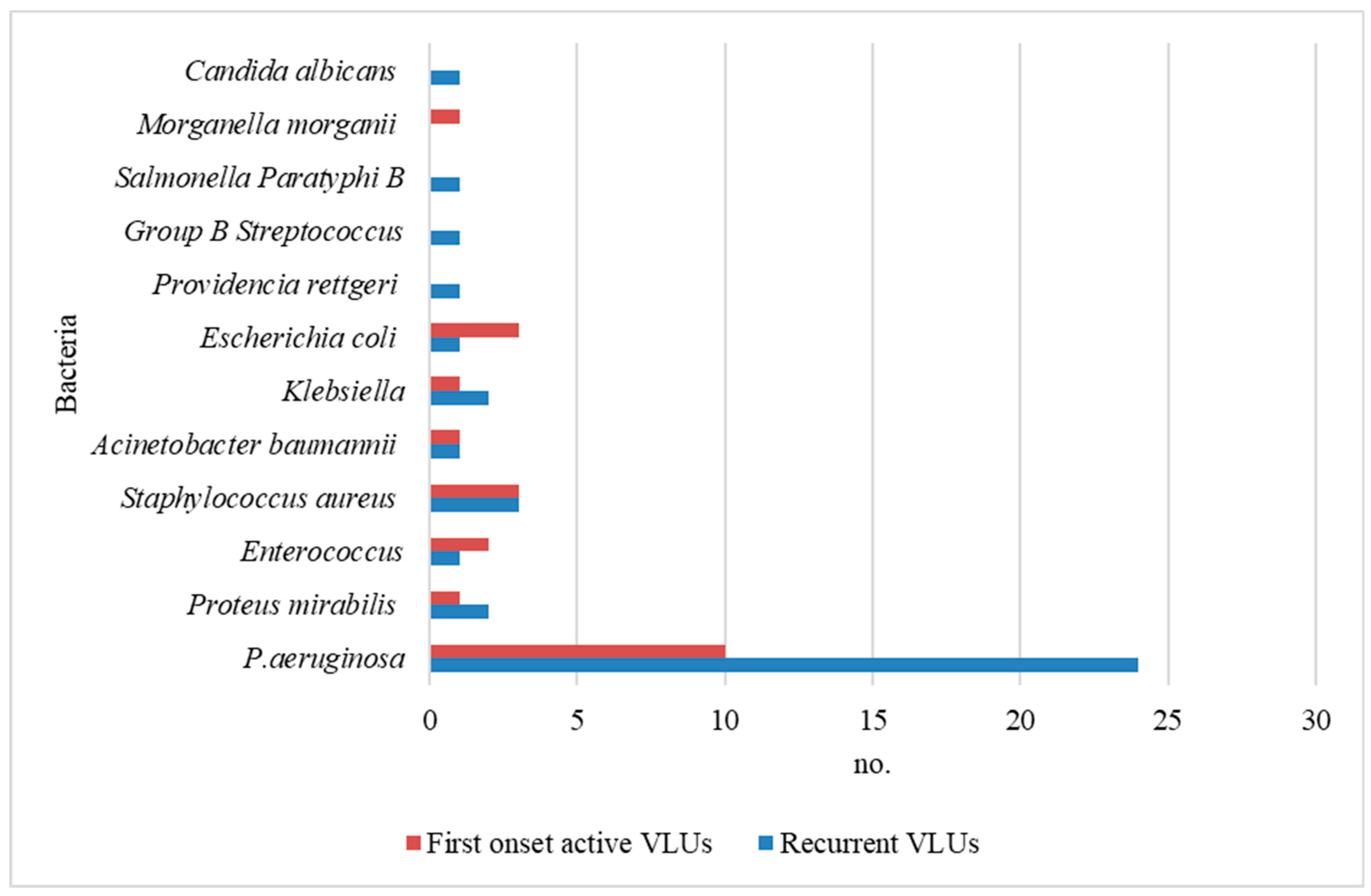
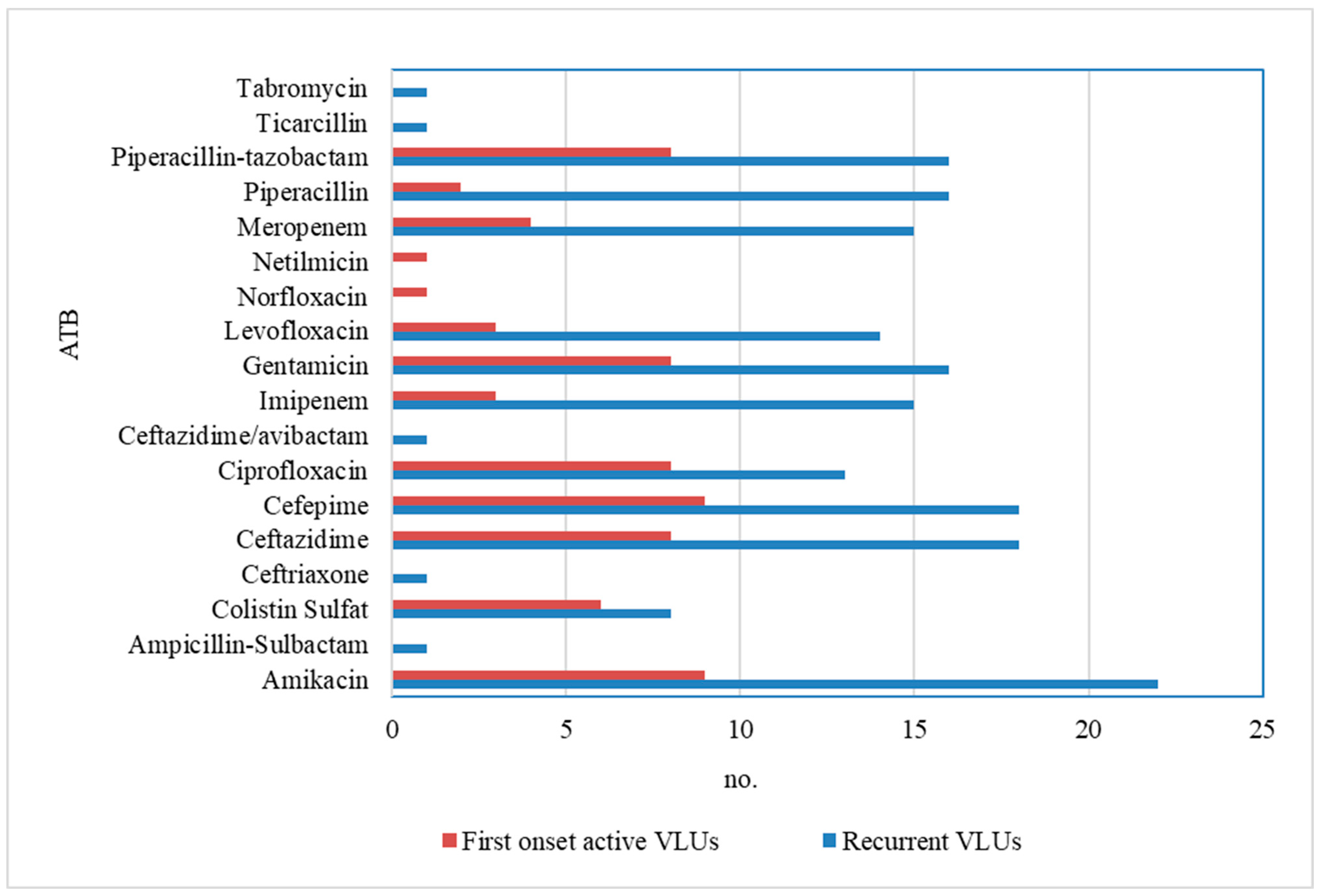
| Infection | No. Study/Control Group | The Antibiotic Sensitivity Profiles | p-Value | ||
|---|---|---|---|---|---|
| Sensitive | Intermediately Sensitive | Resistant | |||
| P. aeruginosa | 24/10 | AK, CAZ, CEF, CIP, IPM, GM, LVX, MERO, PIP, TZP | S, CEF, IPM, LVX | CRO, CZ, CIP, LVX, MERO, PIP, TIC, TM | 0.020 |
| Proteus mirabilis | 2/1 | AK, AM, AMC, CEF, CZ, GM, IPM, SXT | IPM | - | 0.042 |
| Enterococcus sp. | ½ | AM, AMC, CM, E, GM, VA | CIP | - | 0.054 |
| Staphylococcus aureus | 3/3 | GM, CIP, CM, SXT | - | CXM, CM, OX | 0.049 |
| Acinetobacter baumannii | 1/1 | AK, CFP, IPM, MERO | - | CAZ, CEF, CIP | 0.219 |
| Klebsiella spp. | 2/1 | AK, AM, MERO, IPM, GM, PIP, TZP | GM | SXT | 0.015 |
| Escherichia coli | 1/3 | AK, AM, CRO, CEF, CIP, IPM, GM, PIP, TZP | - | LVX, SXT | 0.243 |
| Providencia rettgeri | 1/0 | AK, CAZ, CEF, CIP, IPM, GM | - | - | 1.015 |
| Group B Streptococcus | 1/0 | AM, AMC, CXM, GM, LVX | - | CM, E, SXT | 0.354 |
| Candida albicans | 1/0 | ECN, MIC, AMB, FCZ, NYS; VOR | - | 5-FC | 2.540 |
| Salmonella Paratyphi B | 1/0 | AM, AMC, CRO, GM, LVX | - | CM, E | 1.024 |
| Morganella morganii | 0/1 | AK, CRO, CZ, CEF, CIP, IPM, GM, MERO, LVX, PIP | AMC | SXT | 0.810 |
| Laboratory Tests | Study Group Mean ± SD | Control Group Mean ± SD | p-Value |
|---|---|---|---|
| WBC (n) | 6.4 ± 0.97 | 7.8 ± 2.34 | 0.008 |
| RBC (n) | 4.4 ± 0.50 | 4.4 ± 0.47 | 0.066 |
| Neutrophils (%) | 70.2 ± 8.27 | 64.2 ± 9.09 | 0.011 |
| Lymphocytes (%) | 17.9 ± 6.39 | 23.6 ± 8.02 | 0.003 |
| CK (U/L) | 147.6 ± 79.41 | 101.9 ± 83.02 | 0.035 |
| CK-MB (U/L) | 19.7 ± 4.55 | 22.0 ± 11.05 | 0.307 |
| CRP (mg/L) | 32.8 ± 19.07 | 16.2 ± 10.26 | 0.005 |
| Fibrinogen (mg/L) | 511.4 ± 120.59 | 435.8 ± 112.93 | 0.016 |
| ESR (mm/h) | 48.0 ± 11.34 | 31.0 ± 11.15 | 0.001 |
| Glycemia (mg/dL) | 145 ± 84.94 | 115.8± 35.40 | 0.084 |
| Hba1c (%) | 6.3 ± 1.19 | 6.5 ± 1.17 | 0.433 |
| Creatinine (mg/dL) | 0.9 ± 0.24 | 0.9 ± 0.31 | 0.527 |
| Urea (mg/dL) | 45.4 ± 13.76 | 41.3 ± 13.62 | 0.264 |
Disclaimer/Publisher’s Note: The statements, opinions and data contained in all publications are solely those of the individual author(s) and contributor(s) and not of MDPI and/or the editor(s). MDPI and/or the editor(s) disclaim responsibility for any injury to people or property resulting from any ideas, methods, instructions or products referred to in the content. |
© 2024 by the authors. Licensee MDPI, Basel, Switzerland. This article is an open access article distributed under the terms and conditions of the Creative Commons Attribution (CC BY) license (https://creativecommons.org/licenses/by/4.0/).
Share and Cite
Matei, S.-C.; Dumitru, C.S.; Fakhry, A.M.; Ilijevski, N.; Pešić, S.; Petrović, J.; Crăiniceanu, Z.P.; Murariu, M.-S.; Olariu, S. Bacterial Species Involved in Venous Leg Ulcer Infections and Their Sensitivity to Antibiotherapy—An Alarm Signal Regarding the Seriousness of Chronic Venous Insufficiency C6 Stage and Its Need for Prompt Treatment. Microorganisms 2024, 12, 472. https://doi.org/10.3390/microorganisms12030472
Matei S-C, Dumitru CS, Fakhry AM, Ilijevski N, Pešić S, Petrović J, Crăiniceanu ZP, Murariu M-S, Olariu S. Bacterial Species Involved in Venous Leg Ulcer Infections and Their Sensitivity to Antibiotherapy—An Alarm Signal Regarding the Seriousness of Chronic Venous Insufficiency C6 Stage and Its Need for Prompt Treatment. Microorganisms. 2024; 12(3):472. https://doi.org/10.3390/microorganisms12030472
Chicago/Turabian StyleMatei, Sergiu-Ciprian, Cristina Stefania Dumitru, Ayman Mohamed Fakhry, Nenad Ilijevski, Slobodan Pešić, Jovan Petrović, Zorin Petrişor Crăiniceanu, Marius-Sorin Murariu, and Sorin Olariu. 2024. "Bacterial Species Involved in Venous Leg Ulcer Infections and Their Sensitivity to Antibiotherapy—An Alarm Signal Regarding the Seriousness of Chronic Venous Insufficiency C6 Stage and Its Need for Prompt Treatment" Microorganisms 12, no. 3: 472. https://doi.org/10.3390/microorganisms12030472