Article

Genomic Insights into the Symbiotic and Plant Growth-Promoting Traits of “Candidatus Phyllobacterium onerii” sp. nov. Isolated from Endemic Astragalus flavescens

by
Asiye Esra Eren Eroğlu
1,
Volkan Eroğlu
2 and
İhsan Yaşa
1,*,†
1
Basic and Industrial Microbiology Section, Biology Department, Faculty of Science, Ege University, 35100 Izmir, Türkiye
2
Botany Section, Biology Department, Faculty of Science, Ege University, 35100 Izmir, Türkiye
*
Author to whom correspondence should be addressed.
Current address: Eumicropower Biotechnology Pharmaceutical Chemical Industry Trade Inc., Ege University Campus, EBİLTEM, Incubation Center, 35100 Izmir, Türkiye.
Microorganisms 2024, 12(2), 336; https://doi.org/10.3390/microorganisms12020336
Submission received: 24 December 2023 / Revised: 30 January 2024 / Accepted: 31 January 2024 / Published: 6 February 2024
(This article belongs to the Special Issue Research on Plant—Bacteria Interactions)

Abstract

:
A novel strain of Gram-negative, rod-shaped aerobic bacteria, identified as IY22, was isolated from the root nodules of Astragalus flavescens. The analysis of the 16S rDNA and recA (recombinase A) gene sequences indicated that the strain belongs to the genus Phyllobacterium. During the phylogenetic analysis, it was found that strain IY22 is closely related to P. trifolii strain PETP02T and P. bourgognense strain STM 201T. The genome of IY22 was determined to be 6,010,116 base pairs long with a DNA G+C ratio of 56.37 mol%. The average nucleotide identity (ANI) values showed a range from 91.7% to 93.6% when compared to its close relatives. Moreover, IY22 and related strains had digital DNA-DNA hybridization (dDDH) values ranging from 16.9% to 54.70%. Multiple genes (including nodACDSNZ, nifH/frxC, nifUS, fixABCJ, and sufABCDES) associated with symbiotic nitrogen fixation have been detected in strain IY22. Furthermore, this strain features genes that contribute to improving plant growth in various demanding environments. This study reports the first evidence of an association between A. flavescens and a rhizobial species. Native high-altitude legumes are a potential source of new rhizobia, and we believe that they act as a form of insurance for biodiversity against the threats of desertification and drought.

1. Introduction

The genus Phyllobacterium is a member of the order Rhizobiales and includes Gram-negative, motile, non-spore-forming, aerobic, and chemoorganotrophic species [1]. While Zimmermann (1902) reported the first isolation of strains from the Phyllobacterium genus, it was Knösel (1962) who first used the name Phyllobacterium for bacteria growing in leaf nodules. Many bacteria within this group are associated with plants and can also be found in various habitats such as water, soil, rhizosphere, and root nodules, and in association with single-celled organisms [2,3,4,5,6]. To the present day, sixteen species of Phyllobacterium have been documented in the source provided (https://lpsn.dsmz.de/search?word=Phyllobacterium (accessed on 30 December 2023)) (Table 1).
Rhizobia colonization is a crucial factor that affects the productivity of leguminous plants. These bacteria inhabit symbiotic nodules formed in the roots of leguminous plants [18]. The relationship between the bacteria and the legume in this symbiotic life is highly specific. Specific signal exchanges occur between legumes and the bacteria forming these nodules. Rhizobia synthesize lipochitin oligosaccharides known as Nod factors during these interactions [19]. Rhizobial species trigger the development of root nodules by activating Nod factors. This symbiotic relationship streamlines the fixation of atmospheric dinitrogen within these nodules, thereby aiding rhizobia in generating the necessary nitrogen source for the plant [20].
Legumes and rhizobia have a mutually beneficial relationship that positively impacts the ecosystem in various ways. This includes mechanisms for metal tolerance/resistance, plant growth promotion, nitrogen fixation, phytohormone production, and phosphorus solubility. PGPR (plant growth-promoting rhizobacteria) exert their active principles through enzymes and secondary metabolites [21]. Recently developed genome-based approaches and bioinformatic tools offer the opportunity to examine the entire genome of rhizobial strains. This enables the investigation of genes that encode beneficial enzymes, which facilitate symbiotic interactions and modulate plant hormone levels, as well as biosynthetic gene clusters that encode active compounds such as antibiotics [22,23,24]. Genome-wide analyses enable the unveiling of the entire arsenal of a PGPR strain, thereby revealing the full potential of the strain for future field applications. This allows the characterization of bacterial strains as potential biofertilizer candidates, considering even possible virulence factors for security purposes [25,26].
With the advancement of genomic techniques, the use of bioinformatic approaches based on whole-genome sequence analysis is now recommended as minimal standards for the identification of new prokaryotic taxa, in addition to classical DNA hybridization techniques. Adequate quantitative comparisons should be conducted between the genomes of new species and the type strains of closely related species. Currently, the most common criteria for assessing genomic similarities between strains are obtained through whole-genome average nucleotide identity (ANI) and digital DNA–DNA hybridization (dDDH) techniques [27]. In rhizobia taxonomy, the primary approach also relies on genomic sequences, ANI, and dDDH values among strains [28,29].
In this study, we isolated rhizobial strain IY22 from the root nodules of Astragalus flavescens Boiss., an endemic and rare species found in Bozdağ, Türkiye. The 16S rDNA sequence of strain IY22 exhibited close similarity to P. trifolii strain PETP02T, whereas the recA sequence displayed significant homology with P. bourgognense strain STM 201T. The analysis of complete genome sequences, dDDH, ANI, and biochemical assessments revealed that strain IY22 is a new species within the genus.

2. Materials and Methods

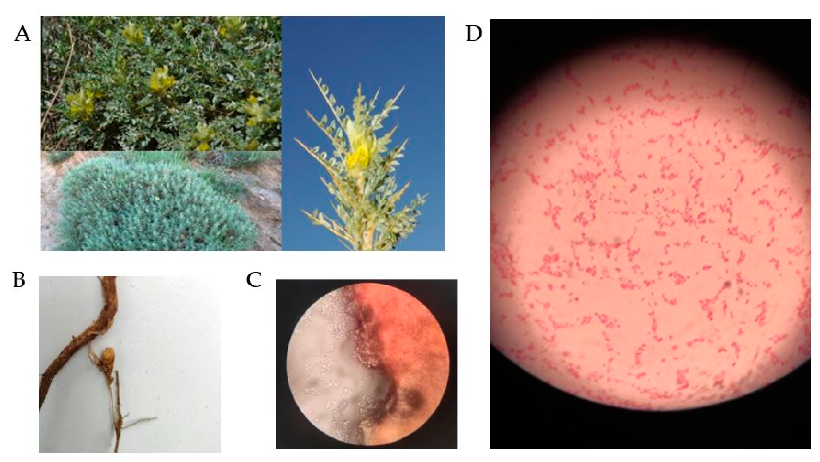
2.1. Sample Collection and Isolation

Astragalus flavescens was collected at an altitude of 1450 m in Ödemiş, Bozdağ (Izmir, Türkiye) at coordinates 38°21′35.94″ N and 28°07′05.90″ E (Figure 1A). A research license to investigate endemic flora was acquired from the Ministry of Agriculture and Forestry’s General Directorate of Agricultural Research and Policies. The study was conducted in compliance with applicable institutional, national, and international protocols and legislation. Plant roots were collected from soil excavated to a depth of approximately 40–50 cm using a hand hoe, and placed in a sample bag containing moistened cotton to prevent the drying of the nodules. Within 2 h, the samples were transported to the Laboratory of Basic and Industrial Microbiology at Ege University. In addition, the plant specimen was identified at the Botanical Garden and Herbarium Research Application Centre of Ege University and registered under the accession number EGE-43727 in the Ege University Herbarium collection.
The nodules were meticulously detached from the plant’s root system and rinsed thoroughly with sterile distilled water to eliminate soil particles (Figure 1B). The nodule surface was sterilized by immersing it in 70% ethanol for 3 min, followed by a 2 min treatment with a mercury (II) chloride solution (HgCl2). Following the procedure, the nodules were washed four times with sterile distilled water. Subsequently, a nodule was sectioned in paraffin and examined under a microscope. At the same time, the nodules were crushed using a sterile glass rod. The homogenized nodule tissue was inoculated onto Yeast Extract Mannitol Agar (YEMA) using the spread plate technique [30]. Petri dishes were incubated at 28 °C for 3–10 days. Bacterial cultures that were to be used for subsequent phenotypic and molecular analysis were purified from a solitary colony after 3–4 days of incubating on YEMA, at a temperature of 28 °C.

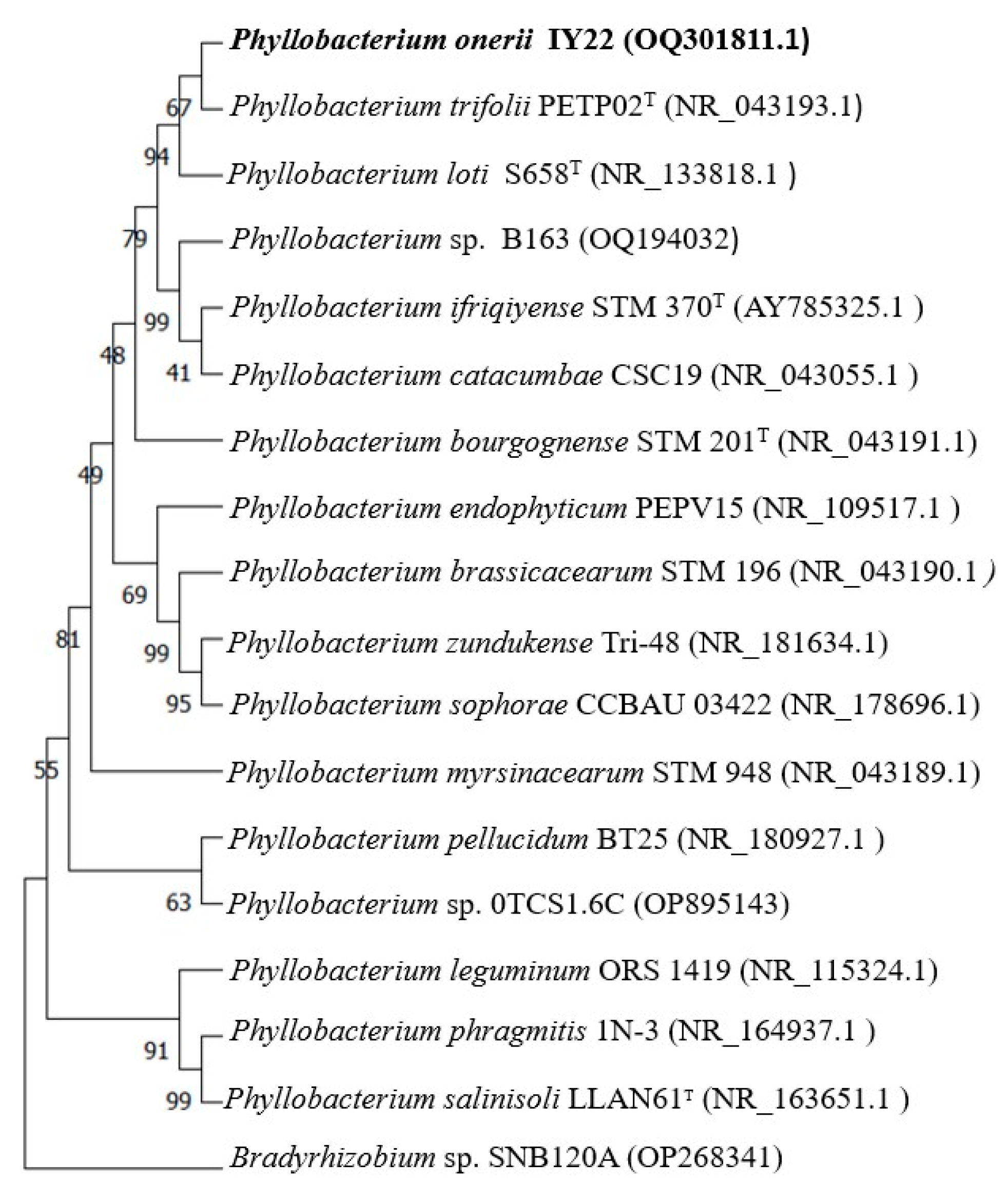
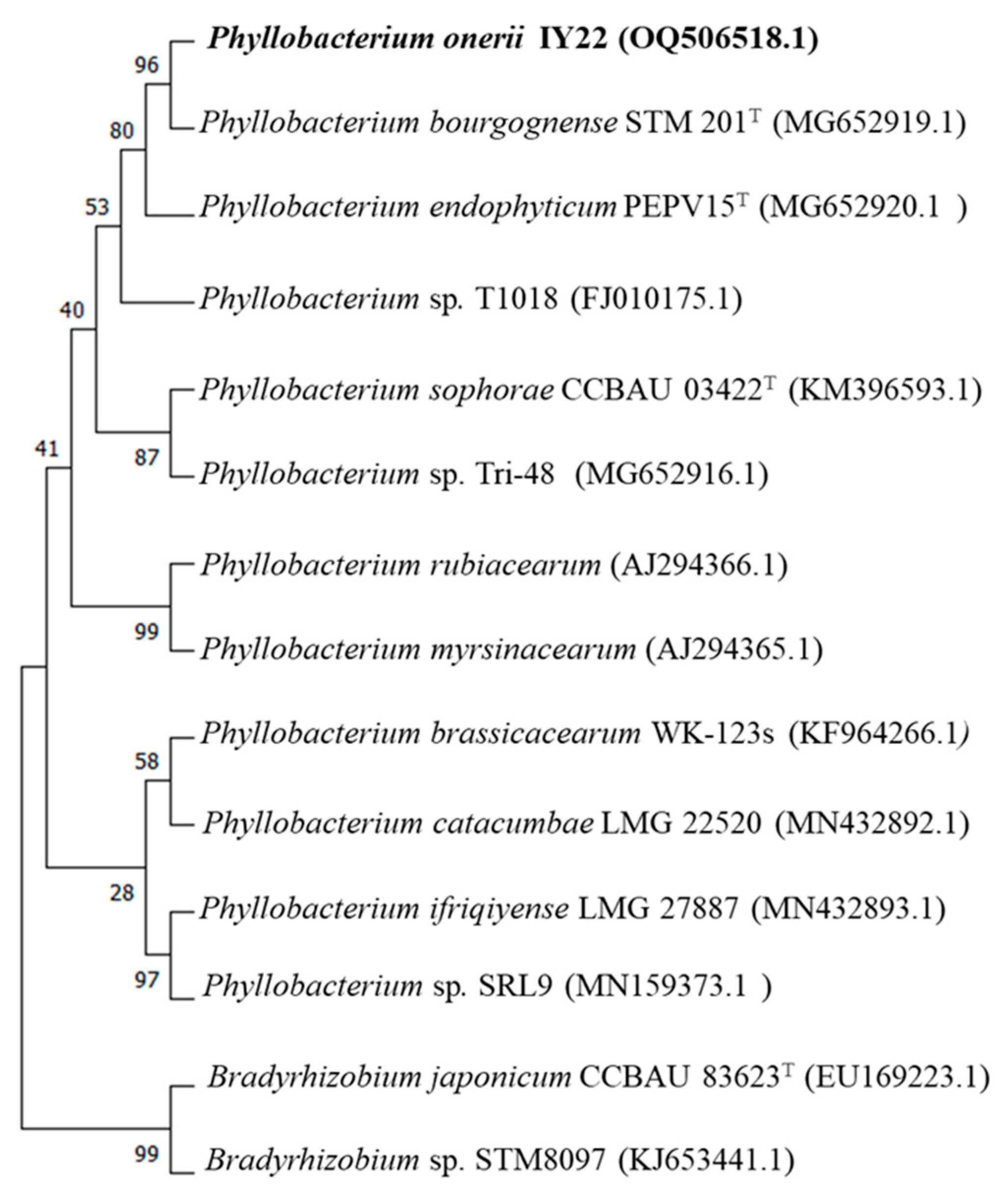
2.2. Phylogeny of 16S rDNA and recA Genes

Genomic DNA was prepared following the High Pure PCR Template Preparation Kit protocol (Roche Applied Science, Mannheim, Germany). The DNA concentration was controlled by the Qubit™ 4 (Invitrogen, New York, NY, USA). The 16S ribosomal DNA (rDNA) and DNA recombinase A (recA) gene sequences of strain IY22 were amplified with PCR using the 27F-1492R and recAF-recAR primer pairs, respectively (Table S1). The FastStartTM Taq DNA Polymerase was used for all PCR amplifications (Roche Applied Science, Mannheim, Germany). The PCR products were sequenced at Letgen Biotechnology Company (Izmir, Türkiye).
Both obtained and related sequences were retrieved from the NCBI (National Center for Biotechnology Information) GenBank databases using the BLASTn search program. Phylogenetic trees were configured separately for the 16S rRNA and recA genes according to the Neighbor joining (NJ) method integrated in Molecular Evolutionary Genetics Analysis (MEGA) software version 11 [31].

2.3. Phenotypic Characterization

Strain IY22 obtained from the root nodule of A. flavescens was characterized. The characterization included Gram test, catalase and oxidase test, and macroscopic and microscopic observations (Olympus CX21,Tokyo, Japan). The growth on YEMA containing Congo red dye (YEMA + CR) was also observed. The ability to grow at different temperatures (22, 28, 37, and 39 °C) in the presence of different NaCl concentrations (1, 2, and 3%) and at different pH (5, 6, 7, and 8) was determined on the YM Broth medium. Other physiological and biochemical tests were also carried out using API (20E, 20NE) test kits (bioMérieux, Marcy l’Etoile, France). API tests were performed according to the manufacturer’s instructions.

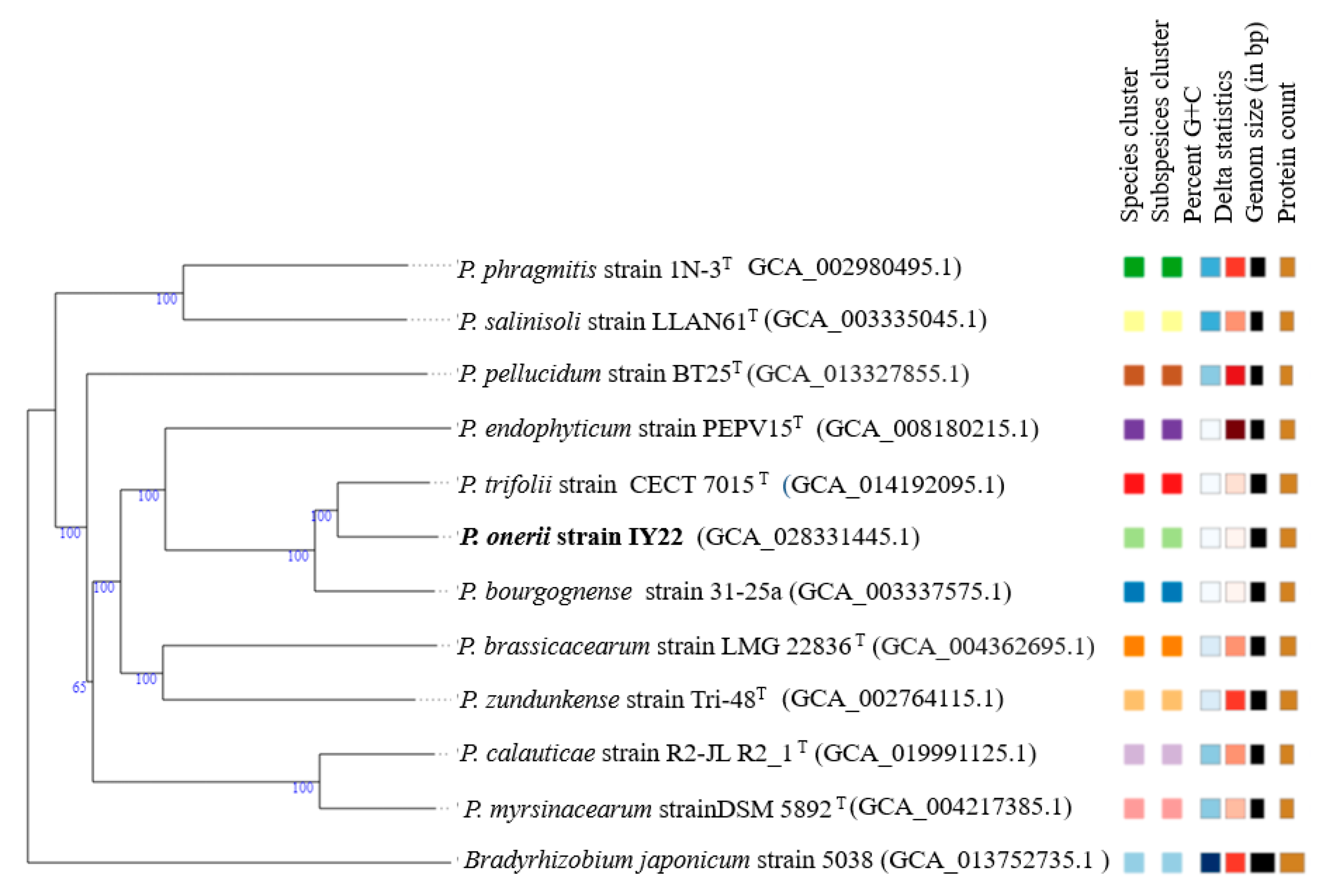
2.4. Genome Analysis

The genomic DNA (50 ng/μL) of strain IY22 was sequenced using the Illumina NovaSeq 6000 sequencing platform with 150 × 2 bp reads with a ~340 bp insert size library by Refgen Biotechnology Inc. (Ankara, Türkiye). Around 2.2 gigabases (Gb) of data were generated, consisting of 7,623,320 reads, using the Illumina NovaSeq 6000 sequencing platform (San Diego, CA, USA). The de novo assembly was carried out using Shovill v1.0.4. with the default settings. Genome annotation was performed using the NCBI Prokaryotic Genome Annotation Pipeline via the NCBI Genome Submission Portal. BioProject ID: PRJNA923719, GenBank Accession number: JAQMHX000000000.1.
The ANI values for strain IY22 and its reference bacteria were computed using the online OrthoANIu algorithm (OrthoANI employing USEARCH) [32]. To calculate the dDDH values, Formula 2 was applied through the server-based Genome-to-Genome Distance Calculator, version 3.0 (http://ggdc.dsmz.de/distcalc2.php (accessed on 20 September 2023)). Taxonomy was determined using the Type (Strain) Genome Server (TGYS) (http://tygs.dsmz.de/ (accessed on 20 September 2023)) with default parameters [33]. Genome sequences of closely related species were acquired from the EzBioCloud and NCBI databases.
Gene functions were determined utilizing the Rapid Annotation Subsystem Technology (RAST) (https://rast.nmpdr.org/ (accessed on 5 February 2023)) and the Pathosystems Resource Integration Center (PATRIC) (https://www.bv-brc.org/ (accessed on 5 February 2023)) web-based pipelines [34,35]. Also on the SEED website, the SEED viewer method was used to perform a functional genomic analysis. Furthermore, the prediction of CRISPR repeats was conducted through the utilization of the CRISPRfinder web server (https://crisprcas.i2bc.paris-saclay.fr (accessed on 10 February 2023)) [36]. Prophages were detected using the Prophage Hunter (https://pro-hunter.genomics.cn (accessed on 10 February 2023)) [37].

3. Results and Discussion

The Sanger sequencing of the 16S rRNA gene sequence of strain IY22 (accession number OQ301811) showed 98.8% similarity to P. trifolii strain PETP02T after BLASTn search of the NCBI nucleotide database. The recA sequence analysis resulted in a close relationship with P. bourgognense strain STM 201T with a similarity of 96.02%. Our findings strongly support the idea that strain IY22, which we investigated in this study, forms a monophyletic group with previously known Phyllobacterium species, indicating its membership of the Phyllobacterium genus (Figure 2 and Figure 3). However, differences in clusters and topology suggest that a phylogenetic analysis based on individual genes may be inconsistent due to their unique evolutionary histories. These paradoxes highlight that constructing an accurate phylogenetic relationship among rhizobial species solely based on individual genes is unlikely [38,39].
Strain IY22 formed a faint pink colony on YEMA medium containing Congo red and formed small colonies (<2 mm in diameter). The cells are Gram-negative, catalase-, and oxidase-positive. Microscopic observations showed that the strain was rod-shaped bacteria (Figure 1C,D). The pH range for growth is pH 5–8 (optimal growth occurs at pH 7). Growth occurs in the presence of 0–2% (w/v) NaCl. The optimal growth temperature is 28 °C. Other physiological and biochemical tests were carried out using API systems following the manufacturer’s instructions, and the results were read after 3 days of incubation at 28 °C. Based on the findings, strain IY22 exhibited differences from the P. trifolii PETP02T and P. bourgognense STM 201T strains in terms of its ability to produce acids from sucrose, trehalose, rhamnose, and raffinose, as well as its indole production and citrate assimilation (Table 2). Specifically, IY22 displayed positive acid production from sucrose, trehalose, and raffinose, while PETP02T exhibited negative results. Furthermore, IY22 showed positive indole production, in contrast to STM 201T which showed a negative result. Additional phenotypic traits of strain IY22 are elaborated upon in the subsequent species description.
The complete genome size of strain IY22 was 6,010,116 bp. The DNA G+C content of strain IY22 was 56.37 mol% (Table 3). In the close relatives of strain IY22, P. trifolii PETP02T and P. bourgognense STM 201T, the G+C contents are 56.4 mol% and 54 mol% (Tm), respectively [9,10]. The ANI values for strain IY22 and its closely related Phyllobacterium species range from 91.7 to 93.6. The dDDH values, calculated using the suggested Formula 2, exhibit a range of 16.9% to 54.70% for the IY22 strain and the closely related Phyllobacterium strains. In terms of the dDDH value, strain IY22’s highest relationship was observed with P. trifolii PETP02T (54.70%). Nevertheless, these values, which are typically utilized as thresholds for bacterial species identification, notably remain below the commonly accepted thresholds of 95–96% (ANI) and 70% (DDH) [40,41]. A phylogenetic tree was generated by analyzing genomes from the nearest relatives of strain IY22 using the TGYS platform (Figure 4).
The genome of strain IY22 consists of 65 contigs, comprising one chromosome and no plasmids. The chromosome contains 3 identical rRNA operons and 46 tRNA genes. Of the 6470 predicted CDS, 2534 were coding hypothetical proteins, 3936 were proteins with functional assignments, 1232 were proteins with EC number assignments, 4252 were proteins with PATRIC genus-specific family (PLfam) assignments, and 4701 were proteins with PATRIC cross-genus family (PGfam) assignments. One CRISPR locus was found in the genome via the CRISPRCasFinder (https://crisprcas.i2bc.paris-saclay.fr (accessed on 10 February 2023)). Six prophages were detected using the Prophage Hunter (https://pro-hunter.genomics.cn (accessed on 10 February 2023)). Genomic and protein characteristics are condensed in Table 3 and Table 4. The genome of strain IY22 was additionally mapped to the seed subsystem to attain high-quality genome annotation via RAST. The distributions of genes linked to subsystems in 27 different categories are shown in Figure 5.
The analysis of carbohydrate metabolism in Phyllobacterium genus species was compared to GenBank reference genomes, revealing similar profiles. In the IY22 genome, 215 genes associated with pathways such as xylose utilization, chitin and N-acetylglucosamine metabolism, maltose and maltodextrin utilization, trehalose biosynthesis, lactate utilization, one-carbon metabolism by tetrahydropterines, and various others were identified (Table S2). The genomic analysis of strain IY22 identified fdoGH, which encodes formate dehydrogenase, a key player in C1 metabolism. Formate metabolism is crucial in Rhizobiales for nitrogen fixation and formate-dependent respiration [42]. Additionally, IY22 also possesses genes for selenocysteine biosynthesis, which were first identified in S. meliloti [43].
In recent times, genomic approaches have been suggested as an alternative to traditional chemotaxonomy for identifying bacterial species [44]. An in silico chemotaxonomy methodology was initially applied to Corynebacterium and Turicella, and subsequently to Aliamphritea species [45,46]. In this study, we used genome sequences to predict chemotaxonomic characteristics of strain IY22. A comparative genomic analysis of closely related Phyllobacterium species revealed that strain IY22 possesses the essential type II fatty acid biosynthesis (FASII) pathway. The fatty acid metabolism pathway based on KEGG is illustrated in Figure 6. This pathway is responsible for synthesizing three major fatty acids: C16:0, C16:1, and C18:1 [47]. The genome of strain IY22 encodes fabA and fabB, indicating the potential to produce C16:1ω7c and C18:1ω7c. The enzymes responsible for the FASII pathway in the IY22 genome were identified and are listed in Table S3. All genes encoding these enzymes are present in P. trifolii PETP02T. The predominant cellular fatty acids produced through these enzymes, crucial to the FASII pathway determinants, including myristic acid (14:0), palmitic acid (16:0), 3-Hydroxypentadecanoic acid (15:0 3-OH), margaric acid (17:0), 3-Hydroxyhexadecanoic acid (16:0 3-OH), stearic acid (18:0), cis-vaccenic acid (C18:1ω7c), cis-9,10-Methyleneoctadecanoic acid (19:0 cyclo ω8c), oleic acid (18:1), eicosadienoic acid (C20:2ω6,9c), and arachidic acid (20:0) [9].
Certain fatty acids, especially polyunsaturated ones, can be synthesized through various enzymatic pathways. For example, stearic acid (18:0) is typically desaturated to produce C18:1ω7c. This desaturation process is catalyzed by an enzyme called stearoyl-ACP desaturase, which converts the methylene group of stearic acid into a double bond, resulting in the production of oleic acid (18:1) and subsequently cis-vaccenic acid (C18:1ω7c) [48]. C20:2ω6,9c is a polyunsaturated fatty acid (PUFA) typically synthesized through the desaturation of linoleic acid (C18:2ω6c). The desaturation of linoleic acid is catalyzed by one or more desaturase enzymes, resulting in the production of oleic acid (C18:1) and subsequently eicosadienoic acid (C20:2ω6,9c) [49]. These metabolic pathways, particularly those involving specific enzymatic steps such as fatty acid desaturation, guide the synthesis of different types of fatty acids. The amino acid sequences of fatty acid desaturases encoded in the genomes of strain IY22 and closely related species were aligned, revealing diversity in conserved histidine clusters (Figure 7). Changes in the active regions of such enzymes can lead to differences in reaction steps, contributing to the uniqueness and diversity of organisms’ lipid metabolism.
The genome of strain IY22 contains all the necessary genes for producing phosphatidylglycerol (PG) and phosphatidylethanolamine (PE), including plsX, plsY, plsC, cdsA, pssA, psd, pgsA, and pgpA. It also has clsA, which is responsible for diphosphatidylglycerol (DPG) production. Additionally, the clsII gene with eukaryotic-like characteristics was identified in the genome (Table S4). Strain IY22 is capable of utilizing two different pathways for cardiolipin synthesis, similar to the mechanisms observed in Streptomyces coelicolor or Agrobacterium tumefaciens [50].
The predominant respiratory quinone in previously described Phyllobacterium species is ubiquinone-10 [9,10,11,12,16]. The comparative analysis of the genome of strain IY22 revealed the presence of the ubiABEGX and pqqC genes. The ubiE gene catalyzes the conversion of demethylmenaquinone to menaquinone (Table S4). These results indicate that, similar to other members of the genus, menaquinone-10 is the predominant quinone in strain IY22.
The genome of strain IY22 contains the crucial LpxABCDH genes necessary for lipid A biosynthesis (Table S4). Notably, these genes exhibit a remarkable level of conservation among rhizobial species, encompassing Mesorhizobium, Bradyrhizobium, and Phyllobacterium [51].
This study presents the initial comparison of in silico chemotaxonomic data for a rhizobial species. As the number of genome sequences in databases increases, in silico chemotaxonomy is expected to become applicable to numerous bacterial species in the future. Additionally, this approach may be effective in predicting chemotaxonomic characteristics of unculturable species using metagenomic data. However, there is a need for further advancements in predicting the structure of enzymes responsible for fatty acid metabolism and their biochemical kinetics. This is crucial because current methods are not well suited to accurately determine the quantitative amounts of predicted fatty acids [46].
A general screening was carried out for genes essential for promoting plant growth under different stresses. Several genes related to iron uptake (17), nitrogen metabolism (8), virulence, disease and defence (32), membrane transport (54), stress response (50), and sulfur metabolism (18) were found in strain IY22 (Table S5). The variety of habitats indicates that Phyllobacterium has acquired an impressive aptitude for adapting to its surroundings. Moreover, their non-pathogenic attributes and capacity to ‘interact’ with plant tissues render them intriguing subjects for research aimed at exploring their potential to promote plant growth [52,53].
In accordance with their previously proposed functions, several genes associated with nodule development (nodACDSNZ) in strain IY22 were identified (Table 5). The β-1,4-linked D-glucosamine residues form the Nod factor backbone, which is N-acylated on the non-reducing end and O-acetylated on the other residues. nodC encodes N-acetylglucosaminyl transferase [54]. The expression of nod genes is controlled by flavonoid-activated NodD proteins, which act as LysR-type transcriptional regulators [55]. NodS is a S-adenosylmethionine-dependent methyltransferase that methylates chitooligosaccharides that are deacetylated at the non-reducing end. NodZ is responsible for the fucosylation of nodulation signals [56,57]. Functionally conserved nod genes are commonly used as target genes in studies of rhizobial diversity [58]. In this study, genes encoding nod factors were identified in the genome of IY22 and a phylogenetic tree was constructed by comparing nodC amino acid sequences, as shown in Figure 8. However, the nodC phylogeny of IY22 displays incongruity with the 16S rRNA taxonomy. Classifying rhizobia according to their symbiotic properties is a major challenge due to the complexity of the plant molecular mechanisms that determine host specificity [59]. This classification is thus largely reliant on the growth conditions of the leguminous plant.
In conjunction with the nod genes, the IY22 genome also harbors key genes responsible for regulating nitrogen fixation, including nifH/frxC, nifUS, sufABCDES, NtrBCYX, fixABCJ, GlnK, glnD, and cysE (Table 5). Biological nitrogen fixation is driven by molybdenum-dependent nitrogenase, a sophisticated enzyme consisting of a two-component protein (MoFe protein and Fe protein). The nifHDK genes encode structural subunits, while the nifBENQVYX genes encode the synthesis and attachment of FeMo-co to nitrogenase [60]. The assembly of the Fe-S cluster is managed by NifU and NifS [61]. The formation and integration of nitrogenase metalloclusters necessitate the production of initial Fe-S units, which can be sourced from either general or specialized biosynthetic systems [62]. Iron–sulfur clusters serve as essential metal cofactors vital for critical biological processes, including DNA repair, respiration, nitrogen fixation, and photosynthesis [63]. The mechanisms underlying Fe-S cluster biosynthesis in various bacterial species involved in nitrogenase are still not well understood. In IY22, a fully functional nif operon was not observed; thus, it is likely that the NifS and SufCDB proteins play the role of a specific sulfur donor and molecular scaffold, sequentially, in the assembly of nitrogenase Fe-S clusters, as observed in P. polymyxa WLY78 [64]. In contrast, the fixABCX operon in IY22 may be associated with nitrogen fixation, as in Azospirillum [65]. Moreover, the genetic constituents and configurations of genes responsible for nitrogen fixation can exhibit significant diversity across various diazotrophic organisms [66].

4. Conclusions

The exploration of legume–plant interactions can provide invaluable insights into the realms of evolutionary biology and fundamental ecological processes. The studies conducted in this regard can act as a form of insurance for biodiversity against the threats of desertification and drought. Moreover, in the context of livestock farming, it is well established that plants cultivated at high altitudes exhibit elevated nutritional values. Additionally, it can be hypothesized that unexplored attributes might exist within plants and microorganisms that have adapted to climatic conditions prevalent at high altitudes, such as low temperatures, altered atmospheric pressure, and heightened UV radiation. Conducting a more comprehensive investigation into the microbial diversity intertwined with high-altitude flora holds paramount importance. Such an endeavor will facilitate the identification of novel bacterial taxa and bacterial–plant symbiotic relationships with the potential to offer valuable contributions to agricultural domains.
Chemical fertilizers have detrimental effects on agricultural land ecosystems. To mitigate these negative impacts, alternative methods such as biological nitrogen fixation are increasingly important. Research on prokaryotic taxa associated with legumes has been intense. Nevertheless, a rhizobial species has not yet been identified in A. flavescens. This study presents the initial findings of an association between A. flavescens and a rhizobial species. This belated discovery may be attributed to the fact that past research has primarily examined diversity at lower altitudes. Our investigation indicates that the indigenous vegetation at high altitudes provides a probable source for the identification of novel rhizobial species.

Description of “Candidatus Phyllobacterium onerii” sp. nov.

Phyllobacterium onerii: o-ne-ri-i N.L. gen. n. onerii named after Mehmet Öner, an important scientist in the field of microbiology, founder of the Basic and Industrial Microbiology Department of Ege University.
The colonies that develop aerobically in YEMA at 28 °C are small, scaly, and white. The cultures grown appear to have a faint pink color when grown in YEMA containing Congo Red. The cells are rod-shaped, Gram-negative, catalase-, and oxidase-positive. The cells can grow in the presence of NaCl concentrations up to 2% (w/v). The temperature range for growth is 4–37 °C; however, the optimal growth temperature is 28 °C. The pH range for growth is 6–8 (optimal growth occurs at pH 7). Arginine dehydrolase, β-galactosidase, and gelatinase were negative. The hydrolysis of aesculin was weak. Lysine decarboxylase, ornithine decarboxylase, tryptophan deaminase, and urease were positive. Indole production and citrate assimilation were positive. Melibiose and amygdalin fermentation were negative. Glucose, L-arabinose, mannose, inositol, sorbitol, rhamnose, sucrose, mannitol, N-acetylglucosamine, maltose, and malate are carbon sources. Gentiobiose is weakly used. It does not grow on caproate, adipate, or phenylacetate. The in silico genome analysis indicates that strain IY22 possesses the FASII biosynthesis pathway, enabling it to produce myristic acid (14:0), palmitic acid (16:0), 3-hydroxypentadecanoic acid (15:0 3-OH), margaric acid (17:0), 3-Hydroxyhexadecanoic acid (16:0 3-OH), stearic acid (18:0), cis-vaccenic acid (C18:1ω7c), cis-9,10-methyleneoctadecanoic acid (19:0 cyclo ω8c), oleic acid (18:1), eicosadienoic acid (C20:2ω6,9c), and arachidic acid (20:0). Strain IY22 encodes a comprehensive set of genes essential for the synthesis of phosphatidylglycerol (PG) and phosphatidylethanolamine (PE), including plsX, plsY, plsC, cdsA, pssA, psd, pgsA, and pgpA. Additionally, it includes clsA, which is responsible for the production of diphosphatidylglycerol (DPG).
Strain IY22 was isolated from the root nodule of the İzmir/Bozdağ endemic legume plant Astragalus flavescens. The DNA G+C content is 56.37 mol%. BioProject ID: PRJNA923719, GenBank Accession number: JAQMHX000000000.1. Genome assembly: GCA_028331445.1.

Supplementary Materials

The following supporting information can be downloaded at: https://www.mdpi.com/article/10.3390/microorganisms12020336/s1, Table S1: Primers and PCR conditions; Table S2: Several genes involved in carbohydrate metabolism; Table S3: Several genes involved in fatty acid metabolism.; Table S4: The genes involved in the biosynthesis of PG, PE, DPG, lipid A, and respiratory quinones.; Table S5: Several genes in strain IY22 that encode PGP traits.

Author Contributions

Conceptualization and methodology; İ.Y., A.E.E.E. and V.E.; writing—original draft preparation, İ.Y. and A.E.E.E. All authors have read and agreed to the published version of the manuscript.

Funding

The funding for this study was provided by Eumicropower Biotechnology Inc. (Izmir, Türkiye).

Data Availability Statement

All sequence data support the findings of this study have been deposited in GenBank (https://www.ncbi.nlm.nih.gov/genbank/ (accessed on 2 February 2023)) with accession numbers OQ301811-OQ506518 and JAQMHX000000000.1. The complete genome data has been assigned BioProject ID PRJNA923719 and is now accessible to the public.

Acknowledgments

This study was conducted using the Industrial and Molecular Microbiology Research Laboratory of Ege University Faculty of Science.

Conflicts of Interest

Author İhsan Yaşa was a co-founder by the company Eumicropower Biotechnology Inc. The remaining authors declare that the research was conducted in the absence of any commercial or financial relationships that could be construed as a potential conflict of interest.

References

  1. Mantelin, S.; Fischer-Le Saux, M.; Zakhia, F.; Béna, G.; Bonneau, S.; Jeder, H.; de Lajudie, P.; Cleyet-Marel, J.C. Emended description of the genus Phyllobacterium and description of four novel species associated with plant roots: Phyllobacterium bourgognense sp. nov., Phyllobacterium ifriqiyense sp. nov., Phyllobacterium leguminum sp. nov. and Phyllobacterium brassicacearum sp. nov. Int. J. Syst. Evol. Microbiol. 2006, 56, 827–839. [Google Scholar] [CrossRef] [PubMed]
  2. Alavi, M.; Miller, T.; Erlandson, K.; Schneider, R.; Belas, R. Bacterial community associated with Pfiesteria-like dinoflagellate cultures. Environ. Microbiol. 2001, 3, 380–396. [Google Scholar] [CrossRef]
  3. Gonzalez-Bashan, L.E.; Lebsky, V.K.; Hernandez, J.P.; Bustillos, J.J.; Bashan, Y. Changes in the metabolism of the microalga Chlorella vulgaris when coimmobilized in alginate with the nitrogen-fixing Phyllobacterium myrsinacearum. Can. J. Microbiol. 2000, 46, 653–659. [Google Scholar] [CrossRef] [PubMed]
  4. Jurado, V.; Laiz, L.; Gonzalez, J.M.; Hernandez-Marine, M.; Valens, M.; Sáiz-Jiménez, C. Phyllobacterium catacumbae sp. nov., a member of the order ‘Rhizobiales’ isolated from Roman catacombs. Int. J. Syst. Evol. Microbiol. 2005, 55, 1487–1490. [Google Scholar] [CrossRef]
  5. Lambert, B.; Joos, H.; Dierickx, S.; Vantomme, R.; Swings, J.; Kersters, K.; Van Montagu, M. Identification and plant interaction of a Phyllobacterium sp., a predominant rhizobacterium of young sugar beet plants. Appl. Environ. Microbiol. 1990, 56, 1093–1102. [Google Scholar] [CrossRef]
  6. Mergaert, J.; Cnockaert, M.C.; Swings, J. Phyllobacterium myrsinacearum (subjective synonym Phyllobacterium rubiacearum) emend. Int. J. Syst. Evol. Microbiol. 2002, 52, 1821–1823. [Google Scholar] [CrossRef]
  7. Knösel, D. Prüfung von bakterien auf Fähigkeit zur Sternbildung. Zentralbl Bakteriol. Parasitenkd. Infekt. Hyg. 1962, 116, 79–100. [Google Scholar]
  8. Knosel, D.H. Genus IV. Phylloacterium (ex Knosel 1962) nom. rev. (Phyllobacterium Knosel 1962, 96). Bergey’s Man. Syst. Bacteriol. 1984, 1, 254–256. [Google Scholar]
  9. Valverde, A.; Velazquez, E.; Fernandez-Santos, F.; Vizcaíno, N.; Rivas, R.; Mateos, P.F.; Martínez-Molina, E.; Igual, J.M.; Willems, A. Phyllobacterium trifolii sp. nov., nodulating Trifolium and Lupinus in Spanish soils. Int. J. Syst. Evol. Microbiol. 2005, 55, 1985–1989. [Google Scholar] [CrossRef]
  10. Flores-Felix, J.D.; Carro, L.; Velazquez, E.; Valverde, A.; Cerda-Castillo, E.; García-Fraile, P.; Rivas, R. Phyllobacterium endophyticum sp. nov., isolated from nodules of Phaseolus vulgaris. Int. J. Syst. Evol. Microbiol. 2013, 63 Pt 3, 821–826. [Google Scholar] [CrossRef]
  11. Sanchez, M.; Ramirez-Bahena, M.H.; Peix, A.; Lorite, M.J.; Sanjuan, J.; Velazquez, E.; Monza, J. Phyllobacterium loti sp. nov. isolated from nodules of Lotus corniculatus. Int. J. Syst. Evol. Microbiol. 2014, 64 Pt 3, 781. [Google Scholar] [CrossRef] [PubMed]
  12. Jiao, Y.S.; Yan, H.; Ji, Z.J.; Liu, Y.H.; Sui, X.H.; Zhang, X.X.; Wang, E.T.; Chen, W.X. Phyllobacterium sophorae sp. nov., a symbiotic bacterium isolated from root nodules of Sophora flavescens. Int. J. Syst. Evol. Microbiol. 2015, 65 Pt 2, 399–406. [Google Scholar] [CrossRef] [PubMed]
  13. León-Barrios, M.; Ramírez-Bahena, M.H.; Igual, J.M.; Peix, Á.; Velázquez, E. Phyllobacterium salinisoli sp. nov., isolated from a Lotus lancerottensis root nodule in saline soil from Lanzarote. Int. J. Syst. Evol. Microbiol. 2018, 68, 1085–1089. [Google Scholar] [CrossRef] [PubMed]
  14. Safronova, V.I.; Sazanova, A.L.; Kuznetsova, I.G.; Belimov, A.A.; Andronov, E.E.; Chirak, E.R.; Popova, J.P.; Verkhozina, A.V.; Willems, A.; Tikhonovich, I.A. Phyllobacterium zundukense sp. nov., a novel species of rhizobia isolated from root nodules of the legume species Oxytropis triphylla (Pall.) Pers. Int. J. Syst. Evol. Microbiol. 2018, 68, 1644–1651. [Google Scholar] [CrossRef]
  15. Liang, L.X.; Sun, Q.W.; Hui, N.; Zhang, X.X.; Li, L.B.; Liu, L. Phyllobacterium phragmitis sp. nov., an endophytic bacterium isolated from Phragmites australis rhizome in Kumtag Desert. Antonie Leeuwenhoek 2019, 112, 661–668. [Google Scholar] [CrossRef]
  16. Park, Y.; Ten, L.N.; Maeng, S.; Chang, Y.; Jung, H.Y.; Kim, M.K. Phyllobacterium pellucidum sp. nov., isolated from soil. Arch. Microbiol. 2021, 203, 2647–2652. [Google Scholar] [CrossRef]
  17. Lustermans, J.J.; Bjerg, J.J.; Schramm, A.; Marshall, I.P. Phyllobacterium calauticae sp. nov. isolated from a microaerophilic veil transversed by cable bacteria in freshwater sediment. Antonie Leeuwenhoek 2021, 114, 1877–1887. [Google Scholar] [CrossRef]
  18. Shumilina, J.; Soboleva, A.; Abakumov, E.; Shtark, O.Y.; Zhukov, V.A.; Frolov, A. Signaling in Legume-Rhizobia Symbiosis. Int. J. Mol. Sci. 2023, 24, 17397. [Google Scholar] [CrossRef]
  19. Singh, J.; Verma, P.K. Role of Nod factor receptors and its allies involved in nitrogen fixation. Planta 2023, 257, 54. [Google Scholar] [CrossRef]
  20. Mbengue, M.D.; Hervé, C.; Debellé, F. Nod factor signaling in symbiotic nodulation. In Advances in Botanical Research; Academic Press: Cambridge, MA, USA, 2020; Volume 94, pp. 1–39. [Google Scholar] [CrossRef]
  21. Bouremani, N.; Cherif-Silini, H.; Silini, A.; Bouket, A.C.; Luptakova, L.; Alenezi, F.N.; Baranov, O.; Belbahri, L. Plant growth-promoting rhizobacteria (PGPR): A rampart against the adverse effects of drought stress. Water 2023, 15, 418. [Google Scholar] [CrossRef]
  22. Wang, D.; Poinsot, V.; Li, W.; Lu, Y.; Liu, C.; Li, Y.; Xie, K.; Sun, L.; Shi, C.; Peng, H.; et al. Genomic insights and functional analysis reveal plant growth promotion traits of Paenibacillus mucilaginosus G78. Genes 2023, 14, 392. [Google Scholar] [CrossRef]
  23. Świątczak, J.; Kalwasińska, A.; Wojciechowska, A.; Brzezinska, M.S. Physiological properties and genomic insights into the plant growth-promoting rhizobacterium Brevibacillus laterosporus K75 isolated from maize rhizosphere. J. Sci. Food Agric. 2023, 103, 1432–1441. [Google Scholar] [CrossRef]
  24. Leontidou, K.; Genitsaris, S.; Papadopoulou, A.; Kamou, N.; Bosmali, I.; Matsi, T.; Madesis, P.; Vokou, D.; Karamanoli, K.; Mellidou, I. Plant growth promoting rhizobacteria isolated from halophytes and drought-tolerant plants: Genomic characterisation and exploration of phyto-beneficial traits. Sci. Rep. 2020, 10, 14857. [Google Scholar] [CrossRef]
  25. Medina-Cordoba, L.K.; Chande, A.T.; Rishishwar, L.; Mayer, L.W.; Valderrama-Aguirre, L.C.; Valderrama-Aguirre, A.; Gaby, J.C.; Kostka, J.E.; Jordan, I.K. Genomic characterization and computational phenotyping of nitrogen-fixing bacteria isolated from Colombian sugarcane fields. Sci. Rep. 2021, 11, 9187. [Google Scholar] [CrossRef]
  26. Zhang, L.; Chen, W.; Jiang, Q.; Fei, Z.; Xiao, M. Genome analysis of plant growth-promoting rhizobacterium Pseudomonas chlororaphis subsp. aurantiaca JD37 and insights from comparasion of genomics with three Pseudomonas strains. Microbiol. Res. 2020, 237, 126483. [Google Scholar] [CrossRef] [PubMed]
  27. Christensen, H. (Ed.) Introduction to Bioinformatics in Microbiology; Springer International Publishing: Cham, Switzerland, 2018; p. 213. [Google Scholar] [CrossRef]
  28. De Lajudie, P.M.; Andrews, M.; Ardley, J.; Eardly, B.; Jumas-Bilak, E.; Kuzmanović, N.; Lassalle, F.; Lindström, K.; Mhamdi, R.; Martínez-Romero, E.; et al. Minimal standards for the description of new genera and species of rhizobia and agrobacteria. Int. J. Syst. Evol. Microbiol. 2019, 69, 1852–1863. [Google Scholar] [CrossRef] [PubMed]
  29. Ormeno-Orrillo, E.; Servín-Garcidueñas, L.E.; Rogel, M.A.; González, V.; Peralta, H.; Mora, J.; Martínez-Romero, J.; Martínez Romero, E. Taxonomy of rhizobia and agrobacteria from the Rhizobiaceae family in light of genomics. Syst. Appl. Microbiol. 2015, 38, 287–291. [Google Scholar] [CrossRef] [PubMed]
  30. Vincent, J.M. A Manual for the Practical Study of the Root-Nodule Bacteria; Blackwell Scientific: Oxford, UK, 1970. [Google Scholar]
  31. Kumar, S.; Stecher, G.; Tamura, K. MEGA7: Molecular evolutionary genetics analysis version 7.0 for bigger datasets. Mol. Biol. Evol. 2016, 33, 1870–1874. [Google Scholar] [CrossRef] [PubMed]
  32. Lee, I.; Ouk Kim, Y.; Park, S.C.; Chun, J. OrthoANI: An improved algorithm and software for calculating average nucleotide identity. Int. J. Syst. Evol. Microbiol. 2016, 66, 1100–1103. [Google Scholar] [CrossRef] [PubMed]
  33. Meier-Kolthoff, J.P.; Göker, M. TYGS is an automated high-throughput platform for state-of-the-art genome-based taxonomy. Nat. Commun. 2019, 10, 2182. [Google Scholar] [CrossRef] [PubMed]
  34. Aziz, R.K.; Bartels, D.; Best, A.A.; DeJongh, M.; Disz, T.; Edwards, R.A.; Formsma, K.; Gerdes, S.; Glass, E.M.; Kubal, M.; et al. The RAST Server: Rapid annotations using subsystems technology. BMC Genom. 2008, 9, 75. [Google Scholar] [CrossRef]
  35. Wattam, A.R.; Davis, J.J.; Assaf, R.; Boisvert, S.; Brettin, T.; Bun, C.; Conrad, N.; Dietrich, E.M.; Disz, T.; Gabbard, J.L.; et al. Improvements to PATRIC, the all-bacterial bioinformatics database and analysis resource center. Nucleic Acids Res. 2017, 45, D535–D542. [Google Scholar] [CrossRef] [PubMed]
  36. Couvin, D.; Bernheim, A.; Toffano-Nioche, C.; Touchon, M.; Michalik, J.; Néron, B.; Rocha, E.P.C.; Vergnaud, G.; Gautheret, D.; Pourcel, C. CRISPRCasFinder, an update of CRISRFinder, includes a portable version, enhanced performance and integrates search for Cas proteins. Nucleic Acids Res. 2018, 46, W246–W251. [Google Scholar] [CrossRef] [PubMed]
  37. Song, W.; Sun, H.X.; Zhang, C.; Cheng, L.; Peng, Y.; Deng, Z.; Wang, D.; Wang, Y.; Hu, M.; Liu, W.; et al. Prophage Hunter: An integrative hunting tool for active prophages. Nucleic Acids Res. 2019, 47, W74–W80. [Google Scholar] [CrossRef] [PubMed]
  38. Ferraz Helene, L.C.; Klepa, M.S.; Hungria, M. New insights into the taxonomy of bacteria in the genomic era and a case study with rhizobia. Int. J. Microbiol. 2022, 2022, 4623713. [Google Scholar] [CrossRef] [PubMed]
  39. González, V.; Santamaría, R.I.; Bustos, P.; Pérez-Carrascal, O.M.; Vinuesa, P.; Juárez, S.; Martínez-Flores, I.; Cevallos, M.; Brom, S.; Martinez-Romero, E.; et al. Phylogenomic Rhizobium species are structured by a continuum of diversity and genomic clusters. Front. Microbiol. 2019, 10, 910. [Google Scholar] [CrossRef] [PubMed]
  40. Goris, J.; Konstantinidis, K.T.; Klappenbach, J.A.; Coenye, T.; Vandamme, P.; Tiedje, J.M. DNA–DNA hybridization values and their relationship to whole-genome sequence similarities. Int. J. Syst. Evol. Microbiol. 2007, 57, 81–91. [Google Scholar] [CrossRef]
  41. Richter, M.; Rosselló-Móra, R. Shifting the genomic gold standard for the prokaryotic species definition. Proc. Natl. Acad. Sci. USA 2009, 106, 19126–19131. [Google Scholar] [CrossRef]
  42. Manian, S.S.; Gumbleton, R.; O’Gara, F. The role of formate metabolism in nitrogen fixation in Rhizobium Spp. Arch. Microbiol. 1982, 133, 312–317. [Google Scholar] [CrossRef]
  43. Pickering, B.S.; Oresnik, I.J. Formate-dependent autotrophic growth in Sinorhizobium meliloti. J. Bacteriol. 2008, 190, 6409–6418. [Google Scholar] [CrossRef]
  44. Vandamme, P.; Sutcliffe, I. Out with the old and in with the new: Time to rethink twentieth century chemotaxonomic practices in bacterial taxonomy. Int. J. Syst. Evol. Microbiol. 2021, 71, 005127. [Google Scholar] [CrossRef]
  45. Baek, I.; Kim, M.; Lee, I.; Na, S.I.; Goodfellow, M.; Chun, J. Phylogeny trumps chemotaxonomy: A case study involving Turicella otitidis. Front. Microbiol. 2018, 9, 834. [Google Scholar] [CrossRef]
  46. Yamano, R.; Yu, J.; Jiang, C.; Harjuno Condro Haditomo, A.; Mino, S.; Sakai, Y.; Sawabe, T. Taxonomic revision of the genus Amphritea supported by genomic and in silico chemotaxonomic analyses, and the proposal of Aliamphritea gen. nov. PLoS ONE 2022, 17. [Google Scholar] [CrossRef]
  47. Yamano, R.; Yu, J.; Haditomo, A.H.C.; Jiang, C.; Mino, S.; Romalde, J.L.; Kang, K.; Sakai, Y.; Sawabe, T. Genome taxonomy of the genus Thalassotalea and proposal of Thalassotalea hakodatensis sp. nov. isolated from sea cucumber larvae. PLoS ONE 2023, 18, e0286693. [Google Scholar] [CrossRef]
  48. Chardot, T. A comparison of natural and induced diversity in plant oils. OCL 2022, 29, 34. [Google Scholar] [CrossRef]
  49. Matsuzawa, T.; Kamisaka, Y.; Maehara, T.; Takaku, H.; Yaoi, K. Identification and characterization of two fatty acid elongases in Lipomyces starkeyi. Appl. Microbiol. Biotechnol. 2020, 104, 2537–2544. [Google Scholar] [CrossRef]
  50. Czolkoss, S.; Fritz, C.; Hölzl, G.; Aktas, M. Two distinct cardiolipin synthases operate in Agrobacterium tumefaciens. PLoS ONE 2016, 11, e0160373. [Google Scholar] [CrossRef]
  51. Zamlynska, K.; Komaniecka, I.; Zebracki, K.; Mazur, A.; Sroka-Bartnicka, A.; Choma, A. Studies on lipid A isolated from Phyllobacterium trifolii PETP02T lipopolysaccharide. Antonie Van Leeuwenhoek 2017, 110, 1413–1433. [Google Scholar] [CrossRef]
  52. Bresson, J.; Vasseur, F.; Dauzat, M.; Labadie, M.; Varoquaux, F.; Touraine, B.; Vile, D. Interact to survive: Phyllobacterium brassicacearum improves Arabidopsis tolerance to severe water Deficit and growth recovery. PLoS ONE 2014, 9, e107607. [Google Scholar] [CrossRef]
  53. Kechid, M.; Desbrosses, G.; Gamet, L.; Castaings, L.; Varoquaux, F.; Djekoun, A.; Touraine, B. Arabidopsis Growth-Promotion and Root Architecture Responses to the Beneficial Rhizobacterium Phyllobacterium brassicacearum Strain STM196 Are Independent of the Nitrate Assimilatory Pathway. Plants 2022, 11, 128. [Google Scholar] [CrossRef]
  54. Wulandari, D.; Songwattana, P.; Gressent, F.; Piromyou, P.; Teamtisong, K.; Boonkerd, N.; Giraud, E.; Tittabutr, P.; Teaumroong, N. Nod-Factor structure and functional redundancy of nod genes contribute the broad host range Bradyrhizobium sp. DOA9. Rhizosphere 2022, 22, 100503. [Google Scholar] [CrossRef]
  55. Peck, M.C.; Fisher, R.F.; Long, S.R. Diverse flavonoids stimulate NodD1 binding to nod gene promoters in Sinorhizobium meliloti. J. Bacteriol. 2006, 188, 5417–5427. [Google Scholar] [CrossRef]
  56. Geelen, D.; Leyman, B.; Mergaert, P.; Klarskov, K.; Van Montagu, M.; Geremia, R.; Holsters, M. NodS is an S-adenosyl-l-methionine-dependent methyltransferase that methylates chitooligosaccharides deacetylated at the non-reducing end. Mol. Microbiol. 1995, 17, 387–397. [Google Scholar] [CrossRef]
  57. Stacey, G. Bradyrhizobium japonicum nodulation genetics. FEMS Microbiol. Lett. 1995, 127, 1–9. [Google Scholar] [CrossRef]
  58. Zhang, J.; Wang, N.; Li, S.; Brunel, B.; Wang, J.; Feng, Y.; Yang, T.; Zong, X. Genotypic composition and performance of pea-nodulating rhizobia from soils outside the native plant-host range. Front. Microbiol. 2023, 14, 1201140. [Google Scholar] [CrossRef]
  59. Kalita, M.; Małek, W.; Coutinho, T.A. Putative novel Bradyrhizobium and Phyllobacterium species isolated from root nodules of Chamaecytisus ruthenicus. Syst. Appl. Microbiol. 2020, 43, 126056. [Google Scholar] [CrossRef]
  60. Jimenez-Vicente, E.; Yang, Z.Y.; Del Campo, J.S.M.; Cash, V.L.; Seefeldt, L.C.; Dean, D.R. The NifZ accessory protein has an equivalent function in maturation of both nitrogenase MoFe protein P-clusters. J. Biol. Chem. 2019, 294, 6204–6213. [Google Scholar] [CrossRef]
  61. Smith, A.D.; Jameson, G.N.; Dos Santos, P.C.; Agar, J.N.; Naik, S.; Krebs, C.; Frazzon, J.; Dean, D.R.; Huynh, B.H.; Johnson, M.K. NifS-mediated assembly of [4Fe− 4S] clusters in the N-and C-terminal domains of the NifU scaffold protein. Biochemistry 2005, 44, 12955–12969. [Google Scholar] [CrossRef]
  62. Ribbe, M.W.; Hu, Y.; Hodgson, K.O.; Hedman, B. Biosynthesis of nitrogenase metalloclusters. Chem. Rev. 2014, 114, 4063–4080. [Google Scholar] [CrossRef]
  63. Maio, N.; Rouault, T.A. Iron–sulfur cluster biogenesis in mammalian cells: New insights into the molecular mechanisms of cluster delivery. Biochim. Biophys. Acta 2015, 1853, 1493–1512. [Google Scholar] [CrossRef]
  64. Li, Q.; Li, Y.; Li, X.; Chen, S. Identification of genes involved in Fe–S cluster biosynthesis of nitrogenase in Paenibacillus polymyxa WLY78. Int. J. Mol. Sci. 2021, 22, 3771. [Google Scholar] [CrossRef]
  65. Yu, Z.; Yang, G.; Liu, X.; Wang, Y.; Zhuang, L.; Zhou, S. Complete genome sequence of the nitrogen-fixing bacterium Azospirillum humicireducens type strain SgZ-5T. Stand. Genom. Sci. 2018, 13, 28. [Google Scholar] [CrossRef]
  66. Koirala, A.; Brözel, V.S. Phylogeny of nitrogenase structural and assembly components reveals new insights into the origin and distribution of nitrogen fixation across bacteria and archaea. Microorganisms 2021, 9, 1662. [Google Scholar] [CrossRef]
Figure 1. Astragalus flavescens, an endemic legume found in Bozdağ (A), is shown with its symbiotic nodule (B). Bacillus-shaped bacteria were observed on microscopic analysis of the nodule section (C). Gram-negative stained bacilli, under 100× magnification (D).
Microorganisms 12 00336 g001
Figure 2. Neighbor-joining 16S rDNA phylogenetic tree of Phyllobacterium onerii sp. nov IY22 and closely related species. The stability of the groupings was estimated using bootstrap analysis with 1000 pseudoreplications. Bradyrhizobium sp. was added as an outgroup.
Microorganisms 12 00336 g002
Figure 3. Phylogenetic tree based on recA gene sequences showing relationships between strain IY22 and strains of related species. The tree was constructed using the neighbor-joining method.
Microorganisms 12 00336 g003
Figure 4. The tree was built with FastME v2.1.6.1 using whole genome-based GBDP distances from the Type Strain Genome Server (TYGS). Branch lengths are scaled with the GBDP distance formula D5. The genome size varies between 4,660,625 and 9,226,248 bp. The protein number ranges from 4558 to 8676. The percent G+C ratios range from 56.37 to 63.66.
Microorganisms 12 00336 g004
Figure 5. Genes related to subsystems and their distribution in different categories.
Microorganisms 12 00336 g005
Figure 6. The fatty acid metabolism is illustrated using a KEGG pathway diagram. The proteins highlighted in red boxes were identified in strain IY22. Numbers indicate E.C. numbers of enzymes.
Microorganisms 12 00336 g006
Figure 7. A multiple alignment of amino acid sequences of fatty acid desaturases (FAD). The conserved residues ‘HXXXH’ and ‘HXXHH’ are highlighted in yellow boxes. Conserved amino acid motifs are highlighted in black. The asterisk (*) indicates residues that are critical for enzyme function.
Microorganisms 12 00336 g007
Figure 8. Phylogenetic tree based on nodC amino acid sequences showing relationships between strain IY22 and strains of related species. The tree was constructed using the neighbor-joining method.
Microorganisms 12 00336 g008
Table 1. List of Phyllobacterium species and sources.
SpeciesSourceReference
P. myrsinacearumArdisia crispa, Germany[7]
P. rubiacearum (junior subjective synonym, P. myrsinacearum)Pavetta leaf nodules, Belgium[8]
P. trifoliiTrifolium pratense, Spain[9]
P. catacumbaeRoman catacombs of Saint Callixtus, Italy[4]
P. bourgognenseBrassica napus, France[1]
P. brassicacearumBrassica napus, France[1]
P. ifriqiyenseAstragalus algerianus and Lathyrus numidicus, Tunisia[1]
P. leguminumArgyrolobium uniflorum and Astragalus algerianus, Tunisia[1]
P. endophyticumPhaseolus vulgaris, Spain[10]
P. lotiLotus corniculatus, Uruguay[11]
P. sophoraeSophora flavescens, China[12]
P. salinisoliLotus lancerottensis, Lanzarote[13]
P. zundukenseOxytropis triphylla (Pall.) Pers, Russia[14]
P. phragmitisPhragmites australis, China[15]
P. pellucidumsoil, Korea[16]
P. calauticaesediment, Denmark[17]
Table 2. Differential phenotypic characteristics of Phyllobacterium onerii sp. Nov. IY22 and its close relatives P. trifolii PETP02T and P. bourgognense STM 201T. +, positive; −, negative; w, weak; nd, no data.
CharacteristicP. onerii IY22
(This Study)
P. trifolii PETP02T
[9]
P. bourgognense STM201T
[1]
Sucrose+nd
Trehalose+w
Rhamnose+w+
Raffinose++
Citrate assimilation++
Indole production+
Table 3. Annotated genome features of P. onerii and close relatives.
AttributeP. onerii
(GCA_028331445.1)
P. trifolii
(GCA_014192095.1)
P. bourgognense
(GCA_003337575.1)
Contigs656961
GC Content56.3756.456.5
Plasmids000
Contig L50597
Genome Length6,010,116 bp6,306,857 bp5,618,596
Contig N50438,728236,202228,269
Chromosomes111
CDS647067155995
tRNA464146
rRNA333
Table 4. Protein features of strain IY22.
Protein FeaturesNumber
Hypothetical proteins2534
Proteins with functional assignments3936
Proteins with EC number assignments1232
Proteins with GO assignments1087
Proteins with pathway assignments952
Proteins with PATRIC genus-specific family (PLfam) assignments4252
Proteins with PATRIC cross-genus family (PGfam) assignments4701
Table 5. Genes associated with symbiotic nitrogen fixation in strain IY22. Identical groups of proteins are indicated by the asterisk (*) symbol.
GenesFunction of Gene Product or IdentityLength (aa)Acc. Number
nodAN-acyltransferase351WP_271896533
nodCN-acetylglucosaminyltransferase423WP_271898196.1
nodDLysR family transcriptional regulator298WP_271898124.1
nodSSAM-dependent methyltransferase281WP_271898982.1
nodNNodulation protein N (MaoC family dehydratase)159WP_271898282.1
nodZGDP-L-fucose synthetase333WP_271892710.1
nifUFe-S cluster biogenesis protein192WP_114431505.1
NZ_JAQMHX010000004.1
217482-218060
nifH/frxC4Fe-4S binding protein666WP_271899276.1
nifSCysteine desulfurase391WP_271893808.1
sufAFe-S cluster assembly protein124WP_271893816.1
sufB *Fe-S cluster assembly protein509WP_112530944.1
NZ_JAQMHX010000001.1
1104702-1106231
sufC *Fe-S cluster assembly ATPase251WP_271893810.1
NZ_JAQMHX010000001.1
1106527-1107282
sufDFe-S cluster assembly protein423WP_271893811.1
sufE *Sulfur acceptor protein146WP_224505514.1
NZ_JAQMHX010000001.1
576195-576635
sufSCysteine desulfurase413WP_271893813.1
NtrBNitrogen metabolism regulation gene384WP_271896598.1
NtrC *Nitrogen metabolism regulation gene486WP_271896598.1
NtrX *Nitrogen metabolism regulation gene486WP_224506676.1
NZ_JAQMHX010000005.1
410594-412054 (+)
GlnK *Nitrogen regulatory protein P-II112WP_008123000
NZ_JAQMHX010000012.1
108434-108772 (+)
glnD[Protein-PII] uridylyltransferase937WP_271895489.1
cysESerine acetyltransferase274WP_271896446.1
fixAElectron transfer flavoprotein beta subunit250WP_271897294.1
fixBElectron transfer flavoprotein alfa subunit309WP_271897292.1
fixCFlavoprotein-ubiquinone oxidoreductase555WP_271893276.1
fixjResponse regulator low-oxygen conditions124WP_271898449.1
Disclaimer/Publisher’s Note: The statements, opinions and data contained in all publications are solely those of the individual author(s) and contributor(s) and not of MDPI and/or the editor(s). MDPI and/or the editor(s) disclaim responsibility for any injury to people or property resulting from any ideas, methods, instructions or products referred to in the content.

