Article

The Phylogeny and Metabolic Potentials of a Lignocellulosic Material-Degrading Aliiglaciecola Bacterium Isolated from Intertidal Seawater in East China Sea

1
Shanghai Engineering Center of Hadal Science and Technology, College of Marine Sciences, Shanghai Ocean University, Shanghai 201306, China
2
Laboratory for Marine Biology and Biotechnology, Qingdao National Laboratory for Marine Science and Technology, Qingdao 266237, China
*
Authors to whom correspondence should be addressed.
These authors contributed equally to this work.
Microorganisms 2024, 12(1), 144; https://doi.org/10.3390/microorganisms12010144
Submission received: 12 December 2023 / Revised: 2 January 2024 / Accepted: 9 January 2024 / Published: 11 January 2024
(This article belongs to the Collection Biodegradation and Environmental Microbiomes)

Abstract

:
Lignocellulosic materials are composed of cellulose, hemicellulose and lignin and are one of the most abundant biopolymers in marine environments. The extent of the involvement of marine microorganisms in lignin degradation and their contribution to the oceanic carbon cycle remains elusive. In this study, a novel lignin-degrading bacterial strain, LCG003, was isolated from intertidal seawater in Lu Chao Harbor, East China Sea. Phylogenetically, strain LCG003 was affiliated with the genus Aliiglaciecola within the family Alteromonadaceae. Metabolically, strain LCG003 contains various extracellular (signal-fused) glycoside hydrolase genes and carbohydrate transporter genes and can grow with various carbohydrates as the sole carbon source, including glucose, fructose, sucrose, rhamnose, maltose, stachyose and cellulose. Moreover, strain LCG003 contains many genes of amino acid and oligopeptide transporters and extracellular peptidases and can grow with peptone as the sole carbon and nitrogen source, indicating a proteolytic lifestyle. Notably, strain LCG003 contains a gene of dyp-type peroxidase and strain-specific genes involved in the degradation of 4-hydroxy-benzoate and vanillate. We further confirmed that it can decolorize aniline blue and grow with lignin as the sole carbon source. Our results indicate that the Aliiglaciecola species can depolymerize and mineralize lignocellulosic materials and potentially play an important role in the marine carbon cycle.

1. Introduction

Lignocellulosic material (LCM) is a class of complex biopolymers on Earth [1]. It is the most abundant plant organic matter, forming 50% of the plant biomass. LCM consists of three main components: cellulose, hemicellulose and lignin [2,3]. Lignin is the second-most abundant terrestrial organic material after cellulose, accounting for 30% of marine sedimentary organic matter [4]. Traditionally, LCM is considered as hard-to-biodegrade organic matter. Most of the hard-to-biodegrade organic compounds contain structures such as single or multiple fused benzene rings, heterocycles and long-chain alkanes and are characterized by high stability and low hydrophilicity [5,6]. Rivers transport large amounts of vascular plant organic matter into the oceans, resulting in a carbon flux that is estimated to make up approximately 0.7% of terrestrial primary production [7]. The marine environment harbors a large and diverse microbial resource, and some microbial genomes have powerful catabolic pathways to degrade hard-to-biodegrade organic compounds [8,9,10]. It has been reported that many marine fungi play important roles in lignocellulose degradation. For example, ascomycetous fungi Halosarpheia ratnagiriensis (strain NIOCC #321) and Sordaria finicola (NIOCC #298) mineralized about 9–10% of the U-ring 14C-labeled lignin to 14CO2 within 21 days [11]. However, only a few marine lignin-degrading bacteria were isolated, including species of Actinobacteria and Bacillota phyla and α- and γ-Proteobacteria, such as Halomonas, Arthrobacter, Pseudoalteromonas, Marinomonas, Thalassospira, Bacillus, Yangia, Pelagibaca, Salipiger, Celeribacter and Vibrio [7,12,13,14,15,16,17,18,19,20,21,22,23]. The extent of marine microbial involvement in lignin degradation and their contribution to the oceanic carbon cycle remains elusive.
Alteromonadaceae, a family belonging to γ-Proteobacteria with the type genus Alteromonas, was proposed in 2001, which initially included the genera of Alteromonas, Pseudoalteromonas, Idiomarina and Colwellia [24]. To date, 25 genera have been proposed belonging to this family in the NCBI taxonomy database, including the genus Aliiglaciecola. Aliiglaciecola was firstly proposed in 2013 to more appropriately accommodate two recognized species of the genera Glaciecola and Aestuariibacter (reclassified as Aliiglaciecola lipolytica E3T and Aliiglaciecola litoralis KMM 3894T) [25,26]. Subsequently, A. coringensis and A. aliphaticivorans were isolated from mangrove forest and sea-tidal flat, respectively [27,28]. Due to limited research, our understanding about the genomic characteristics, metabolic potential and ecological functions of bacteria belonging to the genus Aliiglaciecola is still obscure.
In this study, we report a novel strain, Aliiglaciecola sp. LCG003, isolated from the intertidal seawater of Lu Chao Harbor, East China Sea, as well as its genome sequences, as the first complete genome of genus Aliiglaciecola. Reconstruction of the metabolic pathways, comparative genomics-based approaches and biological experiments were utilized to comprehensively explore the metabolic potentials of strain LCG003, especially in the degradation of lignin, cellulose, protein and other biological macromolecules. The isolation and metabolic characteristics of strain LCG003 present an interesting prospect for the degradation of lignocellulosic material organics, one of Earth’s most abundant biopolymers in the ocean.

2. Materials and Methods

2.1. Sample Description and Bacterial Isolation

Surface seawater samples were collected in the intertidal zone of Lu Chao Harbor of East China Sea (30.846° N, 121.840° E) in December 2022. The samples were filtered with a pore size of 0.2 μm. The microbes on filter membranes were eluted, centrifuged for 3 min (5000 rpm/min) and resuspended using artificial seawater (ASW) three times. Then, the resuspension was cultured in ASW-lignin (Aladdin, Riverside, CA, USA, CAS 9005-53-2) medium (5 mM lignin, pH = 7.6) at room temperature for three weeks. The enrichments were diluted at a final ratio of 1:10,000 using ASW and cultured on marine 2216E-agar plates (1000 mL seawater, 5 g Peptone, 1 g Yeast Extract and 15 g Agar, pH 7.6–7.7) at 28 °C for two weeks. Finally, the colonies were picked and purified via streaking inoculation. The strain LCG003 (=MCCC 1K08888) was routinely cultivated on 2216E medium under aerobic conditions and stored at −80 °C in liquid medium supplemented with 20% (v/v) glycerol.
The artificial seawater (ASW) used in this study contained 52 g/L of NaCl, 10 g/L of MgCl2·6H2O, 8 g/L of Na2SO4, 2.8 g/L of CaCl2·2H2O, 1 g/L of KCl, 0.6 g/L of NH4Cl, 0.2 g/L of KH2PO4 and 2 mM of NaHCO3. The ASW was autoclaved and supplemented with 1:1000 trace elements, including 30 mg/L of FeCl3·6H2O, 2 mg/L of MnCl2·4H2O, 0.23 mg/L of ZnSO4·7H2O, 0.2 mg/L of CoCl2·6H2O, 0.1 mg/L of Na2MoO4·2H2O, 0.2 mg/L of Na2SeO3 and 0.2 mg/L of NiCl2·6H2O. The supplementary vitamin mixture included 1.8 mg/L of thiamine, 0.0984 mg/L of nicotinic acid, 0.03 mg/L of pantothenic acid, 0.1 mg/L of pyridoxine, 0.0001 mg/L of biotin, 0.001 mg/L of folate, 0.001 mg/L of cobalamin, 0.001 mg/L of myo-inositol and 0.08 mg/L of 4-aminobenzoic acid (final concentration). Trace elements and vitamin mixture were filtered through 0.1 μm filters before usage.

2.2. Microbial Utilization of Carbohydrates and Peptones

The microbial utilization of carbohydrates and proteins was tested in triplicate on ASW-agar plates supplemented with peptone, fructose, maltose, glucose, rhamnose, stachyose, cellulose or sucrose as the sole carbon source. The final concentration of peptone and cellulose was 0.5 g/L, and the final concentration of the other carbohydrates was 5 mM. The exponential phase cultures of strain LCG003 cultivated on 2216E (OD600 = 0.4) were centrifugally washed three times with ASW in equal volume, and then, 50 μL of the bacterial solution was coated on different plates with a sole carbon source in triplicate and incubated at 28 °C. The plates were photographed after seven days.

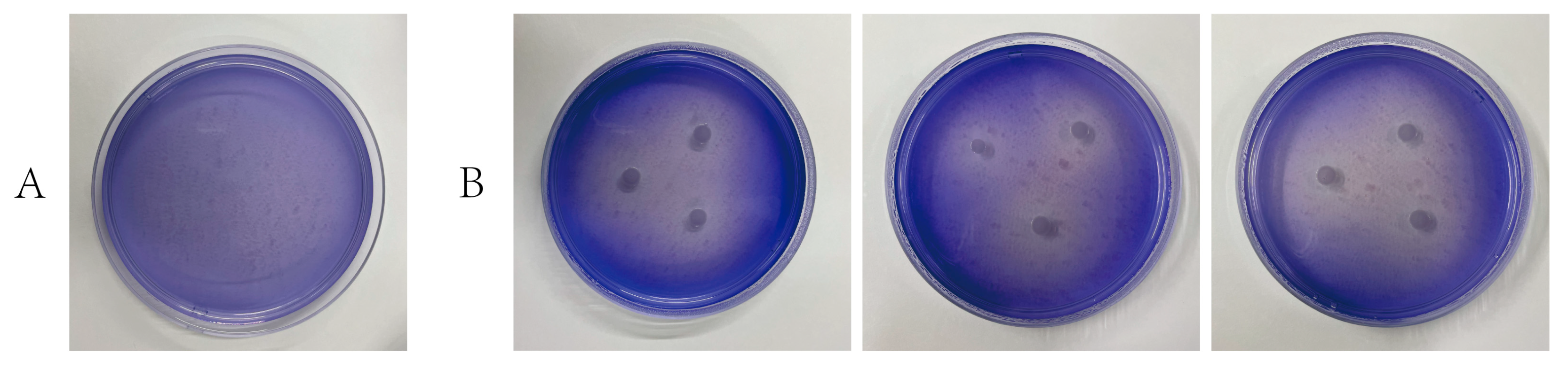
2.3. Microbial Degradation of Lignin

The microbial degradation of lignin was tested in triplicate in 100 mL of ASW liquid medium supplemented with lignin as the sole carbon source at a final concentration of 5 mM. The exponential phase cultures of strain LCG003 were centrifugally washed with ASW liquid medium three times and then inoculated into the ASW liquid medium with and without lignin, respectively, at 28 °C. Live bacteria were counted using a SYTO 9/PI live/dead bacteria double strain kit at 0 and 7 days, respectively. The degradation of lignin by strain LCG003 was verified by comparing the colony numbers of strain LCG003 in the control and experimental groups. Additionally, the experiment of aniline blue decolorization was performed via inoculating strain LCG003 on 2216E-agar plates with 3% aniline blue added at 28 °C. After 2 days, the decolorized circles around the colonies in the plate medium was observed.

2.4. Genomic DNA Extraction, Sequencing and Assembly

The genomic DNA of strain LCG003 was extracted following the method described by Fang et al. [29]. The sequencing of strain LCG003’s genome was conducted by MajorBio (Shanghai Majorbio Bio-pharm Technology Co., Ltd., Shanghai, China) using a combination of the PacBio RS II Single Molecule Real-Time (SMRT) (Pacific Biosciences, Menlo Park, CA, USA) and Illumina HiSeq 2500 platforms (Illumina Inc., San Diego, CA, USA).
For Illumina sequencing, approximately 1 μg of genomic DNA was sheared into 400–500 bp fragments using a Covaris M220 Focused Acoustic Shearer (Covaris, Woburn, MA, USA), according to the manufacturer’s protocol. The sheared fragments were then used to prepare the Illumina sequencing libraries with the NEXTFLEX Rapid DNA-Seq kit (NEXTFLEX, San Jose, CA, USA).
Regarding Pacific Biosciences sequencing, 15 μg of DNA was aliquoted and spun in a Covaris g-TUBE at 6000 RPM for 60 s using an Eppendorf 5424 centrifuge (Eppendorf, Hamburg, Germany). The DNA fragments were subsequently purified, end-repaired and ligated with SMRTbell sequencing adapters following the recommendations of Pacific Biosciences. The resulting sequencing library underwent three rounds of purification using 0.45× volume of Agencourt AMPure XP beads (Agencourt, Scottsdale, AZ, USA), as recommended by the manufacturer. Next, a ~10 kb insert library was prepared and sequenced on one SMRT cell using the standard methods.
The data obtained from both the PacBio and Illumina platforms were utilized for bioinformatics analysis. The complete genome sequence was assembled by combining the reads from PacBio and Illumina. The original image data were converted into sequence data through base calling, resulting in the generation of raw data or raw reads, which were saved as a FASTQ file. Quality trimming was performed to remove low-quality data, and a quality information statistic was employed. The reads were then assembled into a contig using the hierarchical genome assembly process (HGAP) and canu [30]. The final circularization step was manually checked and completed, resulting in a complete genome with a single seamless chromosome. Finally, error correction of the PacBio assembly results was conducted using the Illumina reads with Pilon [31]. The complete genome sequence of Aliiglaciecola sp. LCG003 was deposited in the GenBank database under accession number CP128185.1.

2.5. Gene Annotation and Genomic Comparison

For ORF prediction and gene annotation, we used the NCBI prokaryotic genome annotation pipeline [32]. The predicted protein sequences were aligned with the Clusters of Orthologous Groups of proteins (COG) [33] and TransporterDB 2.0 [34] databases using BLASTp software [35] (version 2.9.0) (identity, 50%; query-cover, 80%; e-value, 1 × 10⁻⁵ and score, 40). BlastKOALA [36] was used to assign the Kyoto Encyclopedia of Genes and Genomes (KEGG) annotation. We utilized IslandViewer 4 [37] for the prediction of genomic islands.
To cluster the protein families of strain LCG003 and phylogenetically related strains, we employed a local OrthoMCL 2.0.9 [38]. The clustering was performed using the following cutoff values: identity, 50%; query coverage, 50%; E value, 1 × 10⁻¹⁰; score, 40 and MCL inflation, 1.5. Protein families that were only present in one strain were considered strain-specific. To calculate the average nucleotide identity (ANI) and average amino acid identity (AAI), we used fastANI [39] and CompareM (https://github.com/dparks1134/CompareM, accessed on 10 December 2023), respectively, with the default parameters. The genome-to-genome distance (DDH) was calculated using GGDC 3.0 [40].

2.6. Phylogenetic Analysis

To study the phylogeny of strain LCG003 and its related strains, we employed 120 conserved bacterial marker genes from the GTDB taxonomy. Firstly, we predicted the sequences of these 120 concentrated proteins in the genomes using GTDB-Tk (database version: Release 07-RS207) [41]. Next, we aligned the sequences individually using Clustal Omega [42]. After manually degapping the aligned sequences, we accounted for any missing concentrated proteins in certain genomes by adding the corresponding number of “-” based on the sequence length after degapping. Subsequently, we tandemly connected the degapped alignments of each concentrated protein [43]. The phylogenetic tree was constructed using the neighbor-joining method with FastTree2 [44]. To evaluate the tree’s robustness, a bootstrap analysis was performed with 1000 replicates. Finally, we plotted the phylogenetic tree using iTOL [45].

3. Results and Discussions

3.1. Description of Strain LCG003

The samples of intertidal surface seawater from Lu Chao Harbor were incubated with lignin as the sole carbon source at room temperature for two weeks. The enriched microbes were then cultured on marine 2216E-agar plates. A colony, strain LCG003, was picked from a marine 2216E-agar plate, which formed a transparent circular colony with regular and slightly raised edges. Transmission electron micrograph (TEM) showed that the cell length of strain LCG003 was 0.9–1.5 μm, the width was 0.4–0.7 μm and the flagellar length was 1.5–3.5 μm (Figure S1). Cultured in the marine 2216E medium, its optimal growth temperature range was 25–28 °C (Figure S2), and biofilms were observed on the walls of conical flasks after 3 days of cultivation at each temperature.

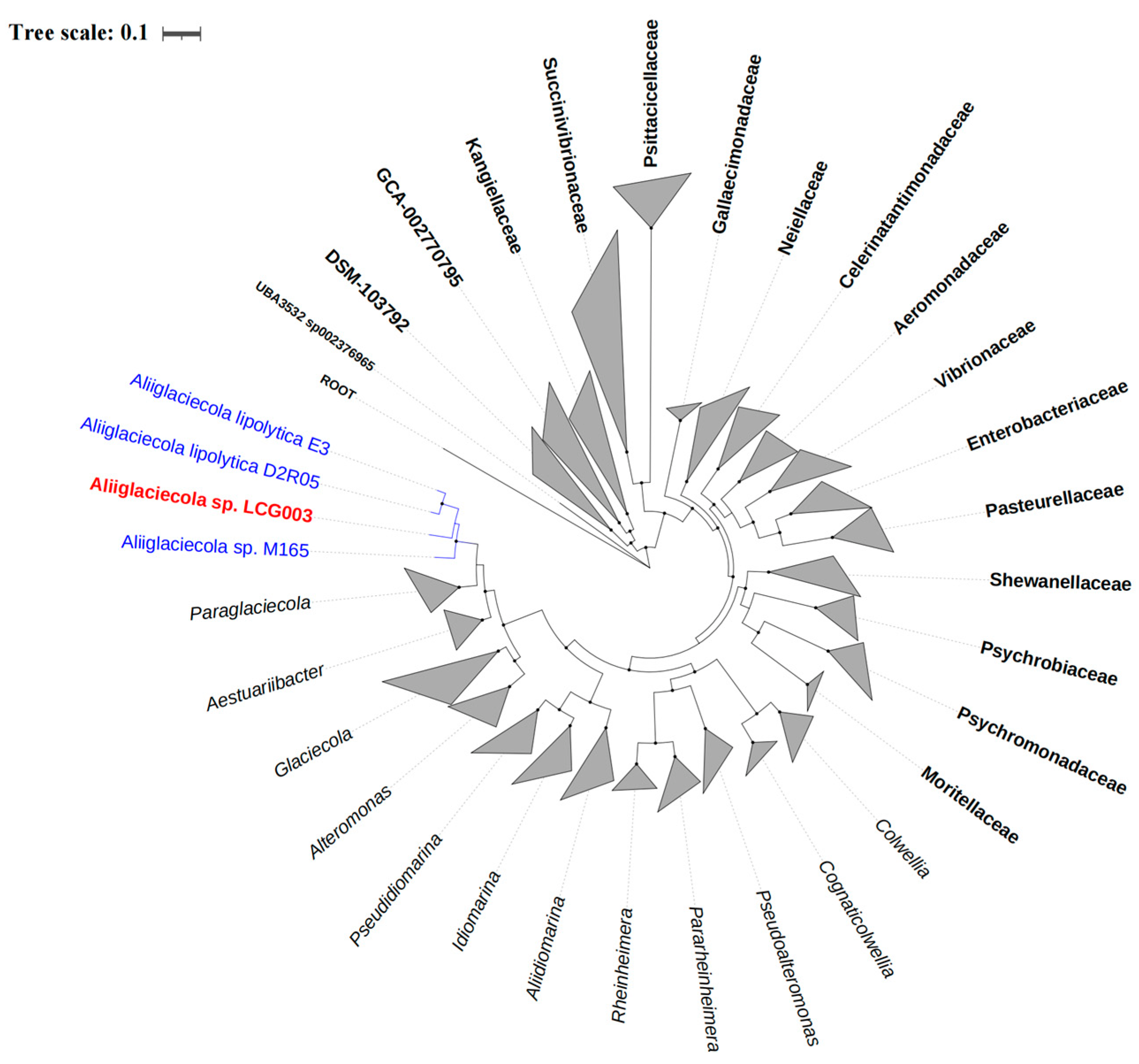
3.2. The Phylogeny of Strain LCG003

The alignments of their 16S rRNA gene sequences showed that strain LCG003 is closely related to Aliiglaciecola sp. W161 (OP344131.1), Aliiglaciecola litoralis SJS3-28 (MG780337.1), Aliiglaciecola sp. M165 (NZ_VKKH01000004.1), Aliiglaciecola lipolytica E3T (NR_044430.1) and Aliiglaciecola lipolytica D2R05 (NZ_JAHKPT010000031.1), with identities of 99.55%, 97.00%, 96.61%, 96.46% and 96.35%, respectively. Moreover, we acquired the complete genome of strain LCG003 (see below) and constructed a phylogenetic tree based on 120 conserved protein sequences (known as GTDB taxonomy). It showed that strain LCG003 formed a coherent phylogenetic cluster associated with Aliiglaciecola lipolytica E3 [25], Aliiglaciecola lipolytica D2R05 and Aliiglaciecola sp. M165 [46] (Figure 1). In addition, strain LCG003 shared ANIb values of 70.67–70.91 with the other three Aliiglaciecola strains, which were higher than those of two neighboring genera, Aestuariibacter and Paraglaciecola (68.2–69.11) (Table S1). The AAI values between strain LCG003 and the other Aliiglaciecola strains were 73.2 to 73.82 but were, at most, 66.34 with the Aestuariibacter and Paraglaciecola species (Table S2). The DDH values between strain LCG003 with and the other Aliiglaciecola strains were merely 14.8 to 14.9 (Table S3). These pieces of evidence indicated that strain LCG003 represents a distinct phyletic lineage in the genus Aliiglaciecola belonging to the family Alteromonadaceae.

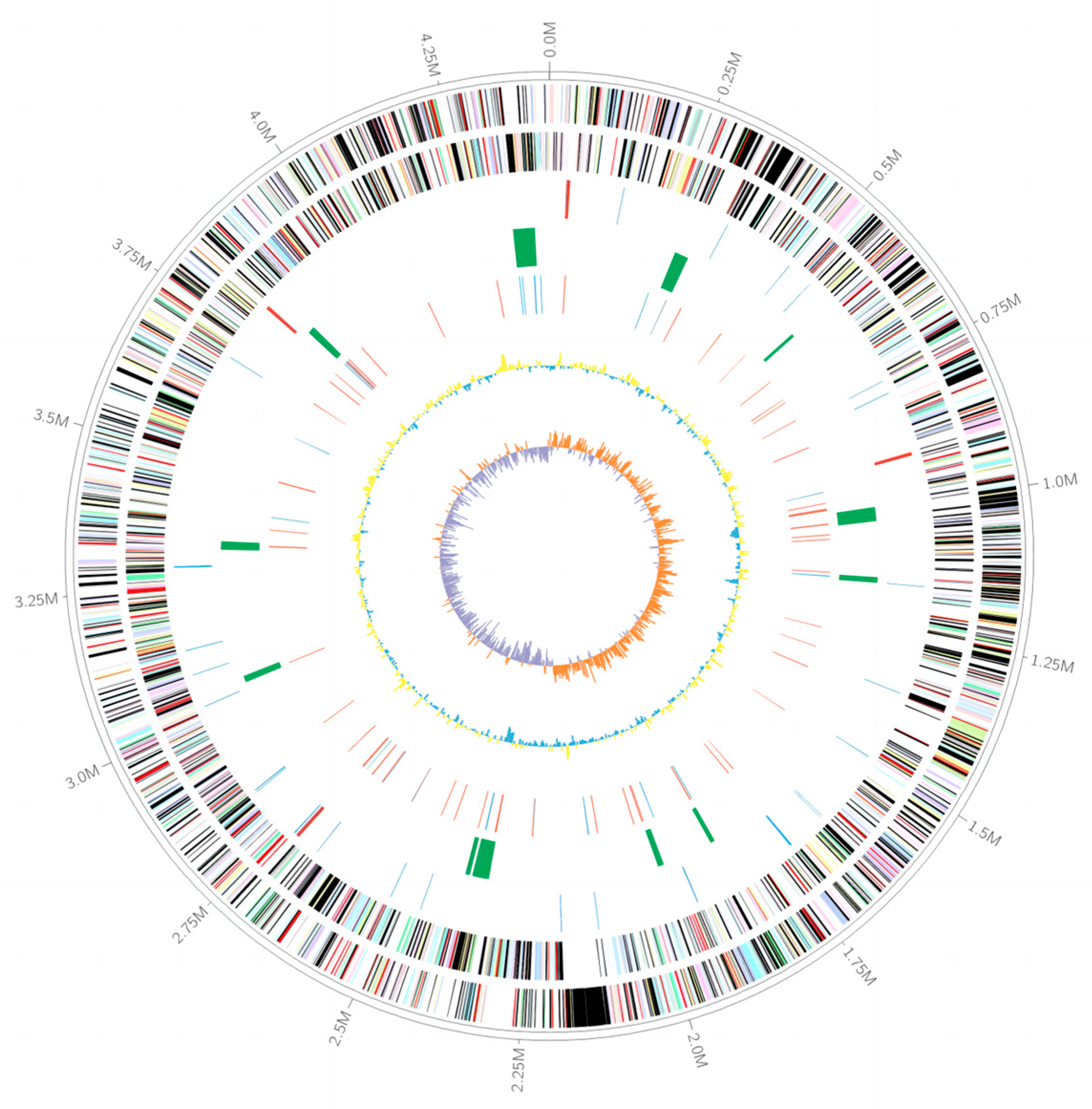
3.3. The Genomic Features of Strain LCG003

The complete genome of strain LCG003 consists of one chromosome with a total length of 4,411,932 base pairs (bp), which is smaller than those of closely related strains, e.g., A. lipolytica E3, A. lipolytica D2R05 and Aliiglaciecola sp. M165. The G + C content of strain LCG003 genome is 42.5%. The genome of strain LCG003 contains 3871 genes, including 3771 protein-coding genes, 58 tRNAs, 4 rRNA operons (including 2 copies of 16S-tRNAIle-tRNAAla-23S-5S and 2 copies of 16S-tRNAAla-tRNAIle-23S-5S), 4 ncRNAs and 26 pseudogenes (Table 1).
The graphical representation of the genome is shown in Figure 2. Upon COG classification, 2421 (63.76%) protein-coding genes were classified into 22 categories (Table S4). The major categories of COG were translation, ribosomal structure and biogenesis (8.20%); general function prediction only (8.09%); amino acid transport and metabolism (7.06%); signal transduction mechanisms (6.55%) and energy production and conversion (6.48%). Moreover, up to 12 genomic islands were found in strain LCG003 (Table S5). In addition, 60 transposases and 13 integrases and/or recombinases were predicted in strain LCG003, of which 58 (79.45%) were absent in the other three Aliiglaciecola strains (Table S6). These pieces of evidence suggest that horizontal gene transfer (HGT) is important for the speciation of this strain.

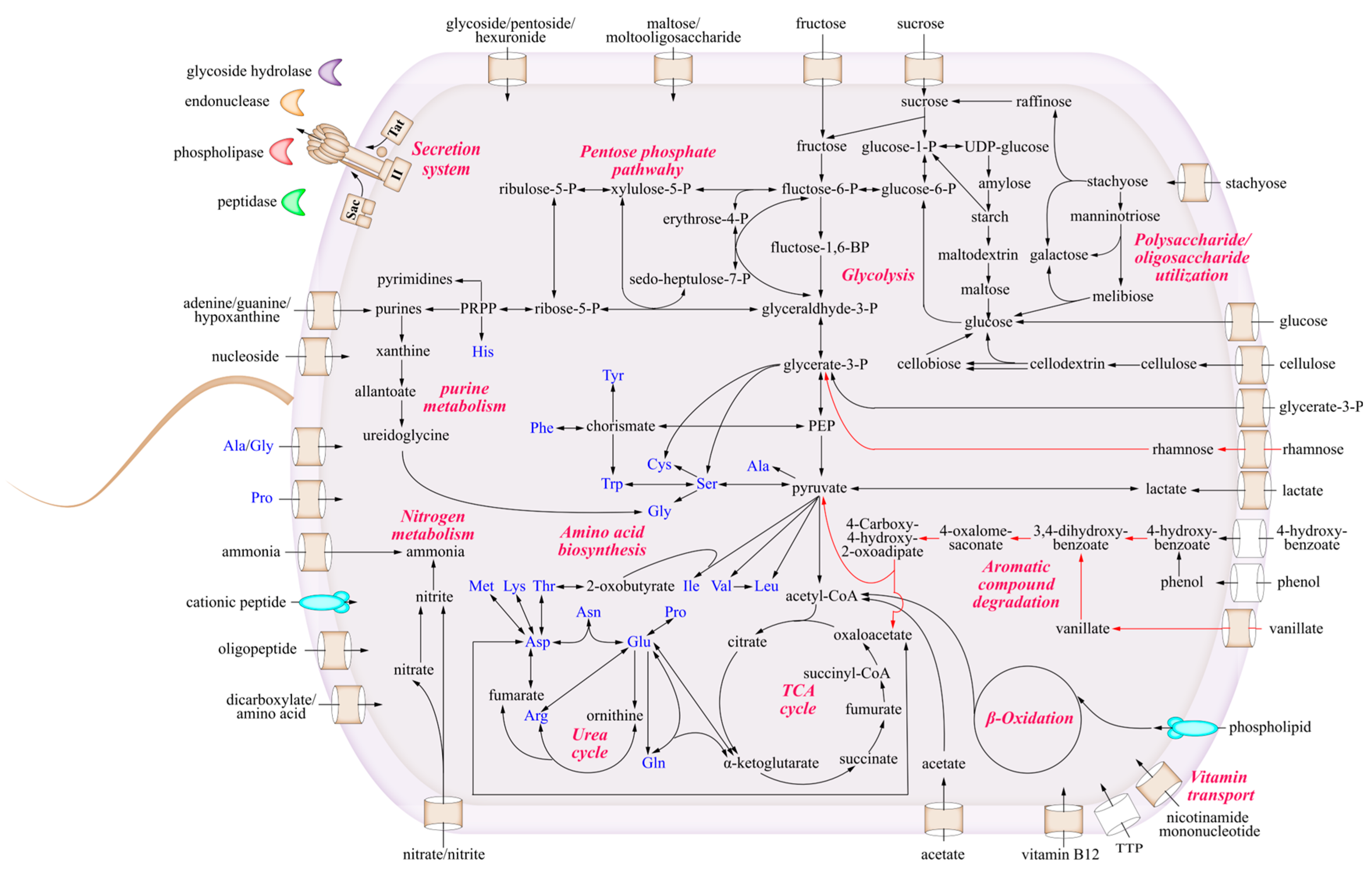
3.4. The Metabolic Characteristics of Strain LCG003

To study the metabolic characteristics and ecological potentials of strain LCG003, we reconstructed the metabolic pathways and compared them with those of the other three Aliiglaciecola strains (Figure 3).

3.4.1. The Utilization of Carbohydrates and Carboxylic Acids

As many as 15 extracellular (signal-fused) and 4 intracellular glycoside hydrolase genes were predicted in strain LCG003. Among these extracellular glycoside hydrolases, 10 genes (66.7%) were absent in the other three Aliiglaciecola strains (BLASTp identity, 50%; query coverage, 50% and e-value, 1 × 10⁻⁵). Also, 11 genes of carbohydrate transporters were predicted in strain LCG003, including glucose, L-rhamnose, maltose/moltooligosaccharide, glycoside/pentoside/hexuronide, arabinose, fucose and glycerol-3-phosphate transporters (Table S7). Moreover, the complete metabolic pathways of glucose, fructose, sucrose, rhamnose, maltose, stachyose, xylose, starch and cellulose were predicted in strain LCG003. In particular, the genes of L-rhamnose/H+ symporter RhaT (QR722_01220), L-rhamnose isomerase (QR722_01230) and bifunctional rhamnulose-1-phosphate aldolase (QR722_01235) were found only in strain LCG003 but were absent in the other three Aliiglaciecola strains, suggesting its unique ecological function of L-rhamnose degradation. Our experiments further confirmed that strain LCG003 could grow with glucose, fructose, sucrose, rhamnose, maltose, stachyose and cellulose used as the sole carbon sources (Figure 4).
Besides carbohydrates, strain LCG003 also contains the transport genes of carboxylic acids, e.g., lactate permease (QR722_RS12480), cation/acetate symporter (QR722_RS15990) and dicarboxylate/amino acid: cation symporter (QR722_RS09570 and QR722_RS10460). Our culture experiments confirmed that strain LCG003 could grow with sodium acetate as the sole carbon source (Figure 4). In addition, polyhydroxybutyrate (PHB) is a microbial bioprocessed polyester belonging to the polyhydroxyalkanoate (PHA) family. The genes of PHB depolymerase (QR722_RS11850) and 3-hydroxybutyrate dehydrogenase (QR722_RS11855) were identified in the four Aliiglaciecola genomes, indicating that PHB could also be a carbon source for Aliiglaciecola species. These lines of evidence suggested significant ecological functions of strain LCG003 in utilizing carbohydrate and (poly)carboxylic acid degradation.

3.4.2. Metabolism of Amino Acids and Extracellular Proteins

In the aspect of biosynthesis, all four Aliiglaciecola strains are able to biosynthesize 20 kinds of amino acids. Also, we predicted seven and six genes involved in the transport of amino acids and oligopeptides in strain LCG003, respectively. Surprisingly, as many as 44 genes of extracellular (SignalP-fused) peptidases (1.16% of all coding genes) were predicted in strain LCG003, which could be classified into 20 peptidase families (Table S8). The genes of the S9, S8 and M28 family peptidase are the most abundant extracellular peptidases, which include six, five and four copy numbers in strain LCG003.
Peptidase family S9 contains a varied set of serine-dependent peptidases. Most members of the family show restricted specificities and are active mainly on oligopeptides [47,48]. Peptidases in family M28 have co-catalytic zinc ions, which have been confirmed to release a variety of N-terminal amino acids [49]. These peptidase families could be important for strain LCG003 to utilize soluble oligopeptides to satisfy its demand of amino acids.
Furthermore, strain LCG003 appears to have the ability to utilize extracellular insoluble proteins as carbon/nitrogen sources. Peptidase family S8 is the second-largest family of serine peptidases, both in terms of the number of sequences and characterized peptidases. Members of family S8 have a catalytic triad in the order Asp, His and Ser in the sequence, and the majority of them are secreted and nonspecific peptidases that may be associated with nutrient intake. Among these S8 family members, we predicted a polycystic kidney disease (PKD) domain from the gene of QR722_RS06975, which was highly conserved in all Aliiglaciecola genomes. The PKD domain as a collagen-binding domain (CBD) can improve the collagenolytic efficiency of the catalytic domain by swelling insoluble collagen and exposing the monomers, as reported in deseasin MCP-01, a bacterial collagenolytic serine protease [50], suggesting the capability of the insoluble collagen degradation of Aliiglaciecola strains. Also, we predicted four extracellular metalloproteases with collagenase activity (G3DSA:3.40.390.10), which belong to the M1A/M12B, M3 and M13 families (Table S8). Such myriad extracellular peptidases illustrated that proteolysis could be one important lifestyle of Aliiglaciecola strains.

3.4.3. Strain-Specific Degradation of Lignin and Aromatic Compounds

To further confirm the lignin-degrading capability of strain LCG003, we cultured strain LCG003 in ASW-lignin medium, then counted the live bacteria using a SYTO 9/Pl live/dead bacteria double strain kit. An obvious cell proliferation was observed, indicating that strain LCG003 was capable of lignin degradation (Figure S3). Furthermore, the initiation of lignin degradation depends on the common action of a series of enzymes, including lignin peroxidase (LiP), manganese peroxidase (MnP) and laccase (Lac), which induce the decolorization of aniline blue [51,52]. We observed a strong decolorization by incubating strain LCG003 on aniline blue-containing 2216E-agar plates (Figure 5), which further indicated that strain LCG003 could produce lignin-degrading enzymes. Regarding the genome, a gene of dyp-type peroxidase (encoded by QR722_RS02795) was predicted in strain LCG003, which initiates lignin decomposition via cleavage of the Cα-Cβ linkage of phenolic lignin compounds [53,54,55,56].
In addition, various genes in the degradation of aromatic compounds were predicted, including 4-hydroxybenzoate and vanillate (Table S9), which were the products of microbial lignin depolymerization [23]. Compared to the other three Aliiglaciecola strains, the genome of strain LCG003 contains several unique genes involved in 4-hydroxybenzoate oxidation, including 4-hydroxybenzoate 3-monooxygenase (pobA), protocatechuate 4,5-dioxygenase alpha chain (ligA) and beta chain (ligB), 2-hydroxy-4-carboxymuconate semialdehyde hemiacetal dehydrogenase (ligC), 2-pyrone-4,6-dicarboxylate lactonase (ligI), 4-oxalomesaconate tautomerase (galD), 4-oxalmesaconate hydratase (ligJ) and 4-hydroxy-4-methyl-2-oxoglutarate aldolase (ligK). Moreover, the genes of the vanillate (4-hydroxy-3-methoxybenzoate) monooxygenase subunits (vanA and vanB), as well as a GntR family transcriptional regulator in vanillate catabolism, were predicted to be approximately 16 k downstream the 4-hydroxybenzoate-degrading genes (Table S9). In addition, a strain-specific gene of benzaldehyde dehydrogenase (xylC) was predicted, which could transform 4-hydroxybenzaldehyde into 4-hydroxybenzoate. These results suggested that strain LCG003 became a novel lignin and aromatic fragments degrader via horizontal gene transfer and could play an important role in the global cycling of lignocellulosic materials.

3.4.4. The Acquirement of Nitrogen and Phosphorus Sources

For inorganic nitrogen metabolism, we predicted ammonia transporters, nitrate/nitrite ABC transporter, nitrate reductase (encoded by nasAB) and nitrite reductase (encoded by nirBD) in strain LCG003. Moreover, many organic nitrogen sources, such as nucleoside (QR722_RS15920) and nicotinamide riboside (QR722_RS13445 and QR722_RS15260), could also be imported by strain LCG003. In addition, the extracellular endonuclease gene (QR722_RS15905) and the complete degrading pathways of purines were found in the genome of strain LCG003, indicating that it could obtain nitrogen from biological macromolecules, such as nucleic acids.
For phosphorus utilization, strain LCG003 predictively has the genes of ABC transporters of phosphate, phosphonate and phospholipids. Moreover, it also contains the genes of extracellular alkaline phosphatase (QR722_RS00805, QR722_RS06260 and QR722_RS14025) and phospholipase A (QR722_RS17455). These enzymes may contribute to the survival of strain LCG003 in a phosphorus-limited surface marine environment.

3.4.5. The Biosynthetic Deficiency of Thiamine and Cobalamin

Although we observed the growth of strain LCG003 with many kinds of carbohydrates as the sole carbon sources, our previous studies showed that all tested carbohydrates did not induce its growth without a supplement of mixed vitamins into the culture medium, implying the biosynthetic deficiency of some vitamins in strain LCG003. Indeed, our genomic study revealed that the biosynthetic pathways of thiamine (vitamin B1) were largely incomplete in Aliiglaciecola strains LCG003 and M165 hosting with red alga (NCBI BioSample: SAMN12241901) but were complete in strains E3 isolated from coastal surface seawater [57] and D2R05 isolated from an intertidal zone (NCBI BioSample: SAMN19350943). Also, the biosynthetic pathways of thiamine triphosphate (TTP) were also missing in strains LCG003 and M165. TTP is an important coenzyme involved in various biochemical processes in microorganisms, especially in glycolysis and the TCA cycle, e.g., enolase, transketolase, pyruvate decarboxylase, malate dehydrogenase and alpha-ketoglutarate dehydrogenase employ TTP in transferring chemical energy. In this study, no TTP transport gene was predicted in strains LCG003 and M165, suggesting some nonclassical TTP transporters that will be revealed in the future.
Vitamin B12 is a member of the corrinoids that contains a corrin ring, which serves as a cofactor for various enzymes involved in important metabolic reactions, including DNA synthesis, amino acid metabolism and fatty acid synthesis [58]. We found that the biosynthetic pathways of the Vitamin B12 coenzyme (cobalamin) were also missing in the four Aliiglaciecola strains. Nevertheless, a gene of the cobalamin-binding protein (QR722_RS17005) was predicted in strain LCG003, which formed an operon with the genes of the iron complex transport system ATP-binding protein (QR722_RS16995) and iron complex permease protein (QR722_RS17000). Taken together, the absence of these coenzyme synthesis capabilities suggested that Aliiglaciecola strains have close interactions with other microorganisms in the marine environment.

4. Conclusions

In this study, a novel lignin-degrading bacteria strain, LCG003 was isolated from intertidal seawater in Lu Chao Harbor, East China Sea. Phylogenetically, strain LCG003 was affiliated with the genus Aliiglaciecola within the family Alteromonadaceae. The strain LCG003 genome represents the first complete genome of the genus. Metabolically, strain LCG003 contains many extracellular (signal-fused) glycoside hydrolase genes and carbohydrate transporter genes and can utilize various carbohydrate substrates, including cellulose. Also, strain LCG003 preoccupies various genes of extracellular peptidases, as well as amino acid and oligopeptide transporters, and can grow with peptone as the sole carbon and nitrogen source, indicating a proteolytic lifestyle of Aliiglaciecola species. Importantly, strain LCG003 contains a gene of dyp-type peroxidase and strain-specific genes involved in the degradation of 4-hydroxybenzoate and vanillate. Experimentally, the LCG003 strain was confirmed to have a strong decolorization ability for aniline blue and could grow with lignin as the sole carbon source, suggesting an important role in the global carbon cycle of lignocellulosic materials. This study expands our knowledge of lignin-degrading microbial species in marine ecosystems and highlights the contribution of horizontal gene transfer to the degradation of lignocellulosic materials.

Supplementary Materials

The following supporting information can be downloaded at https://www.mdpi.com/article/10.3390/microorganisms12010144/s1: Figure S1: The transmission electron micrograph of strain LCG003; Figure S2: The growth curve of strain LCG003 under multiple temperatures; Figure S3: The lignin utilization of strain LCG003; Table S1: The ANIb values and aligned percentages between strain LCG003 and other strains; Table S2: The AAI values and aligned percentages between strain LCG003 and other strains; Table S3: The DDH values and aligned percentages between strain LCG003 and other strains; Table S4: The COG classification of strain LCG003 and the other three Aliiglaciecola strains; Table S5: The 12 genomic islands found in strain LCG003; Table S6: The genes of transposases, integrases and/or recombinases predicted in strain LCG003; Table S7: The glycoside hydrolase genes predicted in strain LCG003; Table S8: The extracellular peptidases predicted in strain LCG003; Table S9: The genes involved in the degradation of 4-hydroxybenzoate and vanillate.

Author Contributions

Conceptualization, J.W.; biological experiment, H.Z. and Z.W.; data analysis, J.W.; writing—original draft preparation, J.W. and H.Z.; writing—review and editing, X.Y., J.W., H.Z., Z.W., J.F., J.C., T.B., J.L. and C.S.; project administration, J.F.; funding acquisition, J.F. All authors have read and agreed to the published version of the manuscript.

Funding

This research was supported by the National Natural Science Foundation of China (No. 92251303), the Marine Biomedical Science and Technology Innovation Platform of Lingang Special Area, Shanghai, China, the Science and Technology Commission of Shanghai Municipality STCSM 20050501700, and the China Postdoctoral Science Foundation (Grant No. 2021M702086).

Data Availability Statement

The GenBank accession number for the complete genome sequence of strain LCG003 is CP128185.1.

Conflicts of Interest

The authors declare no conflicts of interest.

References

  1. Collins, M.N.; Nechifor, M.; Tanasa, F.; Zanoaga, M.; McLoughlin, A.; Strozyk, M.A.; Culebras, M.; Teaca, C.A. Valorization of lignin in polymer and composite systems for advanced engineering applications—A review. Int. J. Biol. Macromol. 2019, 131, 828–849. [Google Scholar] [CrossRef]
  2. Soltanian, S.; Aghbashlo, M.; Almasi, F.; Hosseinzadeh-Bandbafha, H.; Nizami, A.-S.; Ok, Y.S.; Lam, S.S.; Tabatabaei, M. A critical review of the effects of pretreatment methods on the exergetic aspects of lignocellulosic biofuels. Energy Convers. Manag. 2020, 212, 112792. [Google Scholar] [CrossRef]
  3. Isikgor, F.H.; Becer, C.R. Lignocellulosic biomass: A sustainable platform for the production of bio-based chemicals and polymers. Polym. Chem. 2015, 6, 4497–4559. [Google Scholar] [CrossRef]
  4. Cesarino, I.; Araújo, P.; Júnior, A.P.D.; Mazzafera, P. An overview of lignin metabolism and its effect on biomass recalcitrance. Braz. J. Bot. 2012, 35, 303–311. [Google Scholar] [CrossRef]
  5. Sahoo, B.M.; Ravi Kumar, B.V.V.; Banik, B.K.; Borah, P. Polyaromatic Hydrocarbons (PAHs): Structures, Synthesis and their Biological Profile. Curr. Org. Synth. 2020, 17, 625–640. [Google Scholar] [CrossRef]
  6. Dhar, K.; Subashchandrabose, S.R.; Venkateswarlu, K.; Krishnan, K.; Megharaj, M. Anaerobic Microbial Degradation of Polycyclic Aromatic Hydrocarbons: A Comprehensive Review. Rev. Environ. Contam. Toxicol. 2020, 251, 25–108. [Google Scholar] [CrossRef] [PubMed]
  7. Lu, P.; Wang, W.; Zhang, G.; Li, W.; Jiang, A.; Cao, M.; Zhang, X.; Xing, K.; Peng, X.; Yuan, B.; et al. Isolation and characterization marine bacteria capable of degrading lignin-derived compounds. PLoS ONE 2020, 15, e0240187. [Google Scholar] [CrossRef] [PubMed]
  8. Bleem, A.; Kato, R.; Kellermyer, Z.A.; Katahira, R.; Miyamoto, M.; Niinuma, K.; Kamimura, N.; Masai, E.; Beckham, G.T. Multiplexed fitness profiling by RB-TnSeq elucidates pathways for lignin-related aromatic catabolism in Sphingobium sp. SYK-6. Cell Rep. 2023, 42, 112847. [Google Scholar] [CrossRef]
  9. Li, J.; Dong, C.; Lai, Q.; Wang, G.; Shao, Z. Frequent Occurrence and Metabolic Versatility of Marinifilaceae Bacteria as Key Players in Organic Matter Mineralization in Global Deep Seas. mSystems 2022, 7, e0086422. [Google Scholar] [CrossRef]
  10. Zhang, Y.H.; Dong, J.D.; Wang, Y.S.; Gu, J.D.; Yin, J.P.; Ahmad, M.; Ling, J. Comparative genomics reveals the evidence of aromatic hydrocarbons degradation potential in genus Roseovarius in marine environment. Int. Biodeterior. Biodegrad. 2022, 171, 105408. [Google Scholar] [CrossRef]
  11. Raghukumar, C.; Chandramohan, D.; Michel, F.C.; Redd, C.A. Degradation of lignin and decolorization of paper mill bleach plant effluent (BPE) by marine fungi. Biotechnol. Lett. 1996, 18, 105–106. [Google Scholar] [CrossRef]
  12. Li, Y.Q.; Wang, M.J.; Luo, C.B. Highly efficient polyhydroxyalkanoate production from lignin using genetically engineered Halomonas sp. Y3. Bioresour. Technol. 2023, 370, 128526. [Google Scholar] [CrossRef] [PubMed]
  13. Wang, X.; Lin, L.; Zhou, J. Links among extracellular enzymes, lignin degradation and cell growth establish the models to identify marine lignin-utilizing bacteria. Environ. Microbiol. 2021, 23, 160–173. [Google Scholar] [CrossRef]
  14. Tang, H.; Wang, M.J.; Gan, X.F.; Li, Y.Q. Funneling lignin-derived compounds into polyhydroxyalkanoate by Halomonas sp. Y3. Bioresour. Technol. 2022, 362, 127837. [Google Scholar] [CrossRef] [PubMed]
  15. Jiang, C.; Yan, H.H.; Shen, X.H.; Zhang, Y.T.; Wang, Y.; Sun, S.S.; Jiang, H.Y.; Zang, H.L.; Zhao, X.Y.; Hou, N.; et al. Genome Functional Analysis of the Psychrotrophic Lignin-Degrading Bacterium Arthrobacter sp. C2 and the Role of DyP in Catalyzing Lignin Degradation. Front. Microbiol. 2022, 13, 921549. [Google Scholar] [CrossRef] [PubMed]
  16. Jiang, C.; Cheng, Y.; Zang, H.L.; Chen, X.; Wang, Y.; Zhang, Y.T.; Wang, J.M.; Shen, X.H.; Li, C.Y. Biodegradation of lignin and the associated degradation pathway by psychrotrophic Arthrobacter sp. C2 from the cold region of China. Cellulose 2020, 27, 1423–1440. [Google Scholar] [CrossRef]
  17. Woo, H.L.; O’Dell, K.B.; Utturkar, S.; McBride, K.R.; Huntemann, M.; Clum, A.; Pillay, M.; Palaniappan, K.; Varghese, N.; Mikhailova, N.; et al. Near-Complete Genome Sequence of Thalassospira sp. Strain KO164 Isolated from a Lignin-Enriched Marine Sediment Microcosm. Genome Announc. 2016, 4, e01297-16. [Google Scholar] [CrossRef] [PubMed]
  18. Mei, J.F.; Shen, X.B.; Gang, L.P.; Xu, H.J.; Wu, F.F.; Sheng, L.Q. A novel lignin degradation bacteria-Bacillus amyloliquefaciens SL-7 used to degrade straw lignin efficiently. Bioresour. Technol. 2020, 310, 123445. [Google Scholar] [CrossRef]
  19. Khan, S.I.; Zarin, A.; Ahmed, S.; Hasan, F.; Belduz, A.O.; Canakci, S.; Khan, S.; Badshah, M.; Farman, M.; Shah, A.A. Degradation of lignin by Bacillus altitudinis SL7 isolated from pulp and paper mill effluent. Water Sci. Technol. 2022, 85, 420–432. [Google Scholar] [CrossRef]
  20. Chang, Y.C.; Choi, D.; Takamizawa, K.; Kikuchi, S. Isolation of Bacillus sp. strains capable of decomposing alkali lignin and their application in combination with lactic acid bacteria for enhancing cellulase performance. Bioresour. Technol. 2014, 152, 429–436. [Google Scholar] [CrossRef]
  21. Yang, J.; Zhao, J.; Jiang, J.C.; Xu, H.; Zhang, N.; Xie, J.C.; Wei, M. Isolation and Characterization of Bacillus Sp. Capable of Degradating Alkali Lignin. Front. Energy Res. 2021, 9, 807286. [Google Scholar] [CrossRef]
  22. He, X.; Kim, H.; Dong, T.G.; Gates, I.; Lu, Q.Y. Green synthesis of Ag/lignin nanoparticle-loaded cellulose aerogel for catalytic degradation and antimicrobial applications. Cellulose 2022, 29, 9341–9360. [Google Scholar] [CrossRef]
  23. Ley, Y.; Cheng, X.Y.; Ying, Z.Y.; Zhou, N.Y.; Xu, Y. Characterization of Two Marine Lignin-Degrading Consortia and the Potential Microbial Lignin Degradation Network in Nearshore Regions. Microbiol. Spectr. 2023, 11, e0442422. [Google Scholar] [CrossRef] [PubMed]
  24. Ivanova, E.P.; Mikhaĭlov, V.V. A new family of Alteromonadaceae fam. nov., including the marine proteobacteria species Alteromonas, Pseudoalteromonas, Idiomarina and Colwellia. Mikrobiologiia 2001, 70, 15–23. [Google Scholar] [PubMed]
  25. Jean, W.D.; Hsu, C.Y.; Huang, S.-P.; Chen, J.-S.; Lin, S.; Su, M.-H.; Shieh, W.Y. Reclassification of [Glaciecola] lipolytica and [Aestuariibacter] litoralis in Aliiglaciecola gen. nov., as Aliiglaciecola lipolytica comb. nov. and Aliiglaciecola litoralis comb. nov., respectively. Int. J. Syst. Evol. Microbiol. 2013, 63 Pt 8, 2859–2864. [Google Scholar] [CrossRef]
  26. Gago, J.F.; Viver, T.; Urdiain, M.; Pastor, S.; Kampfer, P.; Ferreira, E.; Rossello-Mora, R. Description of three new Alteromonas species Alteromonas antoniana sp. nov., Alteromonas lipotrueae sp. nov. and Alteromonas lipotrueiana sp. nov. isolated from marine environments, and proposal for reclassification of the genus Salinimonas as Alteromonas. Syst. Appl. Microbiol. 2021, 44, 126226. [Google Scholar] [CrossRef] [PubMed]
  27. Gupta, V.; Sharma, G.; Srinivas, T.N.; Anil Kumar, P. Aliiglaciecola coringensis sp. nov., isolated from a water sample collected from mangrove forest in Coringa, Andhra Pradesh, India. Antonie Van. Leeuwenhoek 2014, 106, 1097–1103. [Google Scholar] [CrossRef]
  28. Jin, H.M.; Jeong, H.I.; Jeon, C.O. Aliiglaciecola aliphaticivorans sp. nov., an aliphatic hydrocarbon-degrading bacterium, isolated from a sea-tidal flat and emended description of the genus Aliiglaciecola Jean et al. 2013. Int. J. Syst. Evol. Microbiol. 2015, 65, 1550–1555. [Google Scholar] [CrossRef]
  29. Fang, J.; Kato, C.; Runko, G.M.; Nogi, Y.; Hori, T.; Li, J.; Morono, Y.; Inagaki, F. Predominance of Viable Spore-Forming Piezophilic Bacteria in High-Pressure Enrichment Cultures from ~1.5 to 2.4 km-Deep Coal-Bearing Sediments below the Ocean Floor. Front. Microbiol. 2017, 8, 137. [Google Scholar] [CrossRef]
  30. Koren, S.; Walenz, B.P.; Berlin, K.; Miller, J.R.; Bergman, N.H.; Phillippy, A.M. Canu: Scalable and accurate long-read assembly via adaptive k-mer weighting and repeat separation. Genome Res. 2017, 27, 722–736. [Google Scholar] [CrossRef] [PubMed]
  31. Walker, B.J.; Abeel, T.; Shea, T.; Priest, M.; Abouelliel, A.; Sakthikumar, S.; Cuomo, C.A.; Zeng, Q.; Wortman, J.; Young, S.K.; et al. Pilon: An integrated tool for comprehensive microbial variant detection and genome assembly improvement. PLoS ONE 2014, 9, e112963. [Google Scholar] [CrossRef]
  32. Tatusova, T.; DiCuccio, M.; Badretdin, A.; Chetvernin, V.; Nawrocki, E.P.; Zaslavsky, L.; Lomsadze, A.; Pruitt, K.D.; Borodovsky, M.; Ostell, J. NCBI prokaryotic genome annotation pipeline. Nucleic Acids Res. 2016, 44, 6614–6624. [Google Scholar] [CrossRef]
  33. Galperin, M.Y.; Wolf, Y.I.; Makarova, K.S.; Vera Alvarez, R.; Landsman, D.; Koonin, E.V. COG database update: Focus on microbial diversity, model organisms, and widespread pathogens. Nucleic Acids Res. 2021, 49, D274–D281. [Google Scholar] [CrossRef] [PubMed]
  34. Elbourne, L.D.; Tetu, S.G.; Hassan, K.A.; Paulsen, I.T. TransportDB 2.0: A database for exploring membrane transporters in sequenced genomes from all domains of life. Nucleic Acids Res. 2017, 45, D320–D324. [Google Scholar] [CrossRef] [PubMed]
  35. Ye, J.; McGinnis, S.; Madden, T.L. BLAST: Improvements for better sequence analysis. Nucleic Acids Res. 2006, 34, W6–W9. [Google Scholar] [CrossRef]
  36. Kanehisa, M.; Sato, Y.; Morishima, K. BlastKOALA and GhostKOALA: KEGG Tools for Functional Characterization of Genome and Metagenome Sequences. J. Mol. Biol. 2016, 428, 726–731. [Google Scholar] [CrossRef] [PubMed]
  37. Bertelli, C.; Laird, M.R.; Williams, K.P.; Lau, B.Y.; Hoad, G.; Winsor, G.L.; Brinkman, F.S.L. IslandViewer 4: Expanded prediction of genomic islands for larger-scale datasets. Nucleic Acids Res. 2017, 45, W30–W35. [Google Scholar] [CrossRef]
  38. Li, L.; Stoeckert, C.J., Jr.; Roos, D.S. OrthoMCL: Identification of ortholog groups for eukaryotic genomes. Genome Res. 2003, 13, 2178–2189. [Google Scholar] [CrossRef]
  39. Jain, C.; Rodriguez, R.L.; Phillippy, A.M.; Konstantinidis, K.T.; Aluru, S. High throughput ANI analysis of 90K prokaryotic genomes reveals clear species boundaries. Nat. Commun. 2018, 9, 5114. [Google Scholar] [CrossRef]
  40. Meier-Kolthoff, J.P.; Carbasse, J.S.; Peinado-Olarte, R.L.; Göker, M. TYGS and LPSN: A database tandem for fast and reliable genome-based classification and nomenclature of prokaryotes. Nucleic Acids Res. 2022, 50, D801–D807. [Google Scholar] [CrossRef]
  41. Chaumeil, P.A.; Mussig, A.J.; Hugenholtz, P.; Parks, D.H. GTDB-Tk: A toolkit to classify genomes with the Genome Taxonomy Database. Bioinformatics 2019, 36, 1925–1927. [Google Scholar] [CrossRef] [PubMed]
  42. Sievers, F.; Higgins, D.G. The Clustal Omega Multiple Alignment Package. Methods Mol. Biol. 2021, 2231, 3–16. [Google Scholar] [CrossRef]
  43. Tang, X.; Yu, L.; Yi, Y.; Wang, J.; Wang, S.; Meng, C.; Liu, S.; Hao, Y.; Zhang, Y.; Cao, X.; et al. Phylogenomic analysis reveals a two-stage process of the evolutionary transition of Shewanella from the upper ocean to the hadal zone. Environ. Microbiol. 2021, 23, 744–756. [Google Scholar] [CrossRef]
  44. Price, M.N.; Dehal, P.S.; Arkin, A.P. FastTree 2—Approximately maximum-likelihood trees for large alignments. PLoS ONE 2010, 5, e9490. [Google Scholar] [CrossRef] [PubMed]
  45. Letunic, I.; Bork, P. Interactive Tree Of Life (iTOL) v5: An online tool for phylogenetic tree display and annotation. Nucleic Acids Res. 2021, 49, W293–W296. [Google Scholar] [CrossRef] [PubMed]
  46. Roberts, A.; Nethery, M.A.; Barrangou, R. Functional characterization of diverse type I-F CRISPR-associated transposons. Nucleic Acids Res. 2022, 50, 11670–11681. [Google Scholar] [CrossRef]
  47. Fülöp, V.; Jones, D.T. Beta propellers: Structural rigidity and functional diversity. Curr. Opin. Struct. Biol. 1999, 9, 715–721. [Google Scholar] [CrossRef]
  48. Moss, C.X.; Brown, E.; Hamilton, A.; Van der Veken, P.; Augustyns, K.; Mottram, J.C. An essential signal peptide peptidase identified in an RNAi screen of serine peptidases of Trypanosoma brucei. PLoS ONE 2015, 10, e0123241. [Google Scholar] [CrossRef]
  49. Eisenhaber, B.; Eisenhaber, S.; Kwang, T.Y.; Gruber, G.; Eisenhaber, F. Transamidase subunit GAA1/GPAA1 is a M28 family metallo-peptide-synthetase that catalyzes the peptide bond formation between the substrate protein’s omega-site and the GPI lipid anchor’s phosphoethanolamine. Cell Cycle 2014, 13, 1912–1917. [Google Scholar] [CrossRef]
  50. Wang, Y.-K.; Zhao, G.-Y.; Li, Y.; Chen, X.-L.; Xie, B.-B.; Su, H.-N.; Lv, Y.-H.; He, H.-L.; Liu, H.; Hu, J.; et al. Mechanistic Insight into the Function of the C-terminal PKD Domain of the Collagenolytic Serine Protease Deseasin MCP-01 from Deep Sea Pseudoalteromonas sp. SM9913: BINDING OF THE PKD DOMAIN TO COLLAGEN RESULTS IN COLLAGEN SWELLING BUT DOES NOT UNWIND THE COLLAGEN TRIPLE HELIX*. J. Biol. Chem. 2010, 285, 14285–14291. [Google Scholar] [CrossRef]
  51. Xiong, Y.I.; Zhao, Y.; Ni, K.; Shi, Y.; Xu, Q. Characterization of Ligninolytic Bacteria and Analysis of Alkali-Lignin Biodegradation Products. Pol. J. Microbiol. 2020, 69, 339–347. [Google Scholar] [CrossRef]
  52. Casciello, C.; Tonin, F.; Berini, F.; Fasoli, E.; Marinelli, F.; Pollegioni, L.; Rosini, E. A valuable peroxidase activity from the novel species Nonomuraea gerenzanensis growing on alkali lignin. Biotechnol. Rep. 2017, 13, 49–57. [Google Scholar] [CrossRef] [PubMed]
  53. de Gonzalo, G.; Colpa, D.I.; Habib, M.H.; Fraaije, M.W. Bacterial enzymes involved in lignin degradation. J. Biotechnol. 2016, 236, 110–119. [Google Scholar] [CrossRef]
  54. Lin, L.; Wang, X.; Cao, L.; Xu, M. Lignin catabolic pathways reveal unique characteristics of dye-decolorizing peroxidases in Pseudomonas putida. Environ. Microbiol. 2019, 21, 1847–1863. [Google Scholar] [CrossRef]
  55. Colpa, D.I.; Fraaije, M.W.; van Bloois, E. DyP-type peroxidases: A promising and versatile class of enzymes. J. Ind. Microbiol. Biotechnol. 2014, 41, 1–7. [Google Scholar] [CrossRef] [PubMed]
  56. Catucci, G.; Valetti, F.; Sadeghi, S.J.; Gilardi, G. Biochemical features of dye-decolorizing peroxidases: Current impact on lignin degradation. Biotechnol. Appl. Biochem. 2020, 67, 751–759. [Google Scholar] [CrossRef]
  57. Chen, L.P.; Xu, H.Y.; Fu, S.Z.; Fan, H.X.; Liu, Y.H.; Liu, S.J.; Liu, Z.P. Glaciecola lipolytica sp. nov., isolated from seawater near Tianjin city, China. Int. J. Syst. Evol. Microbiol. 2009, 59, 73–76. [Google Scholar] [CrossRef] [PubMed]
  58. Gruber, K.; Puffer, B.; Kräutler, B. Vitamin B12-derivatives-enzyme cofactors and ligands of proteins and nucleic acids. Chem. Soc. Rev. 2011, 40, 4346–4363. [Google Scholar] [CrossRef] [PubMed]
Figure 1. Phylogeny of the Aliiglaciecola species and their related species based on 120 concentrated proteins. The Aliiglaciecola sp. LCG003 and the other three Aliiglaciecola strains are highlighted in red and blue, respectively. Bold characters represent the family under the order Enterobacterales_A, and italic characters represent other genus in the family Alteromonadaceae. Black points mean bootstrap values ≥ 60.
Microorganisms 12 00144 g001
Figure 2. Graphical representation of the Aliiglaciecola strain LCG003 genomes. Genes on the forward (shown in outer circle) and reverse (shown in inner circle) strands are colored according to their cluster of homologous genes (COG) categories: RNA genes are highlighted in different colors (tRNAs in blue and rRNAs in red), gene islands are shown in green, genes involved in HGT are shown in different colors (transposase red and integrase/recombinase blue), the GC content is shown in yellow/blue and the GC skew is shown in orange/purple.
Microorganisms 12 00144 g002
Figure 3. Metabolic potentials of strain LCG003. The black arrows represent metabolic pathways predicted in strain LCG003 and the red arrows highlight the pathways in strain LCG003 that are absent in the other three Aliiglaciecola strains.
Microorganisms 12 00144 g003
Figure 4. The carbon source utilization of strain LCG003. (AH) represents ASM agar plates with glucose, fructose, sucrose, rhamnose, maltose, stachyose, cellulose and sodium acetate as the sole carbon source, respectively, and (I) represents the blank control. The plates were photographed after seven days. Each sample was with three independent biological replicates.
Microorganisms 12 00144 g004
Figure 5. Decolorization of aniline blue by strain LCG003. (A) means the blank control, and (B) means the decolorization of strain LCG003. Each sample was tested with three independent biological replicates.
Microorganisms 12 00144 g005
Table 1. Genome features of strain LCG003.
ItemsDescription
Size (bp)4,411,932
G + C content (%)42.5
Coding sequence (%)87.51
Total genes3871
Protein-coding genes3771
Genes assigned to COG2732
rRNA operons4
tRNA genes58
ncRNA genes4
Pseudogene26
Gene islands12
Disclaimer/Publisher’s Note: The statements, opinions and data contained in all publications are solely those of the individual author(s) and contributor(s) and not of MDPI and/or the editor(s). MDPI and/or the editor(s) disclaim responsibility for any injury to people or property resulting from any ideas, methods, instructions or products referred to in the content.

