Review

The Good, the Bad, and the Useable Microbes within the Common Alder (Alnus glutinosa) Microbiome—Potential Bio-Agents to Combat Alder Dieback

by
Emma Fuller
1,2,
Kieran J. Germaine
1 and
Dheeraj Singh Rathore
2,*
1
EnviroCore, Dargan Research Centre, Department of Applied Science, South East Technological University, Kilkenny Road, R93 V960 Carlow, Ireland
2
Teagasc, Forestry Development Department, Oak Park Research Centre, R93 XE12 Carlow, Ireland
*
Author to whom correspondence should be addressed.
Microorganisms 2023, 11(9), 2187; https://doi.org/10.3390/microorganisms11092187
Submission received: 17 July 2023 / Revised: 24 August 2023 / Accepted: 25 August 2023 / Published: 30 August 2023
(This article belongs to the Special Issue Biological Control of the Plant Pathogens)

Abstract

:
Common Alder (Alnus glutinosa (L.) Gaertn.) is a tree species native to Ireland and Europe with high economic and ecological importance. The presence of Alder has many benefits including the ability to adapt to multiple climate types, as well as aiding in ecosystem restoration due to its colonization capabilities within disturbed soils. However, Alder is susceptible to infection of the root rot pathogen Phytophthora alni, amongst other pathogens associated with this tree species. P. alni has become an issue within the forestry sector as it continues to spread across Europe, infecting Alder plantations, thus affecting their growth and survival and altering ecosystem dynamics. Beneficial microbiota and biocontrol agents play a crucial role in maintaining the health and resilience of plants. Studies have shown that beneficial microbes promote plant growth as well as aid in the protection against pathogens and abiotic stress. Understanding the interactions between A. glutinosa and its microbiota, both beneficial and pathogenic, is essential for developing integrated management strategies to mitigate the impact of P. alni and maintain the health of Alder trees. This review is focused on collating the relevant literature associated with Alder, current threats to the species, what is known about its microbial composition, and Common Alder–microbe interactions that have been observed worldwide to date. It also summarizes the beneficial fungi, bacteria, and biocontrol agents, underpinning genetic mechanisms and secondary metabolites identified within the forestry sector in relation to the Alder tree species. In addition, biocontrol mechanisms and microbiome-assisted breeding as well as gaps within research that require further attention are discussed.

1. Introduction

Over the last two decades, there has been an elevated interest regarding the significance of native tree species, such as Common Alder (Alnus glutinosa (L.) Gaertn) [1], also known as Black Alder, European Alder, or just Alder. The genus Alnus belongs to the family Betulaceae, the birch clade [2]. Alder is known to be a short-lived, light-demanding, and water-demanding species [1]. In addition, Alder preferentially grows in non-shaded areas with little competition; however, it has the ability to naturally self-prune, as branches tend to die with lack of light [3,4]. This species typically contains short stalked reddish-brown winter buds, small flat waxy winged seeds, and woody cone-shaped fruit (catkins) [4,5]. There are more than thirty Alnus species worldwide, including shrubs and trees, with A. glutinosa being the primary native species within Europe [6,7]. While some other well-known Alder species include Italian Alder (A. cordata), Grey Alder (A. incana), Red Alder (A. rubra), White Alder (A. rhombifolia), Japanese Alder (A. japonica), and American Green Alder (A. viridis) [1,5]. Alder has demonstrated phenomenal resilience due to its adaptation to a variety of climates around the world [8]. This resilient species tolerates frost and survives in an extensive range of temperatures [9]. A. glutinosa has been found on a broad range of sites; however, it favours wet, riverine, and fertile areas with high humidity [3,10]. Preferable growing sites include riverbanks, ponds, surrounding lakes, streams, marshy waterlogged areas, shaded mountainous regions, wet woodlands, wet soils, and highlands with sufficient moisture content [10,11]. This species tends not to grow well on calcareous soils, acidic peat, arid sandy soils, and areas with stagnant water [1,12]. Alder has been found in and/or introduced to regions of Africa, Australia, Canada, India, Japan, Russia, North America, South America, and New Zealand [5,12,13,14].
Historically, Alder was considered to generate an inferior class of wood with low economic value [15]. A black dye, known as ‘poor man’s dye’, was produced from Alder bark and catkins and generally used for tanning leather [15]. Alder had an association with war and was known as the ‘tree of death’ since when cut, the light-coloured wood swiftly oxidises to a vivid red, giving a ‘bleeding’ effect [15,16]. Alder wood was used for items such as war shields, wheels, bowls, tubs, and troughs [15]. Alder has been used within many regions worldwide for various traditional health and healing medicinal practices, due to the presence of chemical constituents and biological components such as flavonoids, terpenes, phenols, saponins, and steroids [17]. For example, Common Alder bark has been shown to have medicinal benefits and has been used for treating burns, diseases, and infections, as well as potential antitumor activity [17]. Acero et al. (2012) conducted an ethno-pharmacological study of Common Alder bark and observed that it potentially has anti-oxidant and anti-inflammatory properties [18]. Fresh Alder catkins have shown antioxidant, antimicrobial, and anti-inflammatory properties due to the presence of polyphenols and certain microorganism strains [19].
Alder tends to have an extensive rooting system and forms large nitrogen-fixing root nodules via symbiosis with nitrogen-fixing filamentous bacteria, such as the genus Frankia [20]. Thus, this actinorhizal species has the pioneering ability to improve soil conditions by increasing organic matter and nitrogen content within soils to improve fertility, contributing to biodiversity so other plant populations can grow in the area. Furthermore, Alder can colonise areas that have previously been subject to significant disturbance and/or degradation, playing a vital role in ecosystem and land restoration [1,21]. Other ecological benefits of Alder roots include the reduction of flooding and soil erosion, water filtration/purification, and riverbank stabilisation [4,5]. Alder is a biodiverse habitat providing shelter for numerous plants, animals, and microbes. For example, otters and fish tend to use Alder roots surrounding waterbodies for nesting purposes as well as shelter to reduce predation [11]. Alder provides an important food source for several birds, fungi, lichens, mosses, and approximately 140 insects and mites, resulting in an additional food source for fish if these insects feed on trees planted beside waterbodies [3,22]. For example, Alder leaves provide a food source for plant-eating invertebrates and Alder catkins provide a source of pollen for bees as well as nectar and seeds for many bird species. The nitrogen-fixing capabilities of Alder allow for the production of nitrogen-rich leaf litter due to the presence of invertebrate detritivores and microbial decomposition, which provides a primary food source for invertebrates that are later eaten by bigger organisms, creating multiple food chains [23].
Its porous wood has various uses for carpentry, building, furniture production, biomass production, instrument manufacturing, cabinetry, dyes, and manufacturing charcoal [3,5,16]. Its timber has high durability and decay resistance beneath water, so it is frequently used for bridge piles, small boats, water structure supports, and jetties [4,24]. The climatic adaptation of A. glutinosa, coupled with the worldwide distribution of this species has substantially increased its importance and promising utilisation [8]. As a result, Alder breeding programs have been established; therefore, greater amounts of A. glutinosa are being cultivated within commercial forests in order to generate trees to produce timber [10,16]. As part of the European Union (EU) forest strategy for 2030, policies on broadleaf and biodiversity have been put in place to improve the quantity and quality of European forestry; thus, since Alder is a broadleaved species, the volumes of Alder plantations throughout Europe has increased [25].
There are numerous existing threats to Alder cultivation, some of which are abiotic such as drought [26,27], whilst others can be biological such as pathogenic infections [28]. Research regarding the relationship between pathogenic microbes and native trees has increased because of the elevated levels of disease spread throughout the world, which has caused damage to and the depletion of numerous tree species. Thus, a greater understanding of Alnus-associated beneficial microbes could provide an ecological, environmentally sustainable solution to the biological control of Phytophthora infection. Therefore, to gain a better understanding of the beneficial and pathogenic microbiota associated with A. glutinosa, this review reports the known microbiome of Alder, pathogenic threats of Alder, as well as any biological solutions available to date for the control of these pathogenic threats.

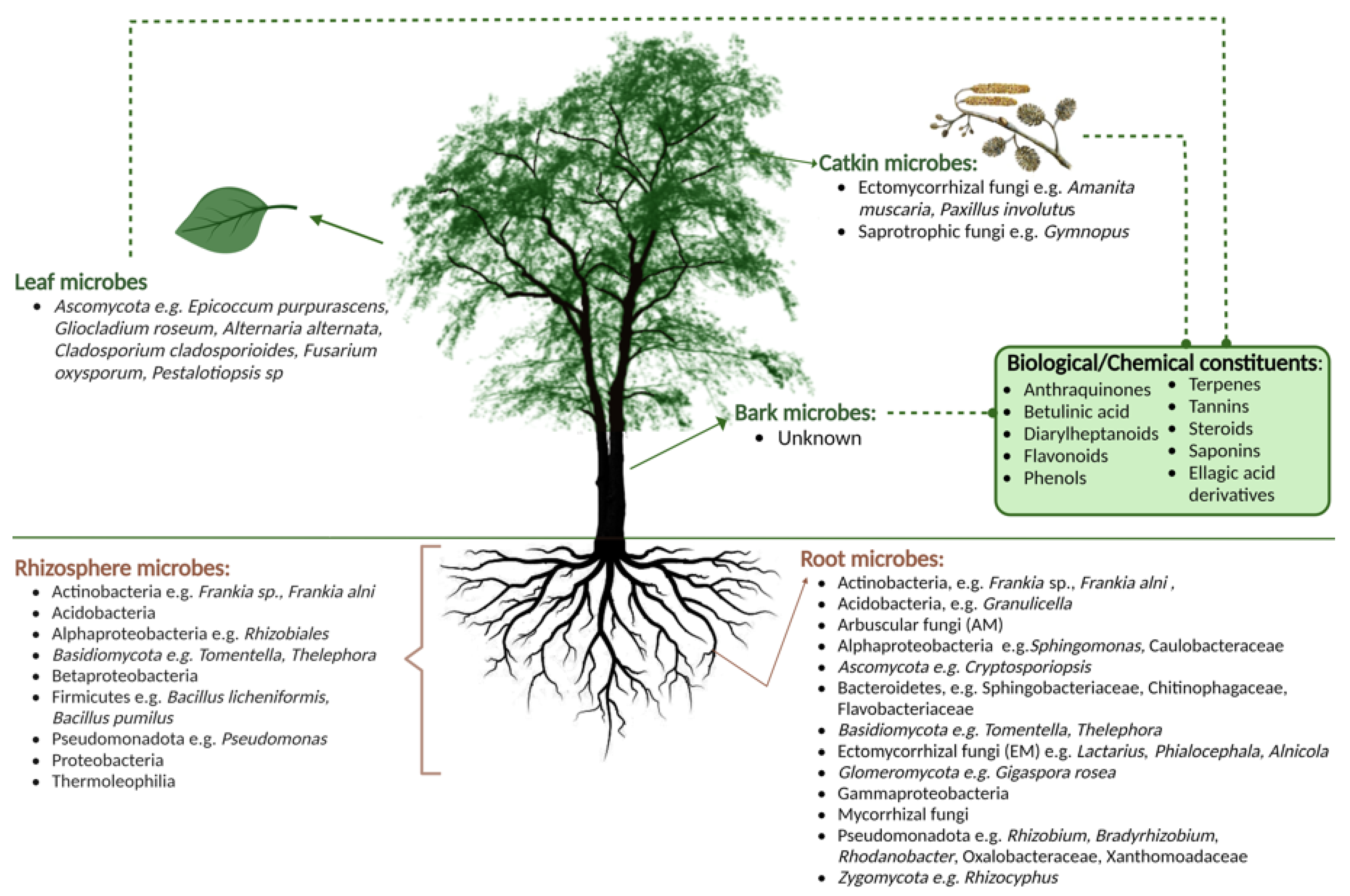
2. The Microbiome of Alder Species

Microorganisms are extremely diverse and plentiful within the environment. The type and abundance of microbial communities that exist within an organism are influenced by the environmental conditions surrounding plant communities as well as soil type, indicating the presence of selective communities associated with different plant species. Forest trees exist in close association with a diverse range of microbial organisms that play a crucial role in maintaining tree health, nutrient conditions, and ecosystem functions. This association can be mutualistic, parasitic, or symbiotic. Combined, these microbial communities associated with the tree are known as its microbiome. The composition of the microbial community can vary due to a range of factors including climate, edaphic conditions, anthropogenic activities, silviculture management practices, and various other events and stressors [29]. Organic matter and associated decomposition products also cause changes within forest soils, causing soil physicochemical alterations, acidification, and supply/leaching of nutrients [30]. The long-lived nature of trees provides a secure food source for beneficial microbiota as well as parasites and pathogens. In comparison to annual crops, underground microbial communities related to trees may be more consistent and have stable interactions due to the deep rooting system, and a constant energy flow system ranging from photosynthesates being pumped into the soil to the abscission of leaf/flower and fruit material, leading to organic matter build up in the soil and the absence of soil disturbance [29].
Tree roots and rhizospheric soil tend to have a particular and plentiful microbial community due to the occurrence of nutrient and mineral exchange between the soil microbiota and tree-component microbiota present in root tissues, ectomycorrhizal fungal roots, arbuscular mycorrhiza roots, and fungal mycelia [31]. These roots have the ability to alter the composition of the microbial community within the soil due to the compounds and root cells that are released into the soil [29]. Throughout tree development, some soil physiochemical properties can change, which causes alterations to the microbial communities present in the rhizosphere that modify tree morphology, promote growth, and enhance nutrient and mineral content [32]. Furthermore, beneficial and/or pathogenic bacterial and fungal communities are also present within the endosphere and phyllosphere microbiota of forest trees, which create ecological interactions such as mutualism, commensalism, and/or antagonism [33]. Beneficial microbiota include plant growth-promoting rhizobacteria (PGPR), plant growth-promoting fungi (PGPF), and biocontrol agents that have the ability to economically and efficiently alter the metabolism of tree substances in order to improve the growth, performance, and resistive properties of the species. Moreover, beneficial microbiota are necessary for the natural degradation of plant residues, since greater amounts of microbial assemblages are present within residues, which additionally degrade them into soil organic matter containing beneficial macromolecules and micromolecules. This process provides increased soil protection and sustains nutrient capacity. Furthermore, when plants develop under stressful environmental conditions, the plant-associated microbiota tends to increase levels of hormones such as abscisic acid and jasmonic acid, which help plants regulate growth and adapt to extreme conditions [33]. Plant growth-promoting microbes tend to be found on the root surface or as endophytes within plant tissues, which have the ability to act as biocontrol agents, bio-fertilisers, and/or bio-stimulants [34]. Due to the complex relationships and interactions within forest ecosystems, there are still many unknowns and potential beneficial microbes associated with the microbiome of trees; therefore, it is important that research on this topic continues.
Plant growth-promoting rhizobacteria (PGPR) play a significant role in the forest ecosystem by releasing numerous regulatory molecules, aiding the growth and development of forest trees [35]. Deciduous forest soils tend to contain a wide array of bacterial phyla, which include Acidobacteria, Actinobacteria, Proteobacteria, Bacteroidetes, and Firmicutes [36,37]. PGPR can be found in the rhizosphere, phyllosphere, and/or endosphere and have the ability to promote plant growth in different ways including nitrogen fixation, the production of the plant hormones such as auxins (e.g., indole acetic acid (IAA)), enzymes (1-aminocyclopropane-1-carboxylic acid (ACC) deaminase, cellulase, chitinases, etc.) and siderophores, as well as the ability to compete with pathogenic microbes to minimise the spread of infection [33]. IAA is a plant hormone that regulates growth and development [38]. ACC deaminase is produced in the presence of ACC, the immediate precursor to the plant stress hormone ethylene, when plants grow within stressful conditions [39]. ACC deaminase aids plant survival by decreasing the amount of ethylene present, as excessive amounts of this plant hormone can have negative effects on plant growth [40]. Siderophores have the ability to uptake iron from the rhizosphere to promote plant growth and contribute to nutrition whilst minimising the amount of iron available for harmful pathogens [41]. Cellulase has been known to promote the breakdown of fungal and plant cell walls, aid soil fertility by speeding up the decomposition of plant residues, reduce spore germination, and reduce fungal growth [42,43,44]. Richter et al. (2010) analysed cellulase activity to suppress P. cinnamomi, a root rot pathogen of avocados and reported that sporangia production was reduced when cellulase enzymes were present, indicating that cellulase enzymes may have the ability to decelerate the spread of P. cinnamomi within avocado sites [45]. Pectinase is an essential enzyme for plant growth and development as it breaks down pectin within cell walls, allowing new plant growth to occur as well as providing entry for beneficial microbes to live endophytically [46]. Other mechanisms that PGPR induce in order to promote the health and growth of plants include the production of exopolysaccaride biofilms, organic acids to increase phosphate solubilisation, hydrogen cyanide to help with biotic stressors, and the production of cytokines and gibberellins which aid growth and development [35]. In addition, some PGPR can reduce oxidative stress by producing abscisic acid as well as stimulating growth by altering physiological mechanisms using emitted bacterial volatile organic components [35]. Some PGPR studies in particular have focused on analysing the interaction between the Betulaceae family and identified beneficial microbes including Actinobacteria genera (Frankia, Streptomyces) [47,48,49,50], an endophytic bacterial genus (Rhizobium) [51], Bacillus isolates (B. licheniformis, B. pumilus, B. megaterium, and B. longisporus) [52,53,54], and Gammaproteobacteria (Pseudomonas) [50,55]. Other known PGPR particularly in agriculture include Arthrobacter, Azospirillum, Azorhizobium, Burkholderia, Flavobacterium, Mesorhizob, and Methylobacterium [56]. With the evident advantages of these microbes in agriculture, there is great potential to exploit these PGPR with further research in the forestry sector.
The inoculation of plants with plant growth-promoting fungi (PGPF) has demonstrated many health benefits including enhanced seed germination, seed vigour, root morphogenesis, pathogenic suppression, the reduction of (a)biotic stressors, as well as an improved process of photosynthesis and mineralization [57]. The roots from the Alnus species have a tendency to form stable long-term biological interactions with mycorrhizae, including ectomycorrhizal fungi and/or arbuscular mycorrhizal fungi [58]. This symbiosis helps to increase the uptake of water and nutrients through the roots, improve tree health, growth, and reproductive ability, as well as provide a degree of tolerance to (a)biotic stressors [58]. For example, Thiem et al. (2020) showed that the inoculation of Alder seedlings with an ectomycorrhizal fungus, Paxillus involutus OW-5, promoted growth and increased tolerance in saline soils [59]. Culturable dark septate endophytes (DSEs) have the potential to promote plant growth particularly in metal-contaminated soils due to their capacity to provide a degree of phytoremediation/phytostabilization of heavy metals, increasing nutrients within soils, and produce the IAA hormone [2,60]. Fungal melanin tends to be found alongside some PGPF, which may help promote plant growth by providing a higher tolerance/resistance to environmental stress and promoting colonisation [58,61]. Fungal melanin exhibits beneficial biological functions including photo-protection, metal binding, mechanical protection, energy harvesting, cell development, antioxidant functions, anti-desiccant functions, chemical protection, as well as thermoregulation [61]. Some examples of identified PGPF that interact with the Betulaceae family include arbuscular mycorrhizal fungi (Gigaspora rosea), ectomycorrhizal fungi (Hebeloma sp., Helotiales sp., Geopora sp., Thelephora sp., Tomentella spp., Paxillus involutus, Tylospora, Leccinum, and Rhizopogon), endophytic fungi (Cryptosporiopsis spp., Rhizoscyphus spp.), DSE (Phialocephala), and ericoid fungi (Oidiodendron) [2,29,47,58,62,63,64,65]. In addition to all the above-mentioned PGPF, other known growth-promoting microbes used in agriculture belong to the genera Alternaria, Chaetomium, Penicillum, Phoma, Serendipita, and Trichoderma [66]. There has been a major growth of interest regarding PGPF acting as bio-fertilisers in agriculture due to their ecological benefits and plant improvement. In particular, the genera Trichoderma, Penicillum, and Aspergillus are the most studied within agriculture as they can promote crop growth and act as eco-friendly biological control agents to promote resistance/tolerance to biotic stresses [67]. These fungal genera may have potential plant growth-promoting properties within forestry, if not already used within this sector. Furthermore, exploring such unknown genera in forest tree species and soils would lead to novel microbes that can benefit both forestry and agriculture for sustainable production.

What Is Known about the Microbiome of Alder?

To date, there have been few studies regarding microbes associated with A. glutinosa. Much of the information that is available focuses on the microbial communities present within Alnus roots and their associated rhizosphere. These studies particularly focus on soil salinity [68], inoculation with nitrogen-fixing bacteria [69], colonisation capabilities [70], plant growth promotion [30], antagonistic effects against pathogenic microbes [71], as well as adaptation to extreme environmental conditions [72]. Alder-associated bacterial microbes that have been identified belong to the phyla Actinobacteria (Frankia alni), Acidobacteria (Granulicella), Alphaproteobacteria (Caulobacteraceae, Rhizobiales, and Sphingomonas), Bacteroidetes (Chitinophagaceae, Flavobacteriaceae, and Sphingobacteriaceae,), Betaproteobacteria, Firmicutes (Bacillus licheniformis and Bacillus pumilus), Gammaproteobacteria, Pseudomonadota (Bradyrhizobium, Oxalobacteraceae, Pseudomonas, Rhizobium, Rhodanobacter, and Xanthomoadaceae), and Thermoleophilia [20,29,30,36,47,48,50,52,53,65,73]. Phyla of fungal microbes associated with Alder species include Ascomycota (Cryptosporiopsis), Basidiomycota (Tomentella, Thelephora), Ectomycorrhizal fungi (Alnicola, Lactarius, and Phialocephala), Glomeromycota (Gigaspora rosea), and Zygomycota (Rhizocyphus) [2,20,47,58,60,65,74]. See Figure 1 for a summary of the microbes identified in the different compartments of the Alnus species. With the economic and environmental importance of Alder coupled with its pathogenic threat, research regarding the root and rhizospheric microbiomes may potentially grow in the future.
Very little is known about the leaf, bark, and catkin microbiomes of Alder trees. A greater focus has been placed on analysing the chemical and biological components, as well as the secondary metabolites present in the bark, leaf, and fruit due to the medicinal value of these components [39,40,41]. Sukhikh et al. (2022) examined the antioxidant properties of A. glutinosa female catkins and found high levels of methanol extracts, ellagic acid, and ethyl acetate indicating their potential use as natural antioxidants [75]. Thiem et al. (2020) analysed the correlation between salt stress and mycorrhizal fungi from A. glutinosa growing in saline soil. Three fungal species were isolated from A. glutinosa catkins, which were Amanita muscaria, Paxillus involutus, and Gymnopus. Seedlings were later inoculated with each fungus to determine their effects against salt stress. It was observed that P. involutus aided in promoting the growth of seedlings and showed a degree of salt tolerance [59]. There have been studies on the metabolites and medicinal extracts of Alder bark, but no studies were found for the analysis of Alder tree bark microbiomes. Alder leaves have been studied regarding their medicinal potential. For example, Mushkina (2021) investigated the ability of A. glutinosa leaf tinctures to be used as wound-healing gels. It was determined that this gel had the ability to regenerate and heal wounds due to the presence of flavonoids, tannins, and phenolic acid [76]. Leaf extracts have been used to analyse their antagonistic effects against pathogens such as Staphylococcus aureus, Bacillus subtilis, Escherichia coli, Pseudomonas aeruginosa, and Candida albicans [77]. Concerning the microbial community within Alder leaves, there are few studies regarding this topic. Kayini and Pandey (2010) isolated fungal microbes from Nepalese Alder leaves including Alternaria alternata, A. raphani, Aspergillus niger, Cladosporium cladosporioides, Epicoccum purpurascens, Fusarium oxysporum, Gliocladium roseum, Mucor hiemalis, and Pestalotiopsis sp. [78]. A greater focus is needed regarding the microbial communities present within Alder leaves, bark, and catkins.

3. Microbial Pathogens of Alder Species

There are several pests and microbial pathogens associated with Alder. For example, Alder yellows phytoplasma disease (caused by a phytoplasma bacterial parasite) has been identified within numerous European countries, which causes stunted growth, yellowing of leaves, reduction of leave size and amount, as well as dieback and/or death [79,80,81,82]. The Ascomycota fungus, Taphrina alni, is a causal agent of Alder tongue galls on female catkins and has been identified throughout Europe. These galls are known as Alder tongues, as they are green-red elongated structures (depending on the season) [83]. The fungus Septoria alnifolia has been known to form leaf spots, stem cankers, and stem breakage on Alder trees [84]. Furthermore, a rust fungus, Melampsoridium hiratsukanum, causing yellow–brown spotting on Alder leaves, followed by early leaf fall, crown thinning, and/or death, has been identified [85]. The fungal pathogen Mycopappus alni tends to cause brown blotches on leaves and defoliation of Alder trees [86]. The bacterium Erwinia alni, has been found to cause bark cankers and bleeding and eventually kill off branches and/or the tree as a whole if the infection is severe [87]. The bacterium Pseudomonas syringae has been reported to cause leaf necrosis and the dieback of Common Alder [88]. With regards to insects, for example, the Alder leaf beetle, Agelastica alni, has been identified in Europe causing damage and defoliation of Common Alder [89]. Several species of Alder sawflies have been identified feeding on Common Alder such as Monsoma pulveratum [90], Eriocampa ovata [91], Hernichroa crocea, and Cimbex conatus [92]. A leaf miner, Fenusa dohrnii, was identified as a pest of Alder that causes damage [93]. There is an extensive list of pests and pathogens associated with Alder; however, not all tend to have damaging effects on this genus. For example, Sims (2014) discusses different types of fungal pathogens and insects that have been identified as associated with red, white, and thin-leaf Alder in western Oregon [94]. McVean (1953) extensively lists the insects and mites that associate with Common Alder as a food source [9], however not all of them are of economic importance and do not pose a severe threat to Alder trees. One of the major pathogens of Alder trees is the plant pathogenic oomycete species Phytophthora.
The species Phytophthora, meaning ‘plant destroyer’, has been associated with the dieback and decline of Alder across Europe [95]. There are approximately 200 identified and accepted species of Phytophthora, with more species unnamed/unidentified and likely to be discovered [96]. The pathogen typically infects the tree roots, causing them to rot, and spreads throughout the tree to cause damaging effects such as crown rot [97]. Infection can be caused via Phytophthora chlamydospores, hyphae, oospores, sporangium, and more frequently zoospores. Flagellated Phytophthora zoospores have a similar morphology to filamentous fungi but mainly have diploid hyphae and a cell wall comprising cellulose and/or β-glucans [97]. Phytophthora show phylogenetic similarities to algae and diatoms due to the structural composition of zoospores as well as the release of similar sexual antheridia (male) and oogonia (female) [96,98]. Phytophthora are soil-borne water moulds that have a strong dependency on water and humidity; therefore, the zoospores typically disperse and spread throughout water systems, eventually infecting tree roots [96,99]. In terms of the pathogen’s life cycle, sporangia tend to form when chlamydospores (asexually) and/or oospores (sexually) are germinated within wet environments which results in zoospores being released from the mature sporangium [97,100]. Oospores are formed when oogonia are fertilised by antheridia of the Phytophthora species. Zoospores swim through water within wet soils where they have the ability to form cysts on susceptible tree roots and bark near the root collar [100]. These cysts germinate, forming mycelia, which allows the pathogen to grow and spread biotrophically throughout the plant tissues where reproduction occurs and the life cycle begins again by producing chlamydospores, oospores, and sporangium via germination within plant tissues, spreading from the roots and further throughout the tree (Figure 2) [97,100]. Infection via tree roots results in damage which causes a reduction in water and nutrient uptake, impairing their availability for the rest of the tree compartments. The lack of minerals causes harmful effects such as leaf stomata closure and a reduction in photosynthesis [101].
Nave et al. (2021) inoculated Alder roots with Phytophthora alni, and after three weeks, oogonia, oospore, sporangium, antheridium, and mycelium were found at different stages of the reproductive cycle throughout the plant tissues [103]. P. alni hyphae has the ability to grow from Alder roots into the bark via secondary growth, travelling throughout the tree and infecting the cambium layer and adjacent phloem and xylem by continuously reproducing [101]. P. alni infects Alder bark via lenticels and/or bark wounds and spreads via secondary growth throughout the plant tissues by medullary rays into the bark xylem [101]. When the pathogen spreads to the bark, damaging effects include the deterioration of phloem tissue and a reduction in mineral transportation. It is unknown whether P. alni directly infects Alder leaves and catkins; however, infection of the roots and bark has damaging effects on all compartments of the tree.
Since the 1990s, P. alni has been a major issue within many countries and has continued to spread worldwide today, causing a range of damage to the Alnus species, which is believed to have emerged due to hybridisation within Europe [104,105,106,107]. The parent species of P. alni are P. uniformis (diploid) and P. multiformis (haploid), with the ploidy level showing that P. alni contains half of each parental genome; therefore P. alni is a triploid homoploid-type taxon [107]. According to Gibbs (2005), this pathogen was first detected in Southern England and has continued to spread throughout Europe [108,109] (Figure 3). It is hypothesised that the European spread of P. alni is due to clonal dispersion, since several mitochondrial haplotype variations have been observed, which may imply that multiple sexual hybridisation incidents generated a hybrid from several clones [107]. P. alni ssp. lat. was formally named by Brasier et al. (2004) and is now divided into three subspecies: P. alni ssp. alni, P. alni ssp. uniformis, and P. alni ssp. multiformis, considering different variants and hybrids amongst the species, with P. alni ssp. alni being the most aggressive one [110,111,112]. The P. alni species complex was later renamed by Husson et al. (2015) to P. × alni, P. × multiformis, and P. uniformis [112]. Only P. alni ssp. uniformis has been identified in North America to date [94,109]. The pathogen has also been identified in Ireland [110]. Symptoms of the P. alni complex species include dieback of branches, crown thinning, bleeding bark, dark spotting on stems, stunted leaf growth, premature leaf abscission, leaf yellowing, root rot, necrotic lesions on Alder bark, an excessive number of catkins on the species due to stress, and/or death [4,11]. Alder trees situated around riverbanks and flood plains have greater exposure to infection as the soil-borne pathogen spreads through water, infecting Alder bark and the roots, thus spreading the disease to a greater extent [3,4]. The pathogen spreads via streams, irrigation, flooding events, canals, drainage water, and other pathways via water systems [113]. Alternative pathways may include the plantation of Alder that originated from an affected nursery, human recreational activities, as well as animal activity [23].
Jung et al. (2018) conducted a thorough review of forestry diseases causing decline and suggested that other Phytophthora species such as P. acerina, P. cactorum, P. chlamydospora, P. gonapodyides, P. lacustris, P. plurivora, P. polonica, P. pseudocryptogea, and P. siskiyouensis may also be responsible for diseases associated with Alder dieback, although P. alni complex species appears to be the main cause of Alder decline [28,94,114,115,116]. For example, O’Hanlon et al. (2020) first reported the presence of P. lacustris causing disease within Alder trees in Ireland, indicating that Alder is at risk of dieback due to other Phytophthora species [28]. In Ukraine, Matsiakh et al. (2020) analysed rhizosphere soils of declining Alder where P. lacustris, P. plurivora, and P. polonica were detected, which further indicates that these pathogens play a role in Alder dieback [117]. Tkaczyk et al. (2023) identified P. plurivora and P. cactorum within soil, root, and water samples associated with declining Alder in Slovakia [118]. Bregant et al. (2020) reported the species P. plurivora, which was isolated from declining Alder trees within areas of Turkey, Italy, Spain, and Poland [111]. Furthermore, Bregant et al. (2020) revealed that P. chlamydospora, P. gonapodyides, and P. siskiyouensis potentially caused disease within Alder trees in North America. Zamora-Ballesteros et al. (2016) found that P. plurivora caused a high mortality rate within inoculated Alder seedlings, indicating that this species is pathogenic to Alder seedlings [119]. Haque and Diez (2015) conducted in vitro pathogenicity tests of Alder leaves, branches, and twigs with P. × alni, P. × multiformis, and P. uniformis, and it was observed that the pathogen caused foliar necrosis, indicating that the roots and root collar may not be the only source of pathogen inoculum [120].
Figure 3. Distribution of Phytophthora alni across Europe [109,121].
Microorganisms 11 02187 g003

4. The Potential of Microbial Biocontrol Agents in the Fight against Alder Diseases

Biocontrol agents tend to be any beneficial organism, including PGPR and PGPF, which have the ability to provide biological control against harmful pathogenic disease, as well as assisting in acquiring nutrients and amplifying resistive traits within a plant species [33]. Furthermore, the use of PGPR/F can act as a biocontrol agent by enhancing the health of a tree species, thus, a stronger tree can fight/withstand pathogenic infections. Moreover, A. glutinosa has many antioxidant, anti-inflammatory, antitumour, and inhibition properties due to the presence of anthraquinones, betulinic acid, diarylheptanoids, flavonoids, phenols, terpenes, tannins, steroids, and saponins; thus, these substances have the potential as BCAs within forestry [17,18,122]. Emerging diseases within forestry are a cause for concern, therefore, the advancement of BCAs within this sector is desirable. BCAs and bio-fertilisers have been used in agriculture for many years for sustainable crop production to minimise the use of harmful pesticides and fertilisers and reduce pathogenic infections, as well as to promote the health and growth of crops [123,124]. This approach is used intermittently within forestry with a few successful applications. Through research, several potential BCAs have been identified within forest ecosystems to reduce pathogenic spread and associated symptoms including dieback and root rot. Utkhede et al. (1997) analysed the antagonistic effects of the bacterium Enterobacter aerogenes against the pathogen P. lateralis, which tends to affect tree roots within soil and water, causing root rot disease of Lawson cypress trees [125]. D’Souza et al. (2005) investigated several potential BCAs including Acacia extensa, A. stenoptera, A. alata, and A. pulchella against the root rot disease caused by P. cinnamomi, to observe that these genera have the potential to provide protection to susceptible species [126]. With the Alnus species being under threat from the harmful effects of Phytophthora, such studies could potentially aid in determining the association between P. alni and the Alnus species and thus determining potential BCAs to prevent infections.
Several bacterial strains could potentially provide BCAs against emerging tree pathogens [127]. For example, for the pathogen Hymenoscyphus fraxineus, a study performed on the microbial community associated with ash tree leaves (Fraxinus excelsior) discovered elevated amounts of isolates within the genera Luteimonas, Aureimonas, Pseudomonas, Bacillus, and Paenibacillus present within tolerant trees, which potentially inhibit the invasion and spread of ash dieback disease (H. fraxineus) by direct competition or inducing systemic resistance, as well as the potential production of antagonistic metabolites and/or antifungal compounds [128,129]. Similarly, Becker et al. (2022) explored the microbiome of tolerant ash trees, and it was determined that the bacterium Aureimonas altamirensis had the ability to reduce H. fraxineus infections by niche exclusion and inducing mechanisms such as exopolysaccharides production and protein secretion [130]. The plant growth-promoting members of the genus Streptomyces have proven to be a prime choice as BCAs since these are readily available in nature and have the capacity to control pathogenic infections [9,131,132,133]. Liu et al. (2009) discovered that the BCA metabolite daidzein was produced by the bacterial genus Streptomyces and isolated it from a root of A. nepalensis alongside the Streptomyces bacterium, which may have potential antioxidant, anti-inflammatory, and anticancer effects [134,135]. The genus Streptomyces can produce the enzyme chitinase, which has been shown to have antagonistic effects against fungi due to the ability to degrade chitin present in the cell walls of fungi as well as reduce spore germination [136].
Biocontrol modes of action can be direct (interaction between pathogens and BCAs) or indirect (interaction between BCAs and plants to enhance their health and resistance to (a)biotic stressors) [137]. Direct biocontrol factors include antimicrobial metabolites, hydrolytic enzymes, quorum sensing, quenching, and resource competition, as well as siderophores. Indirect biocontrol factors include organisms triggering enhanced immunity for plants, environmental adaption, and hormone modulation. Microorganisms have the ability to produce microbe-associated molecular patterns (MAMPs) or damage-associated molecular patterns (DAMPs) like flagellin, glucan, xylan, or chitin [56]. These patterns are recognised by pattern-recognition receptors (PRRs) or other elicitors such as volatile organic compounds (VOCs) or siderophores, which are detected by specific receptors. Activation of these receptors initiates various signalling mechanisms that serve as precursors for the production of phytohormones, triggering defensive pathways. The kinase pathway can phosphorylate transcription factors that regulate the expression of early and late response genes [56]. There are many genes that have an association with biocontrol agents and defence-related genes [138]. Each BCA has thousands of genes present within its genome (depending on its genome size); therefore, genes with biocontrol traits can differ from one species to another. Nelkner et al. (2019) used transcription and genome mining in order to identify genes that are related to biocontrol traits and biosynthetic genes within a Pseudomonas strain [139]. In this study, biocontrol activity included the production of siderophores, secondary metabolites, and antibiotics (2,4-Diacetylphloroglucinol (DAPG), hydrogen cyanide (HCN) synthesis, pathogen inhibition genes (iron acquisition, exoprotease activity, and chitinase activity), resistance genes (PRRs), exopolysaccharides genes, lipopolysaccharides genes, metabolism genes, detoxification genes, and genes related to ISR and growth-promoting compounds (VOCs, acetoin, 2,3-butanediol, growth regulators/plant hormones, and phosphate solubilisation) [139]. Genes also involved in plant defence include the phenylpropanoid pathway gene-expressing phenylalanine ammonia-lyase (PAL), which facilitates the deamination process when phenylalanine is converted into cinnamate and ammonia, as well as the lipoxygenase pathway gene encoding hydroxyperoxide lyase (HPL) [56]. Mitogen-activated protein kinase (MAPK)- and cyclic adenosine monophosphate (cAMP)-associated molecules represent widespread intracellular signalling pathways that integrate extracellular stimuli, alter the expression and functionality of receptors, and regulate processes such as cell survival and neuroplasticity [140]. TGA transcription factors are vital regulators of diverse cellular processes which link to hormonal pathways, interacting proteins, and regulatory elements [141]. In addition, the phytohormones brassinosteroids (BRs) and jasmonates (JAs) also aid in the regulation of plant growth, development, and defensive response [142].
Chitinase has been used in agriculture during crop cultivation for disease management, improved growth, and greater yields [143]. Kumar et al. (2018) highlighted various ways in which chitinase gene expression has been used in agriculture to combat fungal diseases [143]. Auxins such as IAA and phenylacetic acid (PAA) are well-known bio-agents within agriculture due to their plant growth-promoting abilities and provide potential control against Fusarium pathogens, which have been found within soils, causing root rot [144,145]. IAA has been abundantly produced from PGPR isolates associated with forest trees; therefore they may have the potential for BCAs within this sector since they are proven to be advantageous in agriculture [38]. Trinh et al. (2022) discovered that the rhizobacteria Bacillus subtilis, isolated from the rhizosphere roots of black pepper, has potential as a bio-agent for Fusarium antagonism [144]. Fungal endophytes could potentially provide BCAs against emerging tree pathogens [146]. For example, Kosawang et al. (2017) performed in vitro antagonistic assays of Sclerostagonospora sp., Setomelanomma holmii, Epicoccum nigrum, Boeremia exigua, and Fusarium sp. against the dieback-causing fungus, H. fraxineus, and it was observed that these endophytes are potential BCAs [147]. Halecker et al. (2020) analysed the antagonistic effects of the fungal endophyte Hypoxylon rubiginosum on H. fraxineus, and it was determined that this endophyte has potential as a BCA due to the production of metabolites that are toxic to the pathogen [128,148]. This investigative approach is beneficial to determine potential pathogen-fighting microbes present within tolerant tree species that can aid in the protection of trees that are susceptible to certain diseases. See Table 1 for a summary of BCAs.
The most commonly used biocontrol agents belong to the genera Pseudomonas, Bacillus, and Trichoderma [29]. For example, Pseudomonas (P. pudida 06909) was used as a biocontrol agent against Phytophthora root rot within citrus orchids [149], P. chlororaphis has antagonistic effects against the cacao rot pathogen Ph. Palmivora [150], and Bacillus amyloliquefaciens isolated from ginseng rhizosphere induced systemic resistance to Ph. cactorum [151]. In addition, Trichoderma virens, Trichoderma harzianum, Trichoderma asperellum, and Trichoderma spirale isolated from cocoa show antagonistic effects against Ph. Palmivora [152], and Trichoderma saturnisporum also showed antagonistic effects against the Phytophthora spp. [29,153]. Abbas et al. (2022) conducted an extensive review regarding the identification of multiple biocontrol encoding genes within the Trichoderma spp. against R. solani including secondary metabolite genes, siderophores genes, signalling molecular genes, cell wall degradation enzymatic genes, and plant growth regulatory genes [154]. Furthermore, G-protein coupled receptor (GPCR) genes, adenylate cyclase genes, protein kinase-A genes, and transcription factor proteins are important genes found in Trichoderma for biocontrol against R. solani [154]. Moreover, Trichoderma has the ability to generate chitinase, which can minimise the survival of R. solani through the activation of the expression of chitinase genes [155]. Hernando José et al. (2021) discuss several isolated endophytic bacteria, fungi, and metabolites that have shown antagonistic and biocontrol activities as well as induced mechanisms against Phytophthora pathogens [156]. Some bacterial examples include Pseudomonas fluorescence isolated from the flowering vine saw greenbrier, which showed antagonistic effects against P. parasitica, P. cinnamomi and P. palmivora, as well as Burkholderia spp. isolated from the herb huperzine showing the ability to inhibit the growth of P. capsici. The isolate Acinebacter calcoaceticus from soybeans showed antagonistic effects against P. sojae. An isolate from a tomato plant (Bacillus cereus) helped to reduce the infection severity of P. capsici associated with cacao trees. Several strains of bacteria isolated from cucurbits including Bacillus, Cronobacer, Enterobacteriaceae, Lactococcus, Pantoea, and Pediococus showed antagonistic activity against P. capsici using various biocontrol mechanisms. Some fungal examples include species of Trichoderma, Pestalotiopsis, and Fusarium which were isolated from cacao trees and illustrated antagonism against P. palmivora. The fungus Muscodor crispans was isolated from a pineapple plant and showed inhibition properties against P. cinnamomi and P. palmivora. Hernando José et al. (2021) discuss numerous other bacterial and fungal strains that showed biocontrol activities and mechanisms against P. capsici, P. infestans, P. citricola, P. cactorum, and P. pini [156]. It is evident that there are various studies associated with the biocontrol of several Phytophthora species, but there are minimal studies regarding P. alni.

4.1. Improving Plant Resistance via Microbe Inoculation and Genetic Resistant Breeding

In recent studies, a greater focus has been placed on ways to detect and diagnose tree diseases, understand the interactions between trees and pathogens, develop disease-resistant trees, and ultimately optimise soil and tree microbiomes to improve plant health and behaviour [157,158,159]. The microbial communities associated with forest trees provide vital information relative to species health since numerous beneficial microbes have the ability to increase growth, development, productivity, and ecosystem function, as well as improve soil structure, and provide some resistance to pests and pathogenic diseases [30,33]. Alder trees may be inoculated with beneficial microbes in order to improve disease tolerance and environmental stressors, as well as enhance growth and quality. For example, Chandelier et al. (2015) discuss several inoculation methods in order to screen A. glutinosa resistance to P. alni [160]. These included wound inoculation, stem inoculation, seed and seedling inoculation, inoculation by flooding of root cuttings, and zoospore suspension inoculation. It was determined that the combination of zoospore suspension with flooding root systems was the most reliable since unwounded trees better mimicked natural environmental conditions [70]. Zaspel et al. (2014) investigated a way to increase the resistance of Common Alder against P. alni using in vitro and in planta analysis with a cyclolipopeptide (CLP)-producing Pseudomonas veronii metabolite (PAZ1) [70]. It was observed that inoculation with PAZ1 showed some inhibition effects as well as growth-promoting effects on the Alder species.
In order to aid the protection and integrity of forestry, natural genetic resistance breeding programmes of several species against biotic stressors have been studied [161]. Examples of resistance programmes include white pine and white pine blister rust resistance, Port Orford cedar and P. lateralis, Sitka spruce and white pine weevil resistance, Loblolly pine and fusiform rust resistance, Pinus radiata and Dothistroma pini resistance, Koa and koa wilt resistance, American beech and beech bark disease resistance, and Dutch elm disease [161,162,163]. Wei and Jousset (2017) suggest an alternative foundation to reach economically novel phenotypes by altering genetic information coupled with plant-associated microbiota [33,164]. Since pathogenic organisms have caused significant economic losses within forestry, greater research and screening techniques are required in order to expand bio-control procedures against forestry pathogens. Marco et al. (2022) extensively review microbe-assisted breeding programmes to date, particularly those associated with crops, and how this has improved the agricultural sector [165]. The Institute of Forest Genetics in Waldsieversdorf, Germany, was working on a selective breeding programme to improve the resistance of A. glutinosa to P. alni; however, this project appears to be complete since 2018 [166]. A greater focus on the development of natural genetic resistance breeding programmes specifically focused on the Alnus genus coupled with identifying species-specific biocontrol agents to minimise the infection of the pathogen P. alni will greatly aid in the protection of the Alnus species.
Table 1. Bio-control agents (BCAs) and the plant diseases they suppress via associated biocontrol mechanisms.
Potential BCAClassificationPathogens They Potentially ControlMechanism Expressed Showing Bio-Control AbilitiesIn Vitro/In Planta/In Vivo/Field/
Commercial
Reference
A. extensa, A. stenoptera, A. alata, A. pulchellaLegume (Acacia)Phytophthora cinnamomiSuppression and containment of Phytophthora cinnamomi inoculum in soil-infested areas to protect susceptible species and enhance species diversityIn planta[125]
Bacillus amyloliquefaciensFirmicutesPhytophthora cactorumInduces systemic resistanceIn vitro[151]
Pseudomonas chlororaphisGammaproteobacteriaPhytophthora palmivorAntibiosis and suppression; reduce symptom severity; production of lytic enzymes, siderophores, and bio-surfactantsIn vitro/in planta[150]
Pseudomonas putida 06909GammaproteobacteriaPhytophthora spp.Hypovirulence and suppression; colonise pathogenic hyphae and reduce pathogenic populationsField[149]
Enterobacter aerogenesProteobacteriaPhytophthora lateralisSuppression; reduce disease symptoms and promote plant growthField[124]
Hypoxylon rubiginosumAscomycetesHymenoscyphus fraxineusAntibiosis; production of metabolites that are toxic to H. fraxineusIn vitro/in planta[148]
Aureimonas altamirensisAlphaproteobacteriaHymenoscyphus fraxineusColonisation resistance; exopolysaccharide production; protein secretion; stress adaptation genesIn vitro/in planta[129]
Bacillus subtilisFirmicutesFusarium sp.Antibiosis and antagonistic effects In vitro[144]
Pseudomonas fluorescensPseudomonasFusarium oxysporum, Rhizoctonia solani, Macrophomira phaseolina, Fusarium sp.Antibiosis, hypovirulence, and suppression; inhibit plant disease, minimise fungal infections, protect seeds and roots from infection; production of secondary metabolitesIn vitro/field[51,52,167,168]
daidzeinIsoflavonesNo specific pathogenAntibiosis; antioxidant, anti-inflammatory, and anticancer effectsIn vitro/in vivo[133,134]
StreptomycesActinobacteriaAcidovorax spp., Fusarium sp., Ralstonia solanacearum, Xanthomonas spp., Sclerotinia sclerotiorum, Erwinia amylovoraAntibiosis and induced resistance; source of bioactive compounds like antimetabolites, antibiotics, extracellular enzymes, and antitumor agents In vitro/in vivo[48,130,131,132,133]
ChitinaseEnzymePhytophthora cinnamomiSuppression; reduced spread, reduced sporangia production, applied in agriculture during crop cultivation for disease management, improved growth, and greater yieldsCommercial/in vitro[142,143]
IAAAuxin Plant HormoneFusarium sp.Systemic resistance; regulate growth and development processesCommercial/in vitro[38,53,145]

4.2. Microbiome Engineering and Its Potential Phytophtoria Control

The practice of microbiome engineering has been applied recently to enhance human well-being, agricultural efficiency and combat challenges of bioremediation and contamination within the environment [169,170]. This involves various methods for the modification of host-associated microbiota to improve its health, resilience, and disease tolerance, as well as to benefit surrounding ecosystems [170]. These methods include enrichment, artificial selection, direct evolution, population control, pairwise interactions, microbiome transfer, synthetic microbes, and engineered interactions which are used to manipulate a microbiome to obtain desired characteristics [169,170]. In agriculture, a method used to manage plant diseases involves root microbiome transfer by combining fertile disease-suppressive soils with less fertile disease-conductive soils [171]. For example, Mendes et al. (2011) showed that by mixing these soil types, sugar beet soil was suppressive to the pathogen R. solani due to the presence of several species of Proteobacteria, Firmicutes, and Actinobacteria, which play a role in disease suppression. Santhanam et al. (2015) showed that synthetic root-associated microbiota transplants using a mixture of native bacterial isolates (species from Arthrobacter, Bacillus, and Pseudomonas) were effective at reducing the wilt disease of tobacco Nicotiana attenuate and that they increased crop resilience [172]. Mukherjee et al. (2022) used a bio-inoculant containing two bacterial strains isolated from chickpeas, Enterobacter hormaechei and Brevundimonas naejangsanensis, to enhance the productivity of inoculated chickpeas seeds, and it was observed that the consortium increased plant-growth attributes, yields, nutritional content, levels of IAA, siderophore, ammonia, phosphate solubilisation, and potassium solubilisation, as well as antagonistic activity against Fusarium sp. due to successful manipulation of the plant microbiome [173]. Wicaksono et al. (2017) inoculated wounds of kiwi fruit plants with Pseudomonas strains isolated from the medicinal plant Mānuka, and it was determined that the bacterial strains aided the pathogenic resistance of P. syringae, indicating that microbe transfer can be successful in reducing disease severity [174]. There is a great potential for the use of microbiome engineering in forestry as it offers an innovative solution to address various challenges in sustainable forest management. The manipulation of tree-associated microbiota can potentially enhance tree growth and pathogenic resistance as well as optimise nutrient cycles and improve tree tolerance to environmental stressors.

5. Conclusions and Future Perspectives

The Common Alder plays a significant ecological role and has many commercial uses; however, the native species is under threat of decline due to the known causal agent Phytophthora alni. Other Phytophthora species may also contribute to Alder decline, as well as other pests and pathogens to a lesser extent. Understanding the beneficial and pathogenic microbiota associated with Alder is crucial for developing biological solutions to control these threats. Forest trees have many relationships with a wide variety of microbial organisms, which are essential for tree health, nutrient conditions, and ecosystem functionality. Beneficial microbiota, such as PGPR/F, are present in the tree’s endosphere, phyllosphere, and rhizosphere, providing benefits such as nutrient fixation, hormone production, pathogen suppression, and improved plant growth. Research associated with the microbiota of Alnus species is limited and mainly focuses on the root and rhizosphere soil. Bacterial phyla such as Actinobacteria, Acidobacteria, Proteobacteria, Bacteroidetes, and Firmicutes, as well as fungal phyla including Ascomycota, Basidiomycota, Ectomycorrhizal fungi, Glomeromycota, and Zygomycota, have been identified in association with the roots and rhizosphere of Alder trees.
Although, there is limited information available on the microbial communities in other parts of Alder trees, such as the leaves, bark, and catkins. Further research is needed to explore the microbial communities within different compartments of Alder trees and their potential interactions. Understanding the core microbiome of Alder and its functional roles will help improve our knowledge of Alder tree health, growth, and tolerance to (a)biotic stressors. Additionally, exploring the use of plant growth-promoting microbes in the forestry sector may result in beneficial applications for sustainable tree production and protection against pathogens. A greater global focus is needed regarding the breeding of microbe-optimised Alder trees that have a degree of resistance to infection caused by P. alni. Biocontrol agents have the ability to induce mechanisms to suppress pathogenic infections by direct attack on the pathogen, competition for resources, and indirectly due to a systemic stress response resulting in improved defence and resistance. The identification of species-specific biocontrol agents will aid in the protection of Alder trees from this disease threat. Also, the application of PGPR and PGPF to Alder affected by this disease could potentially pave the way to control its spread. The identification of biocontrol agents which can persist in sufficiently large populations within the microbial communities associated with Alder, and are capable of effectively expressing their biocontrol genes, is a very promising approach to minimise the severity and spread of devastating diseases of Alder and other important tree species. Another current knowledge gap that exists within this sector includes a detailed understanding of the systemic Alder microbiome and its seasonal and geographical dynamics. It is important to understand the different varieties and species of Alder as well as the type of microbes present in tolerant versus susceptible genotypes that can be exploited to improve the health of future Alder stands. Furthermore, having the ability to identify effective Phytophthora BCAs that can systematically colonise the tree and persist for long periods of time will aid the long-term survival of Alder to (a)biotic stressors. However, greater knowledge is required to identify effective methods of BCA inoculation of new and existing Alder stands, as well as what positive/negative effects these BCAs have on non-target microbial species within Alder stands.

Author Contributions

Conceptualization, E.F., K.J.G. and D.S.R.; writing—original draft preparation, E.F., K.J.G. and D.S.R.; writing—review and editing, E.F., K.J.G. and D.S.R.; supervision, K.J.G. and D.S.R. All authors have read and agreed to the published version of the manuscript.

Funding

This research was funded by the Irish Research Council Government of Ireland Postgraduate Scholarship GOIPG/2022/2262.

Data Availability Statement

Not applicable.

Acknowledgments

This review article was developed with the help of Kieran J. Germaine and Dheeraj Singh Rathore.

Conflicts of Interest

The authors declare no conflict of interest.

References

  1. Savill, P. The Silviculture of Trees Used in British Forestry. CABI Int. 2013, 2, 29–37. [Google Scholar]
  2. Lalancette, S.; Lerat, S.; Roy, S.; Beaulieu, C. Fungal Endophytes of Alnus incana ssp. rugosa and Alnus alnobetula ssp. crispa and their potential to tolerate heavy metals and to promote plant growth. Mycobiology 2019, 47, 415–429. [Google Scholar] [CrossRef] [PubMed]
  3. Claessens, H.; Oosterbaan, A.; Savill, P.; Rondeux, J. A review of the characteristics of black alder (Alnus glutinosa (L.) Gaertn.) and their implications for silvicultural practices. For. Int. J. For. Res. 2010, 83, 163–175. [Google Scholar] [CrossRef]
  4. Durrant, T.; de Rigo, D.; Caudullo, G. Alnus glutinosa in Europe: Distribution, habitat, usage and threats. In European Atlas of Forest Tree Species; San-Miguel-Ayanz, J., de Rigo, D., Caudullo, G., Houston Durrant, T., Mauri, A., Eds.; Publication Office of the European Union: Luxembourg, 2016; pp. 64–65. [Google Scholar]
  5. Petruzzello, M. Alder. Encyclopedia Britannica. 2021. Available online: https://www.britannica.com/plant/alder (accessed on 26 September 2022).
  6. Mabberley, D.J. Mabberley’s Plant-Book: A Portable Dictionary of Plants, Their Classifications and Uses; Cambridge University Press: Cambridge, UK, 2008; Volume 3, pp. 1–1021. [Google Scholar]
  7. Parnell, J.; Curtis, T.G. Webb’s an Irish Flora; Cork University Press: Cork, Ireland, 2012; pp. 1–556. [Google Scholar]
  8. Roy, S.; Khasa, D.; Greer, C. Combining alders, frankiae, and mycorrhizae for the revegetation and remediation of contaminated ecosystems. Can. J. Bot. 2011, 85, 237–251. [Google Scholar] [CrossRef]
  9. McVean, D.N. Alnus glutinosa (L.) Gaertn. J. Ecol. 1953, 41, 447–466. [Google Scholar] [CrossRef]
  10. Buckley, D.J. Observations on Two Non-Native Alder Species (Betulaceae) Naturalising in Ireland. Br. Ir. Bot. 2021, 3, 90–98. [Google Scholar] [CrossRef]
  11. Woodland Trust. Alder (Alnus glutinosa). 2022. Available online: https://www.woodlandtrust.org.uk/trees-woods-and-wildlife/british-trees/a-z-of-british-trees/alder/ (accessed on 27 September 2022).
  12. Keet, J.H.; Robertson, M.P.; Richardson, D.M. Alnus glutinosa (Betulaceae) in South Africa: Invasive potential and management options. South Afr. J. Bot. 2020, 135, 280–293. [Google Scholar] [CrossRef]
  13. Funk, D.T. Alnus glutinosa (L.) Gaertn. European Alder. In Agriculture Handbook; U.S. Department of Agriculture, Forest Service: Washington, DC, USA, 1990; Volume 654, pp. 239–256. [Google Scholar]
  14. Kajba, D.; Gračan, J. EUFORGEN Technical Guidelines for Genetic Conservation and Use Black Alder (Alnus glutinosa); International Plant Genetic Resources Institute (IPGRI): Rome, Italy, 2003; p. 4. [Google Scholar]
  15. Moore, C. Alder, Why the tree of death? The Pallasboy Project. 2015. Available online: https://thepallasboyvessel.wordpress.com/2015/11/18/alder-why-the-tree-of-death/ (accessed on 9 December 2022).
  16. Sheridan, O. Alder (Alnus glutinosa) More Than Just a Native Species. 2020. Available online: https://www.teagasc.ie/news--events/daily/forestry/alder-alnus-glutinosa-more-than-just-a-native-species.php (accessed on 19 September 2022).
  17. Sati, S.C.; Sati, N.; Sati, O.P. Bioactive constituents and medicinal importance of genus Alnus. Pharmacogn. Rev. 2011, 5, 174–183. [Google Scholar] [CrossRef]
  18. Acero, N.; Muñoz-Mingarro, D. Effect on tumor necrosis factor-α production and antioxidant ability of black alder, as factors related to its anti-inflammatory properties. J. Med. Food 2012, 15, 542–548. [Google Scholar] [CrossRef]
  19. Nawirska-Olszańska, A.; Zaczyńska, E.; Czarny, A.; Kolniak-Ostek, J. Chemical Characteristics of Ethanol and Water Extracts of Black Alder (Alnus glutinosa L.) Acorns and Their Antibacterial, Anti-Fungal and Antitumor Properties. Molecules 2022, 27, 2804. [Google Scholar] [CrossRef] [PubMed]
  20. Thiem, D.; Gołębiewski, M.; Hulisz, P.; Piernik, A.; Hrynkiewicz, K. How Does Salinity Shape Bacterial and Fungal Microbiomes of Alnus glutinosa Roots? Front. Microbiol. 2018, 9, 651. [Google Scholar] [CrossRef]
  21. Fennessy, J. Common alder (Alnus glutinosa) as a forest tree in Ireland. Coford Connect. 2004, 80–84. Available online: http://www.coford.ie/media/coford/content/publications/projectreports/cofordconnects/Alder-reprod.pdf (accessed on 2 November 2022).
  22. Kennedy, C.E.J.; Southwood, T.R.E. The Number of Species of Insects Associated with British Trees: A Re-Analysis. J. Anim. Ecol. 1984, 53, 455–478. [Google Scholar] [CrossRef]
  23. Bjelke, U.; Boberg, J.; Oliva, J.; Tattersdill, K.; Mckie, B.G. Dieback of riparian alder caused by the Phytophthora alni complex: Projected consequences for stream ecosystems. Freshw. Biol. 2016, 61, 565–579. [Google Scholar] [CrossRef]
  24. Klaassen, R.K.W.M.; Creemers, J.G.M. Wooden foundation piles and its underestimated relevance for cultural heritage. J. Cult. Herit. 2012, 13, 123–128. [Google Scholar] [CrossRef]
  25. European Commission. New EU forest Strategy for 2030. 2023. Available online: https://environment.ec.europa.eu/strategy/forest-strategy_en#:~:text=New%20EU%20forest%20strategy%20for%202030&text=The%20strategy%20will%20contribute%20to,and%20climate%20neutrality%20by%202050 (accessed on 18 July 2023).
  26. Valor, T.; Camprodon, J.; Buscarini, S.; Casals, P. Drought-induced dieback of riparian black alder as revealed by tree rings and oxygen isotopes. For. Ecol. Manag. 2020, 478, 1–9. [Google Scholar] [CrossRef]
  27. Teshome, D.T.; Zharare, G.E.; Naidoo, S. The Threat of the Combined Effect of Biotic and Abiotic Stress Factors in Forestry Under a Changing Climate. Front. Plant Sci. 2020, 11, 1874. [Google Scholar] [CrossRef]
  28. O’Hanlon, R.; Wilson, J.; Cox, D. Investigations into Phytophthora dieback of alder along the river Lagan in Belfast, Northern Ireland, ed. B. Tobin. Ir. For. Soc. Ir. For. 2020, 77, 33–43. [Google Scholar]
  29. Mercado-Blanco, J.; Abrantes, I.; Barra Caracciolo, A.; Bevivino, A.; Ciancio, A.; Grenni, P.; Hrynkiewicz, K.; Kredics, L.; Proença, D.N. Belowground Microbiota and the Health of Tree Crops. Front. Microbiol. 2018, 9, 1006. [Google Scholar] [CrossRef] [PubMed]
  30. Gałązka, A.; Marzec-Grządziel, A.; Varsadiya, M.; Niedźwiecki, J.; Gawryjołek, K.; Furtak, K.; Przybyś, M.; Grządziel, J. Biodiversity and Metabolic Potential of Bacteria in Bulk Soil from the Peri-Root Zone of Black Alder (Alnus glutinosa), Silver Birch (Betula pendula) and Scots Pine (Pinus sylvestris). Int. J. Mol. Sci. 2022, 23, 2633. [Google Scholar] [CrossRef] [PubMed]
  31. Baldrian, P. Forest microbiome: Diversity, complexity and dynamics. FEMS Microbiol. Rev. 2016, 41, 109–130. [Google Scholar] [CrossRef] [PubMed]
  32. Lakshmanan, V.; Selvaraj, G.; Bais, H.P. Functional soil microbiome: Belowground solutions to an aboveground problem. Plant Physiol 2014, 166, 689–700. [Google Scholar] [CrossRef] [PubMed]
  33. Tian, L.; Lin, X.; Tian, J.; Ji, L.; Chen, Y.; Tran, L.-S.P.; Tian, C. Research Advances of Beneficial Microbiota Associated with Crop Plants. Int. J. Mol. Sci. 2020, 21, 1792. [Google Scholar] [CrossRef] [PubMed]
  34. Gafur, A. Plant growth promoting microbes (PGPM) for the sustainability of tropical plantation forests in Indonesia. Acad. Open 2021, 1–3. [Google Scholar] [CrossRef]
  35. Vocciante, M.; Grifoni, M.; Fusini, D.; Petruzzelli, G.; Franchi, E. The Role of Plant Growth-Promoting Rhizobacteria (PGPR) in Mitigating Plants Environmental Stresses. Appl. Sci. 2022, 12, 1231. [Google Scholar] [CrossRef]
  36. Lladó, S.; López-Mondéjar, R.; Baldrian, P. Forest Soil Bacteria: Diversity, Involvement in Ecosystem Processes, and Response to Global Change. Microbiol. Mol. Biol. Rev. 2017, 81, e00063-16. [Google Scholar] [CrossRef]
  37. Lauber, C.L.; Hamady, M.; Knight, R.; Fierer, N. Pyrosequencing-based assessment of soil pH as a predictor of soil bacterial community structure at the continental scale. Appl. Environ. Microbiol. 2009, 75, 5111–5120. [Google Scholar] [CrossRef]
  38. Fu, S.F.; Wei, J.Y.; Chen, H.W.; Liu, Y.Y.; Lu, H.Y.; Chou, J.Y. Indole-3-acetic acid: A widespread physiological code in interactions of fungi with other organisms. Plant Signal. Behav. 2015, 10, e1048052. [Google Scholar] [CrossRef]
  39. Polko, J.K.; Kieber, J.J. 1-Aminocyclopropane 1-Carboxylic Acid and Its Emerging Role as an Ethylene-Independent Growth Regulator. Front. Plant Sci. 2019, 10, 1602. [Google Scholar] [CrossRef]
  40. Glick, B.R. Bacteria with ACC deaminase can promote plant growth and help to feed the world. Microbiol. Res. 2014, 169, 30–39. [Google Scholar] [CrossRef]
  41. Sulochana, M.B.; Jayachandra, S.Y.; Kumar, S.A.; Parameshwar, A.B.; Reddy, K.M.; Dayanand, A. Siderophore as a potential plant growth-promoting agent produced by Pseudomonas aeruginosa JAS-25. Appl. Biochem. Biotechnol. 2014, 174, 297–308. [Google Scholar]
  42. Behera, B.C.; Sethi, B.K.; Mishra, R.R.; Dutta, S.K.; Thatoi, H.N. Microbial cellulases—Diversity & biotechnology with reference to mangrove environment: A review. J. Genet. Eng. Biotechnol. 2017, 15, 197–210. [Google Scholar] [PubMed]
  43. Bhattacharyya, C.; Banerjee, S.; Acharya, U.; Mitra, A.; Mallick, I.; Haldar, A.; Haldar, S.; Ghosh, A.; Ghosh, A. Evaluation of plant growth promotion properties and induction of antioxidative defense mechanism by tea rhizobacteria of Darjeeling, India. Sci. Rep. 2020, 10, 15536. [Google Scholar]
  44. Phitsuwan, P.; Laohakunjit, N.; Kerdchoechuen, O.; Kyu, K.L.; Ratanakhanokchai, K. Present and potential applications of cellulases in agriculture, biotechnology, and bioenergy. Folia Microbiol. 2013, 58, 163–176. [Google Scholar]
  45. Richter, B.S.; Ivors, K.; Shi, W.; Benson, D.M. Cellulase activity as a mechanism for suppression of phytophthora root rot in mulches. Phytopathology 2011, 101, 223–230. [Google Scholar]
  46. Confortin, T.C.; Spannemberg, S.S.; Todero, I.; Luft, L.; Brun, T.; Albornoz, E.; Kuhn, R.C.; Mazutti, M. Chapter 21—Microbial Enzymes as Control Agents of Diseases and Pests in Organic Agriculture. In New and Future Developments in Microbial Biotechnology and Bioengineering; Gupta, V.K., Pandey, A., Eds.; Elsevier: Amsterdam, The Netherlands, 2019; pp. 321–332. [Google Scholar]
  47. Callender, K.L.; Roy, S.; Khasa, D.P.; Whyte, L.G.; Greer, C.W. Actinorhizal Alder Phytostabilization Alters Microbial Community Dynamics in Gold Mine Waste Rock from Northern Quebec: A Greenhouse Study. PLoS ONE 2016, 11, e0150181. [Google Scholar]
  48. McEwan, N.R.; Wilkinson, T.J.; Girdwood, S.E.; Snelling, T.J.; Peate, T.; Dougal, K.; Jones, D.L.; Godbold, D.L. Evaluation of the microbiome of decaying alder nodules by next generation sequencing. Endocytobiosis Cell Res. 2017, 28, 14–19. [Google Scholar]
  49. Zappelini, C.; Alvarez-Lopez, V.; Capelli, N.; Guyeux, C.; Chalot, M. Streptomyces Dominate the Soil Under Betula Trees That Have Naturally Colonized a Red Gypsum Landfill. Front. Microbiol. 2018, 9, 1772. [Google Scholar] [PubMed]
  50. Elo, S.; Maunuksela, L.; Salkinoja-Salonen, M.; Smolander, A.; Haahtela, K. Humus bacteria of Norway spruce stands: Plant growth promoting properties and birch, red fescue and alder colonizing capacity. FEMS Microbiol. Ecol. 2000, 31, 143–152. [Google Scholar] [PubMed]
  51. Dukunde, A.; Schneider, D.; Schmidt, M.; Veldkamp, E.; Daniel, R. Tree Species Shape Soil Bacterial Community Structure and Function in Temperate Deciduous Forests. Front. Microbiol. 2019, 10, 1519. [Google Scholar]
  52. Probanza, A.; Lucas, J.A.; Acero, N.; Gutierrez Mañero, F.J. The influence of native rhizobacteria on European alder (Alnus glutinosa (L.) Gaertn.) Growth. Plant Soil 1996, 182, 59–66. [Google Scholar]
  53. Gutiérrez-Mañero, F.J.; Ramos, B.; Probanza, A.; Mehouachi, J.; Tadeo, F.R.; Talon, M. The plant-growth-promoting rhizobacteria Bacillus pumilus and Bacillus licheniformis produce high amounts of physiologically active gibberellins. Physiol. Plant. 2001, 111, 206–211. [Google Scholar]
  54. Egamberdiyeva, D. Effect of plant growth promoting bacteria on growth of Scots pine and silver birch seedlings. Uzb. J. Agric. 2005, 1, 66–69. [Google Scholar]
  55. Imperato, V.; Kowalkowski, L.; Portillo-Estrada, M.; Gawronski, S.W.; Vangronsveld, J.; Thijs, S. Characterisation of the Carpinus betulus L. Phyllomicrobiome in Urban and Forest Areas. Front. Microbiol. 2019, 10, 1110. [Google Scholar]
  56. Lahlali, R.; Ezrari, S.; Radouane, N.; Kenfaoui, J.; Esmaeel, Q.; El Hamss, H.; Belabess, Z.; Barka, E.A. Biological Control of Plant Pathogens: A Global Perspective. Microorganisms 2022, 10, 596. [Google Scholar]
  57. Dąbrowska, G.B.; Garstecka, Z.; Trejgell, A.; Dąbrowski, H.P.; Konieczna, W.; Szyp-Borowska, I. The Impact of Forest Fungi on Promoting Growth and Development of Brassica napus L. Agronomy 2021, 11, 2475. [Google Scholar]
  58. Thiem, D.; Piernik, A.; Hrynkiewicz, K. Ectomycorrhizal and endophytic fungi associated with Alnus glutinosa growing in a saline area of central Poland. Symbiosis 2018, 75, 17–28. [Google Scholar]
  59. Thiem, D.; Tyburski, J.; Golebiewski, M.; Hrynkiewicz, K. Halotolerant fungi stimulate growth and mitigate salt stress in Alnus glutinosa Gaertn. Dendrobiology 2020, 83, 30–42. [Google Scholar]
  60. Xu, R.; Li, T.; Cui, H.; Wang, J.; Yu, X.; Ding, Y.; Wang, C.; Yang, Z.; Zhao, Z. Diversity and characterization of Cd-tolerant dark septate endophytes (DSEs) associated with the roots of Nepal alder (Alnus nepalensis) in a metal mine tailing of southwest China. Appl. Soil Ecol. 2015, 93, 11–18. [Google Scholar]
  61. Cordero, R.J.; Casadevall, A. Functions of fungal melanin beyond virulence. Fungal Biol. Rev. 2017, 31, 99–112. [Google Scholar]
  62. Hrynkiewicz, K.; Szymańska, S.; Piernik, A.; Thiem, D. Ectomycorrhizal Community Structure of Salix and Betula spp. at a Saline Site in Central Poland in Relation to the Seasons and Soil Parameters. Water Air Soil Pollut. 2015, 226, 99. [Google Scholar] [PubMed]
  63. Sousa, N.R.; Franco, A.R.; Ramos, M.A.; Oliveria, R.S.; Castro, P.M.L. The response of Betula pubescens to inoculation with an ectomycorrhizal fungus and a plant growth promoting bacterium is substrate-dependent. Ecol. Eng. 2015, 81, 439–443. [Google Scholar]
  64. Badalamenti, E.; Catania, V.; Sofia, S.; Sardina, M.T.; Sala, G.; La Mantia, T.; Quatrini, P. The Root Mycobiota of Betula aetnensis Raf., an Endemic Tree Species Colonizing the Lavas of Mt. Etna (Italy). Forests 2021, 12, 1624. [Google Scholar]
  65. Orfanoudakis, M.; Wheeler, C.T.; Hooker, J.E. Both the arbuscular mycorrhizal fungus Gigaspora rosea and Frankia increase root system branching and reduce root hair frequency in Alnus glutinosa. Mycorrhiza 2010, 20, 117–126. [Google Scholar]
  66. Karunarathna, S.C.; Ashwath, N.; Jeewon, R. Editorial: The Potential of Fungi for Enhancing Crops and Forestry Systems. Front. Microbiol. 2021, 12, 813051. [Google Scholar] [PubMed]
  67. Argumedo-Delira, R.; Gómez-Martínez, M.J.; Mora-Delgado, J. Plant Growth Promoting Filamentous Fungi and Their Application in the Fertilization of Pastures for Animal Consumption. Agronomy 2022, 12, 3033. [Google Scholar]
  68. Aguiar-Pulido, V.; Huang, W.; Suarez-Ulloa, V.; Cickovski, T.; Mathee, K.; Narasimhan, G. Metagenomics, Metatranscriptomics, and Metabolomics Approaches for Microbiome Analysis. Evol. Bioinform Online 2016, 12 (Suppl. S1), 5–16. [Google Scholar]
  69. Pujic, P.; Alloisio, N.; Miotello, G.; Armengaud, J.; Abrouk, D.; Fournier, P.; Normand, P. The Proteogenome of Symbiotic Frankia alni in Alnus glutinosa Nodules. Microorganisms 2022, 10, 651. [Google Scholar]
  70. Gomes Marques, I.; Faria, C.; Conceição, S.I.R.; Jansson, R.; Corcobando, T.; Milanovic, S.; Laurent, Y.; Bernez, I.; Dufour, S.; Mandák, B.; et al. Germination and seed traits in common alder (Alnus spp.): The potential contribution of rear-edge populations to ecological restoration success. Restor. Ecol. 2022, 30, e13517. [Google Scholar]
  71. Zaspel, I.; Naujoks, G.; Krüger, L.; Pham, L.H. Promotion of resistance of black alder clones (Alnus glutinosa (L.) Gaertn.) against Phytophthora alni ssp. alni by cyclolipopeptide producing bacteria. Silvae Genet. 2014, 63, 222–229. [Google Scholar]
  72. Vacek, Z.; Vacek, S.; Cukor, J.; Bulušek, D.; Slávik, M.; Lukáčik, I.; Štefančík, I.; Sitková, Z.; Eşen, D.; Ripullone, F.; et al. Dendrochronological data from twelve countries proved definite growth response of black alder ([L.] Gaertn.) to climate courses across its distribution range. Cent. Eur. For. J. 2022, 68, 139–153. [Google Scholar] [CrossRef]
  73. Asghari, R.; Rahimian, H.; Babaeizad, V. Isolation of Rhizobium strains from Alder root nodules in Mazandaran. In Proceedings of the 22nd Iran Plant Protection Congress, Karaj, Iran, 27–30 August 2016; Volume 2, p. 103. [Google Scholar]
  74. Bogar, L.M.; Dickie, I.A.; Kennedy, P.G. Testing the co-invasion hypothesis: Ectomycorrhizal fungal communities on Alnus glutinosa and Salix fragilis in New Zealand. Divers. Distrib. 2015, 21, 268–278. [Google Scholar] [CrossRef]
  75. Sukhikh, S.; Ivanova, S.; Skrypnik, L.; Bakhtiyarova, A.; Larina, V.; Krol, O.; Prosekov, A.; Frolov, A.; Povydysh, M.; Babich, O. Study of the Antioxidant Properties of Filipendula ulmaria and Alnus glutinosa. Plants 2022, 11, 2415. [Google Scholar] [CrossRef]
  76. Mushkina, V. Effect of wound healing in gels containing tinctures of Alnus glutinosa (L) leaves. Clin. Phytoscience 2021, 7, 62. [Google Scholar] [CrossRef]
  77. Altınyay, Ç.; Eryılmaz, M.; Yazgan, A.N.; Sever Yılmaz, B.; Altun, M.L. Antimicrobial activity of some Alnus species. Eur. Rev. Med. Pharmacol. Sci. 2015, 19, 4671–4674. [Google Scholar]
  78. Kayini, A.; Pandey, R.R. Phyllosphere Fungi of Alnus nepalensis, Castanopsis hystrix and Schima walichii in a Subtropical Forest of North East India. J. Am. Sci. 2010, 6, 118–123. [Google Scholar]
  79. Berges, R.; Seemüller, E. Impact of phytoplasma infection of common alder (Alnus glutinosa) depends on strain virulence. For. Pathol. 2002, 32, 357–363. [Google Scholar] [CrossRef]
  80. Atanasova, B.; Spasov, D.; Jakovljević, M.; Jović, J.; Krstić, O.; Mitrović, M.; Cvrković, T. First Report of Alder Yellows Phytoplasma Associated with Common Alder (Alnus glutinosa) in the Republic of Macedonia. Plant Dis. 2014, 98, 1268. [Google Scholar] [CrossRef] [PubMed]
  81. Marcone, C.; Franco-Lara, L.; Toševski, I. Major Phytoplasma Diseases of Forest and Urban Trees. In Phytoplasmas: Plant Pathogenic Bacteria—I; Rao, G., Bertaccini, A., Fiore, N., Liefting, L., Eds.; Springer: Singapore, 2018; pp. 287–312. [Google Scholar]
  82. Cvrković, T.; Jović, J.; Mitrović, M.; Petrović, A.; Krnjajić, S.; Malembic-Maher, S.; Toševski, I. First report of alder yellows phytoplasma on common alder (Alnus glutinosa) in Serbia. Plant Pathol. 2008, 57, 773. [Google Scholar] [CrossRef]
  83. Forbes, H. Plant of the Week—16 November—Alder tongue (Taphrina alni) (fungus), in Botany in Scotland. 2020. Available online: https://botsocscot.wordpress.com/2020/11/15/plant-of-the-week-date-alder-tongue-taphrina-alni-fungus/ (accessed on 25 March 2023).
  84. Sims, L. Alder (Alnus spp.)-Leaf Spot. 2012. Available online: https://pnwhandbooks.org/plantdisease/host-disease/alder-alnus-spp-leaf-spot (accessed on 25 March 2023).
  85. Moricca, S.; Benigno, A.; Oliveira Longa, C.M.; Cacciola, S.O.; Maresi, G. First Documentation of Life Cycle Completion of the Alien Rust Pathogen Melampsoridium hiratsukanum in the Eastern Alps Proves Its Successful Establishment in This Mountain Range. J. Fungi 2021, 7, 617. [Google Scholar] [CrossRef]
  86. Tomoshevich, M.; Kirichenko, N.; Holmes, K.A.; Kenis, M.; Stenlid, J. Foliar fungal pathogens of European woody plants in Siberia: An early warning of potential threats? For. Pathol. 2013, 43, 345–359. [Google Scholar] [CrossRef]
  87. Surico, G.; Mugnai, L.; Pastorelli, R.; Giovannetti, L.; Stead, D.E. Erwinia alni, a New Species Causing Bark Cankers of Alder (Alnus Miller) Species. Int. J. Syst. Evol. Microbiol. 1996, 46, 720–726. [Google Scholar] [CrossRef]
  88. Scortichini, M. Leaf necrosis and sucker and twig dieback of Alnus glutinosa incited by Pseudomonas syringae pv. syringae. Eur. J. For. Pathol. 1997, 27, 331–336. [Google Scholar] [CrossRef]
  89. Dolch, R.; Tscharntke, T. Defoliation of alders (Alnus glutinosa) affects herbivory by leaf beetles on undamaged neighbours. Oecologia 2000, 125, 504–511. [Google Scholar] [CrossRef] [PubMed]
  90. The Sawflies (Symphyta) of Britain and Ireland. Monsoma pulveratum (Retzius 1783). 2021. Available online: https://www.sawflies.org.uk/monsoma-pulveratum/ (accessed on 25 March 2023).
  91. The Sawflies (Symphyta) of Britain and Ireland. Eriocampa ovata (Linnaeus 1760). 2021. Available online: https://www.sawflies.org.uk/eriocampa-ovata/ (accessed on 25 March 2023).
  92. Edmunds, H.A.; Springate, N.D. Cimbex connatus (Schrank) (Hymenoptera: Cimbicidae): A rare species of sawfly in the British Isles. Br. J. Entomol. Nat. Hist. 1998, 11, 65–68. [Google Scholar]
  93. Érsek, L. Fenusa Dohrnii (Tischbein 1846) European Alder Leafminer. 2019. Available online: https://bladmineerders.nl/parasites/animalia/arthropoda/insecta/hymenoptera/symphyta/tenthredinoidea/tenthredinidae/heterarthrinae/fenusa/fenusa-dohrnii/ (accessed on 26 March 2023).
  94. Sims, L.L. Phytophthora Species and Riparian Alder Tree Damage in Western Oregon State University. Ph.D. Thesis, Botany and Plant Pathology, Western Oregon State University, Monmouth, OR, USA, 2014; pp. 1–12. [Google Scholar]
  95. Kroon, L.P.; Brouwer, H.; de Cock, A.W.A.M.; Govers, F. The genus Phytophthora anno 2012. Phytopathology 2012, 102, 348–364. [Google Scholar] [CrossRef]
  96. Brasier, C.; Scanu, B.; Cooke, D.; Jung, T. Phytophthora: An ancient, historic, biologically and structurally cohesive and evolutionarily successful generic concept in need of preservation. IMA Fungus 2022, 13, 12. [Google Scholar] [CrossRef]
  97. Haque, M. Identification, characterization and pathogenicity of Phytophthora spp. associated with the mortality of Alnus glutinosa in Spain. Ph.D. Thesis, University of Valladolid, Valladolid, Spain, 2014; p. 6. [Google Scholar]
  98. Cai, G.; Hillman, B.I. Chapter Twelve—Phytophthora Viruses. In Advances in Vrius Research; Ghabrial, S.A., Ed.; Academic Press: Cambridge, MA, USA, 2013; Volume 86, pp. 327–350. [Google Scholar]
  99. Wingfield, M.J. Pathology|Disease Affecting Exotic Plantation Species. In Encyclopedia of Forest Sciences; Burley, J., Ed.; Elsevier: Oxford, UK, 2004; pp. 816–822. [Google Scholar]
  100. Abad, Z.G.; Burgess, T.I.; Redford, A.J.; Bienapfl, J.C.; Srivastava, S.; Mathew, R.; Jennings, K. IDphy: An international online resource for molecular and morphological identification of Phytophthora. Plant Dis. 2023, 107, 987–998. [Google Scholar] [CrossRef]
  101. Oßwald, W.; Fleischmann, F.; Rigling, D.; Coelho, A.C.; Cravador, A.; Diez, J.; Dalio, R.J.; Horta Jung, M.; Pfanz, H.; Robin, C.; et al. Strategies of attack and defence in woody plant–Phytophthora interactions. For. Pathol. 2014, 44, 169–190. [Google Scholar] [CrossRef]
  102. Beckerman, J.; Creswell, T. Phytophthora Diseases in Ornamentals. Plant Pathology in the Landscape Series. 20201-6. Available online: https://www.extension.purdue.edu/extmedia/BP/BP-215-W.pdf (accessed on 11 January 2023).
  103. Nave, C.; Schwan, J.; Werres, S.; Riebesehl, J. Alnus glutinosa Threatened by Alder Phytophthora: A Histological Study of Roots. Pathogens 2021, 10, 977. [Google Scholar] [CrossRef]
  104. Gibbs, J.; van Dijk, C.; Webber, J. Phytophthora disease of alder in Europe. In Forestry Commission Bulletin 126; Forestry Commission: Edinburgh, UK, 2003; pp. 1–82. [Google Scholar]
  105. Thoirain, B.; Husson, C.; Marçais, B. Risk factors for the phytophthora-induced decline of alder in northeastern France. Phytopathology 2007, 97, 99–105. [Google Scholar] [CrossRef]
  106. Černý, K.; Strnadová, V. Phytophthora Alder Decline: Disease Symptoms, Causal Agent and its Distribution in the Czech Republic. Plant Prot. Sci. 2010, 46, 12–18. [Google Scholar] [CrossRef]
  107. Aguayo, J.; Halkett, F.; Husson, C.; Nagy, Z.Á.; Szigethy, A.; Bakonyi, J.; Frey, P.; Marçais, B. Genetic Diversity and Origins of the Homoploid-Type Hybrid Phytophthora alni. Appl. Environ. Microbiol. 2016, 82, 7142–7153. [Google Scholar] [CrossRef]
  108. Gibbs, J.N. Phytophthora root disease of alder in Britain. EPPO Bull. 1995, 25, 661–664. [Google Scholar] [CrossRef]
  109. Marçais, B. Phytophthora Alni Species Complex (Alder Phytophthora); CABI International; CABI Compendium: Wallingford, UK, 2022. [Google Scholar] [CrossRef]
  110. Brasier, C.M.; Kirk, S.A.; Delcan, J.; Cooke, D.E.; Jung, T.; Man in’t Veld, W.A. Phytophthora alni sp. nov. and its variants: Designation of emerging heteroploid hybrid pathogens spreading on Alnus trees. Mycol. Res. 2004, 108 Pt 10, 1172–1184. [Google Scholar] [CrossRef] [PubMed]
  111. Bregant, C.; Sanna, G.P.; Bottos, A.; Maddau, L.; Montecchio, L.; Linaldeddu, B.T. Diversity and Pathogenicity of Phytophthora Species Associated with Declining Alder Trees in Italy and Description of Phytophthora alpina sp. nov. Forests 2020, 11, 848. [Google Scholar] [CrossRef]
  112. Husson, C.; Aguayo, J.; Revellin, C.; Frey, P.; Ioos, R.; Marçais, B. Evidence for homoploid speciation in Phytophthora alni supports taxonomic reclassification in this species complex. Fungal Genet. Biol. 2015, 77, 12–21. [Google Scholar] [CrossRef]
  113. Trzewik, A.; Maciorowski, R.; Orlikowska, T. Pathogenicity of Phytophthora & times; alni isolates obtained from symptomatic trees, soil and water against Alder. Forests 2022, 13, 20. [Google Scholar]
  114. Jung, T.; Pérez-Sierra, A.; Durán, A.; Horta Jung, M.; Balci, Y.; Scanu, B. Canker and decline diseases caused by soil- and airborne Phytophthora species in forests and woodlands. Persoonia 2018, 40, 182–220. [Google Scholar] [CrossRef]
  115. Seddaiu, S.; Linaldeddu, B.T. First Report of Phytophthora acerina, plurivora, and P. pseudocryptogea Associated with Declining Common Alder Trees in Italy. Plant Dis. 2020, 104, 1874. [Google Scholar] [CrossRef]
  116. Hansen, E.; Reeser, P.; Rooney-Latham, S. Forest Phytophthoras of the World: Phytophthora siskiyouensis. For. Phytophthoras 2012, 1. [Google Scholar] [CrossRef]
  117. Matsiakh, I.; Kramarets, V.; Cleary, M. Occurrence and diversity of Phytophthora species in declining broadleaf forests in western Ukraine. For. Pathol. 2021, 51, e12662. [Google Scholar] [CrossRef]
  118. Tkaczyk, M.; Sikora, K.; Galko, J.; Kunka, A. Occurrence of Phytophthora species in riparian stands of black alder (Alnus glutinosa) in Slovakia. For. Pathol. 2023, 53, e12800. [Google Scholar] [CrossRef]
  119. Zamora-Ballesteros, C.; Haque, M.M.U.; Diez, J.J.; Martín-García, J. Pathogenicity of Phytophthora alni complex and P. plurivora in Alnus glutinosa seedlings. For. Pathol. 2017, 47, e12299. [Google Scholar] [CrossRef]
  120. Haque, M.M.U.; Martín-García, J.; Diez, J.J. Variation in pathogenicity among the three subspecies of Phytophthora alni on detached leaves, twigs and branches of Alnus glutinosa. For. Pathol. 2015, 45, 484–491. [Google Scholar] [CrossRef]
  121. Downing, M.C.; Jung, T.; Thomas, V.; Blaschke, M.; Tuffly, M.F.; Reich, R. Estimating the Susceptibility to Phytophthora alni Globally Using Both Statistical Analyses and Expert Knowledge; General Technical Report; Pacific Northwest Research Station, USDA Forest Service: Washington, DC, USA, 2010; Volume 802, pp. 559–570. [Google Scholar]
  122. Smeriglio, A.; D’Angelo, V.; Cacciola, A.; Ingegneri, M.; Raimondo, F.M.; Trombetta, D.; Germanò, M.P. New Insights on Phytochemical Features and Biological Properties of Alnus glutinosa Stem Bark. Plants 2022, 11, 2499. [Google Scholar] [CrossRef] [PubMed]
  123. Pirttilä, A.M.; Mohammad Parast Tabas, H.; Baruah, N.; Koskimäki, J.J. Biofertilizers and Biocontrol Agents for Agriculture: How to Identify and Develop New Potent Microbial Strains and Traits. Microorganisms 2021, 9, 817. [Google Scholar] [CrossRef]
  124. Köhl, J.; Kolnaar, R.; Ravensberg, W.J. Mode of Action of Microbial Biological Control Agents against Plant Diseases: Relevance beyond Efficacy. Front. Plant Sci. 2019, 10, 845. [Google Scholar] [CrossRef] [PubMed]
  125. Utkhede, R.; Stephen, B.; Wong, S. Control of Phytophthora lateralis root rot of Lawson cypress with Enterobacter aerogenes. J. Arboric. 1997, 23, 144–146. [Google Scholar] [CrossRef]
  126. D’Souza, N.; Colquhoun, I.J.; Sheared, B.L.; Hardy, G.E. Assessing the potential for biological control of Phytophthora cinnamomi by fifteen native Western Australian jarrah-forest legume species. Australas. Plant Pathol. 2005, 34, 533–540. [Google Scholar] [CrossRef]
  127. Bonaterra, A.; Badosa, E.; Daranas, N.; Francés, J.; Roselló, G.; Montesinos, E. Bacteria as Biological Control Agents of Plant Diseases. Microorganisms 2022, 10, 1759. [Google Scholar] [CrossRef]
  128. Ulrich, K.; Becker, R.; Behrendt, U.; Kube, M.; Ulrich, A. A Comparative Analysis of Ash Leaf-Colonizing Bacterial Communities Identifies Putative Antagonists of Hymenoscyphus fraxineus. Front. Microbiol. 2020, 11, 966. [Google Scholar] [CrossRef]
  129. Prospero, S.; Botella, L.; Santini, A.; Robin, A. Biological control of emerging forest diseases: How can we move from dreams to reality? For. Ecol. Manag. 2021, 496, 119377. [Google Scholar] [CrossRef]
  130. Becker, R.; Ulrich, K.; Behrendt, U.; Schneck, V.; Ulrich, A. Genomic Characterization of Aureimonas altamirensis C2P003—A Specific Member of the Microbiome of Fraxinus excelsior Trees Tolerant to Ash Dieback. Plants 2022, 11, 3487. [Google Scholar] [CrossRef] [PubMed]
  131. Olanrewaju, O.S.; Babalola, O.O. Streptomyces: Implications and interactions in plant growth promotion. Appl. Microbiol. Biotechnol. 2019, 103, 1179–1188. [Google Scholar] [CrossRef] [PubMed]
  132. Le, K.D.; Yu, N.H.; Park, A.R.; Park, D.J.; Kim, C.J.; Kim, J.C. Streptomyces sp. AN090126 as a Biocontrol Agent against Bacterial and Fungal Plant Diseases. Microorganisms 2022, 10, 791. [Google Scholar] [CrossRef]
  133. Ghanem, G.A.M.; Gebily, D.A.S.; Ragab, M.M.; Ali, A.M.; Soliman, N.E.D.K.; El-Moity, T.H.A. Efficacy of antifungal substances of three Streptomyces spp. against different plant pathogenic fungi. Egypt. J. Biol. Pest Control 2022, 32, 112. [Google Scholar] [CrossRef]
  134. Liu, N.; Wang, H.; Lie, M.; Gu, Q.; Zheng, W.; Huang, Y. Streptomyces alni sp. nov., a daidzein-producing endophyte isolated from a root of Alnus nepalensis D. Don. Int. J. Syst. Evol. Microbiol. 2009, 59, 254–258. [Google Scholar] [CrossRef]
  135. Alshehri, M.M.; Sharifi-Rad, J.; Herrera-Bravo, J.; Jara, E.L.; Salazar, L.A.; Kregiel, D.; Uprety, Y.; Akram, M.; Iqbal, M.; Martorell, M.; et al. Therapeutic Potential of Isoflavones with an Emphasis on Daidzein. Oxid Med. Cell Longev. 2021, 2021, 6331630. [Google Scholar] [CrossRef]
  136. Ekundayo, F.O.; Folorunsho, A.E.; Ibisanmi, T.A.; Olabanji, O.B. Antifungal activity of chitinase produced by Streptomyces species isolated from grassland soils in Futa Area, Akure. Bull. Natl. Res. Cent. 2022, 46, 95. [Google Scholar] [CrossRef]
  137. Legein, M.; Smets, W.; Vandenheuvel, D.; Eilers, T.; Muyshondt, B.; Prinsen, E.; Samson, R.; Lebeer, S. Modes of Action of Microbial Biocontrol in the Phyllosphere. Front. Microbiol. 2020, 11, 1619. [Google Scholar] [CrossRef]
  138. Daguerre, Y.; Siegel, K.; Edel-Hermann, V.; Steinberg, C. Fungal proteins and genes associated with biocontrol mechanisms of soil-borne pathogens: A review. Fungal Biol. Rev. 2014, 28, 97–125. [Google Scholar] [CrossRef]
  139. Nelkner, J.; Tejerizo, G.T.; Hassa, J.; Lin, T.W.; Witte, J.; Verwaaijen, B.; Winkler, A.; Bunk, B.; Spröer, C.; Overmann, J.; et al. Genetic Potential of the Biocontrol Agent Pseudomonas brassicacearum (Formerly P. trivialis) 3Re2-7 Unraveled by Genome Sequencing and Mining, Comparative Genomics and Transcriptomics. Genes 2019, 10, 601. [Google Scholar] [CrossRef] [PubMed]
  140. Funk, A.J.; McCullumsmith, R.E.; Haroutunian, V.; Meador-Woodruff, J.H. Abnormal activity of the MAPK- and cAMP-associated signaling pathways in frontal cortical areas in postmortem brain in schizophrenia. Neuropsychopharmacology 2012, 37, 896–905. [Google Scholar] [CrossRef] [PubMed]
  141. Tomaž, Š.; Gruden, K.; Coll, A. TGA transcription factors—Structural characteristics as basis for functional variability. Front. Plant Sci. 2022, 13, 935819. [Google Scholar] [CrossRef]
  142. Liao, K.; Peng, Y.J.; Yuan, L.B.; Dai, Y.S.; Chen, Q.F.; Yu, L.J.; Bai, M.Y.; Zhang, W.Q.; Xie, L.J.; Xiao, S. Brassinosteroids Antagonize Jasmonate-Activated Plant Defense Responses through BRI1-EMS-SUPPRESSOR1 (BES1). Plant Physiol. 2020, 182, 1066–1082. [Google Scholar] [CrossRef]
  143. Kumar, M.; Brar, A.; Yadav, M.; Chawade, A.; Vivekanand, V.; Pareek, N. Chitinases—Potential Candidates for Enhanced Plant Resistance towards Fungal Pathogens. Agriculture 2018, 8, 88. [Google Scholar] [CrossRef]
  144. Trinh, T.H.T.; Nguyen, V.B.; Tran, D.M.; Doan, C.T.; Tran, T.N.; Wang, S.L.; Kosta, K.; Szkladanyi, S.; Le, M.H.; Nguyen, A.D. A Potent Fusarium Antagonistic Bacterium Bacillus subtilis RB.CJ41 Isolated from the Rhizosphere Roots of Black Pepper (Piper nigrum L.). unpublished manuscript.
  145. Petti, C.; Reiber, K.; Ali, S.S.; Berney, M.; Doohan, F.M. Auxin as a player in the biocontrol of Fusarium head blight disease of barley and its potential as a disease control agent. BMC Plant Biol. 2012, 12, 224. [Google Scholar] [CrossRef]
  146. Rabiey, M.; Hailey, L.E.; Roy, S.R.; Grenz, K.; Al-Zadjali, M.A.S.; Barrett, G.A.; Jackson, R.W. Endophytes vs tree pathogens and pests: Can they be used as biological control agents to improve tree health? Eur. J. Plant Pathol. 2019, 155, 711–729. [Google Scholar] [CrossRef]
  147. Kosawang, C.; Amby, D.B.; Bussaban, B.; McKinney, L.V.; Xu, J.; Kjær, E.D.; Collinge, D.B.; Nielsen, L.R. Fungal communities associated with species of Fraxinus tolerant to ash dieback, and their potential for biological control. Fungal Biol. 2018, 122, 110–120. [Google Scholar] [CrossRef]
  148. Halecker, S.; Wennrich, J.P.; Rodrigo, S.; Andrée, N.; Rabsch, L.; Baschien, C.; Steinert, M.; Standler, M.; Surup, F.; Schulz, B.J. Fungal endophytes for biocontrol of ash dieback: The antagonistic potential of Hypoxylon rubiginosum. Fungal Ecol. 2020, 45, 100918. [Google Scholar] [CrossRef]
  149. Steddom, K.; Becker, O.; Menge, J.A. Repetitive Applications of the Biocontrol Agent Pseudomonas putida 06909 and Effects on Populations of Phytophthora parasitica in Citrus Orchards. Phytopathology 2002, 92, 850–856. [Google Scholar] [CrossRef] [PubMed]
  150. Acebo-Guerrero, Y.; Hernández-Rodríguez, A.; Vandeputte, O.; Miguélez-Sierra, Y.; Heydrich-Pérez, M.; Ye, L.; Cornelis, P.; Bertin, P.; El Jaziri, M. Characterization of Pseudomonas chlororaphis from Theobroma cacao L. rhizosphere with antagonistic activity against Phytophthora palmivora (Butler). J. Appl. Microbiol. 2015, 119, 1112–1126. [Google Scholar] [CrossRef]
  151. Lee, B.D.; Dutta, S.; Ryu, H.; Yoo, S.J.; Suh, D.S.; Park, K. Induction of systemic resistance in Panax ginseng against Phytophthora cactorum by native Bacillus amyloliquefaciens HK34. J. Ginseng. Res. 2015, 39, 213–220. [Google Scholar] [CrossRef] [PubMed]
  152. Mpika, J.; Kébé, I.B.; Issali, A.E.; N’Guessan, F.K.; Druzhinina, S.; Komon-Zélazowska, M.; Kubicek, C.P.; Aké, S. Antagonist potential of Trichoderma indigenous isolates for biological control of Phytophthora palmivora the causative agent of black pod disease on cocoa (Theobroma cacao L.) in Côte d’Ivoire. Afr. J. Biotechnol. 2009, 8, 5280–5293. [Google Scholar]
  153. Diánez Martínez, F.; Santos, M.; Carretero, F.; Marín, F. Trichoderma saturnisporum, a new biological control agent. J. Sci. Food Agric. 2016, 96, 1934–1944. [Google Scholar] [CrossRef]
  154. Abbas, A.; Mubeen, M.; Zheng, H.; Sohail, M.A.; Shakeel, Q.; Solanki, M.K.; Iftikhar, Y.; Sharma, S.; Kashyap, B.K.; Hussain, S.; et al. Trichoderma spp. Genes Involved in the Biocontrol Activity against Rhizoctonia solani. Front. Microbiol. 2022, 13, 884469. [Google Scholar] [CrossRef]
  155. Palmieri, D.; Ianiri, G.; Del Grosso, C.; Barone, G.; De Curtis, F.; Castoria, R.; Lima, G. Advances and Perspectives in the Use of Biocontrol Agents against Fungal Plant Diseases. Horticulturae 2022, 8, 577. [Google Scholar] [CrossRef]
  156. Hernando José, B.-A.; González-Rodríguez, V.E.; Almeida, G.R.; Izquierdo-Bueno, I.; Moraga, J.; Carbú, M.; Cantoral, J.M.; Garrido, C. Endophytic Microorganisms as an Alternative for the Biocontrol of Phytophthora spp. In Agro-Economic Risks of Phytophthora and an Effective Biocontrol Approach; Waleed Mohamed Hussain, A., Ed.; IntechOpen: Rijeka, Croatia, 2021; pp. 1–12. [Google Scholar]
  157. Naidoo, S.; Slippers, B.; Plett, J.M.; Coles, D.; Oates, C.N. The Road to Resistance in Forest Trees. Front. Plant Sci. 2019, 10, 273. [Google Scholar] [CrossRef]
  158. Moricca, S.; Panzavolta, T. Recent Advances in the Monitoring, Assessment and Management of Forest Pathogens and Pests. Forests 2021, 12, 1623. [Google Scholar]
  159. Rabiey, M.; Welch, T.; Sanchez-Lucas, R.; Stevens, K.; Raw, M.; Kettles, G.J.; Catoni, M.; McDonald, M.C.; Jackson, R.W.; Luna, E. Scaling-up to understand tree–pathogen interactions: A steep, tough climb or a walk in the park? Curr. Opin. Plant Biol. 2022, 68, 102229. [Google Scholar]
  160. Chandelier, A.; Husson, C.; Druart, P.; Marçais, B. Assessment of inoculation methods for screening black alder resistance to Phytophthora× alni. Plant Pathol. 2016, 65, 441–450. [Google Scholar]
  161. Sniezko, R.A.; Koch, J. Breeding trees resistant to insects and diseases: Putting theory into application. Biol. Invasions 2017, 19, 3377–3400. [Google Scholar]
  162. Woodcock, P.; Marzano, M.; Quine, C.P. Key lessons from resistant tree breeding programmes in the Northern Hemisphere. Ann. For. Sci. 2019, 76, 51. [Google Scholar]
  163. Pike, C.C.; Koch, J.; Nelson, C.D. Breeding for Resistance to Tree Pests: Successes, Challenges, and a Guide to the Future. J. For. 2020, 119, 96–105. [Google Scholar]
  164. Wei, Z.; Jousset, A. Plant Breeding Goes Microbial. Trends Plant Sci. 2017, 22, 555–558. [Google Scholar] [PubMed]
  165. Marco, S.; Loredana, M.; Riccardo, V.; Raffaella, B.; Walter, C.; Luca, C. Microbe-assisted crop improvement: A sustainable weapon to restore holobiont functionality and resilience. Hortic. Res. 2022, 9, uhac160. [Google Scholar]
  166. Bubner, B. Resistant alder; Selection of Black alder (Alnus glutinosa (L.) resistant against Phytophthora alni. Thünen Institute. 2018. Available online: https://www.thuenen.de/en/institutes/forest-genetics/research-groups/pathogen-resistance-and-seed-quality-research/completed-projects/breeding-of-black-alder (accessed on 26 March 2023).
  167. Rathore, R.; Vakharia, D.N.; Rathore, D.S. In vitro screening of different Pseudomonas fluorescens isolates to study lytic enzyme production and growth inhibition during antagonism of Fusarium oxysporum f. sp. cumini, wilt causing pathogen of cumin. Egypt. J. Biol. Pest Control 2020, 30, 57. [Google Scholar]
  168. Ganeshan, G.; Manoj Kumar, A. Pseudomonas fluorescens, a potential bacterial antagonist to control plant diseases. J. Plant Interact. 2005, 1, 123–134. [Google Scholar]
  169. Foo, J.L.; Ling, H.; Lee, Y.S.; Chang, M.W. Microbiome engineering: Current applications and its future. Biotechnol. J. 2017, 12, 1600099. [Google Scholar]
  170. Hu, H.; Wang, M.; Huang, Y.; Xu, Z.; Xu, P.; Nie, Y.; Tang, H. Guided by the principles of microbiome engineering: Accomplishments and perspectives for environmental use. mLife 2022, 1, 382–398. [Google Scholar]
  171. Mendes, R.; Kruijt, M.; de Bruijn, I.; Dekkers, E.; van der Voort, M.; Schneider, J.H.; Piceno, Y.M.; DeSantis, T.Z.; Andersen, G.L.; Bakker, P.A.; et al. Deciphering the rhizosphere microbiome for disease-suppressive bacteria. Science 2011, 332, 1097–1100. [Google Scholar] [PubMed]
  172. Santhanam, R.; Luu, V.T.; Weinhold, A.; Goldberg, J.; Oh, Y.; Baldwin, I.T. Native root-associated bacteria rescue a plant from a sudden-wilt disease that emerged during continuous cropping. Proc. Natl. Acad Sci. USA 2015, 112, E5013–E5020. [Google Scholar] [PubMed]
  173. Mukherjee, A.; Singh, S.; Gaurav, A.K.; KumarChouhan, G.; Jaiswal, D.K.; Pereira, A.P.A.; KumarPassari, A.; Abdel-Azeem, A.; Verma, J.P. Harnessing of phytomicrobiome for developing potential bio stimulant consortium for enhancing the productivity of chickpea and soil health under sustainable agriculture. Sci. Total Environ. 2022, 836, 2–11. [Google Scholar]
  174. Wicaksono, W.A.; Jones, E.; Casonato, S.; Monk, J.; Ridgway, H.J. Biological control of Pseudomonas syringae pv. actinidiae (Psa), the causal agent of bacterial canker of kiwifruit, using endophytic bacteria recovered from a medicinal plant. Biol. Control 2018, 116, 103–112. [Google Scholar]
Figure 1. Overview of microbial communities present in different compartments of the Alnus species based on published literature (Created with BioRender.com, accessed on 1 February 2023).
Microorganisms 11 02187 g001
Figure 2. Life cycle of Phytophthora showing sexual and asexual phases and associated symptoms on an Alder tree. (Adapted from [97,102] and created with BioRender.com accessed on 17 June 2023).
Microorganisms 11 02187 g002
Disclaimer/Publisher’s Note: The statements, opinions and data contained in all publications are solely those of the individual author(s) and contributor(s) and not of MDPI and/or the editor(s). MDPI and/or the editor(s) disclaim responsibility for any injury to people or property resulting from any ideas, methods, instructions or products referred to in the content.

