Article

Parasite Diversity in a Freshwater Ecosystem

1
Department of Fisheries and Aquaculture, University of Veterinary and Animal Sciences, Lahore 54000, Pakistan
2
Institite of Zoology, University of Punjab, Lahore 54590, Pakistan
3
Department of Zoology, Kinnaird College for Women, Lahore 54000, Pakistan
4
Department of Zoology, Islamia University Bahawalpur (Bahawalnagar Campus), Bahawalnagar 63100, Pakistan
5
Department of Pharmacognosy, College of Pharmacy, King Saud University, Riyadh 11451, Saudi Arabia
6
Department of Biochemical Engineering, College of Engineering, Keimyung University, 1095 Dalgubeol-Daero, Dalseo-gu, Daegu 42601, Republic of Korea
7
Department of Pharmacognosy, Faculty of Pharmaceutical and Allied Health Sciences, Lahore College for Women University, Lahore 54000, Pakistan
8
Department of Zoology, University of Malakand, Chakdara 18800, Pakistan
*
Authors to whom correspondence should be addressed.
Microorganisms 2023, 11(8), 1940; https://doi.org/10.3390/microorganisms11081940
Submission received: 3 July 2023 / Revised: 11 July 2023 / Accepted: 21 July 2023 / Published: 29 July 2023
(This article belongs to the Special Issue Parasitic Diseases in Livestock)

Abstract

:
Parasites are a significant component of biodiversity. They negatively affect fish appearance, growth, and reproduction. In this study, the prevalence of infection, diversity, and mean intensity of parasites were examined in 9 freshwater fish species (45 samples per fish species). Ecto-parasites were examined on the skin, gills, and fins with a hand lens. Wet mounts were prepared using mucosal scrapings from all the external and internal organs of the sampled fish. Microscopy, muscle compression, and the pepsin-HCL artificial digestion technique were also performed. In this study, 26 species of parasites were identified including three taxa belonging to 9 species of protozoan parasites, 11 treamtodes, and 6 monogenean parasites. The identified protozoan parasites were Entamoeba histolitica, Chilodonella sp., Coccidia sp., Costia sp., Cryptobia sp., Ichthyopthiris-multifilis, Microsporidia, Piscinoodinium sp., and Ichthyobodo necator. The identified trematode parasites were Fasciola gigantica, Echinostoma revolutum, Fasciola hepatica, Haplorchis pumilio, Brachylaima cribbi, Echinostoma cinetorchis, Neascus sp., Deropegus sp., Trematode Soldier, Centrocestus formosanus, and Clinostomum marginatum. The identified monogenean parasites were Dactylogyrus limipopoensis, Dactylogyrus anchoratus, Dactylogyrus myersi, Dactylogyrus vastator, Gyrodactylus salaris, and Ancyrocephalus. The diversity of parasites was maximum at the Okara site. The host’s organs that were targeted for parasitic infection included the intestine, liver, gills, fins, skin, and kidneys. The majority of the parasites were identified in Labeo rohita followed by Hypophthalmichthys molitrix, Ctenopharyngodon idella, Oreochromis niloticus, Cyprinus carpio, and Wallagu attu. Two species appeared to be resistant species because none of the parasites were observed in Notopterus notopterus or Sperata seenghala. This study also concluded that the prevalence of parasites increased with increasing length, size, and age of fish.

1. Introduction

Disease outbreaks in fish are the most devastating challenge for aquaculture production. Many freshwater fish species are seriously afflicted with various parasites, which results in high fish mortality and reduced aquaculture productivity and has a negative influence on the economy [1]. Infections caused by numerous fish parasites can impede the development of a culture system. The significance of fish parasites is directly connected to the significance of fish health. The host’s tissues or digested gut contents are the primary sources of nutrition for fish parasites, and upon favorable conditions, the parasites reproduce rapidly [2].
The rate of parasitic infection in fish is high among vertebrates, which is related to the aquatic environment in terms of dispersion, life cycle, and reproduction [3]. Parasites are ubiquitous in Asian countries and thus reduce aquaculture productivity, which is an important source of employment in many countries [4]. Parasites enhance their transmission in fish by altering the host’s eating, mating, and social behavior and migration patterns [5]. They also influence the neurological system of their host, interfere with the secretary functions of the alimentary canal and metabolism, and damage fish skin and gills by causing sores, ulceration, and tissue deterioration [6]. Parasites can influence the structure of fish communities [7]. Adult parasites are more dangerous, depending on the parasite load and size of the host, as well as the form of attachment [8]. In certain circumstances, parasites do not kill fish, but they have a negative impact on the population and individual fish. Protozoans, trematodes, and monogeneans are the most common parasites of fish species [9].
Protozoan parasites are of great importance as they vary in size and shape. Protozoan parasites have diversified classification [10]. Among the protozoan parasites, Ichthyobodo necator (Henneguy, 1883), Ichthyophthirius multifiliis (Fouquet, 1876), Trichodina sp. (Ehrenberg 1831), and Trichophyra and Hexamita (J. R. Uzmann, J.W. Jesse 1963) are some of the most significant pathogens that cause diseases in aquaculture [11]. They raise farm inputs due to increased handling costs and yield insufficient growth rates due to disease outbreaks [12]. Protozoan parasites cause severe diseases in freshwater fishes all over the world including ichtyobodiasis, coccidiosis, ichtyopthiariasis, and trichodiniasis [13]. The mortality rate of infected fish can reach almost 100% [14]. Parasitic diseases have gained much attention in research as fish consumption has increased in the last decade.
Fishbone trematodes cause serious infections in humans if they are consumed improperly [15]. Fish-borne trematodes affect the health of more than 40 million people in the world [4]. In 2005, 56.2 million people were infected with foodborne trematodiasis, including 7158 deaths [16]. Many farmers experience economic losses due to trematode parasites [17]. Trematodes belong to the phylum Platyhelminthes. Adult trematodes are obligatory parasites of many vertebrates. Trematodes complete their life cycle in four hosts [18,19]. Trematodes of the family Heterophyidae are intestinal trematodes. Their final hosts are mammals and birds. There are 22 species in the Heterophyidae family that cause infection in humans worldwide. Some previous studies reported that Haplorchis pumilio and Centrocestus formosanus were zoonotic species found in Sutchi catfish, but some unidentified species were also found, suggesting that there is a need for further investigation [20]. There is a need to record the distribution of parasites in relation to host size, season, and farm management [21]. The effects of the trematode metacercariae on fish include delayed growth of young fish and decreased immunity of fish, due to which secondary infections may also occur. Some other diseases include black spot disease, malformations in fish, inflammation of the liver, necrotic tissue change, displacement of organs, functional morbidity, and severe gill damage [22].
Monogeneans are a typically diverse group of ectoparasites of freshwater and marine fishes [23]. Monogeneans worms and their communities in cultured fish affect ecosystem health [24]. Monogeneans cause an increase in fish mortality due to various infections such as respiratory problems, anemia, and osmoregulatory dysfunction, and they also cause secondary microbial infection. Mongenean infestations in fish aquaculture bring about large financial losses [25]. The monogenean life cycle was found to rapidly increase in an artificial environment, which caused injurious infection in their hosts [26]. Depending on the fish species, monogeneans attach to the gills, the surface of the skin, fins, and eyes and typically fed on the blood, mucus, and epidermal cells of their host. Small- to medium-sized monogenean parasites complete their life cycle in a single host [27]. Different species of monogenean have been involved in the death of wild and cultured fish such as tilapia [28]. Members of Dactylogyridea, Ancyrocephalidae, and Gyrodactylidae have been reported in cultured and wild fish. Transmission of these parasites mostly depends on host-to-host interactions, although parasites may also occupy a new host by drifting with water currents or depending on water quality, which directly affects their infection processes [29]. In polluted water, parasitic infections commonly increase, and they provide an indication of water quality [30].
Relatively little research has been conducted on freshwater fish parasites. The identification of parasites is important for determining the specific etiology of sicknesses. Once the diversity of fish parasites is established, identification of the disease-causing agent and their pathogenicity will be easily accessible. After the identification of parasites, the risk of infection can be determined using the prevalence of parasites in fish and different water bodies. The present study was therefore designed to identify parasites of freshwater fishes in River Ravi, Pakistan

2. Materials and Methods

2.1. Study Site

Fish samples were collected from 3 different harvesting sites (River Ravi Downstream Head Balloki District Kasur, Lower Bari Doab Canal District Kasur, and River Ravi Tehsil Okara) in River Ravi. Samples were collected during the harvesting season (2020–2021).
Microorganisms 11 01940 i001

2.2. Sample Collection

In total, 405 fish samples (45 of each fish species) were collected using a gillnet to investigate the prevalence of infection, mean intensity, and abundance of protozoan, monogenean, and trematode parasites. The examined fishes were Labeo rohita (F. Hamilton, 1822), Hypophthalmichthys molitrix (Valenciennes, 1844), Ctenopharyngodon idella (Valenciennes, 1844), Cirrhinus mrigala (Hamilton, 1822), Cyprinus carpio (C. Linnaeus, 1758), Wallagu attu (Bloch and Schneider, 1801), Notopterus notopterus (Pallas, 1769), Sperata seenghala (Sykes, 1839), and Oreochromis niloticus (Linnaeus, 1758). The samples were examined physically to assess the general health status of the fish. For further investigation, fish samples were kept in plastic bags within ice boxes after labeling and then transported immediately to the Laboratory of Fisheries and Aquaculture, University of Veterinary and Animal Sciences, Ravi campus.

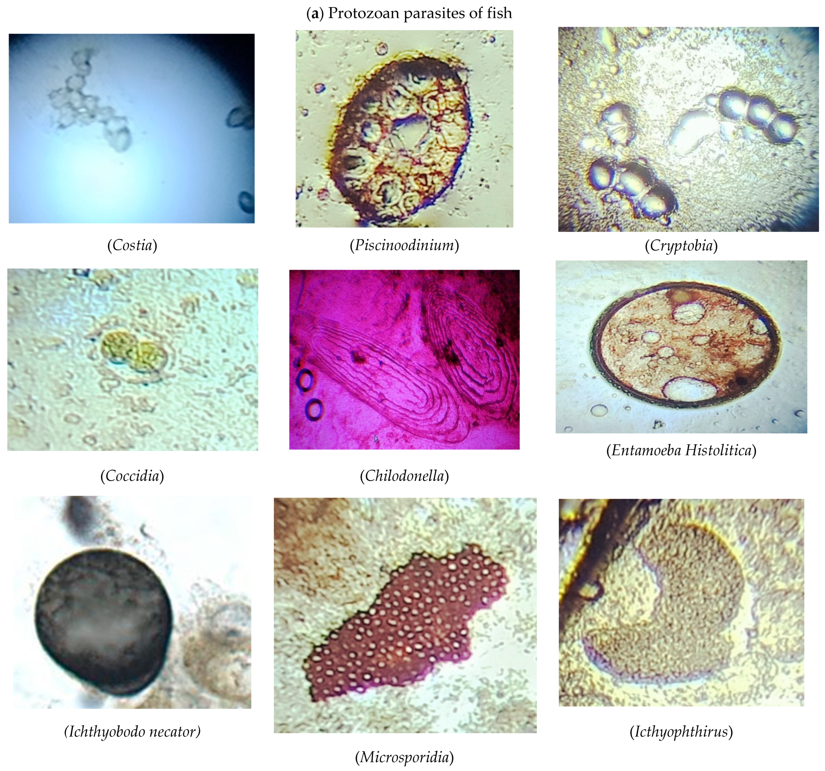
2.3. Examination of Protozoan Parasites

2.3.1. Study of External Protozoan Parasites

The length and weight of all the fish were recorded. For the study of ectoparasites, external organs of the fish such as skin, scales, fins, tails, etc., were examined with a hand lens. Permanent and wet mounts were prepared by scraping the dorsolateral surface of a fish with the dull side of a scalpel blade. Scrapings were obtained from near the base of all the fins [31]. To observe gill parasites, the operculum was removed from the fish. In the case of a small fish, the entire gill arch was removed, transferred to a slide, and examined under a microscope. In the case of a larger fish, the bony arch was also removed and examined under a stereomicroscope. Furthermore, the gills were also dissected from the branchial cavity and placed in a mixture of 1:4000 formalin solution for one hour. The decanted sediments were placed in a Petri dish and analyzed for protozoan parasites under a stereoscopic microscope at low and high magnifications [32]. Photographs of the identified external protozoan parasites are shown in Figure 1a.

2.3.2. Study of Internal Protozoan Parasites

The fish were dissected for the detection of internal protozoan parasites in different organs (heart, liver, intestine, kidney, and spleen). Each organ was removed and separately bathed with a physiological saline solution (0.7% NaCl solution). Intestinal parasites were examined after placing 1–2 drops of intestinal fluid on microscopic slides. In the case of a small fish, the intestinal tract was opened along its entire length and examined under a stereomicroscope by compressing a longer section of the intestine between the glass slides. Wet mounts were prepared using a scraping from the affected fish after fixing it in a 10% buffered neutral formalin solution. These samples were then stained with hematoxylin and counter-stained with eosin for examination under a microscope at both low and high magnification [32]. Photographs of the identified internal protozoan parasites are shown in Figure 1a.

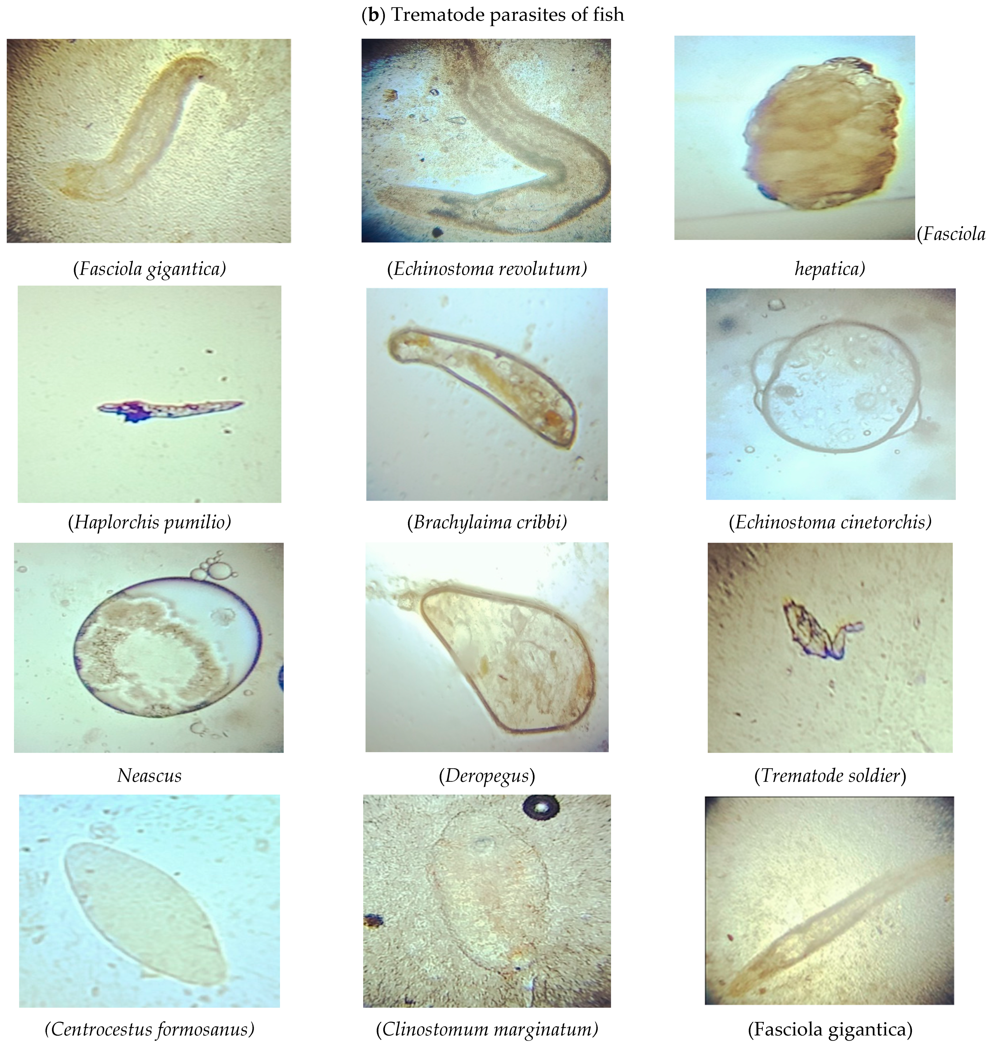
2.4. Examination of Trematode Parasites

2.4.1. Detection of Metacercariae

Two general methods were used to examine the foodborne trematode (FBT) metacercariae in second intermediate hosts.
  • The muscle compression technique.
  • The pepsin-HCL artificial digestion technique.

2.4.2. Muscle Compression

For this technique, samples were obtained from the muscles, intestine, kidney, heart, gills, and liver of the fish. Each sample was weighed to estimate the density of metacercariae. Each organ sample was compressed between two glass slides. Metacercariae were observed and identified under a stereo microscope. The number of metacercariae was counted and their density was calculated [33].

2.4.3. Pepsin-HCL Artificial Digestion Technique

Samples of fish muscles, intestines, kidneys, heart, gills, and liver were ground one by one with a mortar and pestle. The ground samples were transferred to artificial gastric juice {Conc. HCl (10 mL) + pepsin (10 g) + distilled water (1000 mL)}, mixed well, and incubated at 37 °C for two hours with occasional stirring and filtering. Saline (0.85%) was added, the supernatant was thrown away carefully, and the sediments were again washed until the supernatant became clear. A small amount of sediment was transferred to a Petri dish in which physiological saline (6–7 mL) was present. A stereomicroscope and light microscope were used to count and identify the metacercariae on the basis of the shape of cysts, size of suckers, and shape of the excretory bladder, according to [34,35,36]. Photographs of the identified trematode parasites are shown in Figure 1b.

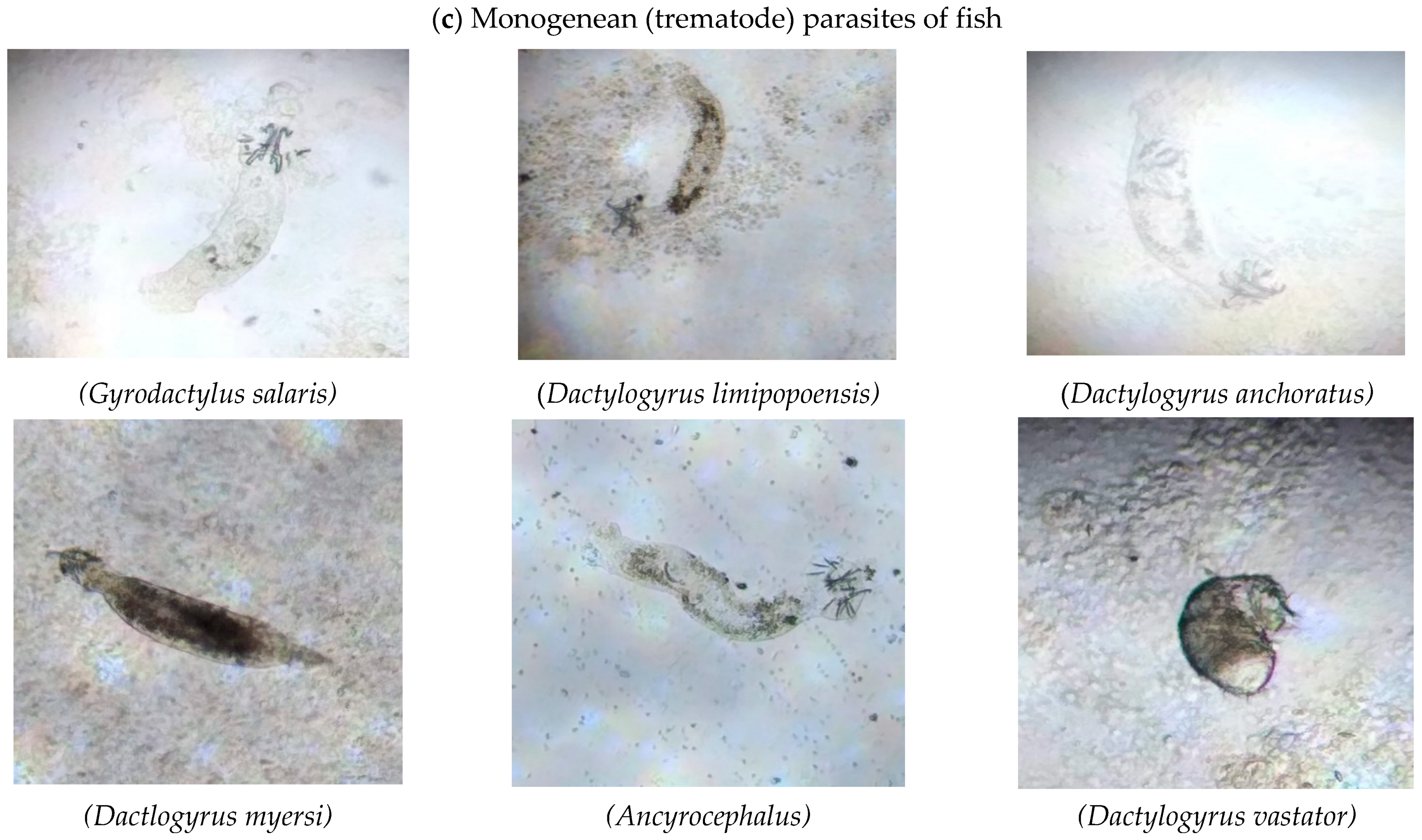
2.5. Examination of Monogenean Trematodes

A magnifying lens was used to check monogenean parasites on the external surface of fins, skin, and gills. Scalpel blades were used to obtain scrapings from the gills, skin, and fins for the examination of attached parasites. These scrapings were transferred to a clean microscopic slide to prepare wet mounts, which were examined under a stereomicroscope. From each gill filament, the gill arches were dissected using surgical scissors and then placed in Petri dishes for microscopic examination. Similarly, from the base of each fin, fin pieces were placed on slides, covered with coverslips, and then observed under a binocular microscope. Monogenean parasites were identified using sclerotized structures (anchors, transverse bar, vestigial ventral bar, hooks, and male copulatory organ) [31]. Photographs of the identified monogenean parasites are shown in Figure 1c.

2.6. Identification of Parasites

The identification of the examined protozoan, monogenean, and trematode parasite was completed using the standard keys [9,31,33,34,35,36,37].

2.7. Prevalence, Intensity, and Density of Parasites

The following formulas were used to estimate the prevalence, density, and intensity of parasites by following [38]:
P r e v a l e n c e = n o . o f   i n d i v i d u a l   o f   a   h o s t   i n f e c t e d   w i t h   a   p a r t i c u l a r   p a r a s i t e   s p e c i e s ÷ n o . o f   h o s t s   e x a m i n e d × 100
I n t e n s i t y = s u m   o f   i n d i v i d u a l s   o f   a   p a r t i c u l a r   p a r a s i t e   s p e c i e s   i n   a   s a m p l e   o f   a   h o s t ÷ t o t a l   o f   i n f e c t e d   i n d i v i d u a l s   o f   t h e   h o s t   i n   t h e   s a m p l e .
D e n s i t y = s u m   o f   i n d i v i d u a l s   o f   a   p a r t i c u l a r   p a r a s i t e   s p e c i e s   i n   a   s a m p l e   o f   h o s t s ÷ t o t a l   n o . o f   i n d i v i d u a l s   o f   h o s t   ( i n f e c t e d + u n i n f e c t e d )   i n   s a m p l e .

2.8. Statistical Analysis

Statistical analyses of the collected data were performed using statistical package for social sciences (SPSS) version 21.0. The chi-square test was used to compare the infection rate of parasites at different sites. A p-value < 0.05 was considered statistically significant for all analyses [39].

3. Results

During the harvesting season (2020–2021), nine species of freshwater fish were sampled from the three study sites. In total, 405 freshwater fish from these nine fish species (45 samples per fish species) were examined, including carp, catfish, and tilapia. The weight of the fish that were examined during this study was between 100 g and 3000 g. The results revealed that parasite prevalence was high in large-sized fish compared to small- or medium-sized fish.
Table 1: In total, 9 species of protozoan parasites, 11 species of trematodes, and six species of monogenean parasites were identified in different organs from seven freshwater fish species. Protozoan parasites were observed on the external and internal organs of fish such as the intestine, liver, kidneys, skin, and fins. Trematodes were found only in the intestine and gills. Monogeneans were found only on the external organs of fish, i.e., the skin, gills, and fins. Two fish species, Sperata seenghala and Notopterus notopterus, were identified as resistant species as none of the parasites was observed in either of these fish species.
Table 2: In total, 573 parasites were identified in 405 fish consisting of 210 protozoan parasites, 185 trematode parasites, and 178 monogenean parasites. The rate of parasite infection was calculated by counting the total number of parasite species for each class at a specific site. The chi-square (Χ2) statistic was used to analyze the relationship between parasites and a specific site. The level of significance was set at p < 5% (Table 2). The statistical analysis revealed that there was not any association between parasites and a specific site. The existence of parasites was not site-specific.
Table 3 lists the infection rate of individual fish species for each protozoan parasite including Chilodonella, Coccidia, Costia, Cryptobia, Entamoeba histolitica, Icthyophthirus multifillus, Microsporidia, Piscinoodinium, and Ichthyobodo necator. The number of infected fish with a certain number of protozoan parasites is also listed. The prevalence of infection, mean intensity, and mean abundance were calculated using the formulas mentioned in Section 2.6. Photographs of each protozoan parasite are shown with their names in Figure 1a.
Table 4 lists the infection rate of individual fish species for each trematode parasite including Brachylaima cribbi, Centrocestus formosanus, Clinostomum marginatum, Deropegus sp., Echinostoma cinetorchis, Echinostoma revolutum, Fasciola hepatica, Fasciola gigantica, Haplorchis pumilio, Neascus, and Trematode Soldier. The number of infected fish with a certain number of trematode parasites is also listed. The prevalence of infection, mean intensity, and mean abundance were calculated using the formulas mentioned in Section 2.6. Photographs of each trematode parasite are shown with their names in Figure 1b.
Table 5 lists the infection rate of individual fish species for each monogenean parasite including Ancyrocephalus sp., Dactylogyrus sp., Gyrodactylus sp. The number of infected fish with a certain number of monogenean parasites is also listed. The prevalence of infection, mean intensity, and mean abundance were calculated using the formulas mentioned in Section 2.6. Photographs of each monogenean parasite are shown with their names in Figure 1c.

4. Discussion

The significance of continuous surveillance of foodborne parasites and their epidemiological dispersion cannot be overstated in developing countries. Parasites cause pathogenic effects and financial damage to fish farming. The data in this study indicated the distribution of parasites in freshwater fish that were collected from three study sites along River Ravi (shown on the map). The fish diagnosed for parasite infestation were Labeo rohita, Hypophthalmichthys molitrix, Ctenopharyngodon idella, Cirrhinus mrigala, Cyprinus carpio, Wallagu attu, Notopterus notopterus, Sperata seenghala, and Oreochromis niloticus. During the fish investigation, nine protozoan parasites, 11 trematode parasites, and 6 monogenean parasites were observed under the microscope. A similar study was conducted in Turkey on the host–parasite relationship, which examined protozoa (14 species), monogenean (12), and trematode (15) parasites and some other taxa [11]. Similarly, another investigation was carried out in Bangladesh on the prevalence of protozoan and monogenean parasites in fish (H. molitrix, C. idella, C. carpio, B. gonionotus, C. catla, L. rohita and C. cirrhosis) by [1], and two species of protozoan parasites (Trichodina and Chilodonella) and one species of monogenean parasites (Dactylogyrus) were examined. The results of the preSSsent study showed some dissimilarities with [1] because Trichodina was not observed in any fish, and some other parasites were also identified in our study. Another study was performed by [40] on large-sized silver and common carp. Their findings identified two protozoans (Trichodina and Ichthyophthirius multifiliis), two monogeneans (Dactylogyrus and Gyrodactylus), and two crustacean parasites, which is pertinent to this study because the majority of parasites were seen in large-sized fish. The photographs of parasites in Figure 1 were similar to the findings of [9,31,33,35,36,37,38]. In this study, the majority of parasites were found in L. rohita, followed by H. molitrix, C. idella, O. niloticus, C. carpio, and W. attu.
This study examined protozoan parasites including Coccidia, Piscinoodinium, Microsporidia, Icthyophthirus multifillus, Costia, Ichthyobodo necator, Cryptobia, Chilodonella, and Entamoeba histolitica. The prevalence of infection, mean intensity, and abundance of protozoan parasites that infected the fish is mentioned in Table 3. In [41], an investigation was carried out on 11 protozoan parasites that had infected Clarias gariepinus. Out of the eleven protozoan parasites, four parasites were also identified in this study, including Piscinoodinium, Coccidia, Chilodonella, and Microsporidians. Another protozoan parasite, Costia, was also identified in the findings of [13,41]. In [42,43,44,45,46,47,48], it was reported that Trichodina and Ichthyophthirius multifiliis were the prevalent protozoan parasites on the skin and gills of cyprinid fish. In the present study, Ichthyophthirius was observed in rohu and silver carp. In [49], the authors described chronic infections of catfish by the protozoan parasite Ichthyophthirius on skin and gills, Ichthyobodo and Chilodonella on the skin, and Cryptobia in the stomach and intestine. As compared to [49], the present study identified that Ichthyophthirius was present in intestine, Chilodonella was identified on skin, and Cryptobia was identified in the intestine. In [11], the authors identified harmful protozoan parasites and their infections, such as Trichodina, Tetrahymena, Ichthyophthirius, and Ichthyobodo necator. This work showed closeness with the current study because the protozoan parasite Ichthyophthirius and its prevalence was identified in different fish species. Our results relate to the findings of [50], which found most protozoan parasites in the intestine. A decline in protozoan parasites in the stomach was found to be due to the acidic nature of the stomach because protozoan parasites occupy a specific pH medium [51,52].
This study examined monogenean parasites including Dactylogyrus limipopoensis, Dactylogyrus anchoratus, Dactylogyrus myersi, Dactylogyrus vastator, Gyrodactylus salaris, and Ancyrocephalus (mentioned in Table 3). The highest prevalence of parasites was observed in Labeo rohita. Gyrodactylus salaris was first discovered in farmed salmonids in Romania [53,54]. In the present study, Gyrodactylus was observed in carp. According to [47], 70 species of Dactylogurus were reported in both wild and farmed common carp in Iran. In the present study, four species of Dactylogyrous (D. limipopoensis, D. anchoratus, D. myersi and D. vastator) were observed in the following freshwater fish from River Ravi: Hypophthalmichthys molitrix, Labeo rohita, Cyprinus carpio, and Oreochromis niloticus. A study by [44] reported that D. anchoratus and D. extensus were observed within a specific range of water quality parameters. Furthermore, [55] described that infection with a monogenean parasite could be dangerous and harmful for fries in hatcheries. The results of the present study also coincide with the findings of [40], which reported that two monogeneans, Dactylogyrus ssp. and Gyrodactylus, were observed in H. molitrix and C. carpio. These results are in conformance with [46], who investigated Dactylogyrus sp. and Gyrodactylus in Channa pleurophtalma in terms of their dominance on the gills.
This study examined trematode parasites including Fasciola hepatica, Trematode Soldier, Haplorchis pumilio, Brachylaima cribbi, Echinostoma cinetorchis, Clinostomum marginatum, Deropegus sp., Neascus sp., Fasciola gigantica, Echinostoma revolutum, and Centrocestus formosanus. A microscopic examination of endoparasites of commercially important fish from Egypt was performed by El-shahawy, in which only one trematode and two cestodes were identified, while in the present study, none of the cestodes was observed. A study by [35] identified the infection of many fish species with metacercariae from four species of trematodes. In the present study, two trematode parasites were identified in freshwater fish that resemble the findings of [35], including Haplorchis pumilio in the intestine of grass carp [56] and Centrocestus formosanus in the intestine of silver carp [57]. Haplorchis pumilio was detected in the intestine of grass carp from the Lower Bari Doab Canal. Thien et al. (2009) analyzed the same parasites in catfish. Metacarcaria of C. formosanus were also observed in fish during a study by [58,59,60]. Two species belonging to two genera of zoonotic trematode parasites were recorded with different prevalence rates (Centrocestus formosanus, Centrocestus sinensis, Haplorchis taichui, and Haplorchis pumilio) in various freshwater fish species from local markets in northern Vietnam [61], while in the present study, only one species from both genera was observed. Two trematode parasites, Clinostomum marginatum and Neascus sp., observed in the present study also relate to the findings of [62]. In the present study, it was also found that large-sized fish had more parasites than smaller fish. This parasitic load in bigger fish was suggested to result from being exposed to a variety of parasites while foraging for food [42]. It was also found that the majority of parasites was observed at the Okara site.

5. Conclusions

A wide diversity of protozoan, trematode, and monogenean parasites was observed in different organs of freshwater fish species. The highest burden was of protozoans in carps, which can cause serious detrimental effects on fish health and the economy. Documentation of these parasites is important to determine their ecological role and the economic value of the losses they cause in natural waters. The identification of fish parasites and their density and diversity is very important to correctly determine the infectious agents and to assess the safety techniques used to improve natural fauna and flora in better way.

Author Contributions

Methodology, B.N.K., A.A., I.A., H., M.S.A., I.K., A.A.K.K., B.A., N.M. and M.A.; Software, R.A.M.; Investigation, A.S.; Resources, M.H.-u.-R.; Supervision, F.A. All authors have read and agreed to the published version of the manuscript.

Funding

The authors extend their appreciation to Researchers Supporting Project number (RSP2023R119), King Saud University, Riyadh, Saudi Arabia, for funding this work.

Institutional Review Board Statement

All methods in this work were carried out in accordance with the “National Biosafety Guidelines” of the Pakistan Environmental Protection Agency, Ministry of Environment, Government of Pakistan. URL: http://www.environment.gov.pk/images/guidelines/BiosftyGlines2005.pdf (accessed on 10 July 2023). All experimental protocols were approved by the Ethical Review Committee, University of Veterinary and Animal Sciences (UVAS), Pakistan. URL: http://www.uvas.edu.pk/doc/directorates/2014/Research/Guidelines-Protocols.pdf (accessed on 10 July 2023).

Data Availability Statement

Not applicable.

Conflicts of Interest

The authors declare no conflict of interest.

References

  1. Hossain, M.K.; Ahmed, A.T.; Khan, M.H.; Rafiquzzaman, S.M.; Begum, F.; Islam, M.A. Distribution, prevalence and intensity of protozoan and monogenean parasites of carp fingerlings in selected nursery ponds. Bangladesh J. Fish. Res. 2007, 11, 37–44. [Google Scholar]
  2. Dogiel, V. Parasitology of Fishes; Oliver and Boyd: London, UK, 1956. [Google Scholar]
  3. Cavalcanti, E.T.S.; Takemoto, R.M.; Alves, L.C.; Chellappa, S. First report of metazoan fish parasites with zoonotic potential in Scomberomorus brasiliensis and Trichiurus lepturus from the coastal waters of Rio Grande do Norte, Brazil. Mar. Biodivers. Rec. 2012, 5, e40. [Google Scholar]
  4. Chai, J.Y.; Murrell, K.D.; Lymbery, A.J. Fish-borne parasitic zoonoses: Status and issues. Parasitol. Int. 2005, 35, 1233–1254. [Google Scholar]
  5. Seppala, O.; Karvonen, A.; Valtonen, E.T. Manipulation of fish host by eye flukes in relation to cataract formation and parasite infectivity. Anim. Behav. 2005, 70, 889–894. [Google Scholar]
  6. Bedasso, G.T. Study on the prevalence and temporal abundance of parasites of fishes in Lake Elan. Int. J. Fish. Aquac. 2015, 3, 265–269. [Google Scholar]
  7. Moyle, P.B.; Cech, J.J. An Introduction to Ichthyology; University of California: Los Angeles, CA, USA, 2004. [Google Scholar]
  8. Tasawar, Z.; Umer, K.; Hayat, C.S. Observations on lernaeid parasites of Catla catla from a fish hatchery in Muzaffargarh, Pakistan. Pak. Vet. J. 2007, 27, 17. [Google Scholar]
  9. Lom, J.; Dykova, I. Protozoan Parasites of Fishes. Dev. Aquac. Fish. Sci. 1992, 26, 312–315. [Google Scholar]
  10. Roberts, R.J. The parasitology of teleosts. In Fish Pathology; W.B. Saunders: London, UK, 2001; pp. 254–296. [Google Scholar]
  11. Kayis, S.; Ozcelep, T.; Capkin, E.; Altinok, I. Protozoan and Metazoan Parasites of Cultured Fish in Turkey and their Applied Treatments. Isr. J. Aquac.—Bamidgeh 2009, 61, 93–102. [Google Scholar]
  12. Schaperclaus, W. Diseases caused by ciliates. In Fish Diseases; Amerind Publishing: New Delhi, India, 1991; pp. 702–725. [Google Scholar]
  13. Abowei, J.F.N.; Briyai, O.F.; Bassey, S.E. A Review of Some Basic Parasite Diseases in Culture Fisheries Flagellids, Dinoflagellides and Ichthyophthriasis, Ichtyobodiasis, Coccidiosis, Trichodiniasis, Heminthiasis, Hirudinea Infestation, Crustacean Parasite and Ciliates. Br. J. Pharmacol. 2011, 2, 213–226. [Google Scholar]
  14. Meyer, F.P.; Hoffman, G.L. Parasites of Freshwater Fishes; II, Protozoa 3. Ichthyophthirius multifiliis; Leaflet No. 2; U.S. Department of the Interior U.S. Fish and Wildlife Service Fish Disease: Washington, DC, USA, 1974.
  15. Li, K.; Clausen, J.H.; Murrell, K.D.; Liu, L.; Dalsgaard, A. Risks for fishborne zoonotic trematodes in Tilapia production systems in Guangdong province, China. Vet. Parasitol. 2013, 198, 223–229. [Google Scholar]
  16. Furst, T.; Keiser, J.; Utzinger, J. Global burden of human food-borne trematodiasis: A systematic review and meta-analysis. Lancet Infect. Dis. 2012, 12, 210–221. [Google Scholar]
  17. Park, C.W.; Kim, J.S.; Joo, H.S.; Kim, J. A human case of Clinostomum complanatum infection in Korea. Korean J. Parasitol. 2009, 47, 401. [Google Scholar] [PubMed]
  18. Hechinger, R.F.; Lafferty, K.D. Host diversity begets parasite diversity: Bird final hosts and trematodes in snail intermediate hosts. Proc. R. Soc. B Biol Sci. 2005, 272, 1059–1066. [Google Scholar]
  19. Chi, T.T.K.; Murrell, K.D.; Madsen, H.; Khue, N.V.; Dalsgaard, A. Fishborne zoonotic trematodes in raw fish dishes served in restaurants in Nam Dinh Province and Hanoi, Vietnam. J. Food Prot. 2009, 72, 2394–2399. [Google Scholar]
  20. Sripa, J.; Kiatsopit, N.; Piratae, S. Prevalence of trematode larvae in intermediate hosts: Snails and fish in Ko Ae sub-district of Khueang Nai, Ubon Ratchathani province, Thailand. Southeast Asian J. Trop. Med. Public Health. 2016, 47, 399–409. [Google Scholar] [PubMed]
  21. Thuy, D.T.; Kania, P.; Buchmann, K. Infection status of zoonotic trematode metacercariae in Sutchi catfish (Pangasianodon hypophthalmus) in Vietnam: Associations with season, management and host age. Aquaculture 2010, 302, 19–25. [Google Scholar]
  22. Pardeshi, P.R.; Hiware, C.J.; Wangswad, C. Histopathology of the liver of Mastacembelus armatus (Lecepede, 1800) due to trematode parasite, Allocreadium khami n. sp. J. Parasit. Dis. 2012, 36, 53–55. [Google Scholar]
  23. Kearn, G.C. Monogeneans-the ultimate fish parasites. Monogenea 2011, 58, 28–32. [Google Scholar]
  24. Sures, B.; Nachev, M.; Selbach, C.; Marcogliese, D.J. Parasite responses to pollution: What we know and where we go in ‘Environmental Parasitology. Parasit. Vectors. 2017, 10, 65. [Google Scholar]
  25. Ogawa, K. Diseases of cultured marine fishes caused by Platyhelminthes (Monogenea, Digenea, Cestoda). Parasitology 2015, 142, 178–195. [Google Scholar]
  26. Rubio-Godoy, M. Fish host-monogenean parasite interactions, with special reference to Polyopisthocotylea. In Advances in the Immunobiology of Parasitic Disease; Research Signpost: Kerala, India, 2007; pp. 91–109. [Google Scholar]
  27. Mishra, S.P. A new monogenean Hamatopeduncularia saketensisn. sp. from fresh water fish Wallago attu. Int. J. Adv. Multidiscip. Res. Dev. 2014, 1, 244–246. [Google Scholar]
  28. Zhi, T.; Huang, C.; Sun, R.; Zheng, Y.; Chen, J.; Xu, X.; Brown, C.L.; Yang, T. Mucosal immune response of Nile tilapia Oreochromis niloticus during Gyrodactylus cichlidarum infection. Fish Shellfish. Immunol. 2020, 106, 21–27. [Google Scholar] [CrossRef] [PubMed]
  29. Cable, J.; Scott, E.C.G.; Tinsley, R.C.; Harris, P.D. Behavior favoring transmission in the viviparous monogenean Gyrodactylus turnbulli. J. Parasitol. 2002, 88, 183–184. [Google Scholar] [CrossRef]
  30. Sahoo, P.K.; Paul, A.; Sahoo, M.K.; Pattanayak, S.; Kumar, P.; Das, B.K. Incidences of infectious diseases in freshwater aquaculture farms of eastern India: A passive surveillance based study from 2014–2018. J. Aquac. Res. Dev. 2020, 11, 579. [Google Scholar]
  31. Hoffman. NWFHS Laboratory Manual. In Parasitology; Becky Lasee Lacrosse Fish Health Center: Washington, DC, USA, 2004; Chapter 8; pp. 18–19. [Google Scholar]
  32. Weber, E.P., 3rd; Govett, P. Parasitology and necropsy of fish. Compend. Yardley PA 2009, 31, 12. [Google Scholar]
  33. Sohn, W.M. Fish-borne zoonotic trematode metacercariae in the Republic of Korea. Korean J. Parasitol. 2009, 47, 103. [Google Scholar] [CrossRef]
  34. Myint, E.E.P.; Sereemaspun, A.; Rocklov, J.; Nithikathkul, C. Discovery of Carcinogenic Liver Fluke Metacercariae in Second Intermediate Hosts and Surveillance on Fish-Borne Trematode Metacercariae Infections in Mekong Region of Myanmar. Int. J. Environ. Health Res. Public. Health. 2020, 17, 4108. [Google Scholar] [CrossRef]
  35. Chai, J.Y.; Sohn, W.M.; Na, B.K.; Park, J.B.; Jeoung, H.G.; Hoang, E.H.; Htoon, T.T.; Tin, H.H. Zoonotic trematode metacercariae in fish from Yangon, Myanmar and their adults recovered from experimental animals. Korean J. Parasitol. 2017, 55, 631. [Google Scholar]
  36. Magda, A.A.; Amer, O.H.; Maysa, A.I.A.; Merwad, A.M.A.; Hassan, M.S.A. Role of Atherina fi sh in transmitting some trema todes of public health importance. In Proceedings of the 3rd Global Fisheries and Aquaculture Research Conference, Foreign Agricultural Relations (FAR), Cairo, Egypt, 29 November–1 December 2010; pp. 120–133. [Google Scholar]
  37. Klinger, R.; Floyd, R.F. Introduction to Freshwater Fish Parasites; IFAS Extension, University of Florida: Gainesville, FL, USA, 1998; Volume 1, pp. 1–14. [Google Scholar]
  38. El-Shahawy, I.S.; El-Seify, M.O.; Metwally, A.M.; Fwaz, M.M. Survey on endoparasitic fauna of some commercially important fishes of the River Nile, southern of Egypt (Egypt). Rev. De Med. Vet. 2017, 168, 126–134. [Google Scholar]
  39. Ashmawy, K.I.; Hiekal, F.A.; Abo-Akadda, S.S.; Laban, N.E. The inter-relationship of water quality parameters and fish parasite occurrence. Alexandr. J. Veter. Sci. 2018, 59, 97. [Google Scholar] [CrossRef]
  40. Prasad, A.; Yadav, S.; Limbu, J.H. Identification of Ecto-parasites in silver carp (Hypophthalmichthys molitrix) and common carp (Cyprinus carpio) at fishery development Center Bhairahawa, Rupandehi, Nepal. Int. J. Fish. Aquat. Stud. 2018, 6, 116–120. [Google Scholar]
  41. Eyiseh, T.E.; Amos, S.O.; Thomas, I. Incidence of parasitic infection in adult and juvenile Clarias gariepinus in a private fish farm, Yola, Adamawa state. Int. J. Fish. Aquat. Stud. 2022, 10, 38–45. [Google Scholar] [CrossRef]
  42. Emere, M.C.; Egbe, N.E.L. Protozoan parasites of Synodonits clarias (a fresh water fish in river Kaduna). Best. J. 2006, 3, 58–64. [Google Scholar]
  43. Hines, R.S.; Spira, D.T. Ichthyophthiriasis in the mirror carp Cyprinus carpio (L.). III. pathology. J. Fish Biol. 1974, 6, 189–196. [Google Scholar] [CrossRef]
  44. Jalali, B.; Barzegar, M. Fish parasites in Zarivar Lake. J. Agric. Sci. Technol. 2018, 8, 47–58. [Google Scholar]
  45. Barzegar, M.; Raeisi, M.; Bozorgnia, A.; Jalali, B. Parasites of the eyes of fresh and brackish water fishes in Iran. Iran. J. Vet. Res. 2008, 9, 256–261. [Google Scholar]
  46. Borji, H.; Naghibi, A.; Nasiri, M.R.; Ahmadi, A. Identification of Dactylogyrus spp. and other parasites of common carp in northeast of Iran. J. Paras. Dis. 2012, 36, 234–238. [Google Scholar] [CrossRef] [Green Version]
  47. Jalali, J.B. Parasites and Parasitic Diseases of Freshwater Fishes of Iran; Iranian Fisheries Organization Press: Tehran, Iran, 1998. [Google Scholar]
  48. Raissy, M.; Ansari, M.; Moumeni, M.; Goudarzi, M.A.; Sohrabi, H.R.; Rashedi, M. An epizootic of Ichthyophthiriasis among fishes in Armand River, Iran. J. Cell Anim. Boil. 2010, 4, 150–153. [Google Scholar]
  49. Omeji, S.; Solomon, S.G.; Idoga, E.S. A comparative study of the common protozoan parasites of Clarias gariepinus from the wild and cultured environments in Benue State, Nigeria. J. Parasitol. Res. 2011, 2011, 916489. [Google Scholar] [CrossRef] [Green Version]
  50. Smith, A.C. Introduction to Parasitology; Willey: New York, NY, USA, 1981. [Google Scholar]
  51. Noble, E.R.; Noble, G.A. Parasitology: The biology of animal parasites. In Parasitology, 3rd ed.; Lea & Febiger: Philadelphia, PA, USA, 1971. [Google Scholar]
  52. Adebanjo, A.O. A Survey of Parasites of Clarias lazera in Dundaye area of Rima River; Sokoto, Project, Zoology Unit of Biological Science, Usman Danfodio University: Sokoto, Nigeria, 1979. [Google Scholar]
  53. Rokicka, M.; Lumme, J.; Ziętara, M.S. Identification of Gyrodactylus ectoparasites in Polish salmonid farms by PCR-RFLP of the nuclear ITS segment of ribosomal DNA (Monogenea, Gyrodactylidae). Acta Parasitol. 2007, 52, 185–195. [Google Scholar] [CrossRef]
  54. Paladini, G.; Gustinelli, A.; Fioravanti, M.L.; Hansen, H.; Shinn, A.P. The first report of Gyrodactylus salaris Malmberg, 1957 (Platyhelminthes, Monogenea) on Italian cultured stocks of rainbow trout (Oncorhynchus mykiss Walbaum). Vet. Parasitol. 2009, 165, 290–297. [Google Scholar] [CrossRef] [PubMed] [Green Version]
  55. Paperna, I. Parasites, Infections and Diseases of Freshwater Fishes in Africa; CIFA Technical Paper; Department of Fisheries, CIFA, FAO: Rome, Italy, 1980. [Google Scholar]
  56. Xu, L.Q.; Yu, S.H.; Chen, Y.D. Clonorchiasis sinensis in China. Parasitology 2005, 1, 1–26. [Google Scholar]
  57. Yu, Z.J.; Cao, H.X.; Lv, G. The current prevalence of clonorchiasis in China. J. Hainan. Med. Uni. 2003, 9, 248–251. [Google Scholar]
  58. Scholz, T.; Ditrich, O.; Giboda, M. Larval stages of medically important flukes (Trematoda) from Vientiane province, Laos Part 1. metacercariae. Ann. De Parasitol. Hum. Et Comp. 1990, 65, 238–243. [Google Scholar] [CrossRef] [Green Version]
  59. Rim, H.J.; Sohn, W.M.; Yong, T.S.; Eom, K.S.; Chai, J.Y.; Min, D.Y.; Lee, S.H.; Hoang, E.H.; Phommasack, B.; Insisengmay, S. Fishborne trematode metacercariae detected in freshwater fish from Vientiane Municipality and Savannakhet Province, Lao PDR. Korean J. Parasitol. 2008, 46, 253. [Google Scholar] [CrossRef] [PubMed]
  60. Han, E.T.; Shin, E.H.; Phommakorn, S.; Sengvilaykham, B.; Kim, J.L.; Rim, H.J.; Chai, J.Y. Centrocestus formosanus (Digenea: Heterophyidae) encysted in the freshwater fish, Puntius brevis, from Lao PDR. Korean J. Parasitol. 2008, 46, 49. [Google Scholar]
  61. Dai, F.; Hong, S.J.; Pak, J.H.; Le, T.H.; Choi, S.H.; Na, B.K.; Sohn, W.M. High prevalence of Clonorchis sinensis and other zoonotic trematode metacercariae in fish from a local market in Yen Bai province, Northern Vietnam. Korean J. Parasitol. 2020, 58, 333. [Google Scholar] [CrossRef]
  62. Waruiru, R.M.; Mbuthia, P.G.; Wanja, D.W.; Mwadime, J.M. Prevalence, intensity and influence of water quality on parasites of farmed fish in Kirinyaga County, Kenya. Livest Res. Rural Devel. 2020, 32, 164. [Google Scholar]
Figure 1. (a) Photographs of protozoan parasites along with their names, which were identified (Table 1) in the freshwater fish from River Ravi. (b) Photographs and names of trematode parasites that were identified in the freshwater fish from River Ravi. (c) Photographs and names of monogenean parasites that were identified in the freshwater fish from the three studied sites in River Ravi.
Microorganisms 11 01940 g001aMicroorganisms 11 01940 g001bMicroorganisms 11 01940 g001c
Table 1. Parasites and their infection sites in the fish species obtained from different harvesting localities in River Ravi.
Identified ParasitesHostInfection SiteLocality
Protozoans
Microsporidia
(Balbiani, 1882)
Labeo rohitaLiverLBD Canal
Icthyophthirus multifillus
(Foquet, 1876)
Intestine
Costia
(Henneguy, 1883)
Cirrhinus mrigalaKidney
Ichthyobodo necator
(Henneguy, 1883)
Cyprinus carpioLiver
Cryptobia
(E. Nohynkova, 1984)
Labeo rohitaIntestineHead Balloki
Chilodonella
(Kiernik, 1909)
Skin
Entamoeba histolitica
(Chatton, 1909)
Ctenopharyngodon idellaLiver
Coccidia
(WT. Johnson, 1892)
Labeo rohitaGillsOkara
IchthyophithirusHypophthalmichthys molitrixIntestine
ChilodonellaSkin
Piscinoodinium
(Schaperclaus, 1954)
Intestine
Entamoeba histoliticaWallagu attuLiver
Trematodes
Fasciola hepatica
(Linnaeus, 1758)
Labeo rohitaGillsLBD Canal
Trematode Soldier
(Gibson, 1996)
Intestine
Haplorchis pumilio
(Looss, 1896)
Ctenopharyngodon idellaIntestine
Brachylaima cribbi
(A.R. Butcher, 2003)
Echinostoma cinetorchis
(Ando & Ozaki, 1923)
Labeo rohitaGillsHead Balloki
Clinostomum marginatum
(Rudolphi, 1819)
Hypophthalmichthys molitrixIntestine
Deropegus sp.
(McCauley, 1961)
Ctenopharyngodon idellaIntestine
Neascus
(Hoffman, 1955)
Fasciola gigantica
(Cobbold, 1853)
Labeo rohitaGillsOkara
Echinostoma revolutum
(Frohlich, 1802)
Centrocestus formosanus
(Nishigori, 1924)
Hypophthalmichthys molitrixIntestine
Monogeneans
Gyrodactylus
(Malmberg, 1957)
Ctenopharyngodon idellaGillsLBD Canal
Dactylogyrus
(Kulwiec, 1927)
Hypophthalmichthys molitrixFins
GyrodactylusOreochromis niloticusSkin
DactylogyrusLabeo rohitaSkinHead Balloki
DactylogyrusFin
Ancyrocephalus
(Creplin, 1839)
Hypophthalmichthys molitrixGills
GyrodactylusCyprinus carpioGillsOkara
DactylogyrusSkin
AncyrocephalusCtenopharyngodon idellaGills
DactylogyrusOreochromis niloticusSkin
Table 2. The rate of parasite infection at three different study sites in River Ravi.
Study SiteObserved ParasitesNumberInfection RateΧ2p-Value
Protozoans
LBDMicrosporidia1636%16.3330.569
Icthyophthirus multifillus12
Costia20
Ichthyobodo necator28
Head BallokiCryptobia1621%
Chilodonella18
Entamoeba histolitica10
OkaraCoccidia2043%
Ichthyophithirus10
Chilodonella20
Piscinoodinium32
Entamoeba histolitica8
Treematodes
LBDFasciola hepatica1534.5%220.341
Trematode Soldier20
Haplorchis pumilio12
Brachylaima cribbi17
Head BallokiEchinostoma cinetorchis2030%
Clinostomum marginatum12
Deropegus sp.16
Neascus8
OkaraFasciola gigantica2335%
Echinostoma revolutum30
Centrocestus formosanus12
Monogeneans
LBDGyrodactylus3032.5%230.310
Dactylogyrus28
Head ballokiDactylogyrus2025%
Ancyrocephalus25
OkaraGyrodactylus2042%
Dactylogyrus27
Ancyrocephalus28
Table 3. Infection rate of each fish species for each protozoan parasite.
ParasiteHost (n = 45/Species)Infected Fish
(%)
Parasite
Number
(N)
Prevalence of Infection
(p)
Mean Intensity
(MI)
Mean Abundance
(MA)
ChilodonellaLabeo rohita10 (0.22)1822.21.80.4
Hypophthalmichthys molitrix8 (0.17)2017.62.50.4
CoccidiaLabeo rohita15 (0.33)2033.21.330.4
CostiaCirrhinus mrigala19 (0.42)2042.21.050.4
CryptobiaLabeo rohita10 (0.22)1622.21.60.35
Entamoeba histoliticaCtenopharyngodon idella10 (0.22)1022.210.22
Wallagu attu4 (0.08)848.820.17
Icthyophthirus multifillusLabeo rohita15 (0.33)1233.20.80.26
Hypophthalmichthys molitrix5 (0.11)101120.22
MicrosporidiaLabeo rohita12 (0.26)1626.91.30.35
PiscinoodiniumHypophthalmichthys molitrix25 (0.55)3255.41.280.71
Ichthyobodo necatorCyprinus carpio18 (0.4)28401.550.62
Table 4. Infection rate of each fish species for each trematode parasite.
ParasiteHost (n = 45/Species)Infected
Fish
(%)
Parasite Number (N)Prevalence of Infection
(p)
Mean Intensity (MI)Mean Abundance (MA)
Brachylaima cribbiCtenopharyngodon idella12
(0.26)
1726.61.410.36
Centrocestus formosanusHypophthalmichthys molitrix10
(0.22)
1222.21.20.26
Clinostomum marginatumHypophthalmichthys molitrix11
(0.24)
1224.41.090.26
Deropegus sp.Ctenopharyngodon idella14
(0.31)
16311.140.34
Echinostoma cinetorchisLabeo rohita18
(0.4)
20401.110.44
Echinostoma revolutumLabeo rohita26
(0.57)
3057.61.150.66
Fasciola hepaticaLabeo rohita12
(0.26)
1526.61.250.32
Fasciola giganticaLabeo rohita19
(0.42)
2342.21.210.5
Haplorchis pumilioCtenopharyngodon idella10
(0.22)
1222.21.20.26
NeascusCtenopharyngodon idella7
(0.15)
815.41.140.16
Trematode SoldierLabeo rohita15
(0.33)
2033.21.330.44
Table 5. Infection rate of each fish species for each monogenean parasite.
ParasiteHost (n = 45/Species)Infected Fish
(%)
Parasite
Number
(N)
Prevalence of Infection
(p)
Mean Intensity
(MI)
Mean Abundance
(MA)
GyrodactylusCtenopharyngodon idella15 (0.33)1233.30.80.26
Ancyrocephalus15 (0.33)1033.30.60.22
DactylogyrusHypophthalmichthys molitrix38 (0.84)2484.40.630.53
Ancyrocephalus38 (0.84)2084.40.50.44
DactylogyrusLabeo rohitaza40 (0.88)4688.81.151
GyrodactylusCyprinus carpio23 (0.51)15510.650.33
Dactylogyrus23 (0.51)13510.50.28
GyrodactylusOreochromis
Niloticus
15 (0.33)1033.30.60.22
Dactylogyrus15 (0.33)833.30.530.17
Disclaimer/Publisher’s Note: The statements, opinions and data contained in all publications are solely those of the individual author(s) and contributor(s) and not of MDPI and/or the editor(s). MDPI and/or the editor(s) disclaim responsibility for any injury to people or property resulting from any ideas, methods, instructions or products referred to in the content.

