Article

Ocular Bacterial Infections: A Ten-Year Survey and Review of Causative Organisms Based on the Oklahoma Experience

by
Roger A. Astley
1,
Md Huzzatul Mursalin
1,
Phillip S. Coburn
1,
Erin T. Livingston
2,
James W. Nightengale
1,
Eddy Bagaruka
3,
Jonathan J. Hunt
3 and
Michelle C. Callegan
1,2,4,*
1
Department of Ophthalmology, University of Oklahoma Health Sciences Center, Oklahoma City, OK 73104, USA
2
Department of Microbiology and Immunology, University of Oklahoma Health Sciences Center, Oklahoma City, OK 73104, USA
3
Department of Biology, Oklahoma Christian University, Edmond, OK 73013, USA
4
Dean McGee Eye Institute, Oklahoma City, OK 73104, USA
*
Author to whom correspondence should be addressed.
Microorganisms 2023, 11(7), 1802; https://doi.org/10.3390/microorganisms11071802
Submission received: 24 May 2023 / Revised: 29 June 2023 / Accepted: 10 July 2023 / Published: 13 July 2023
(This article belongs to the Special Issue Ocular Infections and Microbiota in Health and Disease 2.0)

Abstract

:
Ocular infections can be medical emergencies that result in permanent visual impairment or blindness and loss of quality of life. Bacteria are a major cause of ocular infections. Effective treatment of ocular infections requires knowledge of which bacteria are the likely cause of the infection. This survey of ocular bacterial isolates and review of ocular pathogens is based on a survey of a collection of isolates banked over a ten-year span at the Dean McGee Eye Institute in Oklahoma. These findings illustrate the diversity of bacteria isolated from the eye, ranging from common species to rare and unique species. At all sampled sites, staphylococci were the predominant bacteria isolated. Pseudomonads were the most common Gram-negative bacterial isolate, except in vitreous, where Serratia was the most common Gram-negative bacterial isolate. Here, we discuss the range of ocular infections that these species have been documented to cause and treatment options for these infections. Although a highly diverse spectrum of species has been isolated from the eye, the majority of infections are caused by Gram-positive species, and in most infections, empiric treatments are effective.

1. Introduction

The eye is arguably the most vulnerable organ in the human body. Despite being exposed to organisms, allergens, and physical insults from the external environment, the eye must maintain a healthy and transparent surface in order to allow the retina to be exposed to light for proper vision. The eye must also refrain from responding to these threats as other organs do because those responses can be damaging to the eye’s purpose of sensing light. The surface of the eye is also host to a distinct microbiome which serves to control pathogen growth and provides protection without instigating unnecessary inflammation. Under normal physiological conditions, ocular barriers such as the lids, tear film, and antimicrobial factors physically protect the eye, and the innate and adaptive immune systems, along with the microbiome, prevent the growth of harmful organisms [1,2]. When these protections fail due to systemic illness, physical insult, contact lens wear, or various environmental insults, infection may result [1,2,3]. That said, the normal flora is the predominant source of pathogens that cause bacterial conjunctivitis [4,5], keratitis [6], and postoperative endophthalmitis [7,8].
When an ocular infection does occur, prompt and effective treatment is necessary to prevent damage from both the infection and the immune response. Clinical decisions affecting the management of ocular infections are based on the identification of the pathogen. When culture results return, the rational question is, “is this bacterium a pathogen or a commensal?” Some species of bacteria are always viewed as pathogens, but many ocular bacteria can be pathogenic or commensal depending on the ocular conditions. That is where the science and art of determining an empirical treatment intersect. Knowledge of normal and pathogenic ocular bacteria is vital for prompt and effective treatment [7,9]. This paper reviews common, uncommon, and very rare ocular bacterial isolates and their pathogenic potential and reports comparisons with species included in a collection of ocular isolates banked over a ten-year span.
The data presented below were compiled from a survey of a collection of bacterial species in the Ocular Isolate Bank located at the Dean McGee Eye Institute in Oklahoma City, Oklahoma. Collected isolates from ocular bacterial infections were dated from March 2011 to March 2022. Clinical data were not available in the information provided for the survey, but anatomic location information was available for 71.9% of isolates. Original bacterial identification was performed offsite using a Bruker MALDI-Biotyper and confirmed when necessary in-house using routine microbiological methods. Polymerase chain reaction (PCR) analysis of staphylococcal virulence factors was conducted as previously described [10]. Figure 1 depicts the anatomical diversity of isolates in the collection. Of the isolates with a designated anatomical location, 55.81% were from the cornea, 7.86%from the eyelid, 5.33%from the conjunctiva, 2.55%from the vitreous, 0.44% from the aqueous humor, and 0.22% from the anterior chamber. Other anatomical locations included the lacrimal gland (0.89%) and sac (0.44%), canaliculus (0.22%), and from contact lens (0.44%). Of the 903 isolates, 79.51% were Gram-positive, and 20.38% were Gram-negative (Figure 2). Of the Gram-positive isolates, Staphylococcus and Streptococcus were the most commonly isolated genera (49.39% and 17.39%, respectively). Pseudomonas was the most frequently isolated Gram-negative bacteria (10.08%).
Of the known anatomic sites sampled (Figure 1), the cornea was the source of 55.81% of the bacterial isolates. Eyelid (7.86%), conjunctiva (5.33%), and vitreous (2.55%) were also sampled. At all sampled sites, staphylococci were the predominant bacteria isolated (Figure 2 and Figure 3). Pseudomonads were the most common Gram-negative bacterial isolate, except in vitreous, where Serratia was the most common Gram-negative bacterial isolate. These data generally agree with those of Armstrong [1], Lakhundi et al. [11], and Nair et al. [12], where staphylococci and Pseudomonas were most commonly isolated from ocular infections.

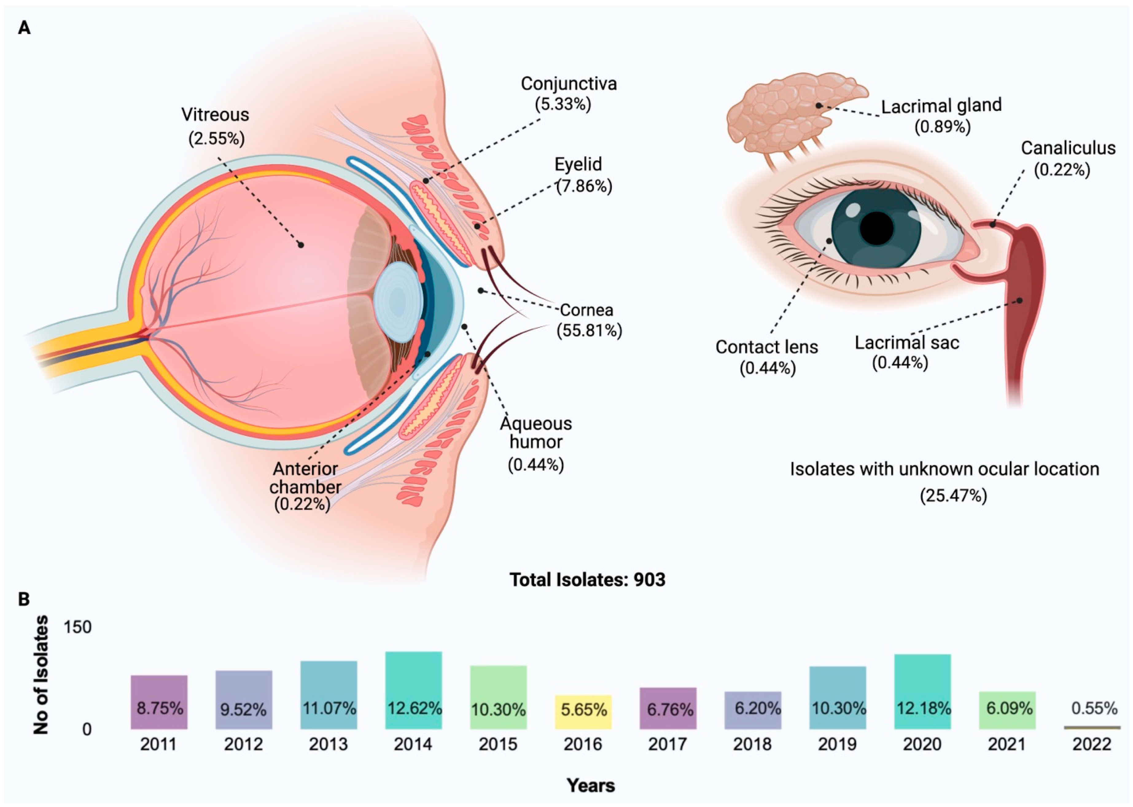
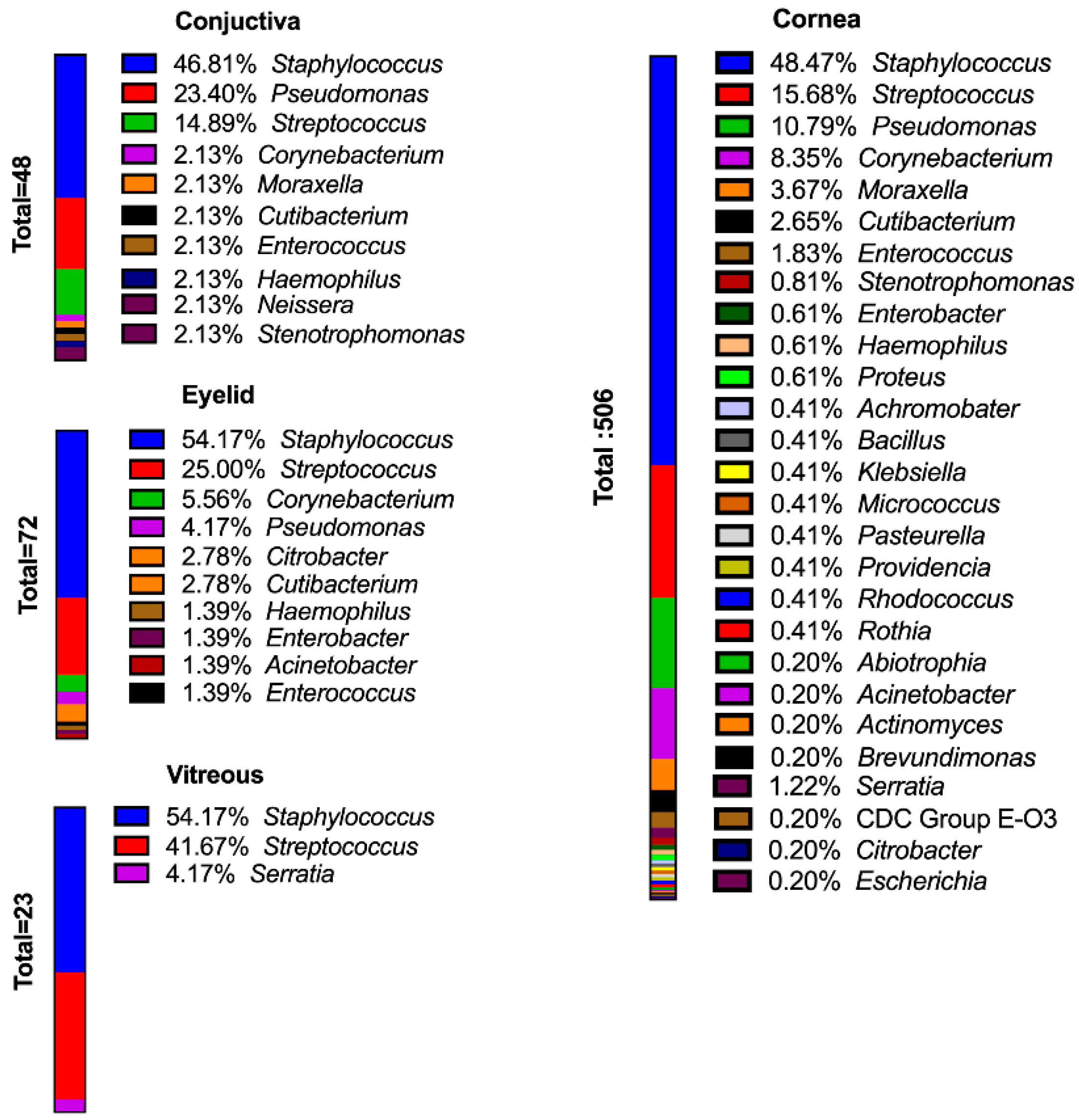
2. Species Distribution of Isolates

Among the Gram-positive bacteria, the most common staphylococcal species was Staphylococcus epidermidis (S. epidermidis) (41.70%). Staphylococcus aureus (S. aureus) (30.94%) was the next most common (Figure 4A). Streptococcus sp. (unidentified species) (50.32%) and Streptococcus pneumoniae (S. pneumoniae) (35.67%) were the most common streptococcal species in the collection (Figure 4B). Corynebacterium were also frequently isolated but were generally not speciated and may have been part of the normal microbiota [13,14]. Corynebacterium macginleyi (18.97%) was the most commonly identified Corynebacterium species isolated from ocular tissues (Figure 4C). The pseudomonad isolated with the greatest frequency was Pseudomonas aeruginosa (P. aeruginosa) (94.51%), and the Morexella species isolated most frequently was Moraxella lucunata (M. lucunata) (51.61%) (Figure 4D,E respectively).
As discussed above, numerous species have been isolated from the surface of the eye. Expectations for isolating a particular group of organisms in an infection are useful, but to make rational care decisions, it is also necessary to know the pathogenic potential of these isolates. Using our survey of ocular isolates as a basis, we provide a review of the basic bacteriology and pathogenic potential of each genus of bacterial isolates, from the most common isolates to those only isolated once (Hapax Legomenon) during the time period of our survey. In this review, we also describe the range of ocular infections these genera are known to cause and their virulence factors, pathogenic potential, and treatment options.

3. Gram-Positive Ocular Pathogens

3.1. Bacillus

Bacillus is a Gram-positive, motile, spore-forming rod-shaped bacteria ubiquitously present in nature and is found most commonly in the soil [15]. Bacillus anthracis and Bacillus cereus are the two major Bacillus species that are medically significant. B. anthracis, the causative agent of anthrax, has historically gained the most attention. B. cereus is well known for its association with food poisoning, and Bacillus species other than anthracis are also associated with various systemic and acute infections [16]. Compared to other anatomical sites where Bacillus causes disease, the eye is extremely sensitive to this pathogen. The reason is two-fold: the eye is an immune-privileged site and inflammation is particularly dangerous to this organ, and B. cereus contains a particularly potent virulence arsenal. Bacillus has been reported to be associated with blinding forms of keratitis, post-traumatic endophthalmitis, and endogenous endophthalmitis. Bacillus keratitis is relatively rare and is commonly associated with ocular trauma and other ocular surface disturbances [17]. To our knowledge, there have been only two reports of Bacillus keratitis associated with contact lens use [18,19]. In ophthalmology, there are valid concerns about Bacillus spore contamination of eye makeup [20] and decorative contact lenses [21]. Although B. cereus is the most commonly encountered species in ocular infections, other Bacillus species have been recovered from keratitis; B. megaterium [17,22], B. subtilis [17,18], B. coagulans, B. firmis, B. lincheniformis, and B. polymyxa [17]. In an analysis of 39 Bacillus ocular isolates [23], 52.6% were B. cereus, 26.3% were B. thuringiensis, and other isolates included B. subtilis, B. mycoides, B. pumilus, B. flexus, and Paenibacillus polymyxa (formerly Bacillus polymyxa). Among all the organisms that cause endophthalmitis, members of the Bacillus cereus sensu lato group cause the most severe form of this disease that results in loss of vision in less than three days in most cases [24]. In addition to its motility, B. cereus possesses adhesive pili [25] and produces numerous toxins and enzymes which are under the control of the quorum-sensing regulator PlcR [25]. Bacillus toxins produced in mouse eyes during experimental endophthalmitis include hemolysin BL, nonhemolytic enterotoxin, cereolysin O, and enterotoxins A, C, and FM [26]. Bacillus rapidly replicates in the ocular environment during an active infection [25]. Therefore, proper treatment should be initiated as soon as possible [27]. B. cereus is inherently beta-lactam resistant but is sensitive to vancomycin. The empiric treatment of Bacillus endophthalmitis includes intravitreal injection of vancomycin and ceftazidime [28]. In experimental models of endophthalmitis, gatifloxacin is able to sterilize rabbit and mouse eyes infected with Bacillus [29,30]. Our survey included six isolates of Bacillus sp.: three from unlisted locations, one from contact lenses, and two from the cornea.

3.2. Corynebacterium

Corynebacterium is a genus composed of rod-shaped or coccobacilli bacteria with a club-shaped appearance, and its members are widely distributed among animals and plants [13]. Some species of Corynebacterium are rare opportunistic pathogens, primarily in immunocompromised individuals [13]. Species that have been isolated from cases of conjunctivitis or keratitis are Corynebacterium accolens, C. amycolatum, C. mastitidis, C. propinquum, C. pseudodiphtheriticum, C. striatum, and C. xerosis [14,31,32,33,34,35]. By far, the most commonly isolated Corynebacterium reported in ocular infections is C. macginleyi [14,35].
Corynebacterium macginleyi is a lipophilic facultative anaerobic rod that has been commonly isolated from healthy eyes [35]. Hoshi et al. [36] sampled the conjunctiva of patients prior to cataract surgery and reported that 84% of the Corynebacterium isolates were C. macginleyi. Although C. macginleyi has been isolated from various types of infections, such as endocarditis, surgical site infections, and bladder catheter infections [37,38,39], the majority of C. macginleyi case reports are of ocular infections [14]. C. macginleyi has been isolated from cases of blebitis, conjunctivitis, endophthalmitis, and keratitis [35,40,41,42,43]. C. macginleyi has few known virulence factors, but this may be due to the lack of functional studies conducted with this species. Sagerfors et al. [35] reported that 37% of C. macginleyi genes have unknown functions. In their study of 29 culture-proven cases of C. macginleyi keratitis, this group also reported that the course of the infections was “uneventful”, with no need for a corneal transplant. However, more serious cases required corneal cross-linking and amniotic membrane transplant [35]. The most common risk factors for C. macginleyi keratitis were contact lens wear (66%) and ocular surface disease (10%) [35].
The antibiotic resistance of C. macginleyi varies depending on the report: Sangerfors et al. [35] reported that all C. macginleyi isolates in their study were susceptible to fluoroquinolones, while Eguchi et al. [41] reported that 68.8% of their C. macginleyi isolates had “high levels of resistance” to all fluoroquinolones tested. Aoki et al. [14] observed that the antibiotic resistance of C. macginleyi varies by region and stated that Corynebacterium species remain susceptible to third-generation cephems, which they recommended as a pragmatic treatment for ocular infection caused by Corynebacterium species.
St. Leger and Caspi [44] isolated Corynebacterium mastitidis (C. mastitidis) from the conjunctiva of a group of C57BL/6 mice housed at the National Institutes of Health and demonstrated that C. mastitidis is able to form a stable ocular colonines in mouse eyes. This species has been used in mouse models to study how the ocular microbiome affects the ocular surface immune homeostasis [45]. To our knowledge, this organism has not been isolated from human ocular infection.
In our survey, there were 58 isolates of Corynebacterium which comprised 6.42% of all the isolates (Figure 2). The most common anatomical site of isolation was the cornea, of which 8.35% of all corneal isolates where Corynebacterium (Figure 3). A total of 74.14% of the Corynebacterium were only identified to the genus, 18.97% (11 isolates) of the isolates were C. macginleyi, two were C. amycolatum, and two were C. striatum (Figure 4C).

3.3. Cutibacterium acnes (Propionibacterium acnes)

Cutibacterium acnes (C. acnes), formerly called Propionibacterium acnes, is an aerotolerant anaerobic, rod-shaped bacterium that is part of the microbiota of the oral cavity, conjunctiva, and skin, and is most commonly known as the causative agent of acne vulgaris [46,47]. C. acnes has also been reported as part of the conjunctival microbiota [48]. With regards to eye infections, C. acnes has been isolated in cases of conjunctivitis, cellulitis, infectious keratitis [49,50], and in delayed-onset post-surgical endophthalmitis following cataract surgery [51,52]. C. acnes has a number of virulence factors, such as Christie-Atckins–Munch–Petersen factors (CAMP factors), porphyrins, hyaluronate lyase, adhesins, and the ability to form biofilms [53]. C. acnes is also naturally resistant to 5-nitromidazole agents, aminoglycosides, sulfonamides, mupirocin, and resistance to erythromycin and clindamycin is developing [53]. Isolates of C. acnes remain sensitive to vancomycin and β-lactams [49].
Because of their protracted time course, descriptions of C. acnes ocular infections commonly include words like “slow”, “chronic”, “indolent”, “smoldering”. In their review of C. acnes endocarditis, Gunthard et al. [54] reported that the average time needed to detect growth in anaerobic or aerobic cultures was 6 days, with a range of 2–15 days. C. acnes keratitis is characterized by small lesions with deep stromal infiltration in the peripheral cornea, and it has been suggested that C. acnes should be considered in cases of negative keratitis cultures after seven days of incubation [50]. In their retrospective review of six cases of C. acnes endophthalmitis following cataract extraction with intraocular lens implantation, Fowler et al. [52] reported that the average time from surgery to diagnosis was 7.4 ± 5.2 months. In the same study, 100% of patients who underwent intraocular lens (IOL) removal achieved complete resolution of their endophthalmitis, compared to 77% of those undergoing pars plana vitrectomy with partial capsulectomy plus intravitreal antibiotics, or 18% receiving antibiotics alone. In general, visual outcomes of C. acnes endophthalmitis are typically good, but IOL removal may be necessary.
In our survey, there were 21 isolates of C. acnes, which comprised 3.21% of all isolates collected (Figure 2). A total of 47.6% of the isolates were from unlisted anatomical locations, 42.8% were collected from the cornea, and 1 isolate was collected from the conjunctiva.

3.4. Enterococcus

Enterococcus are facultative cocci known for their ability to grow and thrive under a variety of harsh environmental conditions, such as high salinity, low pH, and temperatures ranging from 10 to 45 °C [55]. The species E. faecalis and E. faecium rank among the leading causes health-care associated infections, including include urinary tract infections, bacteremia, surgical site infections, and endocarditis [55]. Enterococcus is a feared pathogen because of frequent antibiotic resistance to aminoglycosides, β-lactams, and vancomycin [56]. E. faecalis accounts for 4 to 8% of cases of post-operative endophthalmitis, including filtering bleb infections following glaucoma surgery [57,58,59,60,61]. The visual outcome of endophthalmitis due to E. faecalis is uniformly poor [57]. E. faecalis possesses several virulence factors, such as aggregation substance, enterococcal surface protein, hemolysins, extracellular superoxide, and gelatinase [62]. Cytolysin is one of the most important enterococcal virulence factors [63]. Cytolysin is a pore-forming exotoxin capable of lysing bacterial and eukaryotic cells [63], and this toxin may be responsible for poor visual outcomes in E. faecalis endophthalmitis [55,64]. E. faecalis endophthalmitis, as well as other types of E. faecalis infections, are increasingly caused by strains resistant to multiple antibiotics, including the last resort drug, vancomycin [55,60,65]. As such, E. faecium is a member of the “ESKAPE” group of bacterial pathogens [66]. Because of the increasing threat of multi-drug resistant infections, determining the mechanisms by which E. faecalis causes intraocular infection is vital.
Our survey contained 15 Enterococcus isolates: 11 E. faecalis, three nonspeciated enterococci, and one E. cloacae. Of the 11 E. faecalis isolates, six were isolated from the cornea, one each from the conjunctiva and eye, and three were from unlisted locations. The single E. cloacae isolate was from the cornea. Enterococcus comprised 1.66% of all the species isolated in this study (Figure 2).

3.5. Micrococcus

Micrococcus is a Gram-positive coccus in the Micrococcaceae family and is widely found in water, soil, dust, on the skin, and in the conjunctival microbiota of humans [67]. Micrococcus luteus (M. luteus) is the species most commonly isolated from human skin and infections, with Micrococcus lylae being more rarely isolated from human skin [68]. Although M. luteus is not considered to be pathogenic, there have been reports of various types of infections associated with this organism in the immunocompromised, such as HIV patients [69]. M. luteus has been associated with infections, including septic shock [70], meningitis [71], and catheter infections [72]. Because it is a commensal, the virulence of M. luteus has not been widely studied, but this species is capable of forming biofilms on implanted medical devices [73]. Although M. luteus is usually penicillin–sensitive, biofilm formation can make infections with this commensal more resistant to antibiotic treatment. In those cases, vancomycin and rifampin are recommended for their ability to penetrate biofilms [73]. The association of Micrococcus with ocular infection is rare. M. luteus was reported as the cause of keratitis in the left eye of a patient who underwent simultaneous LASIK for myopia [67]. The isolated M. luteus was resistant to ciprofloxacin and oxacillin and was treated successfully with fortified cefazolin 5% [67].
Micrococcus was an uncommon isolate in this survey, with only three isolates collected, none of which were identified to the species level. Two isolates were collected from the cornea, and one was collected from an unlisted location. Micrococcus comprised only 0.33% of all the genera collected in our survey (Figure 2).

3.6. Staphylococci

3.6.1. Staphylococcus aureus

Staphylococcus aureus (S. aureus) is a nonmotile coccus that occurs in irregular grape-like clusters. S. aureus is the most significant pathogen of the staphylococci group and is a common cause of food poisoning [74], abscess formation, pyogenic infections, and fatal septicemia [75]. S. aureus are characterized by a β-hemolytic phenotype, a positive coagulase reaction, mannitol fermentation, and the golden pigmentation from which their species name is derived [76]. A positive coagulase reaction separates S. aureus from the other staphylococci species isolated from humans (the coagulase-negative staphylococci, CoNS) [77]. S. aureus are part of the human microbiota and have been isolated from the nasal mucosa of a quarter to one-third of healthy individuals [78]. S. aureus has been reported to comprise 1.8% to 25% of species cultured from healthy eyes (Table 1). In our survey, there were 138 S. aureus isolates, comprising 15.39% of all the isolates collected and 30.94% of all staphylococcal isolates (Figure 4A). Additionally, 55.8% of the S. aureus isolates were isolated from the cornea, 6.5% were conjunctival isolates, and 19.62% were from unlisted locations.
S. aureus is a leading cause of a host of eye infections, such as blepharitis, cellulitis, conjunctivitis, keratitis, dacryocystitis, and endophthalmitis [5,6,9,27,79,80,81,82,83,84,85]. Because of its potential for multidrug antibiotic resistance and the impressive array of toxins and enzymes in its arsenal, S. aureus is considered a formidable and often dangerous pathogen [86,87].
Table 1. Isolation of normal ocular flora from different retrospective studies across the world.
LocationStudy PopulationGram-PositiveBacillusCoNSS. epidermidisS. aureusGram-NegativePseudomonasP. aeruginosa
Arantes [88]BrazilPre-cataract88.90% 54% 8%11.10%
Capriotti [89]Sierra LeoneHealthy Individuals78.1%5.5%36.1% 25.1%21.9% 7.8%
Dave [90]USA, NashvilleIntravitreal Injections 45.70%6.50%8.70%
Dorrepaal [91]TorontoIntravitreal Injections 64% 1.80%0.90%
Hsu [92]USA, St. LouisPre-cataract90.5% 74.8%57.2%5.0%9.5%1.8%
Lin [93]TaiwanPre-cataract91.70% 45.20%16.70%2.40%8.30%4.80%
Mamah [94]NigeriaPre-cataract73.70% 34.20%34.20%13.20% 2.60%
Martins [95]Sao PauloHealthy Individuals 61.70% 11.70% 3.40%
Mshangila [96]UgandaPre-cataract 65.90%76.90%21.00%10.10%
Papa [97]ItalyPre-cataract95% 67.90%58.00%15.30%4.60%
Rubio [98]MadridPre-cataract 56.80% 6.40%7.30%
Suto [99]JapanPre-cataract67% 57.20%3.90%6.30%0.70%
We conducted virulence gene analysis by PCR for all of the S. aureus ocular isolates in this survey. The PCR primers are included in Table 2, and isolated DNA preparations of these isolates were subjected to PCR as described in [10]. Ninety-eight percent of all S. aureus isolates were positive for hla (Table 3), the gene coding for α-toxin. Other studies have reported that 95–100% of clinical isolates were positive for hla [10,86]. α-toxin is also known as α-hemolysin because of its ability to produce β-hemolysis on blood agar [100]. α-toxin is a beta-barrel pore-forming toxin that binds to the receptor of a disintegrin and metalloproteinase 10 (ADAM10) [101]. High concentrations of α-toxin result in cell death, but sublytic concentrations of the toxin binding to ADAM10 activate ADAM10 metalloprotease activity, causing cleavage of E-cadherin adherens junctions, resulting in disruption of cellular focal adhesions and tissue breakdown [101]. It is not known if the ADAM10-mediated tissue damage mechanism occurs when S. aureus infects the cornea. However, in a rabbit model of keratitis, isogenic mutants of S. aureus lacking α-toxin injected into corneas caused no epithelial erosions as did the α-toxin-producing parental strain [102]. Similar studies conducted in a mouse model of keratitis confirmed that an α-toxin mutant was less virulent for the cornea [103]. In that study, corneal pathology caused by S. aureus was more severe in aged mice (36–48 weeks old) compared to young mice (6–7 weeks old) [103]. Putra et al. [104] used S. aureus strain JE2 in a corneal debridement model of keratitis in mice in which corneal healing was more rapid following infection with the α-toxin-deficient mutant strain compared to that of the parental strain. In a rabbit model of S. aureus endophthalmitis, infection with the isogenic α-toxin-deficient mutant caused less retinal damage than the parental strain [27]. These findings were later confirmed in a mouse model of S. aureus endophthalmitis [105]. These results indicate that α-toxin plays a major role in S. aureus ocular virulence. This toxin can contribute to pathogenesis either by direct killing of cells and/or by stimulation of the immune response. Blocking the activity of α-toxin would therefore be a rational therapeutic target for improving the visual outcome of keratitis and endophthalmitis. Theoretically, this could be achieved by passive or active anti-toxin immunization or the use of nanoparticles to neutralize the pore-forming toxins [29,106].
The percentage of S. aureus isolates in the current study that were positive for hlb, the gene encoding β-toxin, was 77.5% (Table 3), with 75% of mecA positive isolates also possessing the β-toxin gene. Reports of β-toxin frequency in clinical isolates ranged from 39% [10] to 57% [108]. β-toxin is not a toxin per se but is a neutral sphingomyelinase that hydrolyzes the plasma membrane lipid sphingomyelin, producing α-hemolysis on blood agar plates [109]. Most strains of S. aureus do not produce β-toxin due to the insertion of phage φSa3 into the hlb gene [110]. However, during in vivo growth, the phage φSa3 can excise and restore β-toxin production [111]. The role of β-toxin in S. aureus keratitis is unclear. In a rabbit model of S. aureus keratitis, infection with isogenic β-toxin-deficient mutants resulted in less scleral edema than that observed during infection with the wild-type strain. However, epithelial erosions, intrastromal ulcers, and slit lamp scores were similar in infections caused by hlb-deficient mutants [112]. In the rabbit model of experimental endophthalmitis, injection of an isogenic β-toxin-deficient mutant resulted in significantly less retinal damage compared to the wild-type parental strain [27]. These studies suggest that β-toxin may play a role in the virulence of S. aureus, but damaging activities might be more apparent in the posterior segment.
Panton–Valentine leucocidin (PVL) is a prophage-encoded toxin that binds to the C5a receptor, targeting neutrophils, monocytes, macrophages, natural killer cells, dendritic cells, and T-lymphocytes [113]. The percentage of S. aureus positive for PVL-associated genes in non-ocular clinical samples was reported to be 1.6% to 10% [114,115]. The percentage of all S. aureus positive for PVL-associated genes in staphylococcal isolates in our survey was 23.3% (Table 3). Of the mecA-positive isolates in our study, 37.5% were positive for PVL-associated genes. Bispo et al. [116] examined 68 isolates of MRSA from keratitis and orbital abscess/cellulitis cases and reported that the isolates grouped into two clonal complexes, CC5 and CC8. Isolates in the CC8 linage primarily caused orbital abscess/cellulitis and were predominately composed of USA300 strains in which pvl is more common, while CC5 primarily caused keratitis cases and was populated by USA100 and USA800 strains in which pvl was less common [116]. CC8 isolates were 93.7% positive for pvl, and none of the CC5 isolates were positive for pvl [116]. The role of PVL in ocular infections is unclear. Foster et al. [117] studied isolates from 85 cases of pediatric periorbital or orbital cellulitis and reported that 85% were pvl positive and 78% of these 85 cases were USA300 strains. This group further reported that there was no difference in clinical features or visual outcomes comparing infections with pvl positive or negative isolates or USA300 to non-USA300 isolates [117]. Sueke et al. [118] examined 95 keratitis isolates and reported that 9.5% were pvl-positive, and the pvl-positive cases suffered larger corneal ulcers and required more surgical intervention than pvl-negative cases.
S. aureus is often multidrug-resistant and is a member of the ESKAPE group of bacterial pathogens. In the antibiotic resistance monitoring in ocular microorganisms (ARMOR) study in the United States, ocular isolates of S. aureus were resistant to azithromycin (60.6%), ciprofloxacin (35.8%), and methicillin (36.6%); fewer were also resistant to chloramphenicol (6.1%), trimethoprim (4.4%), tetracycline (4.3%); and all isolates were sensitive to vancomycin [119]. Multidrug resistance was high among S. aureus, with 32.0% of isolates being resistant to three or more drug classes [118]. Bispo et al. [116] reported that 26.7% of their ocular isolates were MRSA. In our survey, of the 129 S. aureus ocular isolates tested, 37.2% were mecA positive (Table 3).

3.6.2. Coagulase-Negative Staphylococci

The coagulase-negative staphylococci (CoNS) group includes more than 50 species of staphylococci, whose species are distinguished from S. aureus by their inability to coagulate plasma [120]. Most members of the CoNS group cause chronic rather than life-threatening acute infections, but because of the high frequency of these infections, the difficulty diagnosing the etiology of an infection with a commensal bacterium, and the high rate of antibiotic resistance in this group, CoNS infections can be a burden on health care systems and have a profound impact on patient health [120]. Several CoNS species have emerged as pathogens of health-care facilities: Staphylococcus capitis, S. epidermidis, S. haemolyticus, S. lugdunensis, and S. saprophyticus [121]. Staphylococcus epidermidis (S. epidermidis) is the most commonly isolated CoNS in clinical samples and is the most widely studied of all CoNS [120,121]. Although S. epidermidis lacks the classical S. aureus virulence factor α-toxin, S. epidermidis isolates produce many potential virulence factors such as metalloproteases, β-hemolysin, δ-hemolysin, phenol-soluble modulins, proteases, numerous adhesion factors, and can form biofilms [121]. Antibiotic resistance is widespread in CoNS, and the group may serve as a reservoir of antibiotic-resistance genes for S. aureus [122]. The propensity of CoNS to form biofilms and their widespread antibiotic resistance make these infections difficult to treat [118,123,124].
CoNS are the most frequently isolated cause of many ocular infections, such as post-injection, post-operative, and post-traumatic endophthalmitis [51] and keratitis (Table 4). Patients recovering from endophthalmitis caused by CoNS are more likely to recover baseline visual acuity than those recovering from endophthalmitis caused by S. aureus or Streptococcus sp. [125]. Among surveys of bacteria from healthy eyes, CoNS comprise 34% to 74.8% of all bacterial isolates (Table 1). Among keratitis isolates, CoNS comprise 5% to 48% of bacterial species isolated. In our survey, 34.1% of all isolates were CoNS, with 44.8% of those being S. epidermidis. Eight different species of CoNS were isolated in this survey (Figure 4A), with 24.68% not identified at the species level (Figure 4A).
Since CoNS are part of the ocular microbiota, their identification as pathogens should be considered in view of the individual patient history [120]. Isolation of CoNS from the eyes of immunocompromised patients, such as those with poorly control diabetes, cancer patients, and chronic ocular corticosteroid use, should be considered in the keratitis diagnosis [2]. The ARMOR study noted above reported that ocular CoNS were resistant to azithromycin (61%), methicillin (48.6%), and ciprofloxacin (34.9%), and had less frequent resistance to tobramycin (17.0%), tetracycline (13.9%) and chloramphenicol (1.2%). All isolates were sensitive to vancomycin [119]. Multidrug resistance was also high among CoNS, with 40.7% of isolates being resistance to three or more drug classes [119]. Most of the CoNS in the ARMOR study were S. epidermidis.
Although CoNS are classically described as being nonhemolytic [138,139], there are reports of a β-hemolytic phenotype among members of this group [140,141,142,143,144]. The α-toxin gene hla has been detected by PCR in S. epidermidis [141,143,144,145]. Okee et al. [141] suggested that the cause of the hemolytic phenotype in these strains may be caused by a combination of factors. In their study of community and ICU isolates of S. epidermidis, this group reported that 70% of isolates from their ICU were β-hemolytic on 5% sheep blood agar, but only 20% were hla positive by PCR [141]. Interestingly, this phenotype was only detected in hospital-acquired isolates; no isolates from the community were β-hemolytic or hla positive. In the first year of our survey (2011), 35% of all isolates were CoNS, and only 3.57% of CoNS were β-hemolytic. In the last full year of our survey (2021), 18.2% of all isolates were CoNS, but 70% of these CoNS were β-hemolytic (Figure 5A). Over the 10-year time span, 40.7% of all CoNS in our survey had a β-hemolytic phenotype. Examples of hemolytic and non-hemolytic CoNS are shown in Figure 5B, illustrating the variation in hemolytic zones by these isolates compared with the hemolysis produced by β-hemolytic and non-β-hemolytic S. aureus laboratory strains (strains 8325-4 and RN4220, respectively). The origin of this evolving and potential pathogenic phenotype is under investigation.

3.6.3. Staphylococcus pseudintermedius

There was a single isolate of Staphylococcus pseudintermedius (S. pseudintermedius) noted in our survey (Figure 4), isolated from the cornea. S. pseudintermedius is a coagulase-positive Staphylococcus that has been isolated from 20.9% of healthy dogs [146] and is the most common staphylococcal species isolated from dogs [147]. S. pseudintermedius has been reported as an emerging zoonosis of canine origin and has been reported as the cause of skin and soft tissue infections in humans [148]. S. pseudintermedius has a high rate of methicillin resistance and multidrug resistance [148,149,150]. There are five main clonal lineages of methicillin-resistant S. pseudintermedius, each of which has distinct antimicrobial resistance profiles, geographic distributions, and SCCmec content [151]. We included S. pseudintermedius in this review as this species is a frequent isolate from canine ulcerative keratitis [149] and purulent soft tissue infections in canines and other domesticated animals [148,150]. S. pseudintermedius virulence mechanisms are not well studied but seem to be primarily due to the production of phenol-soluble modulins [152] and also include biofilm formation, lipase production, and toxins hlgA and hlgB [146]. As noted above, there is a high rate of multidrug resistance among S. pseudintermedius, but Ruiz-Ripa et al. [147] reported that all isolates in their study were susceptible to vancomycin and linezolid.

3.7. Streptococci

On lists of the prevalence of ocular bacterial isolates, streptococci are commonly ranked third, after CoNS and S. aureus, if they are listed at all. Streptococcus are Gram-positive bacteria that grow in chains or pairs and are commensals in the upper respiratory tracts and gastrointestinal tracts of most mammals and birds [153]. Streptococcus can become opportunistic pathogens under suitable conditions, such as in infections in elderly or immunocompromised patients [153]. In studies of the culturable bacterial flora of healthy eyes, streptococci were reported to range from 0% in São Paulo [95] to 13.2% in Nigeria [94]. Haung et al. [154] and Shin et al. [155] detected Streptococcus by 16S rDNA from conjunctival samples of healthy individuals, indicating that Streptococcus forms part of the core microbiome of the conjunctiva. In our survey, 17.39% of all the isolated bacteria were streptococci (Figure 2). Eleven species of streptococci were identified, with Streptococcus pneumoniae (S. pneumoniae) being the most common, comprising 35.67% of the streptococci isolates (Figure 4B). Moreover, 50.32% of the streptococcal isolates were not identified at the species level (Figure 4B).
S. pneumoniae are the most commonly isolated streptococci from ocular infections such as conjunctivitis, endogenous and exogenous endophthalmitis, and keratitis [156,157]. S. pneumoniae is also the most common cause of bacterial keratitis in low-income countries [158]. Among keratitis isolates, S. pneumoniae has been reported to have been isolated in 2% to 46.80% of cases (Table 4). In cases of endophthalmitis, even with prompt treatment, there is a high risk of profound vision loss and enucleation or evisceration in eyes infected with S. pneumoniae [156,157]. Chen et al. [157] reported that of 38 cases of S. pneumoniae endophthalmitis, 84% resulted in light perception to no light perception, and 26% underwent evisceration or enucleation.
S. pneumoniae possesses a number of virulence factors, such as a polysaccharide capsule, neuraminidase, pneumolysin, and zinc metalloproteinases [157]. The polysaccharide capsule enables S. pneumoniae to evade phagocytosis by inhibiting complement-mediated opsonization and is found in the majority of invasive isolates [159]. In animal models, encapsulated and nonencapsulated strains of S. pneumococcus were capable of causing severe keratitis [160], and the capsule was shown to be necessary for full virulence in the rabbit model of endophthalmitis [156]. The pore-forming toxin pneumolysin is highly conserved among pneumococcal isolates, and in addition to being cytotoxic to corneal epithelial cells, pneumolysin is highly immunogenic and causes an intense inflammatory response [161] and may be responsible for the rapid liquefactive necrosis that many pneumococcal ulcers undergo despite prompt treatment [3]. Endophthalmitis and keratitis caused by the Streptococcus genus as a whole are characterized by poor visual outcomes [162]. Gower et al. [163] reported that 70% Streptococcus sp. endophthalmitis cases following cataract surgery were count fingers or worse.
In their study of 271 clinical conjunctivitis isolates, Valentino et al. [164] reported that 90% were nonencapsulated and formed a distinct clade characterized by divergent forms of virulence factors and adhesins not found in encapsulated strains. Andre et al. [165] studied 45 clinical keratitis isolates and reported that 95.2% were encapsulated, but the capsular types in these strains were not covered by the pneumococcal vaccine PCV-13. These isolates were sensitive to fluoroquinolones and vancomycin but showed varying degrees of resistance to macrolides, such as erythromycin and azithromycin. In the ARMOR study noted above, ocular S. pneumoniae isolates reported in vitro resistance to azithromycin (35.9%) and penicillin (33.3%), but resistance to fluoroquinolones was less than 1% [119].

4. Gram-Negative Ocular Pathogens

4.1. Achromobacter

Achromobacter is a multidrug-resistant rod-shaped bacteria found in soil and water that can cause a wide variety of opportunistic infections in immunocompromised patients, such as bacteremia, abscesses, meningitis, urinary tract infections, and pneumonia [166]. Nineteen species of Achromobacter have been described, with Achromobacter xylosoxidans (A. xylosoxidans) being the most commonly isolated species from clinical cases [167]. Achromobacter spp. are predominantly isolated from patients with cystic fibrosis [168,169]. Among patients not suffering from cystic fibrosis, Achromobacter pneumonia or bacteremia are the most common types of infection [169]. Species other than A. xylosoxidans demonstrate a geographical diversity, with A. ruhlandii being the second most commonly isolated species in North America. In Europe, A. dolens and A. insuavis are more common, but it is not known if these species are of clinical significance [170]. Because Achromobacter is infrequently isolated from human infections, its virulence factors, clinical features, and optimal treatments for Acromobacter infections are not well described [170]. Achromocbater produces biofilms, is motile, and is frequently multidrug-resistant [171], with intrinsic resistance to most cephalosporins, aztreonam, and aminoglycosides due to multidrug efflux pumps and chromosomal β-lactamases [170].
A. xylosoxidans is a rare cause of chronic conjunctivitis, keratitis, and post-surgical endophthalmitis [172,173,174]. In their retrospective review of 10 ocular infections caused by A. xylosoxidans, Reddy et al. [173] reported eight cases of keratitis, six of which developed following penetrating keratoplasty and two cases of endophthalmitis. The keratitis infections were characterized by a slowly progressive disease with localized infiltration [173]. This group also reported that 90% of their isolates were sensitive to ceftazidime, and 70% were sensitive to amikacin [173]. In our survey, two Achromobacter isolates were noted, one A. xylosoxidans from an unlisted location and one Achromobacter sp. isolated from the cornea.

4.2. Acinetobacter

Acinetobacter are coccobacilli commonly found in soil and water samples and are frequently isolated from the skin of hospital staff and patients [175]. Although Acinetobacter are considered to be low-virulence opportunistic pathogens, this genus is capable of causing severe infections in immunocompromised patients following invasive procedures [175]. Although the danger of Acinetobacter lies in its high level of multidrug resistance, it is becoming appreciated that this species’ ability to adapt and survive lends to its persistence in hostile environments [176]. The presence of polysaccharide capsules in some Acinetobacter species, its ability to repair its genome during rehydration, and its high tolerance to oxidative stress all contribute to its desiccation resistance [176]. Acinetobacter also forms biofilms, is motile, and uses its capsule to circumvent host immunity. Acinetobacter are becoming increasingly resistant to several antibiotics and, as such, is a member of the ESKAPE group of bacterial pathogens [66]. Talreja et al. [177] reported that all 12 of the ocular Acinetobacter baumannii isolates tested in their study were multidrug resistant. Acinetobacter anitratus [178] and A. lwoffi [179] have been reported as rare causes of post-traumatic endophthalmitis, and Acinetobacter baumannii [177] and A. junii [180] have been reported as the cause of corneal ulcers. In our survey, three Acinetobacter lwoffi isolates were noted and were isolated from the cornea, aqueous, and an unknown region of the eye.

4.3. Citrobacter

Citrobacter sp. are rod-shaped common environmental bacteria comprised of 11 recognized species that have been found in the normal gut microbiota of humans [181,182]. Citrobacter are an increasing problem in human infections, such as urinary tract infections and bacteremia, because of evolving multidrug resistance [183]. Common virulence and adaptation traits, such as polysaccharide capsules, iron acquisition genes, and motility operons, have been reported in this genus, as well as the presence of a high-pathogenicity island that is essential for virulence in mice [181]. Citrobacter freudii (C. freudii) and Citrobacter koseri (C. koseri) have been reported in cases of exogenous and endogenous endophthalmitis [183,184] and keratitis [185,186,187]. Overall, reports of Citrobacter in eye infections have been relatively rare but almost always resulted in severe infections and vision loss [184]. In our survey, we report four Citrobacter isolates: two C. koseri from an unknown area of the eye and one from the cornea, and a C. freudii isolate from an unknown area of the eye.

4.4. Enterobacter

Enterobacter are rod-shaped, facultative bacteria and are members of the family Enterobacteriaceae [188]. The Enterobacter genus consists of 22 species, some of which are members of the normal gastrointestinal microbiota, but can cause healthcare-associated infections such as urinary tract, respiratory, and soft tissue infections, as well as osteomyelitis and endocarditis, especially in immunocompromised individuals [188]. The virulence factors of Enterobacter are poorly understood but include motility, chemotaxis, and capsules [189]. Because of their evolving multidrug resistance, Enterobacter is a member of the ESKAPE group of bacterial pathogens [66]. While intraocular infections with Enterobacter are rare, cases of postoperative and posttraumatic endophthalmitis have been reported [190,191,192]. E. cloacae has been associated with acute postoperative, delayed filtering bleb-related, and posttraumatic endophthalmitis [190,191,192]. In their retrospective study of 44 culture-positive cases of Enterobacter endophthalmitis, Dave et al. [193] reported that 77.27% were from posttraumatic cases, 15.9% were from postoperative cases, and 6.8% were from endogenous endophthalmitis cases. Isolates from cases of Enterobacter endophthalmitis were reported to be susceptible to ciprofloxacin, amikacin, and ceftazidime [193,194]. Enterobacter-associated endophthalmitis uniformly presents as a rapid and severe infection and results in poor visual outcomes despite early and appropriate management [193]. In our survey, seven Enterobacter isolates are reported, five E. cloacae (two from the cornea, one from the eye, and two from unlisted locations), one E. cancerogenus (from the cornea), and one E. aerogenes (from the lacrimal gland).

4.5. Escherichia coli

Escherichia coli (E. coli) is a Gram-negative, facultatively anaerobic, motile, non-spore-forming rod-shaped bacteria [195]. E. coli are mainly commensal members of the large intestine, but certain strains of this species are pathogenic and are classified into pathotypes based on various criteria, such as the target organ, host species, or the presence of specific virulence genes [196]. Pathogenic E. coli can cause serious infections such as urinary tract infections, intra-abdominal, skin, and soft tissue infections, pulmonary infections, newborn meningitis, bacteremia, and hemolytic and uremic syndrome [196]. E. coli possess numerous virulence factors encoded in pathogenicity islands, plasmids, and other mobile genetic elements [197]. These virulence factors include adhesins, toxins such as α-hemolysin and cytotoxic necrotizing factor 1, iron acquisition factors, polysaccharide capsules, and liposaccharide [197]. Antibiotic resistance in this species is widespread and increasing [196]. E. coli have been isolated from ocular infections such as conjunctivitis [5], dacryocystitis [198], keratitis [199,200], and endophthalmitis [200,201]. Jackson et al. [201] reported that seven percent of endogenous endophthalmitis cases were caused by E. coli, which is a rare but not uncommon complication of septicemia. In our survey, two E. coli isolates were noted, one from the cornea and one from an unlisted location in the eye.

4.6. Haemophilus

Haemophilus are Gram-negative coccobacilli that inhabit the upper respiratory tract and are rarely associated with ocular infections [202,203]. Pathogenic strains that cause disease generally enter the upper respiratory tract through droplet inhalation or direct contact [204]. The most common pathogenic Haemophilus are Haemophilus influenzae (H. influenzae), which are characterized by capsular type [205]. H. influenzae virulence factors include polysaccharide capsule, biofilm formation, IgA proteases, and macrophage survival factor [205]. Haemophilus are responsible for a range of mild and serious infections, including sinusitis, conjunctivitis, pneumonia, bacteremia, otitis media, meningitis, cellulitis, and epiglottitis [203]. Conjunctivitis is a common ocular bacterial infection occasionally caused by H. influenzae [206]. Conjunctivitis-otitis syndrome is a manifestation of acute conjunctivitis in infants that can be caused by H. influenzae [207]. Topical antibiotic therapy may reduce the duration of this disease, but it is typically self-limited within a few weeks. In the ARMOR study noted above, H. influenzae were “…susceptible to all antibiotics tested” [119]. In our survey, nine isolates of Hemophilus were collected, comprising 1% of all isolates (Figure 2). Eight of the isolates were H. influenzae, and one was not identified at the species level. Four of the isolates were from unlisted locations, three were from the cornea, and one each was from the conjunctiva and eyelid.

4.7. Klebsiella

Klebsiella species are non-motile, encapsulated rods and are predominantly opportunistic pathogens [208]. Klebsiella infections typically occur in hospital settings among individuals who are immunocompromised and have a severe underlying condition [209,210]. K. pneumoniae and K. oxytoca are responsible for the majority of healthcare-associated infections that include pneumonia, septicemia, soft tissue abscesses, meningitis, and endophthalmitis [208]. K. pneumoniae has become increasingly resistant to multiple antibiotics [211,212,213,214] and is a member of the ESKAPE group of bacterial pathogens [66]. The virulence of K. pneumoniae has been ascribed to the production of cell-wall-associated factors and capsules [215,216]. The hypermucoviscosity phenotype is commonly associated with strains that cause liver abscesses and those that possess enhanced intraocular virulence [217,218]. This hypermucoviscosity phenotype produces a mucopolysaccharide web which, in a mouse model of endophthalmitis, produced rapid retinal function decline and inhibited phagocytosis, as compared to the isotype mutant lacking this phenotype [216,217]. K. pneumoniae currently ranks among the leading causes of endogenous bacterial endophthalmitis and is responsible for 80–90% of cases in Asian countries [218].
In their review of 14 cases of endogenous K. pneumoniae endophthalmitis spanning 12 years, Mak et al. [219] reported that hepatobiliary sepsis was the source of ocular infection in 64% of patients, 14% of patients died, 38% experienced total loss of vision, and 19% required evisceration of the globe. The outcome of endogenous endophthalmitis is often severe, ranging from count fingers visual retention to evisceration or enucleation [218]. In a streptozocin-induced diabetic mouse model of endogenous K. pneumoniae endophthalmitis, intraocular infection incidence correlated with a compromised blood–retinal barrier and increases in vascular permeability [220]. Endogenous endophthalmitis was observed in mice 3 and 5 months following streptozocin injection but not in mice 1 month post-injection or in control, nondiabetic mice. These results suggested that K. pneumoniae requires a compromised blood–retinal barrier in order to gain access to the eye from the bloodstream and pointed towards an underlying mechanism for the increased prevalence of cases of K. pneumoniae endogenous endophthalmitis observed among diabetic patients [220]. In contrast, in a streptozocin-induced diabetic mouse model of endogenous S. aureus endophthalmitis, intraocular infections were observed in both nondiabetic mice, as well as in mice 1, 3, and 5 months post-streptozocin injection [221]. This indicated that S. aureus does not require a compromised blood–retinal barrier and can cause endogenous endophthalmitis in the absence of diabetes.
K. oxytoca has been reported as a rare cause of keratitis [222], and K. pneumoniae has been reported as a rare cause of interface keratitis following lamellar keratoplasty [223]. Although drug-resistant K. pneumoniae have been reported in Asia, 86% of endogenous bacterial endophthalmitis cases responded well to intravitreal ceftazidime [219]. In our survey, there were four isolates of K. oxytoca and one K. pneumoniae. Two of the isolates were from the cornea, and three were from unlisted locations.

4.8. Moraxella

Moraxella sp. are coccobacilli that were first reported from ocular infections in 1896 and 1897 from patients with angular blepharitis [224,225]. Moraxella are considered part of the microbiota of the upper respiratory tract and urogenital tract [226,227,228]. Moraxella sp. are known causes of keratitis [226,227,228,229], conjunctivitis [226,227,228,229,230], and endophthalmitis [226]. In their retrospective review of 101 culture-proven cases of Moraxella keratitis, Hoarau et al. [231] reported that the most common species were M. lucunata (50%) and M. nonliquefasciens (38%). This group also reported that the clinical features, such as ulcer size and healing, treatment duration, and infiltrate size, did not vary with the species causing the infection, and the preferred treatment was fluoroquinolone and rifamycin [231]. Moraxella catarrhalis have virulence factors such as β-lactamases, biofilm formation, MID/Hag, which mediates hemagglutination and non-immune binding of IgD, and a number of outer-membrane proteins involved in adherence to epithelial cells [232]. Thirty-one Moraxella isolates were identified in our survey (Figure 4): 51.6% M. lucunata, 22.6%; M. catarrhalis, 12.9%; M. nonliquefaciens; one M. osloenis; and three unspeciated Moraxella. Although there are reports of increasing frequency of Moraxella isolates in ocular infections [228,233], no increase in the number of isolates with time was observed in our survey. Interestingly, there were no isolates of M. catarrhalis collected after 2014, and all isolates of M. nonliquefactiens, M. osloensis, and unspeciated Moraxella were collected after 2018.

4.9. Pseudomonas

P. aeruginosa are ubiquitous rod-shape bacteria which, due to their simple nutritional needs and innate resistance to antibiotics and antiseptics, have been isolated from soil, water, human gastrointestinal tracts, sinks, showers, distilled water [234] and are commonly isolated from uninfected eyes (Table 1). Pseudomonas sp. have also been identified by 16s rDNA as part of the conjunctival microbiota [154,155]. P. aeruginosa are causes of acute conjunctivitis [4,235], dacryocystitis [84], post-surgical and post-traumatic endophthalmitis [236,237], endogenous endophthalmitis [238], and are the major cause of contact lens-associated keratitis [239,240,241]. Recently, deaths have been reported following P. aeruginosa ocular infections resulting from contaminated eye drops [242]. P. aeruginosa may sequester in niches in ocular glands during keratitis, potentially leading to spread to other extraocular sites [243].
Keratitis infections of contact lens wears are associated with the phylogenetic subgroup encoding the cytotoxin exotoxin U gene (ExoU) [241,244], while keratitis infections of populations with lower contact lens use are predominantly caused by species with genes encoding exotoxin S (ExoS) [2]. Enzymes such as elastase B, protease IV, and P. aeruginosa small protease [245,246,247] have been reported to play a role in experimental Pseudomonas keratitis. Although P. aeruginosa are the most commonly isolated pseudomonads, other species have been recovered from ocular infections, such as and P. putida from a case of conjunctivitis [248] and P. fluorescens from a case of endophthalmitis [249].
The role of the exotoxins mentioned above in P. aeruginosa keratitis have been widely studied, as has the pathogenic profile of cytotoxic and invasive strains and the intracellular nature of P. aeruginosa in the corneal epithelium. Pseudomonas contact lens-associated keratitis is the result of the confluence of the widespread use of ocular prosthetic devices and an opportunistic pathogen with a large and versatile arsenal of virulence factors [250]. The healthy, undamaged cornea is naturally resistant to Pseudomonas infection [251]. A full description of the many virulence factors involved in overcoming the innate resistance of the healthy cornea to infection is beyond the scope of this review but has been ably studied and reviewed by Fleisizg et al. [252]. To our knowledge, investigation into the specific virulence factors involved in the development and pathogenesis of Pseudomonas endophthalmitis has not been undertaken.
Multidrug resistance to antibiotics is common among P. aeruginosa due to chromosomally encoded genes and the ability to acquire mobile genetic elements [253]. Because of this, P. aeruginosa are members of the ESKAPE group of bacterial pathogens [66]. The most effective antibiotics against P. aeruginosa keratitis have been reported to be levofloxacin [92,253,254], ciprofloxacin [92,253], and amikacin [253], with reported sensitivities of 94.6%, 90.9%, and 90.2%, respectively [253]. However, resistance patterns vary from country to country [253]. In the ARMOR study noted above, all ocular isolates of P. aeruginosa were sensitive to all tested antibiotics, with infrequent in vitro resistance to polymyxin B (8.6%), tobramycin (2.5%), and fluoroquinolones (5.2–7.4%) [119]. In our survey, we report 90 isolates of P. aeruginosa: 64.4% from the cornea, 12.22% from the conjunctiva, and the remainder from other ocular sites (Figure 3 and Figure 4).

4.10. Proteus mirabilis

Proteus mirabilis (P. mirabilis) are motile, rod-shaped ubiquitous bacteria found in soil and water and are commensal inhabitants of animal gastrointestinal tracts [255]. P. mirabilis possesses a number of virulence factors, such as motility, proteases, and hemolysins, Proteus has a toxic agglutinin which promotes autoaggregation of bacteria and lysis of bladder cells in vitro, and a ZapA metalloprotease which cleaves IgA, IgG, complement proteins C1q and C3, and proteins such as fibronectin, actin, and collagen [255]. P. mirabilis can cause infections of the gastrointestinal tract and wounds but is most commonly known for catheter-associated urinary tract infections [255]. P. mirabilis is a rare cause of keratitis [256] and endophthalmitis [257]. Mo et al. [256], in their retrospective review of 26 culture-proven cases of P. mirabilis keratitis, reported that all isolates were susceptible to ciprofloxacin, ofloxacin, moxifloxacin, gatifloxacin, and cefazolin. Although capable of causing serious disease, P. mirabilis keratitis has been reported to respond well to prompt and appropriate treatment. Our survey reports four P. mirabilis isolates, three isolated from the cornea and one isolated from the lacrimal gland.

4.11. Serratia

Serratia is a motile, rod-shaped anaerobe widely found in soil, plants, and water [258,259]. The most common species isolated from infections is Serratia marcescens (S. marcescens). Isolation of Serratia liquefactions (S. liquefaciens) is less commonly reported [260]. S. marcescens virulence factors include motility, fimbriae for adherence, several hemolysins which are toxic to different cell types, metalloproteinase, gelatinase, endonuclease, and proteases [260,261,262]. In a mouse model of keratitis, S. marcescens induced corneal inflammation by activating TLR4 and TLR5, inducing CXC chemokine production, which recruited neutrophils to the corneal stroma, resulting in corneal thickening and opacity [263]. Using a rabbit model of keratitis, Romanowski et al. [264] demonstrated that the Serratia Rcs stress response system is regulated by GumB. Mutation of GumB resulted in a greater than 50-fold reduction in S. marcescens proliferation and a reduction in inflammatory markers, indicating that GumB is a key mediator of S. marcescens corneal virulence [264].
Serratia species are inherently resistant to a wide range of antibiotics, including ampicillin, amoxicillin, and colistin [259]. S. marcescens frequently has both chromosomal and plasmid-mediated resistance to multiple antibiotics [260]. S. marcescens cause nosocomial infections ranging from pneumonia and endocarditis to urinary tract infections [265]. In the eye, S. marcescens has been reported to cause keratitis, lacrimal duct infection, endophthalmitis, and conjunctivitis [266]. S. liquefaciens has been reported in a case of keratitis [267]. S. marcescens keratitis is associated with the presence of an abnormal corneal surface, the use of topical medications, and contact lens use [266,268]. In their retrospective study of 24 cases of S. marcescens keratitis, Mah-Sadorra et al. [268] reported that a good clinical response was obtained with topical fluoroquinolones and fortified aminoglycoside drops. In their review of 51 patients, Atta et al. [266] reported that most patients with Serratia keratitis responded well to antibiotic drops and rarely required adjunctive treatment or surgical interventions. Serratia has also been reported to cause endogenous endophthalmitis resulting in visual impairment and loss of vision [269,270]. In our survey, 10 isolates of Serratia are reported: nine S. marcescens (five from the cornea, two from the canaliculus, one from the vitreous, and one from the anterior chamber), and one S. liquefaciens isolate from the cornea.

4.12. Stenotrophomonas maltophilia

Stenotrophomonas maltophilia (S. maltophilia) is a ubiquitous environmental rod-shaped bacterium with intrinsic antibiotic resistance to β-lactams and aminoglycosides. Stenotrophomonas is capable of causing a variety of nosocomial and community-acquired infections, primarily in immunocompromised patient populations [271]. S. maltophilia is frequently isolated from hospitalized pneumonia patients but also causes infections of the respiratory tract, central nervous system, gastrointestinal tract, urinary tract, soft tissues, and bone [271]. The virulence factors of S. maltophilia include motility, biofilm formation, and production of extracellular enzymes, such as DNase, proteases, lipases, hyaluronidase, and hemolysin [271]. S. maltophilia is a rare cause of ocular infections but has been reported as a cause of cellulitis, conjunctivitis, dacryocystitis, endophthalmitis, and keratitis [272,273,274,275]. In pediatric keratitis cases, S. maltophilia was the second most commonly identified species after P. aeruginosa [240]. In their retrospective review of 16 culture-proven cases of S. maltophilia keratitis, Park et al. [275] reported that most cases were associated with ocular surface instability, such as trauma or contact lens use, and that the treatment of choice was a mixed use of a fluoroquinolone, beta-lactam, and aminoglycoside. Our survey reports eight samples of S. maltophilia isolated from the cornea, contact lenses, and conjunctiva.

5. Hapax Legomenon Isolates

5.1. Abiotrophia, Actinomyces, and Brevundimonas diminuta

Eleven genera were only isolated once in our survey (Figure 2). Three of these, Abiotrophia sp. (Gram-positive cocci, corneal isolate), Actinomyces sp. (Gram-positive filamentous, corneal isolate), and Brevundimonas diminuta (Gram-negative bacillus, corneal isolate) have been reported as rare causes of ocular infections [276,277,278]. The isolates of Abiotrophia, Actinomyces, and Brevundimonas diminuta in our survey were all corneal isolates.

5.2. CDC Group EO-3, Cronobacter, and Lactobacillus

CDC Group EO-3 (Gram-negative coccobacillus, corneal isolate) has a single case report of peritonitis [279] in the literature, and to our knowledge, no reports of causing ocular infections. Cronobacter sakazakii (Gram-negative bacillus, unknown ocular location) is a common contaminant of dry plant-based foods and has caused foodborne outbreaks of necrotizing enterocolitis, septicemia, and meningitis among neonates [280]. The most recent US outbreak of Cronobacter infections was linked to contaminated infant formula powder [281]. To our knowledge, there are no literature reports of ocular infections due to Cronobacter. Lactobacillus sp. (Gram-positive bacillus from an unlisted ocular location) is widely distributed among plants and animals [282], is part of the ocular microbiota [254], and is not considered pathogenic [282]. However, Lactobacillus sp. is a rare cause of bacteremia in immunocompromised patients [283]. To our knowledge, there have been no reports of ocular infections caused by Lactobacillus, although there have been reports of the use of Lactobacillus in probiotic preparations to stimulate ocular immunity [284,285].

5.3. Neisseria

Neisseria sp. (Gram-negative diplococcus) is a cause of mucopurulent conjunctivitis and keratitis [286]. Neisseria meningitidis (N. meningitidis) virulence factors include pili, opacity-associated proteins, lipooligosaccharides, and capsular polysaccharides [287]. There are 13 clinically significant serogroups of Neisseria based on their capsular polysaccharides, and each has a specific geographic distribution [287]. In their study of 60 isolates of Neisseria keratitis, Kate et al. [286] reported that forty percent of isolates were only identified to the genus, 21.7% were caused by N. elongata, 16.7% by N. meningitis, 6.6% by N. weaver, and 5% by N. mucosa. Keratitis was often preceded by conjunctivitis, but keratitis also developed following a compromised ocular surface or ocular injury, particularly when topical corticosteroids were in use [286]. Keratitis normally responded well to topical antibiotics, and 100% of Neisseria keratitis isolates were susceptible to gatifloxacin, 94% susceptible to amikacin, 96% susceptible to chloramphenicol, and 96% susceptible to gentamycin [286].
Although a common cause of sexually transmitted diseases, N. gonorrhoeae is also a rare cause of ocular disease. Virulence factors include Type IV pili, lipooligosaccharides, porin, opacity proteins, and efflux pumps [288]. In their five-year retrospective review, Butler et al. [289] reported 15 cases of ocular gonococcal infections. The most common presenting features reported were a purulent discharge (93% of cases), hemorrhagic conjunctivitis (67% of cases), and pre-sepal cellulitis (60% of cases). All patients were treated with systemic antibiotics and topical chloramphenicol or ofloxacin. Corneal involvement was reported in 33% of cases, but there was no “significant” corneal melting or perforation [289]. Because corneal ulceration can rapidly progress to corneal melting and perforation, treatment must be prompt and effective [290,291]. Because of the increasing rates of N. gonorrhoeae resistance to penicillin, tetracycline, and fluoroquinolones, the Center for Disease Control recommends an intramuscular injection of ceftriaxone in a single dose with topical saline lavage of the eye and sexual partners of the patient should be referred for evaluation and treatment [292]. The single isolate of unspeciated Neisseria in our survey was isolated from the conjunctiva.

5.4. Pasteurella

Pasteurella sp. is a common Gram-negative coccobacilli resident of the oral and digestive tract of many birds and mammals, particularly dogs and cats [293]. Pasteurella is not part of normal human flora [294]. Pasteurella infections of soft tissues, bones, and wounds can result from animal bites and scratches [295]. Ocular infections with Pasteurella are rare, but bites and scratches to the eye can result in keratitis, conjunctivitis, or endophthalmitis, primarily from species P. canis and P. multocida [293,294]. Studies of the antibiotic susceptibilities of non-ocular clinical isolates have shown that most human isolates of Pasteurella are susceptible to moxifloxacin, amoxicillin, azithromycin, and clarithromycin [295]. Shah et al. [294] recommended fortified vancomycin and tobramycin for the treatment of Pasteurella keratitis. The single unspeciated Pasteurella isolate in our survey was a corneal isolate.

5.5. Providencia

Providencia sp. are Gram-negative motile rods that have been isolated from water and soil and are most commonly known for causing urinary tract and wound infections [296]. Human isolates of Providencia have been recovered from the axilla, blood, perineum, stool, urine, and wounds [297]. Risk factors for ocular infections with Providencia are immunocompromise and urinary tract infections, particularly among those with long-term indwelling urinary catheters [296]. Providencia form biofilms, have intrinsic resistance to colistin and tigecycline [298], may produce β-lactamase, and resistance to fluoroquinolones is on the increase [296]. Providencia alcalifaciens has been isolated from a case of keratitis [299], and P. rettgeri has been reported to cause keratitis, dacryocystitis, conjunctivitis, and endophthalmitis [296]. The single Providencia isolate in our survey was isolated from a cornea and was not speciated.

5.6. Rhodococcus

Rhodococcus sp. are Gram-positive to Gram-variable actinomycetes, are ubiquitous in soil and water, and are most commonly known for causing opportunistic infections of immunocompromised patients, particularly those with HIV [300,301]. The virulence factors of Rhodococcus equi include polysaccharide capsules, hemolytic enzymes, β-lactamases, and the ability to multiply inside macrophages [302]. Rhodococcus has been detected by 16S rDNA PCR from normal human eyes in repeated samples, indicating that this genus is part of the conjunctival microbiota [303]. R. rhodochrous [300], R. globerulus [304], and R. ruber [305] are reported but uncommon causes of keratitis. R. erythropodes and R. luteus have been isolated from cases of post-surgical endophthalmitis [306]. R. gordoniae was identified as a cause of eyelid infections [307]. A reported case of R. globerulus keratitis was treated with fortified vancomycin, amikacin, and erythromycin [304], and a case of R. ruber keratitis was treated with 2% amikacin [305]. Both cases responded well to the treatments. The single unspeciated Rhodococcus in our survey was a corneal isolate.

5.7. Rothia

Rothia sp. are Gram-positive rods that are resident flora of the upper respiratory tract and are responsible for a variety of infections, such as bacteremia, endocarditis, meningitis, and pneumonia, most commonly in immunocompromised patients [308,309]. Rothia dentocariosa and R. mucilaginosa are rare causes of endophthalmitis [310], and R. dentocariosa is a rare cause of keratitis [311,312]. Keratitis caused by R. dentocariosa has been treated successfully with cefuroxime and penicillin [312], and the case reported by Morley and Tuft [311] was sensitive to ofloxacin, chlorophenol, and cefuroxime. The single unspeciated Rothia isolate in our survey was a corneal isolate.

6. Conclusions

Diagnosing the cause of any infection can be a challenging task. Once the culture results return days or weeks later, the question is whether the bacteria in the report is the cause of the infection. As will have been noted by the reader, many of the bacteria listed in this review are commensal organisms and can be easily isolated from healthy individuals. Complicating the matter is the fact that in keratitis samples, only 40–56% of cultures are positive for growth [313,314], and with endophthalmitis cultures, only 40–70% of cultures are positive for growth [315]. Essential to diagnosing and properly treating the cause of infection is understanding the factors involved, such as which pathogens in that area cause that type of infection, whether multidrug resistance is prevalent in that region, and the immunocompetent state of the patient [315].
Efforts to shorten the diagnostic time for identifying possible pathogens in eye infections by using PCR, whole genome sequencing (WGS), and Nanopore Sequencing [315] are important. In a study of samples from cases of endophthalmitis, WGS was shown to be more sensitive than culturing or 16S sequencing for the identification of pathogens [315]. Methods such as WGS have the potential to detect all organisms (bacterial, fungal, and viral) in environmental samples, which could improve diagnostics and treatment of complex polymicrobial infections. WGS also allows for the analysis of genomes for virulence factors and antibiotic resistance genes [315], which could inform the treating physician about the potential severity of the infection as it evolves. However, as most eye care professionals know, the majority of keratitis and endophthalmitis patients respond very well to empirical treatments, and culture results do not often change the treatment regimen [316]. In one survey, only 35.1% of corneal ulcers were actually cultured [317]. While this may be a cause for concern, it also demonstrates that we have a good understanding of what pathogens are likely causing an infection and that our treatments are currently effective for the majority of cases. Also of concern is complacency in assuming that current treatment regimens will be successful for all ocular infections. Continual monitoring of the resident flora of the eye and the organisms involved in eye infections is essential to identify emerging antibiotic resistance and virulence profiles. As reports of high-throughput sequencing of the ocular microbiota are becoming commonplace, it is important to note that the organisms reported in our survey only covered culturable bacteria. While novel strategies such as rapid Nanopore Sequencing may identify previously undetected organisms [315], this will not circumvent the need to culture bacteria. In order to track antibiotic resistance or to understand the function of genomic data, it is necessary to have an organism to test and assays to determine the extent to which expression of resistance or virulence genes occurs. Pathogens are constantly evolving to adapt to antibiotic treatments, new pathogens are emerging to exploit vulnerable human populations, and some of these vulnerable populations are growing in number.
In this review, we report a survey of the spectrum of bacteria recovered from ocular infections and banked over the course of 10 years. A limitation of this survey is the absence of correlative patient data. This review is by no means an exhaustive summary of all the possible bacteria that can be cultured from human eyes, but it illustrates the variety of species that have been isolated from a population at a single institute over a specific period of time. The phrase “ocular infections” covers a wide scope of diseases, ranging from conjunctivitis to pan-ophthalmitis. While certain ocular infections, such as keratitis and endophthalmitis, have been relatively well studied, that is true only in regard to the most common pathogens which cause the disease. As this review has shown, there is a vast spectrum of bacteria capable of colonizing the ocular surface, and only a few have been studied well enough that treatments have moved beyond “empirical”.
Several studies have sampled the ocular flora of healthy eyes and have shown that the predominant ocular bacteria in healthy conjunctiva are composed of CoNS and Gram-positive cocci (Table 1). In studies of keratitis isolates, Gram-positive cocci and CoNS are also the most common type of bacteria isolated (Table 4). We found the same trend of Gram-positive cocci and CoNS being the most common type of bacteria isolated (Figure 2). Table 1 and Table 4 summarize the results of a number of surveys of ocular bacterial isolates from healthy eyes and isolates from keratitis cases, respectively. Comparisons of our data with the bacteria isolated from healthy and keratitis eyes are difficult because of the large range of numbers in those studies, but some general comparisons can be made. In our survey, 79.51% of the bacteria isolated were Gram-positive bacteria, which is in the upper range of both groups of isolates. However, the 34.1% CoNS and 15.28% S. aureus in our survey are in the lower bounds of both groups, indicating that this population has higher numbers of Gram-positive cocci other than Staphylococcus. Our survey contained 20.38% Gram-negative bacteria, which is in the upper range reported in keratitis eyes and the lower range reported in healthy eyes. These data suggest that the species of bacteria found in our sample of isolates from patients visiting an eye care facility might not be representative of a healthy population.
Studies of bacterial infections tend to focus on single species, although it is well-known that bacteria in the natural environment exist as part of a community of organisms. Delbeke et al. [318], in their systematic review of 11 ocular microbiome studies, define the core ocular microbiome as being composed of Corynebacterium, Acinetobacter, Staphylococcus, Propionibacterium, and Streptococcus. All of these genera have been reviewed above as having pathogenic potential. Prolonged contact lens wear and repeated intravitreal injections have provided normally symbiotic bacteria an avenue to ocular pathogens by altering the epithelial surface of the eye or by breaching the protective layers of the eye. The study by Shin et al. [155] comparing the ocular microbiome of contact lens wears with noncontact lens wear showed that contact lens use was associated with a decrease in the relative abundance of Corynebacterium, Staphylococcus, and Propionibacterium and an increase in the relative abundance of Pseudomonas and Acinetobacter. Understanding the bacterial communities that populate the ocular surface enables us to prevent the unintended enrichment of bacteria with pathogenic potential into the eye and provides us with information as to the likely pathogens when infections do occur.
Ocular infections of any kind are not trivial inconveniences to those who suffer from them. Even a self-limiting case of bacterial conjunctivitis can result in lost wages, time lost from school, parental time away from work, and the social stigma of an unsightly contagious ocular infection. Because of the delicate nature and great importance of ocular tissues, medical care must be prompt and effective. Because of the continual adaptation of known pathogens and the continual emergence of new pathogens, it is vital that the topic of ocular flora continue to be studied.

Author Contributions

Conceptualization, R.A.A. and M.C.C.; writing, R.A.A., E.T.L., P.S.C. and M.H.M.; visualization, M.H.M.; conducting experiments, J.W.N., E.B. and J.J.H. All authors have read and agreed to the published version of the manuscript.

Funding

National Institutes of Health grants R01EY028810, R01EY032073, and P30EY021725 (Molecular Biology Module of the P30 Vision Core), and an unrestricted grant from Research to Prevent Blindness Inc. to the Dean A. McGee Eye Institute. The funders had no role in the design of the study; in the collection, analyses, or interpretation of data; in the writing of the manuscript, or in the decision to publish the results.

Data Availability Statement

The data presented in this study are available on request from the corresponding author.

Conflicts of Interest

The authors declare no conflict of interest.

References

  1. Armstrong, R.A. The microbiology of the eye. Ophthal. Physiol. Opt. 2000, 20, 429–441. [Google Scholar] [CrossRef]
  2. Ung, L.; Chodosh, J. Foundational concepts in the biology of bacterial keratitis. Exper. Eye Res. 2021, 209, 108647. [Google Scholar] [CrossRef] [PubMed]
  3. Miller, D.; Iovieno, A. The Role of microbial flora on the ocular surface. Curr. Opin. Allergy Clin. Immunol. 2009, 9, 466–470. [Google Scholar] [CrossRef] [PubMed]
  4. Hovding, G. Acute bacterial conjunctivitis. Acta Ophthalmol. 2008, 86, 5–17. [Google Scholar] [CrossRef] [PubMed]
  5. Azari, A.A.; Barney, N.P. Conjunctivitis A Systematic Review of Diagnosis and Treatment. JAMA 2013, 310, 1721–1729. [Google Scholar] [CrossRef] [PubMed]
  6. Lichtinger, A.; Yeung, S.N.; Kim, P.; Amiran, M.D.; Iovieno, A.; Elbaz, U.; Ku, J.Y.F.; Wolff, R.; Rootman, D.S.; Slomovic, A.R. Shifting Trends in Bacterial Keratitis in Toronto an 11-Year Review. Ophthalmology 2012, 119, 1785–1790. [Google Scholar] [CrossRef] [PubMed]
  7. Benz, M.S.; Scott, I.U.; Flynn, H.W., Jr.; Unonius, N.; Miller, D. Endophthalmitis Isolates and Antibiotic Sensitivities: A 6-Year Review of Culture-proven Cases. Am. J. Ophthalmol. 2004, 137, 38–42. [Google Scholar] [CrossRef]
  8. Olson, R.; Donnenfeld, E.; Bucci, F.A., Jr.; Raizman, M.; Solomon, K.; Devgan, U.; Trattler, W.; Dell, S.; Wallace, R.B.; Callegan, M.; et al. Methicillin resistance of Staphylococcus species among health care and nonhealth care workers undergoing cataract surgery. Clin. Ophthal. 2010, 4, 1505–1514. [Google Scholar] [CrossRef] [Green Version]
  9. Al-Dhaheri, H.S.; Al-Tamimi, M.D.; Khandekar, R.B.; Dip, P.G.; Khan, M.; Stone, D.U. Ocular Pathogens and Antibiotic Sensitivity in Bacterial Keratitis Isolates at King Khaled Eye Specialist Hospital, 2011 to 2014. Cornea 2016, 35, 789–794. [Google Scholar] [CrossRef]
  10. Booth, M.; Pence, L.; Mahasreshti, P.; Callegan, M.; Gilmore, M. Clonal Associations among Staphylococcus aureus Isolates from Various Sites of Infection. Infect. Immun. 2001, 69, 345–352. [Google Scholar] [CrossRef] [Green Version]
  11. Lakhundi, S.; Siddiqui, R.; Khan, N. Pathogenesis of microbial keratitis. Microb. Pathog. 2016, 104, 97–109. [Google Scholar] [CrossRef] [PubMed] [Green Version]
  12. Nair, K.; Deepthi, G.; Prabagaran, S. Ocular bacterial infections: Pathogenesis and diagnosis. Microb. Pathog. 2020, 145, 104206. [Google Scholar]
  13. Bernard, K. The Genus Corynebacterium and other Medically Relevant Coryneform-Like Bacteria. J. Clin. Microbiol. 2012, 50, 3152–3158. [Google Scholar] [CrossRef] [PubMed] [Green Version]
  14. Aoki, T.; Kitazawa, K.; Deguchi, H.; Sotozono, C. Current Evidence of Corynebacterium on the Ocular Surface. Microorganisms 2021, 9, 254. [Google Scholar] [CrossRef]
  15. Saxena, A.K.; Kumar, M.; Chakdar, H.; Anuroopa, N.; Bagyaraj, D.J. Bacillus species in soil as a natural resource for plant health and nutrition. J. Appl. Microbiol. 2020, 128, 1583–1594. [Google Scholar] [CrossRef] [Green Version]
  16. Ehling-Schulz, M.; Lereclus, D.; Koehler, T.M. The Bacillus cereus Group: Bacillus Species with Pathogenic Potential. Microbiol. Spectr. 2019, 7, 10–1128. [Google Scholar] [CrossRef]
  17. Choudhuri, K.; Sharma, S.; Garg, P.; Rao, G. Clinical and Microbiological Profile of Bacillus Keratitis. Cornea 2000, 19, 301–306. [Google Scholar] [CrossRef]
  18. Donzis, P.; Mondino, B.; Weissman, B. Bacillus Keratitis Associated with Contaminated Contact Lens Care Systems. Am. J. Ophthalmol. 1988, 105, 195–197. [Google Scholar] [CrossRef]
  19. Pinna, A.; Sechi, L.; Zanetti, S.; Usai, D.; Delogu, G.; Capuccinelli, P.; Carta, F. Bacillus cereus Keratitis Associated with Contact Lens Wear. Ophthalmology 2001, 108, 1830–1834. [Google Scholar] [CrossRef]
  20. Pitt, T.; McClure, J.; Parker, M.; Amezquita, A.; McClure, P. Bacillus cereus in personal care products: Risk to consumers. Internat. J. Cosmet. Sci. 2015, 37, 165–174. [Google Scholar] [CrossRef]
  21. Land, A.; Penno, K.; Brzezinski, J. Identification of Microorganisms Isolated from Counterfeit and Unapproved Decorative Contact Lenses. J. Forensic. Sci. 2018, 63, 635–639. [Google Scholar] [CrossRef]
  22. Ramos-Esteban, J.; Servat, J.; Tauber, S.; Bia, F. Bacillus megaterium Delayed Onset Lamellar Keratitis after LASIK. J. Refact. Surg. 2006, 22, 309–312. [Google Scholar] [CrossRef] [PubMed]
  23. Callegan, M.; Cochran, D.; Kane, S.; Ramadan, R.; Chodosh, J.; McLean, C.; Stroman, D. Virulence Factor Profiles and Antimicrobial Susceptibilities of Ocular Bacillus Isolates. Curr. Eye Res. 2006, 31, 693–702. [Google Scholar] [CrossRef] [PubMed]
  24. Das, T.; Choudhury, K.; Sharma, S.; Jalali, S.; Nuthethi, R.; Endophthalmitis Research Group. Clinical Profile and Outcome in Bacillus Endophthalmitis. Am. Acad. Ophthal. 2001, 108, 1819–1825. [Google Scholar] [CrossRef] [PubMed]
  25. Mursalin, M.H.; Livingston, E.T.; Callegan, M.C. The cereus matter of Bacillus endophthalmitis. Exp. Eye Res. 2020, 193, 107959. [Google Scholar] [CrossRef]
  26. Coburn, P.; Miller, F.; Enty, M.; Land, C.; LaGrow, A.; Mursalin, M.; Callegan, M. The Bacillus virulome in endophthalmitis. Microbiology 2021, 167, 001057. [Google Scholar] [CrossRef]
  27. Callegan, M.; Engelbert, M.; Parke, D.; Jett, B.; Gilmore, M. Bacterial Endophthalmitis: Epidemiology, Therapeutics, and Bacterium-Host Interactions. Clin. Microbiol. Rev. 2002, 15, 111–124. [Google Scholar] [CrossRef] [Green Version]
  28. Bhagat, N.; Nagori, S.; Zarbin, M. Post-traumatic Infectious Endophthalmitis. Surv. Ophthalmol. 2011, 59, 214–251. [Google Scholar] [CrossRef]
  29. Coburn, P.; Miller, F.; LaGrow, A.; Land, C.; Mursalin, H.; Livingston, E.; Amayem, O.; Chen, Y.; Gao, W.; Zhang, L.; et al. Disarming Pore-Forming Toxins with Biomimetic Nanosponges in Intraocular Infections. mSphere 2019, 4, e00262-19. [Google Scholar] [CrossRef] [Green Version]
  30. Wiskur, B.J.; Robinson, M.L.; Farrand, A.J.; Novosad, B.D.; Callegan, M.C. Toward improving therapeutic regimens for Bacillus endophthalmitis. Investig. Ophthalmol. Vis. Sci. 2008, 49, 4931–4938. [Google Scholar] [CrossRef] [Green Version]
  31. Sugumaran, R.; Sistla, S.; Chavhan, P.; Deb, A. Corynebacterium amycolatum: An unusual cause of corneal ulcer. BMJ Case Rep. 2020, 13, e237818. [Google Scholar] [CrossRef] [PubMed]
  32. Badenoch, P.; O’Daniel, L.; Wise, R.; Slattery, J.; Mills, R. Corynebacterium propinquum Keratitis Identified Using MALDI-TOF. Cornea 2016, 35, 686–687. [Google Scholar] [CrossRef] [PubMed]
  33. Duignan, E.; Farrell, S.; Treacy, M.; Fulcher, T.; O’Brien, P.; Power, W.; Murphy, C. Corneal inlay, implantation complicated by infectious keratitis. Br. J. Ophthalmol. 2016, 100, 269–273. [Google Scholar] [CrossRef] [PubMed]
  34. Rubinfeld, R.S.; Cohen, E.J.; Arentsen, J.J.; Laibson, P.R. Diphtheroids as Ocular Pathogens. Am. J. Ophthalmol. 1989, 108, 251–254. [Google Scholar] [CrossRef]
  35. Sagerfors, S.; Poehlein, A.; Afshar, M.; Lindblad, B.; Bruggemann, H.; Soderquist, B. Clinical and genomic features of Corynebacterium macginleyi-associated infectious keratitis. Sci. Rep. 2021, 11, 6015. [Google Scholar] [CrossRef]
  36. Hoshi, S.; Hashida, M.; Urabe, K. Risk factors for aerobic bacterial conjunctival flora in preoperative cataract patients. Eye 2016, 30, 1439–1446. [Google Scholar] [CrossRef] [Green Version]
  37. Tejada, E. Endocarditis and aortic absess due to Corynebacterium macginleyi. Rev. Clin. Esp. 2007, 207, 478–479. [Google Scholar] [CrossRef]
  38. Cacopardo, B.; Stefani, S.; Cardi, F.; Cardi, C.; Pinzone, M.; Nunnari, G. Surgical Site Infection by Corynebacterium macginleyi in a Patient with Neurofibromatosis Type 1. Case Rep. Infect. Dis. 2013, 2013, 970678. [Google Scholar]
  39. Villanueva, J.; Dominguez, A.; Rios, M.; Iglesias, C. Corynebacterium macginleyi Isolated from Urine in a Patient with a Permanent Bladder Catheter. Scand. J. Infect. Dis. 2002, 34, 699–700. [Google Scholar] [CrossRef]
  40. Qin, V.; Laurent, T.; Ledoux, A. Corynebacterium macginleyi-associated Blebitis: A Case Report. J. Glauc. 2018, 27, e174–e176. [Google Scholar] [CrossRef]
  41. Eguchi, H.; Kuwahara, T.; Miyamoto, T.; Nakayama-Imaohji, H.; Ichimura, M.; Hayashi, T.; Shiota, H. High-Level Fluoroquinolone Resistance in Ophthalmic Clinical Isolates Belonging to the Species Corynebacterium macginleyi. J. Clin. Microbiol. 2008, 46, 527–532. [Google Scholar] [CrossRef] [PubMed] [Green Version]
  42. Ferrer, C.; Ruiz-Moreno, J.M.; Rodriquez, A.; Montero, J.; Alio, J.L. Postoperative Corynebacterium macginleyi endophthalmitis. J. Cataract. Refract. Surg. 2004, 30, 2441–2444. [Google Scholar] [CrossRef] [PubMed]
  43. Ruoff, K.; Toutain-Kidd, C.; Srinivasan, M.; Lalitha, P.; Acharya, N.; Zegans, M.; Schwartzman, J. Corynebacterium macginleyi isolated from a corneal ulcer. Inf. Dis. Rep. 2010, 2, e3. [Google Scholar] [CrossRef] [Green Version]
  44. St. Leger, A.; Caspi, R.R. Visions of Eye Commensals: The Known and Unknown About How the Microbiome Affects Eye Disease. BioEssays 2018, 40, e1800046. [Google Scholar] [CrossRef]
  45. Rigas, Y.; Treat, B.R.; Shane, J.; Shanks, R.; St. Leger, A.J. Genetic Manipulation of Corynebacterium mastitidis to Better Understand the Ocular Microbiome. Investig. Ophthalmol. Vis. Sci. 2023, 64, 19. [Google Scholar] [CrossRef]
  46. Perry, A.; Lambert, P. Propionibacterium acnes. Lett. Appl. Microbiol. 2006, 42, 185–188. [Google Scholar] [CrossRef]
  47. Achermann, Y.; Goldstein, E.; Coenye, T.; Shirtliff, M. Propionibacterium acnes: From Commensal to Opportunistic Biofilm-Associated Implant Pathogen. Clin. Microbiol. Rev. 2014, 27, 419–440. [Google Scholar] [CrossRef] [Green Version]
  48. Doan, T.; Akileswaran, L.; Andersen, D.; Johnson, B.; Ko, N.; Shrestha, A.; Shestopalov, V.; Lee, C.; Lee, A.; Van Gelder, R. Paucibacterial Microbiome and Resident DNA Virome of the Healthy Conjunctiva. Investig. Ophthalmol. Vis. Sci. 2016, 57, 5116–5126. [Google Scholar] [CrossRef] [Green Version]
  49. Ovodenko, B.; Seedor, J.; Ritterband, D.; Shah, M.; Yang, R.; Koplin, R. The Prevalence and Pathogenicity of Propionibacterium acnes Keratitis. Cornea 2009, 28, 36–39. [Google Scholar] [CrossRef]
  50. Lim, S.; Na, K.; Joo, C. Clinical Features of Infectious Keratitis Caused by Propionibacterium Acnes. Eye Contact Lens 2017, 43, 330–333. [Google Scholar] [CrossRef]
  51. Durand, M.L. Bacterial and Fungal Endophthalmitis. Clin. Microbiol. Rev. 2017, 30, 597–613. [Google Scholar] [CrossRef] [PubMed] [Green Version]
  52. Fowler, B.; Miller, D.; Yan, X.; Yannuzzi, N.; Flynn, H. Postoperative Endophthalmitis Caused by Cutibacterium (Formerly Propionibacterium) Acnes: Case Series and Review. Case Rep. Ophthalmol. 2021, 12, 1–10. [Google Scholar] [CrossRef]
  53. Dreno, B.; Pecastaings, S.; Corvec, S.; Veraldi, S.; Khammari, A.; Roques, C. Cutibacterium acne (Propionibacterium acnes) and acne vulgaris: A brief look at the latest updates. JEADV 2018, 32, 5–14. [Google Scholar] [CrossRef] [Green Version]
  54. Gunthard, H.; Hany, A.; Turina, M.; Wust, J. Propionibacterium acnes as a Cause of Aggressive Aortic Valve Endocarditis and Importance of Tissue Grinding: Case Report and Review. J. Clin. Microbiol. 1994, 32, 3043–3045. [Google Scholar] [CrossRef] [PubMed] [Green Version]
  55. Fiore, E.; Van Tyne, D.; Gilmore, M.S. Pathogenicity of Enterococci. Microbiol. Spectr. 2019, 7, 1–23. [Google Scholar] [CrossRef] [PubMed]
  56. Vu, J.; Carvalho, J. Enterococcus: Review of its physiology, pathogenesis, diseases and the challenges it poses for clinical microbiology. Front. Biol. 2011, 6, 357–366. [Google Scholar] [CrossRef]
  57. Endophthalmitis Vitrectomy Study Group. Microbiologic factors and visual outcome in the Endophthalmitis Vitrectomy Study. Am. J. Ophthalmol. 1996, 122, 830–846. [Google Scholar] [CrossRef]
  58. Lemley, C.; Han, D. Endophthalmitis: A review of current evaluation and management. Retina 2007, 27, 662–680. [Google Scholar] [CrossRef]
  59. Mandelbaum, S.; Forster, R.K. Endophthalmitis associated with filtering blebs. Int. Ophthalmol. Clin. 1987, 27, 107–111. [Google Scholar] [CrossRef]
  60. Scott, I.U.; Loo, R.H.; Flynn, R.W.; Miller, D. Endophthalmitis caused by Enterococcus faecalis. Ophthalmology 2003, 110, 1573–1577. [Google Scholar] [CrossRef]
  61. Weber, D.J.; Hoffman, K.L.; Thoft, R.A.; Baker, A.S. Endophthalmitis following intraocular lens implantation: Report of 30 cases and review of the literature. Rev. Infect. Dis. 1986, 8, 12–20. [Google Scholar] [CrossRef] [PubMed]
  62. Archimbaud, C.; Shankar, N.; Forestier, C.; Baghdayan, A.; Gilmore, C.F.; Joly, B. In vitro adhesive properties and virulence factors of Enterococcus faecalis strains. Res. Microbiol. 2002, 153, 75–80. [Google Scholar] [CrossRef]
  63. Van Tyne, D.; Martin, M.; Gilmore, M. Structure, Function, and Biology of the Enterococcus faecalis Cytolysin. Toxins 2013, 5, 895–911. [Google Scholar] [CrossRef] [Green Version]
  64. Jett, B.; Jensen, H.; Nordquist, R.; Gilmore, M. Contribution of the pAD1-Encoded Cytolysin to the Severity of Experimental Enterococcus faecalis Endophthalmitis. Infect. Immun. 1992, 60, 2445–2452. [Google Scholar] [CrossRef] [Green Version]
  65. Gilmore, M.S.; Coburn, P.S.; Nallapareddy, S.R.; Murray, B.E. Enterococcal virulence. In The Enterococci: Pathogenesis, Molecular Biology, and Antibiotic Resistance; ASM Press: Washington, DC, USA, 2002; pp. 301–354. [Google Scholar]
  66. De Oliveira, D.; Forde, B.; Kidd, T.; Harris, P.; Schembri, M.; Beatson, S.; Paterson, D.; Walker, M. Antimicrobial Resistance in ESKAPE Pathogens. Clin. Microbiol. Rev. 2020, 33, e00181-19. [Google Scholar] [CrossRef] [PubMed]
  67. Taneja, M.; Rathi, V.M.; Bagga, B.; Murthy, S.I.; Ashar, J.; Reddy, A.K.; Vaddavalli, P.K. Micrococcus keratitis following microkeratome-assisted laser in situ keratomileusis. Oman J. Ophthalmol. 2019, 12, 203–205. [Google Scholar] [PubMed]
  68. Busse, H.  Micrococcus. In Bergey’s Manual of Systematics of Archaea and Bacteria, Online; John Wiley & Sons, Inc.: Hoboken, NJ, USA, 2015. [Google Scholar]
  69. Zhu, M.; Zhu, Q.; Yang, Z.; Liang, Z. Clinical characteristics of patients with Micrococcus luteus bloodstream infection in a Chinese Tertiary-Care Hospital. Pol. J. Microbiol. 2021, 70, 321–326. [Google Scholar] [CrossRef]
  70. Albertson, D.; Natisos, G.; Gleckman, R. Septic Shock with Micrococcus luteus. Arch. Inter. Med. 1978, 138, 487–488. [Google Scholar] [CrossRef]
  71. Fosse, T.; Peloux, Y.; Granthil, C.; Toga, B.; Bertrando, J.; Sethian, M. Meningitis due to Micrococcus luteus. Infection 1985, 13, 280–281. [Google Scholar] [CrossRef]
  72. Oudiz, R.; Widlitz, A.; Beckmann, X.; Camnga, D.; Alfie, J.; Brundage, B.; Barst, R. Micrococcus-Associated Central Venous Catheter Infection in Patients with Pulmonary Arterial Hypertension. Chest 2004, 126, 90–94. [Google Scholar] [CrossRef] [Green Version]
  73. Rodriguez-Nava, G.; Mohamed, A.; Yanez-Bello, M.; Trelles-Garcia, D. Advances in medicine and positive natural selection: Prosthetic valve endocarditis due to biofilm producer Micrococcus luteus. IDCases 2020, 20, e00743. [Google Scholar] [CrossRef] [PubMed]
  74. Hennekinne, J.; De Buyser, M.; Dragacci, S. Staphylococcus aureus and its food poisoning toxins: Characterization and outbreak investigation. FEMS Microbiol. Rev. 2012, 36, 815–836. [Google Scholar] [CrossRef] [PubMed] [Green Version]
  75. Ondusko, D.; Nolt, D. Staphylococcus aureus. Ped. Rev. 2018, 39, 287–298. [Google Scholar] [CrossRef] [PubMed] [Green Version]
  76. Brooks, G.; Carrol, K.; Butel, J.; Morse, S.J. Melnick & Adelberg’s Medical Microbiology, 26th ed.; McGraw Hill: New York, NY, USA, 2012. [Google Scholar]
  77. Otto, M. Staphylococcus epidermidis—The ‘accidental’ pathogen. Nat. Rev. 2009, 7, 555–567. [Google Scholar] [CrossRef] [PubMed] [Green Version]
  78. Eriksen, N.; Espersen, F.; Rosdahl, V.; Jensen, K. Carrage of Staphyloccus aureus among 104 healty persons during a 19-month period. Epidemiol. Infect. 1995, 115, 51–60. [Google Scholar] [CrossRef] [Green Version]
  79. Putnam, C.M. Diagnosis and management of blepharitis: An optometrist’s prospective. Clin. Optom. 2016, 8, 71–78. [Google Scholar] [CrossRef] [Green Version]
  80. Chira, S.; Miller, L. Staphylococcus aureus is the most common identified cause of cellulitis: A systemic review. Epidemiol. Infect. 2010, 138, 313–317. [Google Scholar] [CrossRef] [Green Version]
  81. Harford, D.; Greenan, E.; Knowles, S.; Fitzgerald, S.; Murphy, C. The burden of methicillin-resistant, Staphylococcus aureus in the delivery of eye care. Eye 2021, 36, 1368–1372. [Google Scholar] [CrossRef]
  82. Bourcier, T.; Thomas, F.; Borderie, V.; Chaumeil, C.; Laroche, L. Bacterial keratitis: Predisposing factors, clinical and microbiological review of 300 cases. Br. J. Ophthalmol. 2003, 87, 834–838. [Google Scholar] [CrossRef] [Green Version]
  83. Mitra, S.; Chayani, N.; Mohapatra, D.; Barik, M.R.; Sharma, S.; Basu, S. High prevalence of biofilm-forming MRSA in the conjunctival flora in chronic dacryocystitis. Semin. Ophthalmol. 2019, 34, 74–79. [Google Scholar] [CrossRef]
  84. Luo, B.; Li, M.; Xiang, N.; Hu, W.; Liu, R.; Xiaoqin, Y. The microbiologic spectrum of dacryocystitis. BMC Ophthalmol. 2021, 21, 29. [Google Scholar] [CrossRef]
  85. Yannuzzi, N.A.; Si, N.; Relhan, N.; Kuriyan, A.E.; Albini, T.A.; Berrocal, A.M.; Davis, J.L.; Smiddy, W.E.; Townsend, J.; Miller, D.; et al. Endophthalmitis After Clear Corneal Cataract Surgery: Outcomes Over Two Decades. Am. J. Ophthalmol. 2016, 174, 155–159. [Google Scholar] [CrossRef] [Green Version]
  86. Grumann, D.; Nubel, U.; Broker, B. Staphylococcus aureus toxins—Their functions and genetics. Infect. Genet. Evol. 2013, 21, 583–594. [Google Scholar] [CrossRef] [PubMed] [Green Version]
  87. Astley, R.; Miller, F.; Mursalin, M.; Coburn, P.; Callegan, M. An Eye on Staphylococcus aureus Toxins: Roles in Ocular Damage and Inflammation. Toxins 2019, 11, 356. [Google Scholar] [CrossRef] [PubMed] [Green Version]
  88. Arantes, T.; Cavalcanti, R.; de Fatima Alves Diniz Severo, M.; Neto, J.; Machado Barbosa de Castro, C. Conjunctival bacterial flora and antibiotic resistance patterns in patients undergoing cataract surgery. Arq. Bras. Oftamol. 2006, 69, 33–36. [Google Scholar] [CrossRef] [PubMed] [Green Version]
  89. Capriotti, J.A.; Pelletier, J.S.; Shah, M.; Caivano, D.M.; Ritterband, D.C. Normal ocular flora in heathy eyes from a rural population in Sierra Leone. Int. Ophthalmol. 2009, 29, 81–84. [Google Scholar] [CrossRef]
  90. Dave, S.B.; Hassanain, S.T.; Stephen, J.K.; Kim, M.D. Changes in Ocular Flora in Eyes Exposed to Ophthalmic Antibiotics. Ophthalmology 2013, 120, 937–941. [Google Scholar] [CrossRef]
  91. Dorrepaal, S.J.; Gale, J.; El-Defrawy, S.; Sharma, S. Resistance of ocular flora to gatifloxacin in patients undergoing intravitreal injections. Can. J. Ophthalmol. 2014, 49, 66–71. [Google Scholar] [CrossRef]
  92. Hsu, H.; Lind, J.T.; Tseng, L.; Miller, D. Ocular Flora and Their Antibiotic Resistance Patterns in the Midwest: A Prospective Study of Patients Undergoing Cataract Surgery. Am. J. Ophthalmol. 2013, 155, 36–44. [Google Scholar] [CrossRef]
  93. Lin, Y.; Kang, Y.; Hou, C.; Huang, Y.; Chen, C.; Shu, J.; Hsieh, P.; Hsiao, C. Antibiotic susceptibility profiles of acular and nasal flora in patients undergoing cataract surgery in Tiawan: An observational and cross-sectional study. BMJ Open 2017, 7, 66–77. [Google Scholar] [CrossRef] [Green Version]
  94. Mamah, C.; Anyalebechi, O.; Onwubiko, S.; Okoloagu, M.; Maduka-Okafor, F.; Ebede, S.; Umeh, R. Conjunctival bacterial flora and their antibiotic sensitivity among patients scheduled for cataract surgery in a tertiary hospital in south-east Nigeria. Graefes Arch. Clin. Exp. Ophthalmol. 2020, 259, 443–448. [Google Scholar] [CrossRef] [PubMed]
  95. Martins, E.N.; Alvarenga, L.S.; Hofing-Lima, A.L.; Freitas, D.; Zorat-Yu, M.C.; Farah, M.E.; Mannis, M.J. Aeraobic Bacterial Conjunctival Flora in Diabetic Patients. Cornea 2004, 2, 136–142. [Google Scholar] [CrossRef]
  96. Mshangila, B.; Paddy, M.; Kajumbula, H.; Ateenyi-Agaba, C.; Kahwa, B.; Seni, J. External ocular surface bacterial isolates and their antimicrobial susceptibility patterns among pre-operative cataract patients at Mulago National Hospital in Kampala, Uganda. BMC Ophthalmol. 2013, 13, 71. [Google Scholar] [CrossRef] [Green Version]
  97. Papa, V.; Blanco, A.R.; Santocono, M. Ocular flora and their antibiotic susceptibility in patients having cataract surgery in Italy. J. Cataract Refract. Surg. 2016, 42, 1312–1317. [Google Scholar] [CrossRef]
  98. Rubio, E. Climatic influence on conjunctival bacteria of patients undergoing cataract surgery. Eye 2004, 18, 778–784. [Google Scholar] [CrossRef] [PubMed] [Green Version]
  99. Suto, C.; Morinaga, M.; Yagi, T.; Tsuji, C.; Toshida, H. Conjunctival sac bacterial flora isolated prior to cataract surgery. Infect. Drug Resist. 2012, 5, 37–41. [Google Scholar] [PubMed] [Green Version]
  100. Glenny, A.T.; Stevens, M.F. Staphylococcus Toxins and Antitoxins. J. Pathol. Bacteriol. 1935, 40, 201–210. [Google Scholar] [CrossRef]
  101. Berube, B.; Wardenburg, J. Staphylococcus aureus—Toxin: Nearly a Century of Intrigue. Toxins 2013, 5, 1140–1166. [Google Scholar] [CrossRef] [Green Version]
  102. Callegan, M.C.; Engel, L.S.; Hill, J.M.; O’Callaghan, R.J. Corneal Virulence of Staphylococcus aureus: Roles of Alpha-Toxin and Protein A in Pathogenesis. Infect. Immun. 1994, 62, 2478–2482. [Google Scholar] [CrossRef] [Green Version]
  103. Girgis, D.O.; Sloop, G.D.; Reed, J.M.; O’Callaghan, R.J. Effects of Toxin Production in a Murine Model of Staphylococcus aureus Keratitis. Investig. Ophthalmol. Vis. Sci. 2005, 46, 2064–2070. [Google Scholar] [CrossRef] [Green Version]
  104. Putra, I.; Rabiee, B.; Anwar, K.N.; Gidfar, S.; Shen, X.; Babalooee, M.; Grassemi, M.; Afsharkhamseh, N.; Bakhsh, S.; Missiakas, D.; et al. Staphylococcus aureus Alpha-Hemolysin Impairs Corneal Epithelial Wound Healing and Promotes Intracellular bacterial Invasion. Exp. Eye Res. 2019, 181, 263–270. [Google Scholar] [CrossRef] [PubMed]
  105. Kumar, A.; Kumar, A. Role of Staphylococcus aureus Virulence Factors in Inducing Inflammation and Vascular Permeability in a Mouse Model of Bacterial Endophthalmitis. PLoS ONE 2015, 10, e0128423. [Google Scholar] [CrossRef] [PubMed] [Green Version]
  106. Wang, S.; Wang, D.; Duan, Y.; Zhou, Z.; Gao, W.; Zhang, L. Cellular Nanosponges for Biological Neutralization. Adv. Mater. 2022, 34, 2107719. [Google Scholar] [CrossRef] [PubMed]
  107. Jarraud, S.; Mougel, C.; Thioulouse, J.; Lina, G.; Meugnier, H.; Forey, F.; Nesme, X.; Etienne, J.; Vandernesch, F. Relationships between Staphylococcus aureus Genetic Background, Virulence Factors agr Groups (Alleles), and Human Disease. Infect. Immun. 2002, 70, 631–641. [Google Scholar] [CrossRef] [Green Version]
  108. Aarestrup, F.; Larsen, H.; Eriksen, N.; Elsberg, C.; Jensen, N. Frequency of α- and β- haemolysin in Staphylococcus aureus of bovine and human origin. Apmis 1999, 170, 425–430. [Google Scholar] [CrossRef]
  109. Freer, J.H.; Arbuthnott, J.P. Toxins of Staphylococcus aureus. Pharmacol. Ther. 1983, 19, 55–106. [Google Scholar] [CrossRef]
  110. Goerke, C.; Wirtz, C.; Fluckiger, U.; Wolz, C. Extensive phage dynamics in Staphylococcus aureus contributes to adaptation to the human host during infection. Mol. Microbiol. 2006, 61, 1673–1685. [Google Scholar] [CrossRef]
  111. Salgado-Pabon, W.; Herrera, A.; Vu, B.; Stach, C.; Merriman, J.; Spaulding, A.; Schlievert, P. Staphylococcus aureus β-toxin Production is Common in Strains With the β-toxin Gene Inactivation by Bacteriophage. J. Infect. Dis. 2014, 210, 784–792. [Google Scholar] [CrossRef] [Green Version]
  112. O’Callaghan, R.J.; Callegan, M.C.; Moreau, J.M.; Green, L.C.; Foster, T.J.; Hartford, O.M.; Engel, L.S.; Hill, J.M. Specific roles of alpha-toxin and beta-toxin during Staphylococcus aureus corneal infection. Infect. Immun. 1997, 65, 1571–1578. [Google Scholar] [CrossRef] [Green Version]
  113. Spann, A.; Schiepers, A.; de Haas, C.; van Hooijdonk, D.; Badiou, C.; Contamin, H.; Vandenesch, F.; Lina, G.; Gerard, N.; Gerard, C.; et al. Differential Interactions of the Staphylococcal Toxins Panton-Valentine Leucocidin and Hemolysin CG with Human C5a Receptors. J. Immunol. 2015, 195, 1034–1043. [Google Scholar] [CrossRef] [Green Version]
  114. Wannet, W. Virulent MRSA strains containing Panton Valentine Leukociden gene in the Netherlands. Euro Surveil. 2003, 7, 2173. [Google Scholar]
  115. Holmes, A.; Ganner, M.; McGaune, P.B.; Kearns, A. Staphylococcus aureus Isolates Carrying Panton-Valentine Leucocidin Genes in England and Wales: Frequency, Characterizations, and Association with Clinical Disease. J. Clin. Microbiol. 2005, 43, 2384–2390. [Google Scholar] [CrossRef] [PubMed] [Green Version]
  116. Bispo, P.; Ung, L.; Chodosh, J.; Gilmore, M. Hospital-Associated Multidrug-Resistant MRSA Lineages Are Tropic to the Ocular Surface and Cause Severe Microbiocidal Keratitis. Front. Public Health 2020, 8, 204. [Google Scholar] [CrossRef]
  117. Foster, C.; Yarotsky, E.; Mason, E.; Kaplan, S.; Hulten, K. Molecular Characterization of Staphylococcus aureus Isolates from Children With Periorbital or Orbital Cellulitis. J. Ped. Infect. Dis. Soc. 2017, 3, 205–209. [Google Scholar] [CrossRef] [Green Version]
  118. Sueke, H.; Shankar, J.; Neal, T.; Winstanley, C.; Tuft, S.; Coates, R.; Horsbourgh, M.J.; Kaye, S.; Microbiology Ophthalmic Group. lukSF-PV in Staphylococcus aureus Keratitis Isolates and Association with Clinical Outcomes. Investig. Ophthalmol. Vis. Sci. 2013, 54, 3410–3416. [Google Scholar] [CrossRef] [Green Version]
  119. Thomas, R.; Melton, R.; Asbell, P. Antibiotic resistance among ocular pathogens: Current trends from the ARMOR surveillance study (2009–2016). Clin. Optomet. 2019, 11, 15–26. [Google Scholar] [CrossRef] [PubMed] [Green Version]
  120. Franca, A.; Gaio, V.; Lopes, N.; Melo, L. Virulence Factors in Coagulase-Negative Staphylococci. Pathogens 2021, 10, 170. [Google Scholar] [CrossRef]
  121. Argemi, X.; Hansmann, Y.; Prola, K.; Prevost, G. Coagulase-Negative Staphylococci Pathogenomics. Int. J. Mol. Sci. 2019, 20, 1215. [Google Scholar] [CrossRef] [PubMed] [Green Version]
  122. Otto, M. Coagulase-negative staphylococcus reservoirs of genes facilitating MRSA infection. Bioessays 2012, 35, 4–11. [Google Scholar] [CrossRef] [PubMed] [Green Version]
  123. Lee, J.Y.; Monk, I.R.; Goncalves da Silva, A.; Seemann, T.; Chua, K.; Kearns, A.; Hill, R.; Woodford, N.; Bartels, M.D.; Strommernger, B.; et al. Global spread of three multidrug-resistant lineages of Staphylococcus epidermidis. Nat. Microbiol. 2018, 3, 1175–1185. [Google Scholar] [CrossRef]
  124. Sievert, D.; Ricks, P.; Edwards, J. Antimicrobial-Resistance Pathogens Associated with Healthcare-Associated Infections: Summary of Data Reported to the National Healthcare Safety Network at the Centers for Disease Control and Prevention, 2009–2010. Infect. Control Hosp. Epidemiol. 2013, 34, 188–1301. [Google Scholar] [CrossRef] [PubMed] [Green Version]
  125. Rayness, N.; Obeid, A.; Storey, P.; Juliano, J.; Rahimy, E.; Moshfeghi, A.; Garg, S.; Hsu, J. Long-Term Visual Outcomes and Clinical Features After Anti-Vascular Endothelial Growth Factor Injection-Related Endophthalmitis. Retina 2019, 39, 2070–2076. [Google Scholar] [CrossRef] [PubMed]
  126. Alexandrakis, G.; Eduardo, M.; Alfonso, M.; Miller, D. Shifting Trends in Bacterial Keratitis in South Florida and Emerging Resistance to Fluoroquinolones. Ophthalmology 2000, 107, 1497–1502. [Google Scholar] [CrossRef] [PubMed]
  127. Schaefer, F.; Bruttin, O.; Zografos, L.; Guex-Crosier, Y. Bacterial keratitis: A prospective clinical and microbiological study. Br. J. Ophthalmol. 2001, 85, 842–847. [Google Scholar] [CrossRef] [PubMed] [Green Version]
  128. Leck, A.; Thomas, P.; Hagan, M.; Kaliamurthy, J.; Ackuaku, E.; John, M.; Newman, M.; Codjoe, F.; Opintan, J.; Kalavathy, C.; et al. Aetiology of suppurative corneal ulcers in Ghana and south India, and epidemiology of fungal keratitis. Br. J. Ophthalmol. 2002, 86, 1211–1215. [Google Scholar] [CrossRef] [PubMed]
  129. Lam, D.; Houang, E.; Fan, D.; Lyon, D.; Seal, D.; Wong, E.; Hong Kong Microbial Keratitis Study Group. Incidence and risk factors for microbial keratitis in Hong Kong: Comparison with Europe and North America. Eye 2002, 16, 608–618. [Google Scholar] [CrossRef]
  130. Zhang, C.; Liang, Y.; Deng, S.; Wang, Z.; Li, R.; Sun, X. Distribution of bacterial keratitis and emerging resistance to antibiotics in China from 2001 to 2004. Clin. Ophthalmol. 2008, 2, 575–579. [Google Scholar]
  131. Geethankumari, P.V.; Remya, R.; Girijadevi, M.S.; Reena, A. Bacterial Keratitis and Fungal Keratitis in South Kerala: A Comparative Study. KJO 2011, 23, 43–46. [Google Scholar]
  132. Orlans, H.O.; Hornby, S.J.; Bowler, I. In vitro antibiotic susceptibility patterns of bacterial keratitis isolates in Oxford, UK: A 10-year review. Eye 2011, 25, 489–493. [Google Scholar] [CrossRef] [Green Version]
  133. Tan, S.; Walkden, A.; Au, L.; Fullwood, C.; Hamilton, A.; Qamruddin, A.; Armstrong, M.; Brahma, A.; Carley, F. Twelve-year analysis of microbial keratitis trends at a UK tertiary hospital. Eye 2017, 31, 1229–1236. [Google Scholar] [CrossRef]
  134. Lin, C.; Prajna, L.; Srinivasan, M.; Prajna, N.; McLeod, S.; Acharya, N.; Lietman, T.; Porco, T. Seasonal trends of microbial keratitis in south India. Cornea 2012, 31, 1123–1127. [Google Scholar] [CrossRef] [Green Version]
  135. Ting, D.; Settle, C.; Morgan, S.; Baylis, O.; Ghosh, S. A 10-year analysis of microbiological profiles of microbial keratitis: The North East England Study. Eye 2018, 32, 1416–1417. [Google Scholar] [CrossRef] [PubMed] [Green Version]
  136. Hsiao, C.; Sun, C.; Yeh, L.; Ma, D.H.; Chen, P.Y.; Lin, H.; Tan, H.; Chen, H.; Chen, S.; Huang, Y. Shifting Trends in Bacterial Keratitis in Taiwan: A 10-Year Review in a Tertiary-Care Hospital. Cornea 2016, 35, 313–317. [Google Scholar] [CrossRef]
  137. Gautam, P.; Dahal, P.; Patel, S.; Singh, S. Epidemiology of Bacterial Keratitis in Lumbini Eye Institute. J. Chitwon Med. Coll. 2019, 9, 69–73. [Google Scholar] [CrossRef]
  138. Huebner, J.; Goldmann, D. Coagulase-Negative Staphylococci: Role as Pathogens. Annu. Rev. Med. 1999, 50, 223–236. [Google Scholar] [CrossRef]
  139. Schoenfelder, S.; Lange, C.; Eckart, M.; Hennig, S.; Kozytska, S.; Ziebuhr, W. Success through diversity—How Staphylococcus epidermidis establishes as a nosocomial pathogen. Internat. J. Med. Microbiol. 2010, 300, 380–386. [Google Scholar] [CrossRef]
  140. Kleck, J.; Donahue, J. Production of Thermostable Hemolysin by Cultures of Staphylococcus epidermidis. J. Infect. Dis. 1968, 317, 317–323. [Google Scholar] [CrossRef] [PubMed]
  141. Okee, M.; Joloba, M.; Okello, M.; Najjuka, F.; Katabazi, F.; Bwanga, F.; Nanteza, A.; Kateete, D. Prevalence of virulence determinants in Staphylococcus epidermidis from ICU patients in Kampala, Uganda. J. Infect. Dev. Ctries. 2012, 6, 242–250. [Google Scholar] [CrossRef] [Green Version]
  142. Becker, K.; Heilmann, C.; Peters, G. Coagulase-Negative Staphylococci. Clin. Microbiol. Rev. 2014, 27, 870–926. [Google Scholar] [CrossRef] [PubMed] [Green Version]
  143. Moraveji, Z.; Tabatabaei, M.; Shirzad, A.; Khoshbakht, R. Characterization of hemolysins of Staphylococcus strains isolated from human and bovine, southern Iran. Iran. J. Vet. Res. 2014, 15, 326–330. [Google Scholar] [PubMed]
  144. Nasaj, M.; Saeidi, Z.; Asghari, B.; Roshanaei, G.; Arabestani, M. Identification of hemolysin encoding genes and their association with antimicrobial resistance pattern amoung clinical isolates of coagulase-negative Staphylococci. BMC Res. Notes 2020, 13, 68. [Google Scholar] [CrossRef] [Green Version]
  145. Pinheiro, L.; Brito, C.; de Oliveria, A.; Martins, P.; Pereira, V.; da Cunha, M. Staphylococcus epidermidis and Staphylococcus haemolyticus: Molecular Detection of Cytotoxin and Enterotoxin Genes. Toxins 2015, 7, 3688–3699. [Google Scholar] [CrossRef] [Green Version]
  146. Wang, Z.; Guo, L.; Li, J.; Li, J.; Cui, L.; Dong, J.; Meng, X.; Qian, C.; Wang, H. Antibiotic resistance, biofilm formation, and virulence factors of isolates of Staphylococcus pseudintermedius from healthy dogs and dogs with keratitis. Front. Vet. Sci. 2022, 9, 903633. [Google Scholar] [CrossRef]
  147. Ruiz-Ripa, L.; Simon, C.; Ceballos, S.; Ortega, C.; Zarazaga, M.; Torres, C.; Gomez-Sanz, E. S. pseudintermedius and S. aureus lineages with transmission ability circulate as causative agents of infections in pets for years. BMC Vet. Res. 2021, 17, 42. [Google Scholar] [CrossRef] [PubMed]
  148. Somyaji, R.; Priyantha, M.A.R.; Rubin, J.E.; Church, D. Human infections due to Staphylococcus pseudintermedius, an emerging zoonosis of canine origin: Report of 24 cases. Diagn. Microbiol. Infect. Dis. 2016, 85, 471–476. [Google Scholar] [CrossRef] [PubMed]
  149. Hewitt, J.S.; Allbaugh, R.A.; Kenne, D.E.; Sebbag, L. Prevalence and Antibiotic Susceptibility of Bacterial Isolates from Dogs with Ulcerative Keratitis in Midwestern United States. Front. Vet. Sci. 2020, 7, 583965. [Google Scholar] [CrossRef] [PubMed]
  150. Smith, J.T.; Amador, S.; McGonagle, C.J.; Needle, D.; Gibson, R.; Andam, C.P. Population genomics of Staphylococcus pseudintermedius in companion animals in the United States. Commun. Biol. 2020, 3, 282. [Google Scholar] [CrossRef]
  151. Pires dos Santos, T.; Damborg, P.; Moodley, P.; Guardabassi, L. Systematic Review on Global Epidemiology of Methicillin-Resistant Staphylococcus pseudintermedius: Inference of Population Structure from Multilocus Sequence Typing Data. Front. Microbiol. 2016, 7, 1599. [Google Scholar] [CrossRef] [Green Version]
  152. Maali, Y.; Badiou, C.; Martins-Simoes, P.; Hodille, E.; Bes, M.; Vandenesch, F.; Lina, G.; Diot, A.; Laurent, F.; Trouillet-Assant, S. Understanding the Virulence of Staphylococcus pseudintermedius: A Major Role of Pore-Forming Toxins. Front. Cell. Infect. Microbiol. 2018, 8, 221. [Google Scholar] [CrossRef]
  153. Whiley, R.; Hardie, J.  Streptococcus. In Bergey’s Manual of Systematics of Archaea and Bacteria, Online; John Wiley & Sons, Inc.: Hoboken, NJ, USA, 2015. [Google Scholar]
  154. Huang, Y.; Yang, B.; Li, W. Defining the normal core microbiome of conjunctival microbial communities. Clin. Microbiol. Infect. 2016, 22, 643.e7–643.e12. [Google Scholar] [CrossRef] [PubMed] [Green Version]
  155. Shin, H.; Price, K.; Albert, L.; Dodick, J.; Park, L.; Dominguez-Bello, M. Changes in the Eye Microbiota Associated with Contact Lens Wearing. mBio 2016, 7, e00198-16. [Google Scholar] [CrossRef] [PubMed] [Green Version]
  156. Sanders, M.; Norcross, E.; Robertson, Z.; Moore, Q.; Fratkin, J.; Marquart, M. The Streptococcus pneumoniae Capsule Is Required for Full Virulence in Pneumococcal Endophthalmitis. Investig. Ophthalmol. Vis. Sci. 2011, 52, 865–872. [Google Scholar] [CrossRef] [Green Version]
  157. Chen, K.; Chong, Y.; Sun, M.; Chen, H.; Liu, L.; Chen, Y.; Wu, W.; Kang, Y.; Lai, C. Streptococcus pneumoniae endophthalmitis: Clinical settings, antibiotic susceptibility, and visual outcomes. Sci. Rep. 2021, 11, 6195. [Google Scholar] [CrossRef]
  158. Bharathi, M.; Ramakrishnan, R.; Meenakshi, R.; Padmavathy, S.; Shivakumar, C.; Srivinasan, M. Microbial Keratitis in South India: Influence of Risk Factors, Climate, and Geographical Variation. Ophthal. Epidemiol. 2009, 14, 61–69. [Google Scholar] [CrossRef] [PubMed]
  159. Hyams, C.; Camberlein, E.; Cohen, J.; Bax, K.; Brown, J. The Streptococcus pneumoniae Capsule Inhibits Complement Activity and Neutrophil Phagocytosis by Multiple Mechanisms. Infect. Immun. 2010, 78, 704–715. [Google Scholar] [CrossRef] [PubMed] [Green Version]
  160. Reed, J.; O’Callaghan, R.; Girgis, D.; McCormick, C.; Caballero, A.; Marquart, M. Ocular Virulence of Capsule-Deficient Streptococcus pneumoniae in a Rabbit Keratitis Model. Investig. Ophthalmol. Vis. Sci. 2005, 46, 604–608. [Google Scholar] [CrossRef] [Green Version]
  161. Johnson, M.; Hobden, J.; Hagenah, M.; O’Callaghan, R.; Hill, J.; Chen, S. The Role of Pneumonolysin in Ocular Infections with Streptococcus pneumomiae. Curr. Eye Res. 1990, 9, 1107–1114. [Google Scholar] [CrossRef]
  162. Schmelter, V.; Schneider, F.; Priglinger, S.; Liegl, R.; Kreutzer, T.; Foerster, P. Pars-plana-vitrectomy for endophthalmitis treatment and the role of standardized ultrasound. Int. Ophthalmol. 2023, 43, 1111–1119. [Google Scholar] [CrossRef]
  163. Gower, E.; Keay, L.; Stare, D.; Arora, P.; Cassard, S.; Behrens, A.; Tielsch, J.; Schein, O. Characteristics of Endophthalmitis after Cataract Surgery in the United States Medicare Population. Ophthalmology 2015, 122, 1625–1632. [Google Scholar] [CrossRef] [Green Version]
  164. Valentino, M.; McGuire, A.; Rosch, J.; Bispo, P.; Burnham, C.; Sanfilippo, C.; Carter, R.; Zegans, M.; Beall, B.; Earl, A.; et al. Unencapsulated Streptococcus pneumoniae from conjunctivitis encode variant traits and belong to a distinct phylogenetic cluster. Nat. Commun. 2014, 5, 5411. [Google Scholar] [CrossRef] [Green Version]
  165. Andre, C.; Rouhana, J.; Scarpa de Mello, S.; Rosa da Cunha, G.; Van Camp, A.; Gilmore, M.; Bispo, P. Population structure of ocular Streptococcus pneumoniae is highly diverse and formed by lineages that escape current vaccines. Microb. Genom. 2022, 8, 000763. [Google Scholar] [CrossRef] [PubMed]
  166. Barragan, E.; Perez, J.; Corbella, L.; Orellana, M.; Fernandez-Ruiz, M. Achromobacter xylosoxidans bacteremia: Clinical and microbiological features in a 10-year case series. Rev. Esp. Quimioter 2018, 31, 268–273. [Google Scholar]
  167. Paplia, M.; Figueroa-Espinosa, R.; Steffanowski, C.; Barberis, C.; Almuzara, M.; Barrios, R.; Vay, C.; Gutkind, G.; Di Conza, J.; Radice, M. Expansion and improvement of MALDI-TOF MS databases for accurate identification of Achromobacter species. J. Microbiol. Meth. 2020, 172, 105889. [Google Scholar] [CrossRef] [PubMed]
  168. Amoureux, L.; Sauge, J.; Sarret, B.; Lhoumeau, M.; Bajard, A.; Tetu, J.; Bador, J.; Neuwirth, C.; MucoMicrobes group. Study of 109 Achromobacter spp. isolates from 9 French CF centres reveals the circulation of a multiresistant clone of A. xylosoxidans belonging to ST 137. J. Cyst. Fibros. 2019, 18, 804–807. [Google Scholar] [CrossRef] [PubMed]
  169. Marion-Sanchez, K.; Pailla, K.; Olive, C.; Le Coutour, X.; Derancourt, C. Achromobacter spp. healthcare associated infections in the French West Indies: A longitudinal study from 2006–2016. BMC Infect. Dis. 2019, 19, 795. [Google Scholar] [CrossRef] [PubMed]
  170. Isler, B.; Kidd, T.; Stewart, A.; Harris, P.; Paterson, D. Achromobacter Infections and Treatment Options. Antimicrob. Agents Chemother. 2020, 64, e01025-20. [Google Scholar] [CrossRef]
  171. Gabrielaite, M.; Bartell, J.; Norskov-Lauritsen, N.; Pressler, T.; Nielsen, F.; Johansen, M.R. Transmission and Antibiotic Resistance of Achromobacter in Cystic Fibrosis. J. Clin. Microbiol. 2021, 59, e02911-20. [Google Scholar] [CrossRef]
  172. Arshad, J.; Saud, A.; White, D.; Afshari, N.; Sayegh, R. Chronic Conjunctivitis from a Retained Contact Lens. Eye Contact Lens 2020, 46, e1–e4. [Google Scholar] [CrossRef]
  173. Reddy, A.; Garg, P.; Shah, V.; Gopinathan, U. Clinical, Microbiological Profile and Treatment Outcome of Ocular Infections Caused by Achromobacter xylosoxidans. Cornea 2009, 28, 1100–1103. [Google Scholar] [CrossRef]
  174. Lazzarini, T.; Al-khersan, H.; Patel, N.; Yannuzzi, N.; Martinez, J.; Altamirano, D.; Torres, L.; Miller, D.; Batlle, J.; Amescua, G.; et al. Chronic, Recurrent Bacterial Endophthalmitis Caused by Achromobacter xylosoxidans: Clinical Features and Management. Int. Med. Case Rep. J. 2020, 13, 265–269. [Google Scholar] [CrossRef]
  175. Bergogne-Berezin, E.; Tower, K. Acinetobacter spp. as Nosocomial Pathogens: Microbiological, Clinical, and Epidemiological Features. Clin. Microbiol. Rev. 1996, 9, 148–165. [Google Scholar] [CrossRef]
  176. Harding, C.; Hennon, S.; Feldman, M. Uncovering the mechanisms of Acinetobacter baumannii virulence. Nat. Rev. Microbiol. 2018, 16, 91–102. [Google Scholar] [CrossRef]
  177. Talreja, D.; Muraleedharan, C.; Gunathilaka, G.; Zhang, Y.; Kaye, K.; Walia, S.; Kumar, A. Virulence Properties of Multidrug Resistant Ocular Isolates of Acinetobacter baumannii. Curr. Eye Res. 2014, 39, 695–704. [Google Scholar] [CrossRef] [PubMed]
  178. Mark, D.; Gaynon, M. Trauma-induced endophthalmitis caused by Acinetobacter anitratus. Br. J. Ophthalmol. 1983, 67, 124–126. [Google Scholar] [CrossRef]
  179. Crawford, P.; Conway, M.; Peyman, G. Trauma-Induced Acinetobacter iwoffi Endophthalmitis with Multi-Organism Recurrence: Strategies with Intravitreal Treatment. Eye 1997, 11, 863–864. [Google Scholar] [CrossRef] [PubMed]
  180. Broniek, G.; Langwinska-Wosko, E.; Szaflik, J.; Wroblewska, M. Acinetobacter junii as an aetiological agent of corneal ulcer. Infection 2014, 42, 1051–1053. [Google Scholar] [CrossRef] [PubMed] [Green Version]
  181. Yuan, C.; Yin, Z.; Wang, J.; Qian, C.; Wei, Y.; Zhang, S.; Jiang, L.; Liu, B. Comparative Genomic Analysis of Citrobacter and Key Genes Essential for the Pathogenicity of Citrobacter koseri. Front. Microbiol. 2019, 10, 2774. [Google Scholar] [CrossRef] [PubMed] [Green Version]
  182. Amaretti, A.; Righini, L.; Candeliere, F.; Musmeci, E.; Bonvicini, F.; Gentilomi, G.; Rossi, M.; Raimondi, S. Antibiotic Resistance, Virulence Factors, Phenotyping, and Genotyping of Non-Escherichia coli Enterobacterales from the Gut Microbiota of Healthy Subjects. Int. J. Mol. Sci. 2020, 21, 1847. [Google Scholar] [CrossRef] [Green Version]
  183. Wong, D.; Liu, C.; Tong, J.; Luk, W.; Li, K. Citrobacter Endogenous Endophthalmitis: A Case Report and Review of the Literature. Retin. Cases Br. Rep. 2020, 14, 187–191. [Google Scholar] [CrossRef]
  184. Chen, K.; Sun, M.; Hwang, Y.; Chen, T.; Lai, C.; Chen, Y. Endophthalmitis Caused by Citrobacter Species. Ocul. Immunol. Inflam. 2008, 16, 147–153. [Google Scholar] [CrossRef]
  185. Goold, L.A.; Warrier, S.K.; Wittles, N.K.; Nathan, F. Microbial Keratitis Secondary to Infection with Citrobacter koseri. Cornea 2010, 29, 479. [Google Scholar] [CrossRef]
  186. Khanam, Z.; Gujral, G.; Khan, S. Infectious crystalline keratitis induced by Citrobacter. GMS Ophthalmol. Cases 2021, 11, Doc09. [Google Scholar] [PubMed]
  187. Balamurugan, R.; Gupta, P.; Raghulnadhan, R.; Angrup, A.; Yadav, B.; Ram, J. Microbial keratitis due to infection with Citrobacter freundii: A rare entity. Indian J. Ophthalmol. Case Rep. 2021, 1, 93. [Google Scholar]
  188. Ramirez, D.; Giron, M. Enterobacter Infections; StatPearls Publishing: Treasure Island, FL, USA, 2021. [Google Scholar]
  189. Mishra, M.; Panda, S.; Barik, S.; Sarkar, A.; Singh, D.; Mohapatra, H. Antibiotic Resistance Profile, Outer Membrane Proteins, Virulence Factors and Genomic Sequence Analysis Reveal Clinical Isolates of Enterobacter Are Potential Pathogens Compared to Environmental Isolates. Front. Cell. Infect. Microbiol. 2020, 10, 54. [Google Scholar] [CrossRef] [PubMed] [Green Version]
  190. Okhravi, N.; Ficker, L.; Matheson, M.M.; Lightman, S. Enterobacter cloacae endophthalmitis: Report of four cases. J. Clin. Microbiol. 1998, 36, 48–51. [Google Scholar] [CrossRef] [Green Version]
  191. Boldt, H.C.; Pulido, J.S.; Blodi, C.F.; Folk, J.C.; Weingeist, T.A. Rural endophthalmitis. Ophthalmology 1989, 12, 1722–1726. [Google Scholar] [CrossRef]
  192. Puliafito, C.A.; Baker, A.S.; Haaf, J.; Foster, C.S. Infectious endophthalmitis. Review of 36 cases. Ophthalmology 1982, 89, 921–929. [Google Scholar] [CrossRef]
  193. Dave, V.; Pathengay, A.; Behera, S.; Joseph, J.; Sharma, S.; Pappuru, R.; Das, T. Enterobacter endophthalmitis: Clinical settings, susceptibility profile, and management outcomes across two decades. Indian J. Ophthalmol. 2020, 68, 112–116. [Google Scholar] [CrossRef]
  194. Pathengay, A.; Trehan, H.; Mathai, A.; Jalali, S.; Majji, A.; Das, M.; Sharma, S.; Das, T. Enterobacter Endophthalmitis Clinicomicrobiologic Profile and Outcomes. Retina 2012, 32, 558–562. [Google Scholar] [CrossRef]
  195. Blount, Z.D. The unexhausted potential of E. coli. eLife 2015, 4, e05826. [Google Scholar] [CrossRef]
  196. Denamur, E.; Clermont, O.; Bonacorsi, S.; Gordon, D. The population genetics of pathogenic Escherichia coli. Nat. Rev. Microbiol. 2021, 19, 37–54. [Google Scholar] [CrossRef]
  197. Sarowsha, J.; Futoma-Kolock, B.; Jama-Kmiecik, A.; Frej-Madrzak, M.; Ksiazczyk, M.; Bugla-Ploskonska, G.; Choroszy-Krol, I. Virulence factors, prevalence and potential transmission of extraintestinal pathogenic Escherichia coli isolated from different sources: Recent reports. Gut Pathog. 2019, 11, 10. [Google Scholar] [CrossRef] [PubMed] [Green Version]
  198. Ranjith, K.; Sharma, S.; Shivaji, S. Microbes of the human eye: Microbiome, antimicrobial resistance and biofilm formation. Exp. Eye Res. 2021, 205, 108476. [Google Scholar] [CrossRef] [PubMed]
  199. Zhai, H.; Bispo, P.; Kobashi, H.; Jacobs, D.; Gilmore, M.; Ciolino, J. Resolution of fluoroquinolone-resistant Escherichia coli keratitis with a PROSE device for enhanced targeted antibiotic delivery. Am. J. Ophthalomol. Case Rep. 2018, 12, 73–75. [Google Scholar] [CrossRef]
  200. Ranjith, K.; Abhilash, C.; Prashanthi, G.; Padakandla, S.; Sharma, S.; Shivaji, S. Phylogenetic Grouping of Human Ocular Escherichia coli based on Whole-Genome Sequence Analysis. Microorganisms 2020, 8, 422. [Google Scholar] [CrossRef] [PubMed] [Green Version]
  201. Jackson, T.; Paraskevopoulos, T.; Georgalas, I. Systematic review of 342 cases of endogenous bacterial endophthalmitis. Surv. Ophthalmol. 2014, 59, 627–635. [Google Scholar] [CrossRef]
  202. Mihancea, N. Haemophilus infections in humans. Rom. Arch. Microbiol. Immunol. 1993, 52, 5–13. [Google Scholar]
  203. Kilian, M.  Haemophilus. In Bergey’s Manual of Systematics of Archaea and Bacteria, Online; John Wiley & Sons, Inc.: Hoboken, NJ, USA, 2015. [Google Scholar]
  204. Connor, J.D.; Nyhan, W.L. Haemophilus influenzae infections. N. Engl. J. Med. 1972, 286, 107–108. [Google Scholar]
  205. Wen, S.; Feng, D.; Chen, D.; Yang, L.; Xu, Z. Molecular epidemiology and evolution of Haemophilus influenzae. Infect. Genet. Evolut. 2020, 80, 1–6. [Google Scholar] [CrossRef]
  206. Leung, A.K.C.; Hon, K.L.; Wong, A.H.C.; Wong, A.S. Bacterial Conjunctivitis in Childhood: Etiology, Clinical Manifestations, Diagnosis, and Management. Recent Pat. Inflamm. Allergy Drug Discov. 2018, 12, 120–127. [Google Scholar] [CrossRef]
  207. Jin, Z.; Romero-Steiner, S.; Carlone, G.M.; Robbins, J.B.; Schneerson, R. Haemophilus influenzae type a infection and its prevention. Infect Immun. 2007, 75, 2650–2654. [Google Scholar] [CrossRef] [Green Version]
  208. Podschun, R.; Ullmann, U. Klebsiella spp. as nosocomial pathogens: Epidemiology, taxonomy, typing methods, and pathogenicity factors. Clin. Microbiol. Rev. 1998, 11, 589–603. [Google Scholar] [CrossRef] [PubMed] [Green Version]
  209. Magill, S.; Edwards, J.; Stat, M.; Bamberg, W.; Beldavs, Z.G.; Dumyati, G.; Kainer, M.A.; Lynfield, R.; Maloney, M.; McAllister-Hollod, L.; et al. Multistate Point-Prevalence survey of Health Care-Associated Infections. N. Engl. J. Med. 2014, 370, 1198–1208. [Google Scholar] [CrossRef] [PubMed] [Green Version]
  210. Bengoechea, J.; Sa Pessoa, J. Klebsiella pneumoniae infection biology: Living to counteract host defenses. FEMS Microbiol. Rev. 2018, 43, 123–144. [Google Scholar] [CrossRef] [PubMed] [Green Version]
  211. Bratu, S.; Landman, D.; Haag, R.; Recco, R.; Eramo, A. Rapid spread of carbapenem-resistant Klebsiella pneumoniae in New York City: A new threat to our antibiotic armamentarium. Arch. Intern. Med. 2005, 165, 1430–1435. [Google Scholar] [CrossRef] [Green Version]
  212. Mazzariol, A.; Roelofsen, E.; Koncan, R.; Voss, A.; Cornaglia, G. Detection of a new SHV-Type extended-spectrum ß-lactamase, SHV-31, in a Klebsiella pneumoniae strain causing a large nosocomial outbreak in the Netherlands. Antimicrol. Agents Chemother. 2007, 51, 1082–1084. [Google Scholar] [CrossRef]
  213. Snitkin, E.S.; Zelazny, A.M.; Thomas, P.J.; Stock, F.; NISC Comparative Sequencing Program Group; Henderson, D.K.; Palmore, T.N.; Segre, J.A. Tracking a hospital outbreak of carbapenem-resistant Klebsiella pneumoniae with whole-genome sequencing. Sci. Transl. Med. 2012, 4, 148ra116. [Google Scholar] [CrossRef] [Green Version]
  214. Favre-Bonté, S.; Licht, T.R.; Forestier, C.; Krogfelt, K.A. Klebsiella pneumoniae capsule expression is necessary for colonization of large intestines of streptomycin-treated mice. Infect. Immun. 1999, 67, 6152–6156. [Google Scholar] [CrossRef] [Green Version]
  215. Pomakova, D.K.; Hsiao, C.-B.; Beanan, J.M.; Olson, R.; Macdonald, U.; Keynan, Y.; Russo, T.A. Clinical and phenotypic differences between classic and hypervirulent Klebsiella pneumonia: An emerging and under-recognized pathogenic variant. Eur. J. Clin. Microbiol. Infect. Dis. 2012, 31, 981–989. [Google Scholar] [CrossRef]
  216. Wiskur, B.; Hunt, J.; Callegan, M. Hypermucoviscosity as a virulence factor in experimental Klebsiella pneumoniae endophthalmitis. Investig. Ophthalmol. Vis. Sci. 2008, 49, 4931–4938. [Google Scholar] [CrossRef] [Green Version]
  217. Hunt, J.; Wang, J.; Callegan, M. Contribution of mucoviscosity associated gene A (magA) to virulence in experimental Klebsiella pneumoniae endophthalmitis. Investig. Opthalmol. Vis. Sci. 2011, 52, 6860–6866. [Google Scholar] [CrossRef] [PubMed] [Green Version]
  218. Jackson, T.; Eykyn, S.; Graham, E.; Stanford, M. Endogenous bacterial endophthalmitis: A 17-year prospective series and review of 267 reported cases. Surv. Ophthalmol. 2003, 48, 403–423. [Google Scholar] [CrossRef] [PubMed]
  219. Mak, C.; Ho, M.; Iu, L.; Sin, H.; Chen, L.; Lui, G.; Brelen, M.; Young, A. Clinical features and treatment outcomes of endogenous Klebsiella endophthalmitis: A 12-year review. Int. J. Ophthalmol. 2020, 13, 1933–1940. [Google Scholar] [CrossRef] [PubMed]
  220. Coburn, P.S.; Wiskur, B.J.; Christy, E.; Callegan, M.C. The Diabetic Ocular Enviroment Facilitates the Development of Endogenous Bacterial Endophthalmitis. Investig. Ophthalmol. Vis. Sci. 2012, 53, 7426–7431. [Google Scholar] [CrossRef] [PubMed] [Green Version]
  221. Coburn, P.S.; Wiskur, B.J.; Astley, R.A.; Callegan, M.C. Blood-Retinal Barrier Compromise and Endogenous Staphylococcal aureus Endophthalmitis. Investig. Ophthalmol. Vis. Sci. 2015, 56, 7303–7311. [Google Scholar] [CrossRef] [Green Version]
  222. Dago, T.; Zewudie, A.; Mamo, Y.; Feyissa, D.; Geleta, S. Multi-Drug Resistant Post Corneal Repair Klebsiella oxytoca Keratitis. Intern. Med. Case Rep. J. 2020, 13, 537–541. [Google Scholar]
  223. Gao, Y.; Li, C.; Bu, P.; Zhang, L.; Bouchard, C. Infectious interface keratitis (IIK) following keratoplasty; A literature review. Ocul. Surf. 2019, 17, 635–643. [Google Scholar] [CrossRef]
  224. Morax, V. Note sur un diplobacilli pathogen pour la conjonctive humain. Ann. Inst. Pasteur 1896, 10, 337–345. [Google Scholar]
  225. Axenfeld, T. Uber die Chronishe Diplobacillenconjunctivitis. Zentralbl. Bakteriol. Abt. 1897, 21, 1–9. [Google Scholar]
  226. LaCroce, S.; Wilson, M.; Romanowski, J.; Newman, J.; Jhanji, V.; Shanks, R.; Kowalski, R. Moraxella nonliquefaciens and M. osloensis Are Important Moraxella Species That Cause Ocular Infections. Microorganisms 2019, 7, 163. [Google Scholar] [CrossRef] [Green Version]
  227. Durrani, A.; Faith, S.; Kowalski, R.; Yu, M.; Romanowski, E.; Shanks, R.; Dhaliwal, D.; Jhanji, V. Moraxella Keratitis: Analysis of Risk Factors, Clinical Characteristics, Management, and Treatment Outcomes. Am. J. Ophthalmol. 2018, 197, 17–22. [Google Scholar] [CrossRef] [PubMed]
  228. McSwiney, T.; Knowles, S.; Murphy, C. Clinical and microbiological characteristics of Moraxella keratitis. Br. J. Ophthalmol. 2019, 103, 1704–1709. [Google Scholar] [CrossRef]
  229. Das, S.; Constantinou, M.; Daniell, M.; Taylor, H. Moraxella keratitis: Predisposing factors and clinical review of 95 cases. Br. J. Ophthalmol. 2006, 90, 1236–1238. [Google Scholar] [CrossRef] [PubMed]
  230. Schwartz, B.; Harrison, L.; Motter, J.; Motter, R.; Hightower, M.; Broome, C. Investigation of an Outbreak of Moraxella Conjunctivitis at a Navajo Boarding School. Am. J. Ophthalmol. 1989, 107, 341–347. [Google Scholar] [CrossRef] [PubMed]
  231. Hoarau, G.; Merabet, L.; Brignole-Baudouin, F.; Mizrahi, A.; Borderie, V.; Bouheraoua, N. Moraxella keratitis: Epidemiology and outcomes. Eur. J. Clin. Microbiol. Infect. Dis. 2020, 39, 2317–2325. [Google Scholar] [CrossRef]
  232. Blakeway, L.; Tan, A.; Peak, I.; Seib, K. Virulence determinates of Moraxella catarrhalis: Distribution and considerations for vaccine development. Microbiology 2017, 163, 1371–1384. [Google Scholar] [CrossRef]
  233. Zafar, H.; Tan, S.; Walkden, A.; Fullwood, C.; Au, L.; Brahma, A.; Carley, F. Clinical Characteristics and Outcomes of Moraxella Keratitis. Cornea 2018, 37, 1551–1554. [Google Scholar] [CrossRef]
  234. Pachori, P.; Gothalwal, R.; Gandhi, P. Emergence of antibiotic resistance Pseudomonas aeruginosa in intensive care unit; a critical review. Genes Dis. 2019, 6, 109–119. [Google Scholar] [CrossRef]
  235. Pandey, C.; Kumari, S.; Tiwari, R. To determine the bacteriological profile of acute conjunctivitis in cases in NMCH. Eur. J. Mol. Clin. Med. 2020, 7, 3345–3350. [Google Scholar]
  236. Chen, K.; Sun, M.; Lai, C.; Wu, W.; Chen, T.; Kuo, Y.; Chao, A.; Hwang, Y.; Chen, Y.; Wang, N.; et al. Endophthalmitis Caused by Pseudomonas aeruginosa in Taiwan. Retina 2011, 31, 1193–1198. [Google Scholar] [CrossRef]
  237. Sridhar, J.; Kuriyan, A.; Flynn, H.; Miller, D. Endophthalmitis Caused by Pseudomonas aeruginosa Clinical Features, Antibiotic Susceptibilities, and Treatment Outcomes. Retina 2015, 35, 1101–1106. [Google Scholar] [CrossRef]
  238. Reedy, J.; Wood, K. Endogenous Pseudomonas aeruginosa endophthalmitis: A case report and literature review. Inten. Care Med. 2000, 26, 1386–1389. [Google Scholar] [CrossRef]
  239. Willcox, M. Pseudomonas aeruginosa Infection and Inflammation During Contact Lens Wear: A Review. Optom. Vis. Sci. 2007, 84, 273–278. [Google Scholar] [CrossRef]
  240. Rossetto, J.; Cavuoto, K.; Osigian, C.; Chang, T.; Miller, D.; Capo, H.; Spierer, O. Paediatric infectious keratitis: A case series of 107 children presenting to a tertiary referral centre. Br. J. Ophthalmol. 2017, 101, 1488–1492. [Google Scholar] [CrossRef] [PubMed]
  241. Hilliam, Y.; Kaye, S.; Winstanley, C. Pseudomonas aeruginosa and microbial keratitis. J. Med. Microbiol. 2020, 69, 3–13. [Google Scholar] [CrossRef] [Green Version]
  242. Centers for Disease Control and Prevention. Available online: cdc.gov/hai/outbreaks/crpa-artificial-tears.html#print (accessed on 30 March 2023).
  243. Montgomery, M.L.; Callegan, M.C.; Fuller, K.K.; Carr, D.J.J. Ocular Glands Become Infected Secondarily to Infectious Keratitis and Play a Role in Corneal Resistance to Infection. J. Virol. 2019, 93, e00314-19. [Google Scholar] [CrossRef] [PubMed] [Green Version]
  244. Shankar, J.; Sueke, H.; Wiehlmann, L.; Horsburgh, M.J.; Tuft, S.; Neal, T.J.; Baye, S.B.; Winstanley, C. Genotypic analysis of UK keratitis-associated Pseudomonas aeruginosa suggests adaptation to environmental water as a key component in the development of eye infections. FEMS Microbiol. Lett. 2012, 334, 79–86. [Google Scholar] [CrossRef] [PubMed] [Green Version]
  245. Thibodeaux, B.A.; Caballero, A.R.; Marquart, M.E.; Tommassen, J.; O’Callaghan, R.J. Corneal Virulence of Pseudomonas aeruginosa Elastase B. and Alkaline Protease Produced by Pseudomonas putida. Curr. Eye Res. 2007, 32, 373–386. [Google Scholar] [CrossRef]
  246. O’Callaghan, R.; Caballero, A.; Tang, A.; Bierdeman, M. Pseudomonas aeruginosa keratitis: Protease IV and PASP as Corneal Virulence Mediators. Microorganisms 2019, 7, 281. [Google Scholar] [CrossRef] [Green Version]
  247. Engel, L.S.; Hobden, J.A.; Moreau, J.M.; Callegan, M.C.; Hill, J.M.; O’Callaghan, R.J. Pseudomonas deficient in protease IV has significantly reduced corneal virulence. Investig. Ophthalmol. Vis. Sci. 1997, 38, 1535–1542. [Google Scholar]
  248. Zuberbuhler, B.; Carifi, G. Pseudomonas putida infection of the conjunctiva. Infection 2012, 40, 579–580. [Google Scholar] [CrossRef]
  249. Deshmukh, D.; Joseph, J.; Chakrabarti, M.; Sharma, S.; Jayasudha, R.; Sama, K.; Sontam, B.; Tyagi, M.; Narayanan, R.; Shivaji, S. New insights into culture negative endophthalmitis by unbiased next generation sequencing. Sci. Rep. 2019, 9, 844. [Google Scholar] [CrossRef] [Green Version]
  250. Fleiszig, S.; Evans, D. The pathogenesis of bacterial keratitis: Studies with Pseudomonas aeruginosa. Elin. Exp. Optom. 2002, 85, 271–278. [Google Scholar] [CrossRef]
  251. Evans, E.; Fleiszig, S. Why Does the Healthy Cornea Resist Pseudomonas aeruginosa Infection? Am. J. Ophthalmol. 2013, 155, 961–970. [Google Scholar] [CrossRef] [PubMed] [Green Version]
  252. Fleiszig, S.; Kroken, A.; Nieto, V.; Grosser, M.; Wan, S.; Metruccio, M.; Evans, D. Contact lens-related corneal infection: Intrinsic resistance and its compromise. Prog. Ret. Eye Res. 2020, 76, 100804. [Google Scholar] [CrossRef] [PubMed]
  253. Subedi, D.; Vijay, A.; Willcox, M. Overview of mechanisms of antibiotic resistance in Pseudomonas aeruginosa: An ocular perspective. Clin. Exp. Optom. 2018, 101, 162–171. [Google Scholar] [CrossRef] [Green Version]
  254. Soleimani, M.; Tabatabaei, S.; Masoumi, A.; Mirshahi, R.; Ghahvechian, H.; Tayebi, F.; Momenaei, B.; Mahdizad, Z.; Mohammadi, S. Infectious keratitis: Trends in microbiological and antibiotic sensitivity patterns. Eye 2021, 35, 3110–3115. [Google Scholar] [CrossRef]
  255. Armbruster, C.; Mobley, H.; Pearson, M. Pathogenesis of Proteus mirabilis Infection. EcoSal Plus 2018, 8, 8. [Google Scholar] [CrossRef] [PubMed] [Green Version]
  256. Mo, S.; Durrani, A.; Safiullah, Z.; Kowalski, R.; Jhanji, V. Proteus mirabilis Keratitis: Risk Factors, Clinical Features, Treatment Outcomes, and Microbiological Characteristics. Cornea 2021, 40, 704–709. [Google Scholar] [CrossRef]
  257. Lam, D.; Kwok, A.; Chew, S. Post-keratoplasty endophthalmitis caused by Proteus mirabilis. Eye 1998, 12, 139–140. [Google Scholar] [CrossRef]
  258. Khanna, A.; Khanna, M.; Aggarwal, A. Serratia marcescens—A rare opportunistic nosocomial pathogen and measures to limit its spread in hospitalized patients. J. Clin. Diagn. Res. 2013, 7, 243–246. [Google Scholar]
  259. Grimont, F.; Grimont, P. The Genus Serratia. In The Prokaryotes: A Handbook on the Biology of Bacteria Volume 6: Proteobacteria: Gamma Subclass; Springer: New York, NY, USA, 2006; pp. 219–244. [Google Scholar]
  260. Mahlen, S. Serratia infections: From military experiments to current practice. Clin. Microbiol. Rev. 2011, 24, 755–791. [Google Scholar] [CrossRef] [Green Version]
  261. Shanks, R.; Stella, N.; Hunt, K.; Brothers, K.; Zhang, L.; Thibodeau, P. Identification of SlpB, a Cytotoxic Protease from Serratia marcescens. Infect. Immun. 2015, 83, 2907–2916. [Google Scholar] [CrossRef] [Green Version]
  262. Vafina, G.; Zainutdinova, E.; Bulatov, E.; Filimonova, M.N. Endonuclease from Gram-Negative Bacteria Serratia marcescens Is as Effective as Pulmozyme in the Hydrolysis of DNA in Sputum. Front. Pharmacol. 2018, 9, 114. [Google Scholar] [CrossRef] [PubMed]
  263. Zhou, R.; Zhang, R.; Sun, Y.; Platt, S.; Szczotka-Flynn, L.; Pearlman, E. Innate Immune Regulation of Serratia marcescens—Induced Corneal Inflammation and Infection. Investig. Ophthalmol. Vis. Sci. 2012, 53, 7382–7388. [Google Scholar] [CrossRef] [Green Version]
  264. Romanowski, E.; Stella, N.; Romanowski, J.; Yates, K.; Dhaliwal, D.; St. Leger, A.; Shanks, R. The Rcs Stress Response System Regulator GumB Modulates Serratia marcescens-Induced Inflammation and Bacterial Proliferation in a Rabbit Keratitis Model and Cytotoxicity In Vitro. Infect. Immun. 2021, 89, e00111-21. [Google Scholar] [CrossRef] [PubMed]
  265. Hejazi, A.; Falkiner, F. Serratia marcescens. J. Med. Microbiol. 1997, 46, 903–912. [Google Scholar] [CrossRef] [Green Version]
  266. Atta, S.; Perera, C.; Nayyar, S.; Kowalski, R.; Jhanji, V. An 18-Year Overview of Serratia marcescens Ocular Infection. Eye Contact Lens 2021, 47, 471–475. [Google Scholar] [CrossRef] [PubMed]
  267. Ranganatha, A.; Rana, M.; Murray, A. Serratia liquefactions Causing Severe Ocular Damage in Noncontact Lens Wearer. Eye Contact Lens 2018, 44, 320–321. [Google Scholar] [CrossRef] [PubMed]
  268. Mah-Sadorra, J.; Najjar, D.; Rapuano, J.; Laibson, P.; Cohen, E. Serratia Corneal Ulcers A Retrospective Clinical Study. Cornea 2005, 24, 793–800. [Google Scholar] [CrossRef] [PubMed]
  269. Mariella, M.A.; Warwar, R. Endogenous endophthalmitis due to Serratia marcescens. South. Med. J. 1998, 91, 388–391. [Google Scholar] [CrossRef] [PubMed]
  270. Equi, R.; Green, W. Endogenous Serratia marcescens Endophthalmitis with Dark Hypopyon: A Case Report and Review. Surv. Ophthalmol. 2001, 46, 259–268. [Google Scholar] [CrossRef]
  271. Brooke, J. Advances in the Microbiology of Stenotrophomonas maltophilia. Clin. Microbiol. Rev. 2021, 34, e0003019. [Google Scholar] [CrossRef] [PubMed]
  272. Das, T.; Deshmukh, H.; Mathai, A.; Reddy, A. Stenotrophomonas maltophilia endogenous endophthalmitis: Clinical presentation, sensitivity spectrum and management. J. Med. Microbiol. 2009, 58, 837–838. [Google Scholar] [CrossRef] [PubMed] [Green Version]
  273. Chang, J.; Flynn, H.; Miller, D.; Smiddy, W. Stenotrophomonas maltophilia endophthalmitis following cataract surgery: Clinical and microbiological results. Clin. Ophthalmol. 2013, 7, 771–777. [Google Scholar] [CrossRef] [Green Version]
  274. Wu, A.; Yeh, L.; Ma, D.; Chen, P.; Lin, H.; Sun, C.; Tan, H.; Chen, H.; Chen, S.; Hsiao, C. Clinical Characteristics of Stenotrophomonas maltophilia Keratitis. Cornea 2016, 16, 795–800. [Google Scholar] [CrossRef]
  275. Park, B.; Lim, H.; Park, S.; Koh, J. Clinical Features and Management of Stenotrophomonas maltophilia Keratitis. Ophthalmol. Ther. 2021, 10, 525–533. [Google Scholar] [CrossRef]
  276. Anukolu, R.; Kura, Y.; Kumar, R.; Manderwad, G. Role of Abiotrophia defective in Ocular Infections—A Review. Intern. J. Cell Biol. Cell. Process. 2017, 3, 9–12. [Google Scholar]
  277. Mehrotra, N.; Baidya, A.; Brijwal, M.; Aggarwal, R.; Chaudhry, R. Actinomycosis of the eye: Forgotten but not uncommon. Anaerobe 2015, 35, 1–2. [Google Scholar] [CrossRef]
  278. Pandit, R.T. Brevundimonas diminuta Keratitis. Eye Contact Lens 2012, 38, 63–65. [Google Scholar] [CrossRef]
  279. Daley, D.; Neville, S.; Kociuba, K. Peritonitis Associated with a CDC Group EO-3 Organism. J. Clin. Microbiol. 1997, 35, 3338–3339. [Google Scholar] [CrossRef] [Green Version]
  280. Forsythe, S.J. Updates on the Cronobacter Genus. Annu. Rev. Food Sci. Technol. 2018, 9, 23–44. [Google Scholar] [CrossRef] [PubMed]
  281. Strysko, J.; Cope, J.; Martin, H.; Tarr, C.; Hise, K.; Collier, S.; Bowen, A. Food Safety and Invasive Cronobacter Infections during Early Infancy, 1961–2018. Emerg. Infect. Dis. 2020, 26, 857–865. [Google Scholar] [CrossRef] [PubMed] [Green Version]
  282. Duar, R.M.; Lin, X.B.; Zheng, J.; Martino, M.E.; Grenier, T.; Perez-Munoz, M.E.; Leulier, F.; Ganzle, M.; Walter, J. Lifestyles in translocation: Evolution and natural history of the genus Lactobacillus. FEMS Microbiol. Rev. 2017, 41, 27–48. [Google Scholar] [CrossRef] [PubMed] [Green Version]
  283. Kullar, R.; Goldenstein, E.; Johnson, S.; McFarland, L.V. Lactobacillus Bactermia and Probiotics: A Review. Microorganisms 2023, 11, 896. [Google Scholar] [CrossRef]
  284. Iovieno, A.; Lambiase, A.; Sacchetti, M.; Stampachiacchiere, B.; Micera, A.; Bonini, S. Preliminary evidence for the efficacy of probiotic eye-drop treatment in patients with vernal keratoconjunctivitis. Graefes Arch. Clin. Exp. Ophthalmol. 2007, 246, 435–441. [Google Scholar] [CrossRef]
  285. Hu, J.; Chen, F.; Kan, T.; Zhuang, H.; Zhang, J.; Han, X. Inhibition of Fusarium solani Infection in Murine Keratocytes by Lactobacillus salivarius ssp. salivarius JC1231 Culture In Vitro. Curr. Eye Res. 2017, 42, 1339–1347. [Google Scholar] [CrossRef]
  286. Kate, A.; Bagga, B.; Ramazonova, K.; Joshi, V.; Mohamed, A.; Sharma, S. Risk factors, clinical features and outcomes of Neisseria keratitis. Int. Ophthalmol. 2021, 41, 3361–3369. [Google Scholar] [CrossRef] [PubMed]
  287. Takada, S.; Fujiwara, S.; Inoue, T.; Kataoka, Y.; Hadano, Y.; Matsumoto, K.; Morino, K.; Shimizu, T. Meningococcemia in Adults: A Review of the Literature. Intern. Med. 2016, 55, 567–572. [Google Scholar] [CrossRef] [Green Version]
  288. Quillin, S.; Seifert, H. Neisseria gonorrhoeae host adaptation and pathogenesis. Nat. Rev. Microbiol. 2018, 16, 226–240. [Google Scholar] [CrossRef] [PubMed]
  289. Butler, L.; Shah, M.; Cottom, L.; Winter, A.; Lockington, D. Five-year review of ocular Neisseria gonorrhoeae infections presenting to ophthalmology departments in Greater Glasgow & Clyde, Scotland. Eye 2021, 36, 1442–1447. [Google Scholar]
  290. Kawashima, M.; Kawakita, T.; Den, S.; Tomitia, M.; Shimazaki, J. Surgical management of corneal perforation secondary to gonococcal keratoconjunctivitis. Eye 2009, 23, 339–344. [Google Scholar] [CrossRef] [Green Version]
  291. Tipple, C.; Smith, A.; Bakowska, E.; Corbett, M. Corneal perforation requiring corneal grafting: A rare complication of gonococcal eye infection. Sex Transm. Infect. 2010, 86, 447–448. [Google Scholar] [CrossRef] [Green Version]
  292. Center for Disease Control and Prevention. Sexually Transmitted Diseases Treatment Guidelines. 2022. Available online: cdc.gov/std/treatment-guidelines/gonorrhea-adults.htm (accessed on 15 March 2022).
  293. Mochizuki, Y.; Ishikawa, H.; Sato, A.; Yamada, K.; Takesue, Y.; Gomi, F. Pasteurella multocida-induced endophthalmitis after a cat scratch. Am. J. Ophthalmol. Case Rep. 2020, 18, 100711. [Google Scholar] [CrossRef] [PubMed]
  294. Shah, A.; Talati, M.; Mauger, T. Medical and surgical management of Pasteurella canis infectious keratitis. IDCases 2017, 9, 42–44. [Google Scholar] [CrossRef]
  295. Lion, C.; Conroy, M.; Carpentier, A.; Lozniewski, A. Antimicrobial susceptibilities of Pasteurella strains isolated from humans. Int. J. Antimicrob. Agents 2006, 27, 290–293. [Google Scholar] [CrossRef] [PubMed]
  296. Koreishi, A.; Schechter, M.; Karp, C. Ocular Infections Caused by Providencia rettgeri. Ophthalmology 2006, 113, 1463–1466. [Google Scholar] [CrossRef] [PubMed]
  297. O’Hara, C.M.; Brenner, F.W.; Miller, J.M. Classification, Identification, and Clinical Significance of Proteus, Providencia, and Morganella. Clin. Microbiol. Rev. 2000, 12, 534–546. [Google Scholar] [CrossRef] [PubMed]
  298. Abdallah, M.; Balshi, A. First literature review of carbapenem-resistant Providencia. New Microbes New Infect. 2018, 25, 16–23. [Google Scholar] [CrossRef]
  299. Choi, W.; Ji, Y.; Yoon, K. A case of bilateral keratitis caused by Providencia alcalifaciens: A rarely encountered ocular pathogens. Int. Ophthalmol. 2018, 38, 1325–1328. [Google Scholar] [CrossRef]
  300. Gopaul, D.; Ellis, C.; Maki, A.; Joseph, M. Isolation of Rhodococcus rhodochrous from a Chronic Corneal Ulcer. Diagn. Microbiol. Infect. Dis. 1988, 10, 185–190. [Google Scholar] [CrossRef] [PubMed]
  301. Jones, A.L.; Goodfellow, M.  Rhodococcus. In Bergey’s Manual of Systematics of Archaea and Bacteria, Online; John Wiley & Sons, Inc.: Hoboken, NJ, USA, 2015. [Google Scholar]
  302. Bell, K.; Philip, J.; Aw, D.; Christofi, N. The genus Rhodococcus. J. Appl. Microbiol. 1998, 85, 195–210. [Google Scholar] [CrossRef] [PubMed]
  303. Graham, J.E.; Moore, J.E.; Jiru, X.; Moore, J.E.; Goodall, E.A.; Dooley, J.S.G.; Hayes, V.E.A.; Dartt, D.A.; Downes, C.S.; Moore, T.C.B. Ocular Pathogen or Commensal: A PCR-Based Study of Surface Bacterial Flora in Normal and Dry Eyes. Investig. Ophthalmol. Vis. Sci. 2007, 49, 5616–5623. [Google Scholar] [CrossRef] [PubMed]
  304. Cuello, O.; Caorlin, J.; Reviglio, V.; Carvajal, L.; Juarez, C.; de Guerra, E.; Luna, J. Rhodococcus globerulus keratitis after laser in situ keratomileusis. J. Cataract. Refract. Surg. 2001, 28, 2235–2237. [Google Scholar] [CrossRef]
  305. Lalitha, P.; Srinivasan, M.; Prajna, V. Rhodococcus ruber as a Cause of Keratitis. Cornea 2006, 25, 238–239. [Google Scholar] [CrossRef] [PubMed]
  306. Rummelt, W.; Naumann, K. Chronic postoperative endophthalmitis following cataract extraction and intraocular lens implantation. Report on nine patients. German J. Ophthalmol. 1993, 2, 419–425. [Google Scholar]
  307. Vosoghi, H.; Lyle, C.E.; Fleming, J.C. Recurrent Rhodococcus gordoniae Eyelid Infection After Combined Septoplasty and Blepharoplasty. Am. Soc. Ophthal. Plastic Recon. Surg. 2010, 26, 127–129. [Google Scholar] [CrossRef]
  308. Abidi, M.; Ledeboer, N.; Banerjee, A.; Hari, P. Morbidity and mortality attributable to Rothia bacteremia in neutropenic and nonneutropenic patients. Diagn. Microbiol. Infect. Dis. 2016, 85, 116–120. [Google Scholar] [CrossRef]
  309. Oie, S.; Mochizuki, K.; Ishida, K.; Nakayama, A.; Ohkusu, K. Case of late-onset bleb associated endophthalmitis caused by Rothia mucilaginosa. J. Infect. Chemother. 2016, 22, 645–647. [Google Scholar] [CrossRef]
  310. Alvarez-Ramos, P.; Del Moral-Ariza, A.; Alonso-Moroto, J.; Marin-Casanova, P.; Calandria-Amigueti, J.; Rodriguez-Inglesias, M.; Rodriguez de la Rua, E. First report of acute postoperative endophthalmitis caused by Rothia mucilaginosa after phacoemulsification. Infect. Dis. Rep. 2016, 8, 6–7. [Google Scholar] [CrossRef] [Green Version]
  311. Morley, A.; Tuft, S. Rothia dentocariosa Isolated from a Corneal Ulcer. Cornea 2006, 25, 1128–1129. [Google Scholar] [CrossRef] [PubMed]
  312. Williams, B.; Jafri, M.; Arfeen, S.; Reidy, J.; Hariprasad, S.; Farooq, A. Atypical Keratitis Caused by Rothia Dentocariosa. Ocul. Immunol. Inflam. 2019, 29, 26–28. [Google Scholar] [CrossRef]
  313. O’Brian, T.; Maguire, M.; Fink, N.; Alfonso, E.; McDonnell, P. Efficacy of Ofloxacin Vs Cefazolin and Tobramycin in the Therapy for Bacterial Keratitis: Report from the Bacterial Keratitis Study Research Group. Arch. Ophthalmol. 1995, 113, 1257–1265. [Google Scholar]
  314. Ofloxacin Study Group. Ofloxacin Monotherapy for the Primary Treatment of Microbial Keratitis. A Double-masked, Randomized, Controlled Trial with Conventional Dual Therapy. Ophthalmology 1996, 104, 1902–1909. [Google Scholar]
  315. Low, L.; Nakamichi, K.; Akileswaran, L.; Lee, C.; Lee, A.; Moussa, G.; Murray, P.; Wallace, G.; Van Gelder, R.; Rauz, S.; et al. Deep metagenomic sequencing for endophthalmitis pathogen detection using a nanopore platform. Am. J. Ophthalmol. 2022, 242, 243–251. [Google Scholar] [CrossRef]
  316. Moshirfar, M.; Hopping, G.; Vaidyanathan, U.; Liu, H.; Somani, A.; Ronquillo, Y.; Hoopes, P. Biological Staining and Culturing in Infectious Keratitis: Controversy in Clinical Utility. Med. Hypothesis Dis. Innov. Ophthalmol. 2019, 8, 145–151. [Google Scholar]
  317. Park, J.; Lee, K.M.; Zhou, H.; Rabin, M.; Jwo, K.; Burton, W.B.; Gritz, D.C. Community Practice Patterns for Bacterial Corneal Ulcer Evaluation and Treatment. Eye Contact Lens 2015, 41, 12–18. [Google Scholar] [CrossRef]
  318. Delbeke, H.; Younas, S.; Casteels, I.; Joosens, M. Current knowledge on the human eye microbiome: A systematic review of available amplicon and metagenomic sequencing data. Acta Ophthalmol. 2021, 99, 16–25. [Google Scholar] [CrossRef]
Figure 1. Distribution of ocular bacterial isolates collected according to anatomic site (A), and distribution of the number of ocular bacterial isolates collected over the ten-year period of March 2011 to March 2022 (B).
Microorganisms 11 01802 g001
Figure 2. Distribution of bacteria, staining reaction, and morphology. Data are represented as the percentage of bacterial genera (A), Gram stain reaction (B), and bacterial morphology (C) of isolates in the ocular bacterial isolate survey.
Microorganisms 11 01802 g002
Figure 3. Distribution of bacterial genera and species at anatomic sites of the eye. Data are represented as percent of genera and species isolated from a particular ocular site.
Microorganisms 11 01802 g003
Figure 4. Distribution of specific species within a genus of ocular bacterial isolates. Data are represented as a percent of isolates of Staphylococcus (A), Streptococcus (B), Corynebacterium (C), Pseudomonas (D), and Moraxella (E) noted in this survey.
Microorganisms 11 01802 g004
Figure 5. (A) Distribution of hemolytic coagulase-negative staphylococci (CoNS) compared with CoNS isolates over a ten-year span. Data are demonstrated as percent CoNS in the total isolate pool and percent hemolytic CoNS in the total isolate pool. (B) Representative examples of the variety of hemolysis phenotypes of CoNS isolates after overnight incubation at 37 °F on 5% sheep blood agar. Direct hemolysis surrounding a colony is likely derived from synthesis of a hemolytic toxin or enzyme from growing CoNS, while synergistic hemolysis between colonies may be derived from the interaction of one or more of these hemolysins. Hemolysis of S. aureus lab strains 8325-4 and RN4220 are also shown for comparison.
Microorganisms 11 01802 g005
Table 2. Primer sequences for amplification of virulence genes in PCR of ocular bacterial isolates.
Gene Sequences (5′-3′)
Clumping factor [10]clfFwdCGA TTG GCG TGG CTT CAG
RevGCC AGT AGC CAA TGT CAC
Fibronectin-binding protein B [10]fnbAFwdGCG GAG ATC AAA GAC AA
RevCCA TCT ATA GCT GTG TGG
Fibronectin-binding protein A [10]fnbBFwdGGA GAA GGA ATT AAG GCG
RevGCC GTC GCC TTG AGC GT
Gamma-hemolysin A, B. and C [107]hlgFwdGTC AYA GAG TCC ATA ATG CA TTT AA
RevCAC CAA ATG TAT AGC CTA AAG TG
Alpha-hemolysin [10]hlaFwdGGT TTA GCC TGG CCT TC
RevCAT CAC GAA CTC GTT CG
Beta-hemolysin [10]hlbFwdGCC AAA GCC GAA TCT AAG
RevCGC ATA TAC ATC CCA TGG C
LukE-LukD [107]lukE-lukDFwdTGAAAAAGGTTCAAAGTTGATACGAG
RevTGTATTCGATAGCAAAAGCAGTGCA
Mec A [10]mecAFwdGTA GAA ATG ACT GAA CGT CCG ATA A
RevCCA ATT CCA CAT TGT TTC GGT CTA A
PVL [107]lukS-PV-lukF-PVFwdATC ATT AGG TAA AAA TGT CTG GAC ATGATC CA
RevGCATCAASTGTATTGGATAGCAAAAGC
TSST-1 [10]tstFwdAAG CCC TTT GTT GCT TGC G
RevATC GAA CTT TGG CCC ATA CTT T
Table 3. Virulence genes of MRSA and MSSA S. aureus ocular isolates, as determined by PCR. The number of isolates is designated in parentheses. Gene identities can be found in Table 2.
Gene
clffnbAfnbBhlahlbmecAtsthlglukPVL
MRSA (48)100.0%95.8%77.1%85.4%75.0%100.0%0.0%10.4%85.4%37.5%
MSSA (81)97.5%97.5%58.0%80.2%79.0%0.0%6.2%24.7%71.6%14.8%
All (129)98.4%96.9%65.1%82.2%77.5%37.2%3.9%19.4%76.7%23.3%
Table 4. Species isolated from keratitis cases in various studies across the world.
Period of StudyLocationGram-PositiveBacillusCoNSS. aureusS. epidermidisStreptoccociS. pneumoniaeGram-negativeMoraxellaPseudomonas sp. P. aeruginosaSerratia
Alexandrakis [126]1990–1998USA, Florida48.0% 19.4%1.3%6.7% 49.6% 25.7%7.6%
Schaefer [127]1997–1998Switzerland 1% 22%40%5%8% 5%9% 5%
Leck [128]1999–2001India, Tamil Nadu 0.9%24.7%2.1% 46.80%26.4% 14.9%14%
Leck [128]1999–2001Ghana 0.0%5.0%5.0% 20.0%15.0% 52.5%
Lam [129]1997–1998Hong Kong46.8% 11.4% 53.2% 6.3%
Bourcier [82]1998–1999France, Paris83.1% 48.3%7.7% 9.2%3.4%16.9%0.5% 10.1%5.3%
Zhang [130]2001–2002China, Beijing67.62%2.16% 6%15.83% 7.91%32.38% 17.99% 0.71%
2003–2004China, Beijing59.28%0.71% 8%12.14% 7.14%40.72% 22.15% 2.14%
Geethakumari [131]2007–2009India, Kerala 9.09%15.9% 26.14% 26.14%
Orlans [132]1999–2004UK, Oxford56.1%1%20.1%18.7% 3.6%43.9%3.6%25.9%20.9%3.5%
2004–2009UK, Oxford52.4%0%32.0%9.4% 2.3%47.7%6.2%31.20%28.1%0%
Lichtinger [6]2000–2003Canada, Toronto81.4% 40.3%20.0% 16.3% 19%3% 7.2%3.2%
2004–2007Canada, Toronto74% 33.7%5.7% 20.0% 26%5.1% 15.6%3.8%
2008–2010Canada Toronto69% 33.1%16.9% 15.2% 31%8.4% 21.3%2.8%
Tan [133]2004–2006UK, Manchester71.9%4.4%35.0%14.5% 12.6% 28.1%4.4%12.6% 3.6%
2007–2009UK, Manchester72.7%5.6%30.6%15.9% 12.9% 27.3%5.6%9.4% 2.3%
2010–2012UK, Manchester68.0%1.9%23.9%15.6% 17.9% 32.3%4.8%12.9% 3.5%
2013–2015UK, Manchester66.2%1.7%15.4%21.7% 16.1% 36.1%13.0%10.0% 3.3%
Lin [134]2006–2007India, Southeast 37.0% 21.6%
2007–2008India, Southeast 32.5% 27.0%
2008–2009India, Southeast 35.5% 24.5%
Ting [135]2008–2012UK, England65.8%5.8%21.1%17.4% 16.8% 34.2%10.0%16.3%
2013–2017UK, England74.7%8.6%34.3%13.1% 10.6% 25.3%10.6%7.8%
Al-Dhahari [9]2011–2014Saudi Arabia91.4% 61.4%11.6%47.2% 7.8% 2.2% 6%
Hsiao [136]2003–2007Taiwan46.2%8.3%15.50%9.40% 4.4%53.8% 26.1%5.7%
2008–2012Taiwan54.8%7.4%16.40%7.60% 1.7%45.2% 22.9%4.8%
Gautam [137]2017–2018Nepal 21%56.00% 2.0% 12.0%
Sagerfors [35]2004–2014Sweden79.1% 38.0%14.5% 3.0%20.9%7% 6.4%
Disclaimer/Publisher’s Note: The statements, opinions and data contained in all publications are solely those of the individual author(s) and contributor(s) and not of MDPI and/or the editor(s). MDPI and/or the editor(s) disclaim responsibility for any injury to people or property resulting from any ideas, methods, instructions or products referred to in the content.

