
Blood Biomarkers from the Emergency Department Disclose Severe Omicron COVID-19-Associated Outcomes
 , , , , , , , ,
Abstract
:1. Introduction
2. Materials and Methods
2.1. Participants’ Selection and Study Design
2.2. Data Collection
2.3. Laboratory Examination
2.4. Statistical Analysis
3. Results
4. Discussion
5. Conclusions
Author Contributions
Funding
Institutional Review Board Statement
Informed Consent Statement
Data Availability Statement
Acknowledgments
Conflicts of Interest
References
- World Health Organization (WHO). WHO Director-General’s Opening Remarks at the Mission briefing on COVID-19—16 April 2020. Who 2020:1. Available online: https://www.who.int/director-general/speeches/detail/who-director-general-s-opening-remarks-at-the-mission-briefing-on-covid-19---16-april-2020 (accessed on 1 March 2023).
- World Health Organization (WHO). Clinical Management of Severe Acute Respiratory Infection When Novel Coronavirus (2019-nCoV) Infection Is Suspected. Interim Guidance; World Health Organization (WHO): Geneva, Switzerland, 2020; Volume 2019, pp. 1–11. [Google Scholar]
- Dong, E.; Du, H.; Gardner, L. An interactive web-based dashboard to track COVID-19 in real time. Lancet Infect. Dis. 2020, 20, 533–534. [Google Scholar] [CrossRef]
- Harvey, W.T.; Carabelli, A.M.; Jackson, B.; Gupta, R.K.; Thomson, E.C.; Harrison, E.M.; Ludden, C.; Reeve, R.; Rambaut, A.; Consortium, C.-G.U.; et al. SARS-CoV-2 variants, spike mutations and immune escape. Nat. Rev. Microbiol. 2021, 19, 409–424. [Google Scholar] [CrossRef] [PubMed]
- Kannan, S.; Shaik Syed Ali, P.; Sheeza, A. Omicron (B.1.1.529)—variant of concern—molecular profile and epidemiology: A mini review. Eur. Rev. Med. Pharmacol. Sci. 2021, 25, 8019–8022. [Google Scholar] [CrossRef]
- Ren, S.-Y.; Wang, W.-B.; Gao, R.-D.; Zhou, A.-M. Omicron variant (B.1.1.529) of SARS-CoV-2: Mutation, infectivity, transmission, and vaccine resistance. World J. Clin. Cases 2022, 10, 1–11. [Google Scholar] [CrossRef] [PubMed]
- Petrone, D.; Mateo-Urdiales, A.; Sacco, C.; Riccardo, F.; Bella, A.; Ambrosio, L.; Presti, A.L.; Di Martino, A.; Ceccarelli, E.; Del Manso, M.; et al. Reduction of the risk of severe COVID-19 due to Omicron compared to Delta variant in Italy (November 2021 –February 2022). Int. J. Infect. Dis. 2023, 129, 135–141. [Google Scholar] [CrossRef]
- Romagnoli, S.; Peris, A.; De Gaudio, A.R.; Geppetti, P. SARS-CoV-2 and COVID-19: From the bench to the bedside. Physiol. Rev. 2020, 100, 1455–1466. [Google Scholar] [CrossRef] [PubMed]
- Siddiqi, H.K.; Mehra, M.R. COVID-19 illness in native and immunosuppressed states: A clinical–therapeutic staging proposal. J. Hear. Lung Transplant. 2020, 39, 405–407. [Google Scholar] [CrossRef] [Green Version]
- Yan, R.; Zhang, Y.; Li, Y.; Xia, L.; Guo, Y.; Zhou, Q. Structural basis for the recognition of SARS-CoV-2 by full-length human ACE2. Science 2020, 367, 1444–1448. [Google Scholar] [CrossRef] [Green Version]
- Hu, Y.; Sun, J.; Dai, Z.; Deng, H.; Li, X.; Huang, Q.; Wu, Y.; Sun, L.; Xu, Y. Prevalence and severity of corona virus disease 2019 (COVID-19): A systematic review and meta-analysis. J. Clin. Virol. 2020, 127, 104371. [Google Scholar] [CrossRef]
- Huang, C.; Wang, Y.; Li, X.; Ren, L.; Zhao, J.; Hu, Y.; Zhang, L.; Fan, G.; Xu, J.; Gu, X.; et al. Clinical features of patients infected with 2019 novel coronavirus in Wuhan, China. Lancet 2020, 395, 497–506. [Google Scholar] [CrossRef] [Green Version]
- Lin, L.; Lu, L.; Cao, W.; Li, T. Hypothesis for potential pathogenesis of SARS-CoV-2 infection—A review of immune changes in patients with viral pneumonia. Emerg. Microbes Infect. 2020, 9, 727–732. [Google Scholar] [CrossRef] [Green Version]
- Henry, B.M.; de Oliveira, M.H.S.; Benoit, S.; Plebani, M.; Lippi, G. Hematologic, biochemical and immune biomarker abnormalities associated with severe illness and mortality in coronavirus disease 2019 (COVID-19): A meta-analysis. Clin. Chem. Lab. Med. 2020, 58, 1021–1028. [Google Scholar] [CrossRef] [Green Version]
- Rana, R.; Kant, R.; Huirem, R.S.; Bohra, D.; Ganguly, N.K. Omicron variant: Current insights and future directions. Microbiol. Res. 2022, 265, 127204. [Google Scholar] [CrossRef] [PubMed]
- Liang, H.-Y.; Wu, Y.; Yau, V.; Yin, H.-X.; Lowe, S.; Bentley, R.; Ahmed, M.A.; Zhao, W.; Sun, C. SARS-CoV-2 Variants, Current Vaccines and Therapeutic Implications for COVID-19. Vaccines 2022, 10, 1538. [Google Scholar] [CrossRef] [PubMed]
- Bazargan, M.; Elahi, R.; Esmaeilzadeh, A. OMICRON: Virology, immunopathogenesis, and laboratory diagnosis. J. Gene Med. 2022, 24, e3435. [Google Scholar] [CrossRef]
- Zhang, M.; Gong, Y.; Jiao, S. Neutralization heterogeneity of circulating SARS-CoV-2 variants to sera elicited by a vaccinee or convalescent. Future Virol. 2022, 17, 403–413. [Google Scholar] [CrossRef] [PubMed]
- Brüssow, H. COVID-19: Omicron—The latest, the least virulent, but probably not the last variant of concern of SARS-CoV-2. Microb. Biotechnol. 2022, 15, 1927–1939. [Google Scholar] [CrossRef] [PubMed]
- Aliabadi, H.A.M.; Eivazzadeh-Keihan, R.; Parikhani, A.B.; Mehraban, S.F.; Maleki, A.; Fereshteh, S.; Bazaz, M.; Zolriasatein, A.; Bozorgnia, B.; Rahmati, S.; et al. COVID-19: A systematic review and update on prevention, diagnosis, and treatment. Medcomm 2022, 3, e115. [Google Scholar] [CrossRef]
- Peeling, R.W.; Heymann, D.L.; Teo, Y.-Y.; Garcia, P.J. Diagnostics for COVID-19: Moving from pandemic response to control. Lancet 2021, 399, 757–768. [Google Scholar] [CrossRef]
- Altmann, D.M.; Boyton, R.J. COVID-19 vaccination: The road ahead. Science 2022, 375, 1127–1132. [Google Scholar] [CrossRef]
- Fiolet, T.; Kherabi, Y.; MacDonald, C.-J.; Ghosn, J.; Peiffer-Smadja, N. Comparing COVID-19 vaccines for their characteristics, efficacy and effectiveness against SARS-CoV-2 and variants of concern: A narrative review. Clin. Microbiol. Infect. 2022, 28, 202–212. [Google Scholar] [CrossRef] [PubMed]
- Ceci, F.M.; Fiore, M.; Gavaruzzi, F.; Angeloni, A.; Lucarelli, M.; Scagnolari, C.; Bonci, E.; Gabanella, F.; Di Certo, M.G.; Barbato, C.; et al. Early routine biomarkers of SARS-CoV-2 morbidity and mortality: Outcomes from an emergency section. Diagnostics 2022, 12, 176. [Google Scholar] [CrossRef]
- Parasher, A. COVID-19: Current understanding of its pathophysiology, clinical presentation and treatment. Postgrad. Med. J. 2021, 97, 312–320. [Google Scholar] [CrossRef] [PubMed]
- Petrella, C.; Nenna, R.; Petrarca, L.; Tarani, F.; Paparella, R.; Mancino, E.; Di Mattia, G.; Conti, M.G.; Matera, L.; Bonci, E.; et al. Serum NGF and BDNF in Long-COVID-19 adolescents: A pilot study. Diagnostics 2022, 12, 1162. [Google Scholar] [CrossRef] [PubMed]
- Fiore, M.; Ceci, F.M.; Ferraguti, G.; Lucarelli, M.; Angeloni, A.; Bonci, E.; Petrella, C.; Francati, S.; Barbato, C.; Di Certo, M.G.; et al. Investigating biomarkers for COVID-19 morbidity and mortality. Curr. Top. Med. Chem. 2023. online ahead of print. [Google Scholar] [CrossRef]
- Qiu, W.; Shi, Q.; Chen, F.; Wu, Q.; Yu, X.; Xiong, L. The derived neutrophil to lymphocyte ratio can be the predictor of prognosis for COVID-19 Omicron BA.2 infected patients. Front. Immunol. 2022, 13, 1065345. [Google Scholar] [CrossRef]
- Wei, T.; Li, J.; Cheng, Z.; Jiang, L.; Zhang, J.; Wang, H.; Zhou, L. Hematological characteristics of COVID-19 patients with fever infected by the Omicron variant in Shanghai: A retrospective cohort study in China. J. Clin. Lab. Anal. 2023, 37, e24808. [Google Scholar] [CrossRef]
- Pasculli, P.; Zingaropoli, M.A.; Masci, G.M.; Mazzuti, L.; Perri, V.; Paribeni, F. Chest computed tomography score, cycle threshold values and secondary infection in predicting COVID-19 mortality. New Microbiol. 2021, 44, 145–154. [Google Scholar]
- Payán-Pernía, S.; Pérez, L.G.; Sevilla, F.R.; Gil, J.S.; Canales, S.N. Absolute lymphocytes, ferritin, C-reactive protein, and lactate dehydrogenase predict early invasive ventilation in patients with COVID-19. Lab. Med. 2021, 52, 141–145. [Google Scholar] [CrossRef]
- Ceccanti, M.; Coriale, G.; Hamilton, D.A.; Carito, V.; Coccurello, R.; Scalese, B.; Ciafrè, S.; Codazzo, C.; Messina, M.P.; Chaldakov, G.N.; et al. Virtual Morris task responses in individuals in an abstinence phase from alcohol. Can. J. Physiol. Pharmacol. 2018, 96, 128–136. [Google Scholar] [CrossRef]
- Angelucci, F.; Piermaria, J.; Gelfo, F.; Shofany, J.; Tramontano, M.; Fiore, M.; Caltagirone, C.; Peppe, A. The effects of motor rehabilitation training on clinical symptoms and serum BDNF levels in Parkinson’s disease subjects. Can. J. Physiol. Pharmacol. 2016, 94, 455–461. [Google Scholar] [CrossRef] [Green Version]
- Hong, L.-Z.; Shou, Z.-X.; Zheng, D.-M.; Jin, X. The most important biomarker associated with coagulation and inflammation among COVID-19 patients. Mol. Cell. Biochem. 2021, 476, 2877–2885. [Google Scholar] [CrossRef]
- Marfia, G.; Navone, S.; Guarnaccia, L.; Campanella, R.; Mondoni, M.; Locatelli, M.; Barassi, A.; Fontana, L.; Palumbo, F.; Garzia, E.; et al. Decreased serum level of sphingosine-1-phosphate: A novel predictor of clinical severity in COVID-19. EMBO Mol. Med. 2021, 13, e13424. [Google Scholar] [CrossRef] [PubMed]
- Yamamoto, A.; Wada, H.; Ichikawa, Y.; Mizuno, H.; Tomida, M.; Masuda, J.; Makino, K.; Kodama, S.; Yoshida, M.; Fukui, S.; et al. Evaluation of biomarkers of severity in patients with COVID-19 infection. J. Clin. Med. 2021, 10, 3775. [Google Scholar] [CrossRef] [PubMed]
- Wang, J.; Choy, K.W.; Lim, H.Y.; Ho, P. Laboratory markers of severity across three COVID-19 outbreaks in Australia: Has Omicron and vaccinations changed disease presentation? Intern. Emerg. Med. 2022, 18, 43–52. [Google Scholar] [CrossRef]
- Suzuki, K.; Ichikawa, T.; Suzuki, S.; Tanino, Y.; Kakinoki, Y. Clinical characteristics of the severe acute respiratory syndrome coronavirus 2 omicron variant compared with the delta variant: A retrospective case-control study of 318 outpatients from a single sight institute in Japan. PeerJ Comput. Sci. 2022, 10, e13762. [Google Scholar] [CrossRef] [PubMed]
- Nyberg, T.; Ferguson, N.M.; Nash, S.G.; Webster, H.H.; Flaxman, S.; Andrews, N.; Hinsley, W.; Bernal, J.L.; Kall, M.; Bhatt, S.; et al. Comparative analysis of the risks of hospitalisation and death associated with SARS-CoV-2 omicron (B.1.1.529) and delta (B.1.617.2) variants in England: A cohort study. Lancet 2022, 399, 1303–1312. [Google Scholar] [CrossRef]
- Liu, X.; Chen, M.; Zhou, Z.; Chen, D.; Mo, J.; Liu, J. Epidemiological characteristics of 17 imported patients infected with SARS-CoV-2 Omicron variant. J. Cent. South Univ. Medical. Sci. 2022, 47, 344–351. [Google Scholar] [CrossRef]
- Butt, A.A.; Dargham, S.R.; Loka, S.; Shaik, R.M.; Chemaitelly, H.; Tang, P.; Hasan, M.R.; Coyle, P.V.; Yassine, H.M.; Al-Khatib, H.A.; et al. Coronavirus disease 2019 disease severity in children infected with the omicron variant. Clin. Infect. Dis. 2022, 75, e361–e367. [Google Scholar] [CrossRef]
- Ferraguti, G.; Ciolli, P.; Carito, V.; Battagliese, G.; Mancinelli, R.; Ciafrè, S.; Tirassa, P.; Ciccarelli, R.; Cipriani, A.; Messina, M.P.; et al. Ethylglucuronide in the urine as a marker of alcohol consumption during pregnancy: Comparison with four alcohol screening questionnaires. Toxicol. Lett. 2017, 275, 49–56. [Google Scholar] [CrossRef]
- Pérez-Then, E.; Lucas, C.; Monteiro, V.S.; Miric, M.; Brache, V.; Cochon, L.; Vogels, C.B.F.; Malik, A.A.; De la Cruz, E.; Jorge, A.; et al. Neutralizing antibodies against the SARS-CoV-2 Delta and Omicron variants following heterologous CoronaVac plus BNT162b2 booster vaccination. Nat. Med. 2022, 28, 481–485. [Google Scholar] [CrossRef] [PubMed]
- Andrews, N.; Stowe, J.; Kirsebom, F.; Toffa, S.; Rickeard, T.; Gallagher, E.; Gower, C.; Kall, M.; Groves, N.; O’Connell, A.-M.; et al. COVID-19 Vaccine Effectiveness against the Omicron (B.1.1.529) Variant. N. Engl. J. Med. 2022, 386, 1532–1546. [Google Scholar] [CrossRef]
- Lauring, A.S.; Tenforde, M.W.; Chappell, J.D.; Gaglani, M.; A Ginde, A.; McNeal, T.; Ghamande, S.; Douin, D.J.; Talbot, H.K.; Casey, J.D.; et al. Clinical severity of, and effectiveness of mRNA vaccines against, COVID-19 from omicron, delta, and alpha SARS-CoV-2 variants in the United States: Prospective observational study. BMJ 2022, 376, e069761. [Google Scholar] [CrossRef]
- Bouzid, D.; Visseaux, B.; Kassasseya, C.; Daoud, A.; Fémy, F.; Hermand, C.; Truchot, J.; Beaune, S.; Javaud, N.; Peyrony, O.; et al. Comparison of patients infected with delta versus omicron COVID-19 variants presenting to Paris emergency departments. Ann. Intern. Med. 2022, 175, 831–837. [Google Scholar] [CrossRef]
- Mason, K.E.; Maudsley, G.; McHale, P.; Pennington, A.; Day, J.; Barr, B. Age-adjusted associations between comorbidity and outcomes of COVID-19: A review of the evidence from the early stages of the pandemic. Front. Public Health 2021, 9, 584182. [Google Scholar] [CrossRef]
- Marin, B.G.; Aghagoli, G.; Lavine, K.; Yang, L.; Siff, E.J.; Chiang, S.S.; Salazar-Mather, T.P.; Dumenco, L.; Savaria, M.C.; Aung, S.N.; et al. Predictors of COVID-19 severity: A literature review. Rev. Med. Virol. 2021, 31, 1–10. [Google Scholar] [CrossRef]
- Zinatizadeh, M.R.; Zarandi, P.K.; Ghiasi, M.; Kooshki, H.; Mohammadi, M.; Amani, J.; Rezaei, N. Immunosenescence and inflamm-ageing in COVID-19. Ageing Res. Rev. 2023, 84, 101818. [Google Scholar] [CrossRef] [PubMed]
- Fericean, R.M.; Oancea, C.; Reddyreddy, A.R.; Rosca, O.; Bratosin, F.; Bloanca, V.; Citu, C.; Alambaram, S.; Vasamsetti, N.G.; Dumitru, C. Outcomes of elderly patients hospitalized with the SARS-CoV-2 Omicron B.1.1.529 variant: A systematic review. Int. J. Environ. Res. Public Health 2023, 20, 2150. [Google Scholar] [CrossRef] [PubMed]
- Gain, C.; Song, S.; Angtuaco, T.; Satta, S.; Kelesidis, T. The role of oxidative stress in the pathogenesis of infections with coronaviruses. Front. Microbiol. 2023, 13, 1111930. [Google Scholar] [CrossRef]
- Badaras, I.; Laučytė-Cibulskienė, A. Vascular aging and COVID-19. Angiology 2022, 74, 308–316. [Google Scholar] [CrossRef]
- Mandal, S.M. Nitric oxide mediated hypoxia dynamics in COVID-19. Nitric Oxide 2023, 133, 18–21. [Google Scholar] [CrossRef]
- Tan, C.; Huang, Y.; Shi, F.; Tan, K.; Ma, Q.; Chen, Y.; Jiang, X.; Li, X. C-reactive protein correlates with computed tomographic findings and predicts severe COVID-19 early. J. Med. Virol. 2020, 92, 856–862. [Google Scholar] [CrossRef] [PubMed] [Green Version]
- Velavan, T.P.; Meyer, C.G. Mild versus severe COVID-19: Laboratory markers. Int. J. Infect. Dis. 2020, 95, 304–307. [Google Scholar] [CrossRef]
- Mortality in Patients of COVID-19 Infection: Biochemical Markers and its Cut-off Values for Predicting Outcome. J. Coll. Physicians Surg. Pak. 2022, 32, 37–41. [CrossRef]
- Akter, A.; Ahmed, T.; Tauheed, I.; Akhtar, M.; Rahman, S.I.A.; Khaton, F.; Ahmmed, F.; Ferdous, J.; Afrad, M.H.; Kawser, Z.; et al. Disease characteristics and serological responses in patients with differing severity of COVID-19 infection: A longitudinal cohort study in Dhaka, Bangladesh. PLoS Negl. Trop. Dis. 2022, 16, e0010102. [Google Scholar] [CrossRef]
- Sun, J.-T.; Huang, C.-Y.; Tsai, H.-W.; Liu, C.-Y.; Liu, T.-H.; Huang, H.-L.; Chang, C.-C.; Chen, W.-C. The predictive and prognostic role of hematologic and biochemical parameters in the emergency department among coronavirus disease 2019 patients. Chin. J. Physiol. 2021, 64, 306. [Google Scholar] [CrossRef] [PubMed]
- Korobova, Z.R.; Arsentieva, N.A.; Liubimova, N.E.; Batsunov, O.K.; Dedkov, V.G.; Gladkikh, A.S.; Sharova, A.A.; Adish, Z.; Chernykh, E.I.; Kaschenko, V.A.; et al. Cytokine Profiling in Different SARS-CoV-2 Genetic Variants. Int. J. Mol. Sci. 2022, 23, 14146. [Google Scholar] [CrossRef] [PubMed]
- Park, C.; Tavakoli-Tabasi, S.; Sharafkhaneh, A.; Seligman, B.J.; Hicken, B.; Amos, C.I.; Chou, A.; Razjouyan, J. Inflammatory Biomarkers Differ among Hospitalized Veterans Infected with Alpha, Delta, and Omicron SARS-CoV-2 Variants. Int. J. Environ. Res. Public Health 2023, 20, 2987. [Google Scholar] [CrossRef]
- Gorgojo-Galindo, Ó.; Martín-Fernández, M.; Peñarrubia-Ponce, M.J.; Álvarez, F.J.; Ortega-Loubon, C.; Gonzalo-Benito, H.; Martínez-Paz, P.; Miramontes-González, J.P.; Gómez-Sánchez, E.; Poves-Álvarez, R.; et al. Predictive Modeling of Poor Outcome in Severe COVID-19: A Single-Center Observational Study Based on Clinical, Cytokine and Laboratory Profiles. J. Clin. Med. 2021, 10, 5431. [Google Scholar] [CrossRef]
- Garrafa, E.; Vezzoli, M.; Ravanelli, M.; Farina, D.; Borghesi, A.; Calza, S.; Maroldi, R. Early prediction of in-hospital death of COVID-19 patients: A machine-learning model based on age, blood analyses, and chest X-ray score. eLife 2021, 10, e70640. [Google Scholar] [CrossRef]
- Lin, S.; Mao, W.; Zou, Q.; Lu, S.; Zheng, S. Associations between hematological parameters and disease severity in patients with SARS-CoV-2 infection. J. Clin. Lab. Anal. 2021, 35, e23604. [Google Scholar] [CrossRef] [PubMed]
- Raschke, R.A.; Agarwal, S.; Rangan, P.; Heise, C.W.; Curry, S.C. Discriminant Accuracy of the SOFA Score for Determining the Probable Mortality of Patients With COVID-19 Pneumonia Requiring Mechanical Ventilation. JAMA 2021, 325, 1469. [Google Scholar] [CrossRef] [PubMed]


| Emergency | Hospital Ward | ICU | Deceased | |||||
|---|---|---|---|---|---|---|---|---|
| M (77) | F (107) | M (105) | F (100) | M (14) | F (11) | M (23) | F (12) | |
| Vaccinated | 65 (84.4%) | 81 (75.7%) | 69 (65.7%) | 65 (65%) | 10 (71.4%) | 8 (72.7%) | 14 (60.8%) | 4 (33.3%) |
| 1 dose | 4 (6.15%) | 8 (9.88%) | 3 (4.35%) | 4 (6.15%) | 2 (20.00%) | 0 (0.00%) | 0 (0.00%) | 0 (0.00%) |
| 2 doses | 26 (40.00%) | 33 (40.74%) | 24 (34.78%) | 29 (44.62%) | 3 (30.00%) | 3 (37.50%) | 4 (28.57%) | 1 (25.00%) |
| 3 doses | 38 (55.07%) | 27 (41.54%) | 5 (50.00%) | 27 (41.54%) | 5 (50.00%) | 5 (62.50%) | 10 (71.43%) | 3 (75.00%) |
| 4 doses | 2 (3.08%) | 0 (0.00%) | 0 (0.00%) | 0 (0.00%) | 0 (0.00%) | 0 (0.00%) | 0 (0.00%) | 0 (0.00%) |
| Unknown n° of doses | 6 (9.23%) | 5 (6.17%) | 4 (5.80%) | 5 (7.69%) | 0 (0.00%) | 0 (0.00%) | 0 (0.00%) | 0 (0.00%) |
| Unvaccinated | 12 (15.6%) | 26 (24.3%) | 36 (34.3%) | 35 (35%) | 4 (28.6%) | 3 (27.3%) | 9 (39.2%) | 8 (66.7%) |
| Emergency | Hospital Ward | ICU | Deceased | |
|---|---|---|---|---|
| N. of patients | 184 | 205 | 25 | 35 |
| Ferritin | 161 | 179 | 21 | 31 |
| CK | 178 | 197 | 25 | 33 |
| CK-MB | 171 | 185 | 24 | 31 |
| TnT | 178 | 193 | 24 | 32 |
| FBG | 178 | 194 | 25 | 33 |
| GGT | 161 | 187 | 23 | 32 |
| Glycemia | 178 | 199 | 25 | 33 |
| CRP | 157 | 163 | 15 | 28 |
| INR | 177 | 197 | 25 | 33 |
| aPTT | 177 | 196 | 25 | 32 |
| LDH | 179 | 198 | 25 | 33 |
| Albumin | 157 | 179 | 22 | 33 |
| D-dimer | 170 | 186 | 24 | 31 |
| MGB | 111 | 128 | 22 | 22 |
| AST | 179 | 198 | 25 | 33 |
| ALT | 178 | 198 | 25 | 33 |
| Emergency | Hospital Ward | ICU | Deceased | |||||
|---|---|---|---|---|---|---|---|---|
| M (77) | F (107) | M (105) | F (100) | M (14) | F (11) | M (23) | F (12) | |
| COVID-19 Symptoms | ||||||||
| Fever | 31 (40.26%) | 60 (56.07%) | 55 (52.38%) | 43 (43.00%) | 5 (35.71%) | 5 (45.45%) | 11 (47.83%) | 7 (58.33%) |
| Cough | 27 (35.06%) | 51 (47.66%) | 36 (34.29%) | 27 (27.00%) | 1 (7.14%) | 2 (18.18%) | 8 (34.78%) | 3 (25.00%) |
| Dyspnea | 14 (18.26%) | 30 (28.04%) | 42 (40.00%) | 31 (31.00%) | 2 (14.29%) | 6 (54.55%) | 15 (65.22%) | 9 (75.00%) |
| Asthenia | 10 (12.99%) | 23 (21.50%) | 8 (7.62%) | 12 (12.00%) | 0 (0.00%) | 3 (27.27%) | 3 (13.04%) | 2 (16.67%) |
| Rhinitis | 6 (7.89%) | 5 (4.67%) | 6 (5.61%) | 2 (2.00%) | 0 (0.00%) | 0 (0.00%) | 0 (0.00%) | 1 (8.33%) |
| Memory deficits | 0 (0.00%) | 0 (0.00%) | 1 (0.95%) | 0 (0.00%) | 0 (0.00%) | 0 (0.00%) | 1 (4.35%) | 1 (8.33%) |
| Vertigo | 2 (2.60%) | 3 (2.80%) | 0 (0.00%) | 4 (4.00%) | 0 (0.00%) | 0 (0.00%) | 0 (0.00%) | 0 (0.00%) |
| Anosmia | 1 (1.30%) | 2 (1.87%) | 1 (0.95%) | 4 (4.00%) | 0 (0.00%) | 0 (0.00%) | 0 (0.00%) | 1 (8.33%) |
| Ageusia | 1 (1.30%) | 2 (1.87%) | 1 (0.95%) | 2 (2.00%) | 0 (0.00%) | 0 (0.00%) | 0 (0.00%) | 1 (8.33%) |
| Depression or anxiety | 3 (3.90%) | 2 (1.87%) | 1 (0.95%) | 4 (4.00%) | 1 (7.14%) | 0 (0.00%) | 0 (0.00%) | 1 (8.33%) |
| Brain fog | 1 (1.30%) | 0 (0.00%) | 1 (0.95%) | 0 (0.00%) | 0 (0.00%) | 0 (0.00%) | 1 (4.35%) | 0 (0.00%) |
| Epistaxis | 0 (0.00%) | 0 (0.00%) | 1 (0.95%) | 0 (0.00%) | 0 (0.00%) | 0 (0.00%) | 0 (0.00%) | 1 (8.33%) |
| Arthralgia or myalgia | 13 (16.88%) | 32 (29.91%) | 7 (6.67%) | 7 (7.00%) | 2 (14.29%) | 1 (9.09%) | 0 (0.00%) | 2 (16.67%) |
| Headache | 9 (11.69%) | 13 (12.15%) | 6 (5.71%) | 9 (9.00%) | 0 (0.00%) | 1 (9.09%) | 0 (0.00%) | 0 (0.00%) |
| Paresthesia | 3 (3.90%) | 0 (0.00%) | 0 (0.00%) | 2 (2.00%) | 1 (7.14%) | 0 (0.00%) | 0 (0.00%) | 0 (0.00%) |
| Sore throat | 11 (14.29%) | 5 (4.67%) | 6 (5.71%) | 8 (8.00%) | 0 (0.00%) | 0 (0.00%) | 0 (0.00%) | 0 (0.00%) |
| Comorbidities | ||||||||
| Lung diseases | 7 (9.09%) | 11 (10.28%) | 12 (11.43%) | 21 (21.00%) | 4 (28.57%) | 3 (27.27%) | 2 (8.70%) | 2 (16.67%) |
| Cardiac diseases | 15 (19.48%) | 23 (21.50%) | 54 (51.43%) | 54 54.00%) | 9 (64.29%) | 6 (54.55%) | 16 (69.57%) | 10 (83.33%) |
| Dyslipidemia | 3 (3.90%) | 2 (1.87%) | 11 (10.48%) | 9 (9.00%) | 2 (14.29%) | 0 (0.00%) | 0 (0.00%) | 1 (83.33%) |
| Chronic Renal Failure | 0 (0.00%) | 1 (0.93%) | 11 10.48%) | 11 (11.00%) | 2 (14.29%) | 1 (9.09%) | 6 (26.09%) | 2 (16.67%) |
| Oncological diseases | 3 (3.90%) | 12 (11.21%) | 13 (12.38%) | 15 (15.00%) | 1 (7.14%) | 2 (18.18%) | 9 (39.13%) | 3 (25.00%) |
| Diabetes | 2 (2.60%) | 2 (1.87%) | 19 (18.10%) | 18 (18.00%) | 3 (21.43%) | 2 (18.18%) | 2 (8.70%) | 3 (25.00%) |
| Gastrointestinal diseases | 9 (40.26%) | 9 (8.41%) | 11 (10.48%) | 10 (10.00%) | 4 (28.57%) | 2 (18.18%) | 4 (17.39%) | 3 (25.00%) |
| Neurological or psychiatric diseases | 6 (40.26%) | 4 (3.74%) | 14 (13.33%) | 22 (22.00%) | 3 (21.43%) | 5 (45.45%) | 8 (34.78%) | 3 (25.00%) |
| Urologic diseases | 5 (6.49%) | 5 (4.67%) | 9 (8.57%) | 3 (3.00%) | 3 (21.43%) | 1 (9.09%) | 6 (26.09%) | 0 (0.00%) |
| Ophthalmological diseases | 0 (0.00%) | 1 (0.93%) | 3 (2.86%) | 3 (3.00%) | 1 (7.14%) | 0 (0.00%) | 0 (0.00%) | 0 (0.00%) |
| Immunological, rheumatological, or hematological diseases | 7 (9.09%) | 19 7.76%) | 16 (15.24%) | 25 (25.00%) | 1 (7.14%) | 0 (0.00%) | 4 (17.39%) | 2 (16.67%) |
| Ventilated patients | 0 (0.00%) | 0 (0.00%) | 0 (0.00%) | 0 (0.00%) | 0 (0.00%) | 0 (0.00%) | 5 (21.74%) | 0 (0.00%) |
| Omicron COVID-19 Effect | |||
|---|---|---|---|
| dF | F-Value | p-Value | |
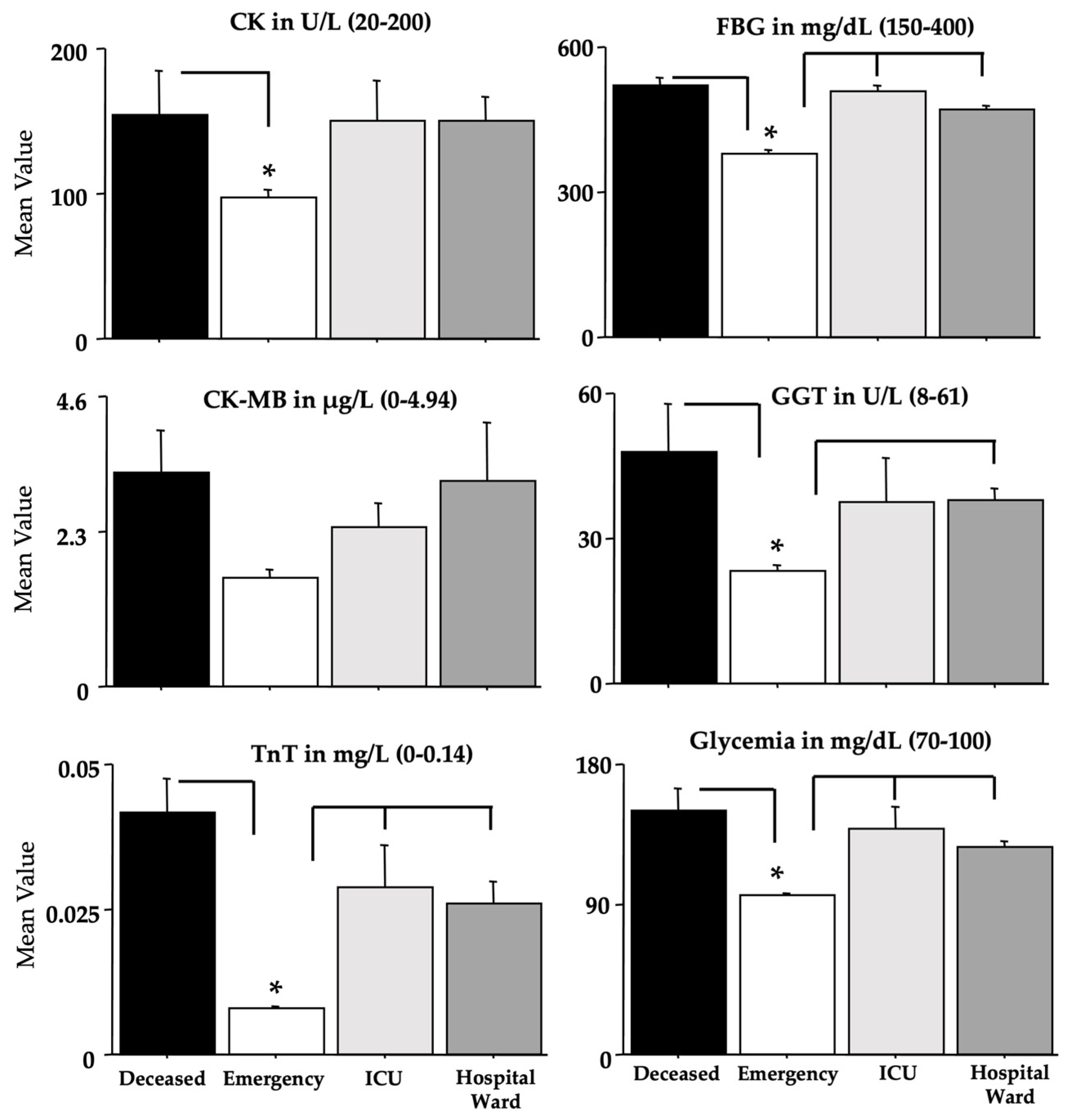
| CK | 3,425 | 2.74 | =0.042 |
| CK-MB | 3,403 | 1.03 | 0.375 |
| TnT | 3,419 | 12.12 | <0.001 |
| FBG | 3,422 | 36.03 | <0.001 |
| GGT | 3,395 | 6.98 | <0.001 |
| Glycemia | 3,427 | 18.59 | <0.001 |
| CRP | 3,355 | 46.41 | <0.001 |
| INR | 3,424 | 1.16 | 0.323 |
| aPTT | 3,422 | 8.08 | <0.001 |
| LDH | 3,427 | 31.40 | <0.001 |
| Albumin | 3,383 | 1.97 | 0.117 |
| D-dimer | 3,403 | 29.05 | <0.001 |
| MGB | 3,275 | 8.60 | <0.001 |
| AST | 3,427 | 2.76 | =0.041 |
| ALT | 3,426 | 1.15 | 0.325 |
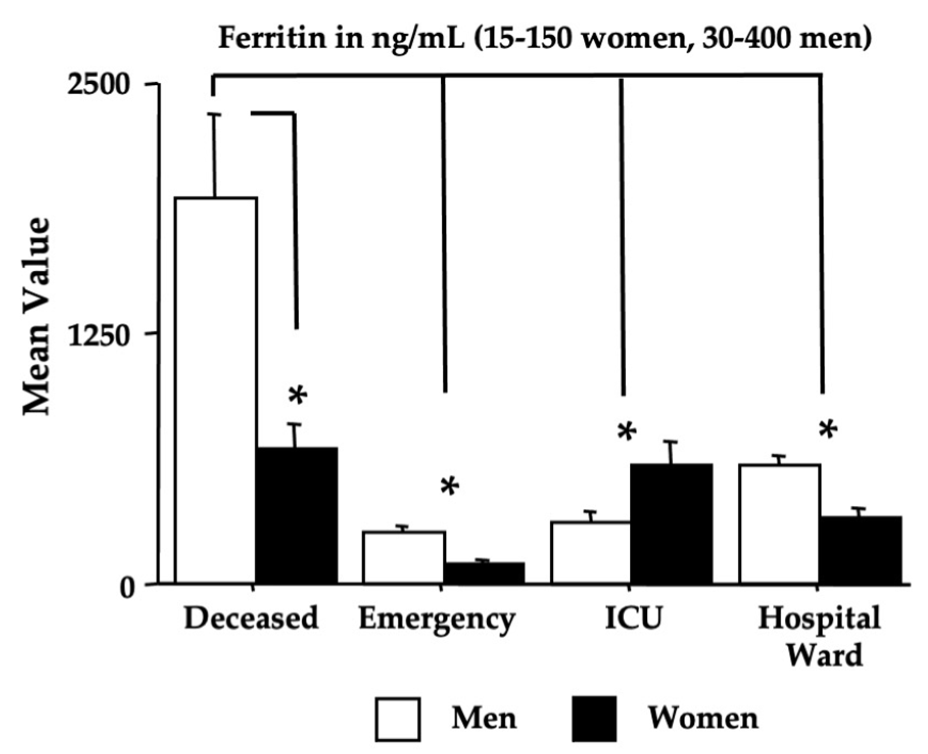
| Ferritin sex sex x morbidity | 3,384 | 32.27 | <0.001 |
| 1,384 | 15.99 | <0.001 | |
| 3,384 | 9.35 | <0.001 | |
| Deceased vs. Emergency | ICU vs. Emergency | |||
|---|---|---|---|---|
| Area under the Curve (AUC) | 95% CI for AUC | Area under the Curve (AUC) | 95% CI for AUC | |
| P/F ratio | 0.792 | 0.695–0.889 | 0.294 | 0.189–0.399 |
| CK | 0.542 | 0.413–0.672 | 0.453 | 0.305–0.601 |
| CK-MB | 0.664 | 0.547–0.780 | 0.618 | 0.478–0.757 |
| TnT | 0.920 | 0.858–0.982 | 0.782 | 0.702–0.862 |
| FBG | 0.844 | 0.746–0.942 | 0.852 | 0.771–0.932 |
| GGT | 0.680 | 0.575–0.784 | 0.661 | 0.481–0.739 |
| Glycemia | 0.805 | 0.708–0.902 | 0.740 | 0.622–0.859 |
| CRP | 0.925 | 0.856–0.994 | 0.787 | 0.646–0.929 |
| INR | 0.653 | 0.540–0.765 | 0.504 | 0.379–0.629 |
| aPTT | 0.756 | 0.645–0.867 | 0.599 | 0.473–0.726 |
| LDH | 0.871 | 0.781–0.961 | 0.693 | 0.579–0.807 |
| Albumin | 0.957 | 0.922–0.993 | 0.764 | 0.635–0.893 |
| D-dimer | 0.921 | 0.881–0.961 | 0.822 | 0.731–0.913 |
| MGB | 0.910 | 0.833–0.986 | 0.756 | 0.637–0.876 |
| AST | 0.774 | 0.677–0.870 | 0.605 | 0.472–0.739 |
| ALT | 0.484 | 0.362–0.606 | 0.539 | 0.404–0.674 |
| Ferritin | 0.948 | 0.907–0.989 | 0.812 | 0.725–0.899 |
| Ferritin (men) | 0.962 | 0.924–1 | 0.585 | 0.411–0.759 |
| Ferritin (women) | 0.956 | 0.907–1 | 0.953 | 0.896–1 |
| PPV (Deceased) | PPV (ICU) | PPV (Hospital Ward) | NPV (Emergency) | |
|---|---|---|---|---|
| P/F ratio (≥400) | 0.571 | 0.360 | 0.410 | 0.908 |
| CK (20–200 U/L) | 0.212 | 0.280 | 0.223 | 0.916 |
| CK-MB (0–4.94 µg/L) | 0.156 | 0.041 | 0.070 | 0.965 |
| TnT (0–0.014 mg/L) | 0.781 | 0.458 | 0.482 | 0.893 |
| FBG (150–400 mg/dL) | 0.878 | 0.920 | 0.176 | 0.932 |
| GGT (8–61 U/L) | 0.181 | 0.130 | 0.758 | 0.635 |
| Glycemia (70–100 mg/dL) | 0.818 | 0.720 | 0.548 | 0.657 |
| CRP (0.1–6 mg/L) | 0.785 | 0.333 | 0.294 | 0.815 |
| INR (0.8–1.2) | 0.182 | 0.120 | 0.584 | 0.938 |
| aPTT (ratio 0.86–1.14) | 0.531 | 0.160 | 0.362 | 0.785 |
| LDH (135–225 U/L) | 0.878 | 0.600 | 0.616 | 0.670 |
| Albumin (3.5–5 g/dL) | 0.625 | 0.181 | 0.196 | 0.968 |
| D-dimer (50–420 µg/L) | 1 | 0.869 | 0.796 | 0.565 |
| MGB (28–72 µg/L) | 0.591 | 0.318 | 0.586 | 0.304 |
| AST (9–45 U/L) | 0.242 | 0.160 | 0.141 | 0.961 |
| ALT (10–40 U/L) | 0.090 | 0.240 | 0.263 | 0.837 |
| Ferritin (ng/mL) | ||||
| Ferritin (men 30–400) | 0.900 | 0.250 | 0.515 | 0.800 |
| Ferritin (women 15–150) | 0.909 | 0.899 | 0.595 | 0.714 |
| Men | Women | |||||
|---|---|---|---|---|---|---|
| SSD | Rho | p-Value | SSD | Rho | p-Value | |
| P/F ratio | 3108.50 | −0.352 | 0.918 | 243.50 | 0.149 | 0.622 |
| CK | 1598.00 | 0.210 | 0.323 | 144.00 | 0.345 | 0.274 |
| CK-MB | 1052.00 | 0.406 | 0.062 | 128.00 | 0.224 | 0.501 |
| TnT | 349.00 | 0.803 | 0.001 | 243.00 | −0.107 | 0.735 |
| FBG | 2040.00 | −0.008 | 0.970 | 297.00 | −0.350 | 0.268 |
| GGT | 2611.50 | −0.290 | 0.173 | 174.00 | −0.055 | 0.870 |
| Glycemia | 1739.00 | 0.141 | 0.509 | 268.00 | −0.218 | 0.490 |
| CRP | 1009.50 | 0.114 | 0.627 | 102.00 | 0.382 | 0.252 |
| INR | 1732.50 | 0.144 | 0.499 | 214.00 | 0.027 | 0.931 |
| aPTT | 1198.00 | 0.324 | 0.138 | 196.00 | 0.109 | 0.730 |
| LDH | 2483.50 | −0.227 | 0.286 | 136.50 | 0.380 | 0.230 |
| Albumin | 2018.00 | 0.003 | 0.988 | 268.50 | −0.220 | 0.485 |
| D-dimer | 1123.00 | 0.366 | 0.093 | 102.00 | 0.382 | 0.252 |
| MGB | 193.00 | 0.470 | 0.103 | 146.00 | 0.115 | 0.729 |
| AST | 2631.00 | −0.300 | 0.159 | 220.00 | 0.001 | 0.999 |
| ALT | 2722 | −0.345 | 0.105 | 183.00 | 0.168 | 0.532 |
| Ferritin | 2306.50 | −0.498 | 0.025 | 256.00 | −0.164 | 0.604 |
| Omicron COVID-19 and Vaccination Effects | |||||||||
|---|---|---|---|---|---|---|---|---|---|
| Vaccination (Yes/No) | Omicron | Vaccination x Omicron | |||||||
| dF | F-Value | p-Value | dF | F-Value | p-Value | dF | F-Value | p-Value | |
| P/F ratio | 1,438 | 3.092 | 0.079 | 3 | 29.12 | 0.001 | 3 | 1.805 | 0.145 |
| CK | 1,423 | 0.731 | 0.393 | 3 | 2.741 | 0.043 | 3 | 1.247 | 0.292 |
| CK-MB | 1,401 | 0.154 | 0.695 | 3 | 0.760 | 0.517 | 3 | 0.199 | 0.897 |
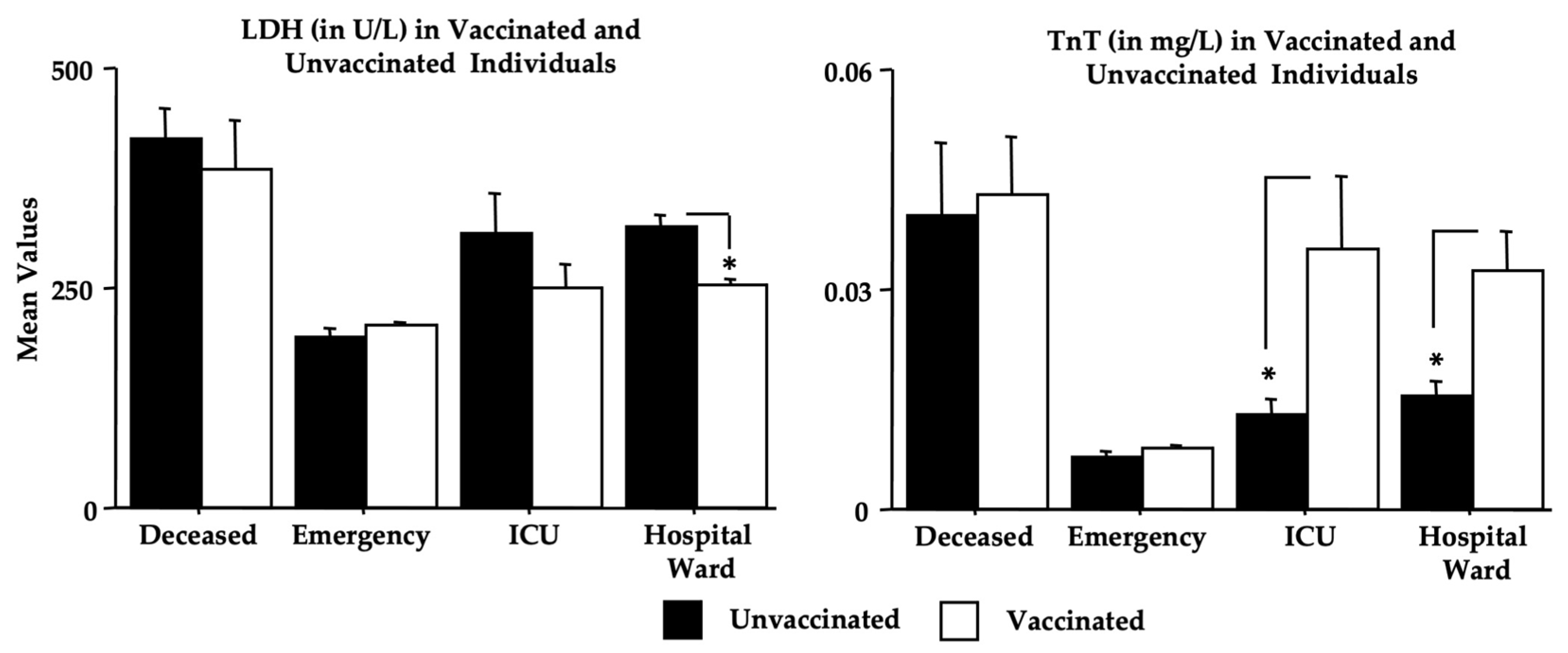
| TnT | 1,417 | 3.927 | 0.048 | 3 | 9.233 | 0.001 | 3 | 1.378 | 0.249 |
| FBG | 1,420 | 1.302 | 0.255 | 3 | 37.44 | 0.001 | 3 | 4.042 | 0.007 |
| GGT | 1,393 | 1.250 | 0.264 | 3 | 8.478 | 0.001 | 3 | 3.261 | 0.022 |
| Glycemia | 1,425 | 2.844 | 0.092 | 3 | 14.90 | 0.001 | 3 | 2.089 | 0.101 |
| CRP | 1,353 | 1.368 | 0.243 | 3 | 52.06 | 0.001 | 3 | 0.891 | 0.446 |
| INR | 1,422 | <0.001 | 1.000 | 3 | 1.526 | 0.207 | 3 | 0.301 | 0.825 |
| aPTT | 1,420 | 1.587 | 0.209 | 3 | 7.636 | 0.001 | 3 | 0.759 | 0.518 |
| LDH | 1,425 | 6.831 | 0.009 | 3 | 35.54 | 0.001 | 3 | 3.396 | 0.018 |
| Albumin | 1,381 | 0.085 | 0.771 | 3 | 1.637 | 0.180 | 3 | 0.261 | 0.853 |
| D-dimer | 1,401 | 0.010 | 0.919 | 3 | 28.99 | 0.001 | 3 | 3.758 | 0.011 |
| MGB | 1,274 | 1.864 | 0.173 | 3 | 6.686 | 0.001 | 3 | 0.767 | 0.513 |
| AST | 1,425 | 2.017 | 0.156 | 3 | 2.114 | 0.098 | 3 | 0.288 | 0.834 |
| ALT | 1,424 | 1.318 | 0.252 | 3 | 0.781 | 0.505 | 3 | 0.590 | 0.622 |
| Ferritin | 1,382 | 0.195 | 0.659 | 3 | 38.78 | 0.001 | 3 | 2.248 | 0.082 |
Disclaimer/Publisher’s Note: The statements, opinions and data contained in all publications are solely those of the individual author(s) and contributor(s) and not of MDPI and/or the editor(s). MDPI and/or the editor(s) disclaim responsibility for any injury to people or property resulting from any ideas, methods, instructions or products referred to in the content. |
© 2023 by the authors. Licensee MDPI, Basel, Switzerland. This article is an open access article distributed under the terms and conditions of the Creative Commons Attribution (CC BY) license (https://creativecommons.org/licenses/by/4.0/).
Share and Cite
Pennacchia, F.; Rusi, E.; Ruqa, W.A.; Zingaropoli, M.A.; Pasculli, P.; Talarico, G.; Bruno, G.; Barbato, C.; Minni, A.; Tarani, L.; et al. Blood Biomarkers from the Emergency Department Disclose Severe Omicron COVID-19-Associated Outcomes. Microorganisms 2023, 11, 925. https://doi.org/10.3390/microorganisms11040925
Pennacchia F, Rusi E, Ruqa WA, Zingaropoli MA, Pasculli P, Talarico G, Bruno G, Barbato C, Minni A, Tarani L, et al. Blood Biomarkers from the Emergency Department Disclose Severe Omicron COVID-19-Associated Outcomes. Microorganisms. 2023; 11(4):925. https://doi.org/10.3390/microorganisms11040925
Chicago/Turabian StylePennacchia, Fiorenza, Eqrem Rusi, Wael Abu Ruqa, Maria Antonella Zingaropoli, Patrizia Pasculli, Giuseppina Talarico, Giuseppe Bruno, Christian Barbato, Antonio Minni, Luigi Tarani, and et al. 2023. "Blood Biomarkers from the Emergency Department Disclose Severe Omicron COVID-19-Associated Outcomes" Microorganisms 11, no. 4: 925. https://doi.org/10.3390/microorganisms11040925