Article

Biocontrol of Soft Rot Dickeya and Pectobacterium Pathogens by Broad-Spectrum Antagonistic Bacteria within Paenibacillus polymyxa Complex

1
State Key Laboratory of Rice Biology and Breeding, Ministry of Agriculture Key Laboratory of Molecular Biology of Crop Pathogens and Insects, Zhejiang Province Key Laboratory of Biology of Crop Pathogens and Insects, Institute of Biotechnology, Zhejiang University, Hangzhou 310058, China
2
Department of Plant Pathology, Bangabandhu Sheikh Mujibur Rahman Agricultural University, Gazipur 1706, Bangladesh
3
Sugarcane Research Institute, Guangxi Academy of Agricultural Sciences, Nanning 530007, China
4
Department of Plant Quarantine, Shanghai Extension and Service Center of Agriculture Technology, Shanghai 201103, China
5
Agricultural Technology Extension Center of Fuyang District, Hangzhou 311400, China
6
State Key Laboratory for Quality and Safety of Agro-Products, Institute of Plant Protection and Microbiology, Zhejiang Academy of Agricultural Sciences, Hangzhou 310021, China
*
Authors to whom correspondence should be addressed.
These authors contributed equally to this work.
Microorganisms 2023, 11(4), 817; https://doi.org/10.3390/microorganisms11040817
Submission received: 15 February 2023 / Revised: 16 March 2023 / Accepted: 20 March 2023 / Published: 23 March 2023
(This article belongs to the Section Plant Microbe Interactions)

Abstract

:
Polymyxin-producing bacteria within the Paenibacillus polymyxa complex have broad-spectrum activities against fungi and bacteria. Their antibacterial activities against soft rot Dickeya and Pectobacterium phytopathogens containing multiple polymyxin-resistant genes were not clear. Here, we selected nine strains within the P. polymyxa complex having broad-spectrum antagonistic activities against phytopathogenic fungi and a polymyxin-resistant D. dadantii strain causing stem and root rot disease of sweet potato and did antagonistic assays on nutrient agar and sweet potato tuber slices. These strains within the P. polymyxa complex showed clear antagonistic activities against D. dadantii in vitro and in vivo. The most effective antagonistic strain P. polymyxa ShX301 showed broad-spectrum antagonistic activities against all the test Dickeya and Pectobacterium strains, completely eliminated D. dadantii from sweet potato seed tubers, and promoted the growth of sweet potato seedlings. Cell-free culture filtrate of P. polymyxa ShX301 inhibited D. dadantii growth, swimming motility, and biofilm formation and disrupted D. dadantii plasma membranes, releasing nucleic acids and proteins. Multiple lipopeptides produced by P. polymyxa ShX301 may play a major role in the bactericidal and bacteriostatic actions. This study clarifies that the antimicrobial spectrum of polymyxin-producing bacteria within the P. polymyxa complex includes the polymyxin-resistant Dickeya and Pectobacterium phytopathogens and strengthens the fact that bacteria within the P. polymyxa complex have high probability of being effective biocontrol agents and plant growth promoters.

1. Introduction

Gram-negative Pectobacterium and Dickeya bacteria produce multiple pectinases and cause destructive soft rot diseases of numerous crops and ornamental plants and great economic losses worldwide [1,2]. While the worldwide distribution of soft rot Pectobacterium was well-known, soft rot Dickeya was previously described as being present mainly in tropical and subtropical regions but is expanding its global distribution. For example, Dickeya spp. associated with blackleg of potato and foot rot of rice spread rapidly in Europe as far north as Finland near the Arctic Circle [3,4] and in China from South China to Northeast China [5]. Importantly, soft rot Dickeya is threatening staple food crops, including potato [3,6], rice [7], maize [8], sweet potato [9,10], and banana [11,12].
Management of soft rot diseases on crops has not been successful due to the lack of resistant crop varieties and the end of large-scale use of effective antibiotics for the risk of introducing resistance to bacterial pathogens in humans or animals [1]. Biological control is an alternative to the breeding of resistant crops and chemical control the diseases and has been increasingly tested to control Pectobacterium and Dickeya pathogens, particularly using bacteriophages with restricted bacterial hosts [13] and broad-spectrum antagonistic bacteria [14,15].
Gram-positive Bacillus and Paenibacillus bacteria, which produce multiple antimicrobial compounds against broad phytopathogens and produce endospores resistant to heating and dryness for the formulation of stable products, are high-profile biological control agents [16,17,18]. Paenibacillus polymyxa is well-known for its production of the antibiotic polymyxins, which are cyclic lipopeptides and used as last-resort agents against notorious Gram-negative multi-drug resistant human pathogens, including Acinetobacter baumannii, Pseudomonas aeruginosa, and Klebsiella pneumoniae [19]. P. polymyxa forms a monophyletic species complex with P. peoriae, P. kribbensis, P. ottowii, P. brasilensis, P. terrae, and “P. maysiensis,” most of which also produce the cyclic lipopeptides-fusaricidins against broad-spectrum fungi, oomycetes, and Gram-positive bacteria, including important phytopathogens [16,20].
Polymyxins are a group of decapeptides containing five to six residues of the nonproteogenic amino acid 2,4-diaminobutyric acid, resulting in a high positive charge density. Polymyxins bind to the lipid A component of lipopolysaccharides on the outer membrane of Gram-negative bacteria and disrupt the outer membrane and then permeabilize and disrupt the inner membrane [16,21]. The general mechanism of bacterial resistance to polymyxins involves modification of lipid A, such as the addition of 4-aminoarabinose by the arnB operon products, the addition of phosphoethanolamine by EptA, hydroxylation of lipid A acyl chains by LpxO, and acylation or deacylation of lipid A by PagP or PagL [22], thereby reducing the net negative charge and polymyxin-binding affinity [16,21]. In Gram-positive bacteria, resistance to antimicrobial peptides occurs in the esterification of phosphate with alanine of teichoic acids by the dltXABCD operon products [23]. In all the sequenced genomes of Dickeya and Pectobacterium, not only arnB operon and eptA are present similar to other enterobacteria, but also dlt operon in Gram-positive bacteria is present [24]. These genes confer resistance to polymyxins.
Polymyxin-resistant Dickeya and Pectobacterium may resist the antagonistic activity from polymyxin-producing strains within the P. polymyxa complex. However, previous studies have not intentionally tested this hypothesis. Here, we tested if the antagonism of the P. polymyxa complex is negative on the polymyxin-resistant Dickeya and Pectobacterium and see if the antagonistic spectrum of the P. polymyxa complex can extend to the polymyxin-resistant Dickeya and Pectobacterium. We selected strains within the P. polymyxa complex previously screened out for their broad-spectrum antagonistic activities against phytopathogenic fungi [20,25] to test their antagonistic activities against Dickeya and Pectobacterium. A polymyxin-resistant D. dadantii strain was used for the first-round screening. Contrary to the hypothesis, all the test strains within the P. polymyxa complex showed clear antagonistic activities against D. dadantii, and the most effective strain P. polymyxa ShX301 showed antagonism to all the tested Dickeya and Pectobacterium strains. We further studied the antibacterial profiles of P. polymyxa ShX301 and showed its application potential to control diseases in fields.

2. Materials and Methods

2.1. Bacterial Strains and Media

Three strains isolated from the rhizosphere soils of cotton plants [25] and six strains isolated from legume nodules [20] were recently screened out for their broad-spectrum antagonistic activities against phytopathogenic fungi. Dickeya dadantii strain CZ1501 is a causal agent of the bacterial stem, and the root disease of sweet potato occurred in Hangzhou, Zhejiang province, China. Its whole genome sequence (GenBank accession number MPDL00000000.1) contains the arnB, eptA, and dlt operons for resistance of polymyxins. It can grow in nutrient broth with 2 μg·mL−1 of polymyxin B sulfate. Six other Dickeya and two Pectobacterium strains were obtained from culture collections (Table 1).
Bacterial strains were cultured in nutrient broth (10 g tryptone, 3 g beef extract, 2.5 g glucose, 5 g NaCl per liter, pH 7.0) or on nutrient agar (nutrient broth with 20 g agar per liter). Bacterial cultures in nutrient broth were washed with sterile water and adjusted to an optical density of 0.6 at 600 nm (OD600); the cell number of the suspension was counted by serial dilution and plating on nutrient agar. Bacterial suspensions were finally adjusted to the concentration of 1 × 10⁸ CFU·mL−1 for use.

2.2. In Vitro and In Vivo Screening of Antagonistic Paenibacillus Strains

Paenibacillus strains against D. dadantii CZ1501 were screened using the in vitro overly culture assay on nutrient agar plates and the in vivo tuber slice assay as previously described [15]. Antagonistic activities of the P. polymyxa stain ShX301 were further tested on other Dickeya and Pectobacterium strains (Table 1) using the in vitro overlay culture assay on both the nutrient agar and the M9 minimal agar [15]. The in vitro inhibition rate (%) on target strains by Paenibacillus was calculated by [1-(diameter of Paenibacillus colony/diameter of inhibition zone)] × 100. The in vivo inhibition rate (%) was calculated by [1-(diameter of maceration zone by D. dadantii with Paenibacillus/diameter of maceration zone by D. dadantii without Paenibacillus] × 100.

2.3. In Vivo Biocontrol of D. dadantii by P. polymyxa ShX301

The biocontrol potential of the P. polymyxa stain ShX301 was determined using the in vivo assay with sweet potato seed tubers as previously described [15]. Surface-sterilized seed tubers were immersed in sterile distilled water (control), D. dadantii suspension (1 × 10⁸ CFU·mL−1), P. polymyxa suspension (1 × 10⁸ CFU·mL−1), or both D. dadantii and P. polymyxa at 1 × 10⁸ CFU·mL−1 for 4 h at 25 °C and then kept under 28 °C, a photoperiod of 12-h light and 12-h dark, and 80% relative humidity for 21 d.

2.4. Antibacterial Activities of Cell-Free Culture Supernatant (CFCS) of P. polymyxa ShX301 against D. dadantii

P. polymyxa ShX301 was cultured in nutrient broth at 30 °C with shaking at 200 rpm for 48 h and adjusted to about 1 × 10⁸ CFU·mL−1 with the nutrient broth. CFCS of the bacterial suspension was obtained by centrifugation and filtering through a 0.22-µm filter and confirmed by incubating 100 µL of the CFCS on nutrient agar at 30 °C for 48 h.
CFCS effects on D. dadantii growth in wells of sterile polystyrene flat-bottom 96-well microplates, biofilm formation on the surface of the microplate wells, and swimming motility in 0.3% (w/v) agar were determined as previously described [15].
CFCS effects on D. dadantii cell integrity were observed by transmission electron microscopy on D. dadantii cells (1 × 10⁸ CFU·mL−1) at mid-exponential phase grown with CFCS (50% volume) for 4 h. D. dadantii cells were washed twice with 0.1 mol·L−1 phosphate buffer (pH 7) and fixed in 2.5% (v/v) glutaraldehyde in the phosphate buffer at 4 °C overnight. After washing with the phosphate buffer, D. dadantii cells were fixed in 1% (w/v) OsO4 dissolved in the phosphate buffer for 1 h at room temperature. D. dadantii cells were then washed with distilled water and dehydrated by a graded series of ethanol. Dehydrated D. dadantii cells were infiltrated by Spurr’s resin at room temperature and embedded in Spurr’s resin at 70 °C for 9 h. Ultrathin sections were cut with glass knives on an ultramicrotome (Reichert-Jung, Vienna, Austria), collected on copper grids, stained with uranyl acetate and lead citrate, and observed with the JEM-1230 transmission electron microscope (JEOL, Tokyo, Japan).
CFCS effects on D. dadantii cell integrity were determined by measuring the release of nucleic acids (absorbance at 260 nm, OD260) and proteins (absorbance at 280 nm, OD280) from D. dadantii cells (1 × 10⁸ CFU·mL−1) at mid-exponential phase grown with CFCS (50% volume) for 4 h as previously described [15].

2.5. Detection of Lipopeptides Produced by P. polymyxa ShX301

Lipopeptides in the CFCS of P. polymyxa ShX301 were detected by matrix-assisted laser desorption/ionization coupled with time-of-flight mass spectrometry (MALDI-TOF MS) as previously described [15].

2.6. Statistical Analysis

Data were subjected to a one-way analysis of variance, and means were compared by Duncan’s multiple range test using the SPSS software version 16 (SPSS, Chicago, IL, USA). The significance was set at p < 0.05.

3. Results

3.1. Strains within the P. polymyxa Complex Inhibited D. dadantii Growth and Maceration of Sweet Potato Tuber Slices

All tested strains within the P. polymyxa complex inhibited D. dadantii growth in the nutrient agar and generated clearing zones around the Paenibacillus colonies. P. polymyxa ShX301 generated relatively larger clearing zones and showed a significantly higher rate (53%) of in vitro inhibition of D. dadantii growth than other Paenibacillus strains did.
D. dadantii CZ1501 degraded plant cell walls and generated maceration zones about 36 mm in diameter at 24 h after inoculation into the sweet potato tuber slices. All tested Paenibacillus strains did not macerate the tuber tissues but inhibited the maceration from D. dadantii. The in vivo inhibition of the maceration from D. dadantii was consistent with the in vitro inhibition of D. dadantii growth in the nutrient agar (Table 2). P. polymyxa ShX301 showed the highest in vivo inhibition rate (80%) at the D. dadantii maceration of sweet potato tuber slices. Therefore, P. polymyxa ShX301 was the most effective strain against D. dadantii and was used for further analyses.

3.2. P. polymyxa ShX301 Showed Broad-Spectrum Antagonistic Activities against Dickeya and Pectobacterium

P. polymyxa ShX301 showed antagonistic activities against all the tested Dickeya and Pectobacterium strains on the nutrient agar and the M9 minimal agar (Table 3). It showed more potent antagonistic activities on the M9 minimal agar than on the nutrient agar.

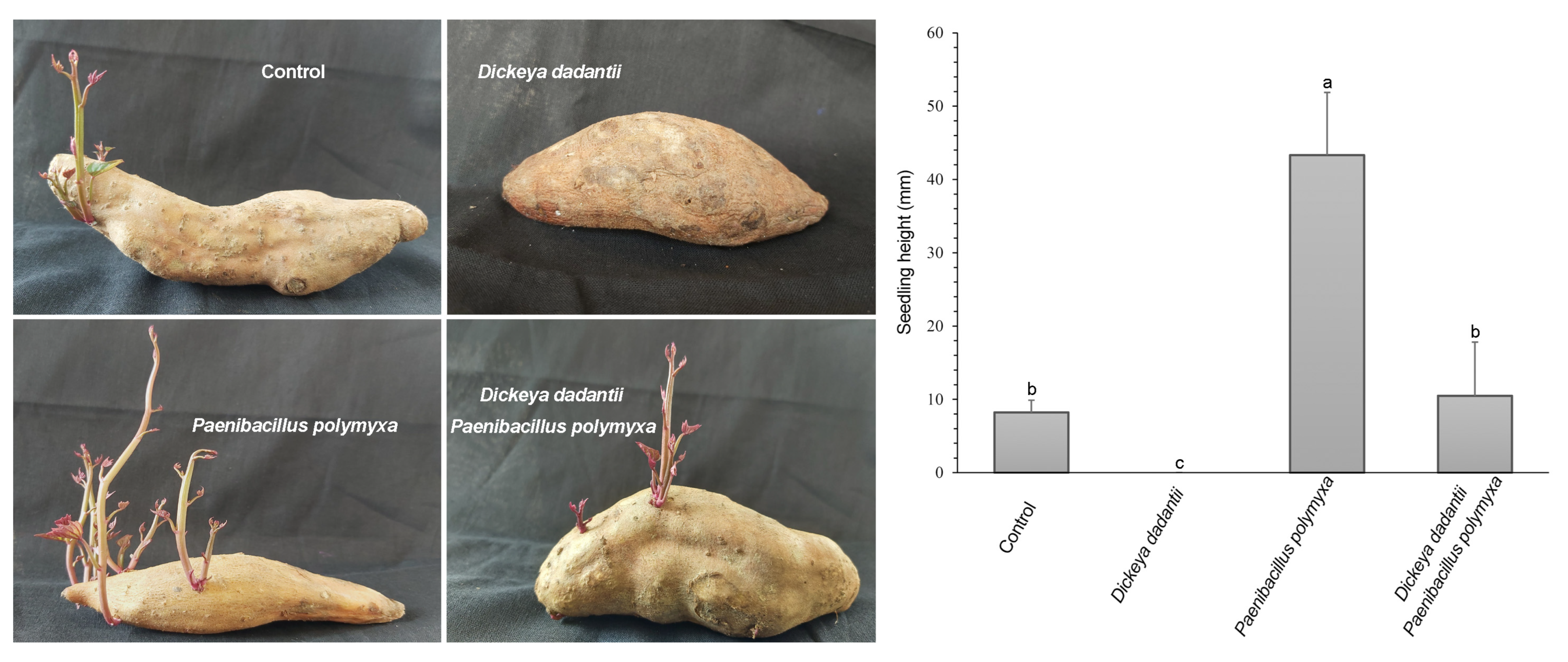
3.3. P. polymyxa ShX301 Protected Seed Tubers and Promoted Seedling Growth

Seed tubers inoculated with only D. dadantii did not germinate and were rotten; D. dadantii was isolated from the rotten seed tubers at 21 d after inoculation. In contrast, seed tubers inoculated with only P. polymyxa ShX301 germinated and grew seedlings significantly higher than the control seedlings. Seed tubers inoculated with D. dadantii along with P. polymyxa ShX301 germinated and grew seedlings slightly higher than the control seedlings (Figure 1). D. dadantii was not isolated from the seedlings inoculated with P. polymyxa ShX301.

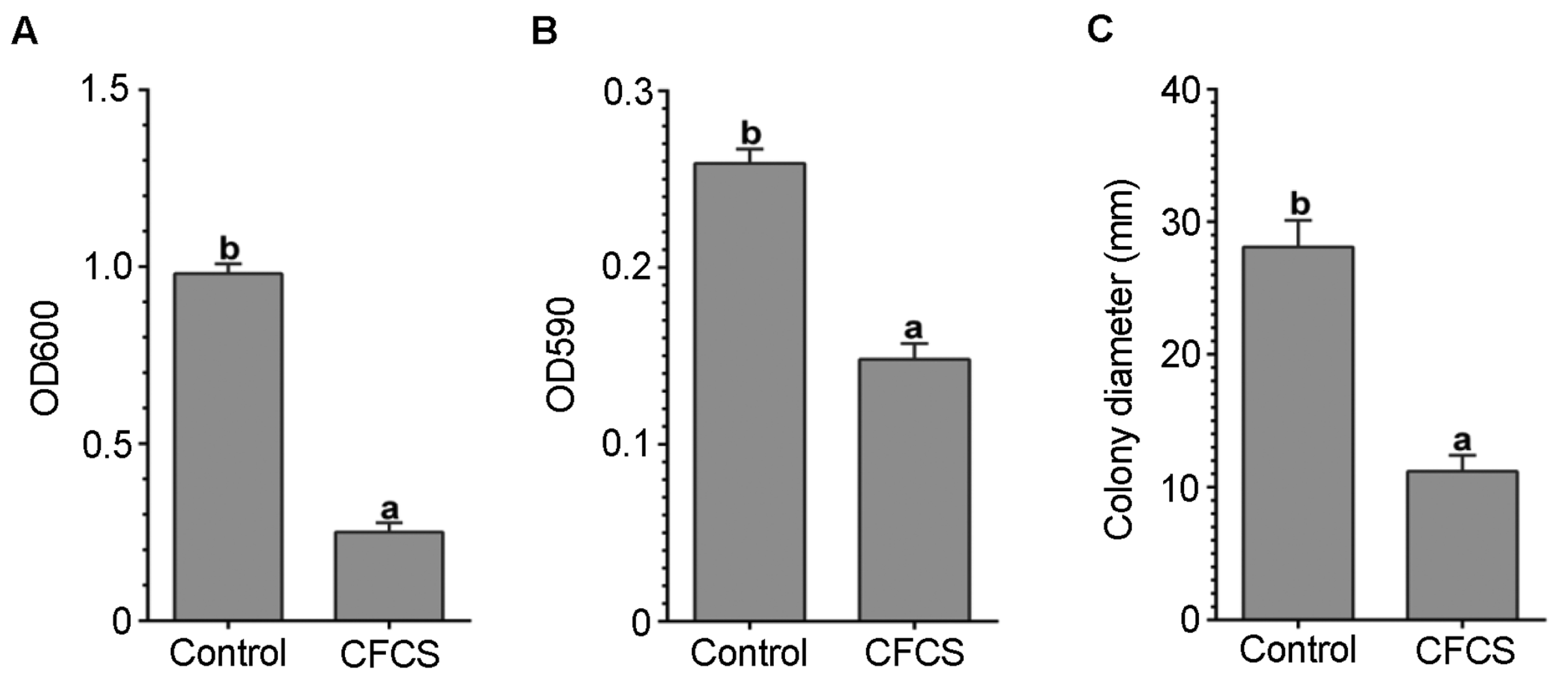
3.4. CFCS of P. polymyxa ShX301 Inhibited D. dadantii Growth, Biofilm Formation, and Swimming Motility

D. dadantii growth in nutrient broth in the microplate wells and biofilm formation on the surface of the microplate wells were significantly inhibited when the CFCS of P. polymyxa ShX301 was present at 50% volume (Figure 2A,B). D. dadantii CZ1501 swam via flagella in 0.3% (w/v) agar and formed haloes about 28 mm in diameter after 48 h. The swimming motility of D. dadantii was almost lost and formed haloes about 11 mm in diameter when the CFCS was present at 50% volume (Figure 2C).

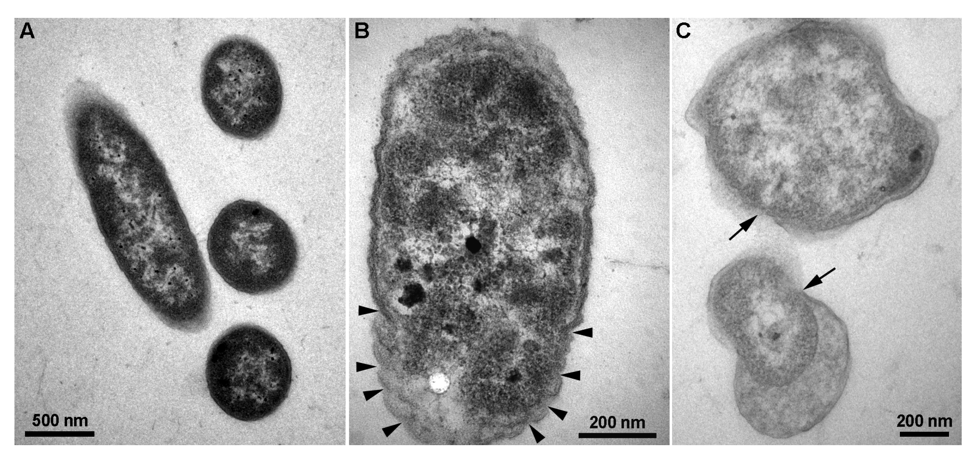
3.5. CFCS of P. polymyxa ShX301 Breached D. dadantii Cells

Transmission electron microscopy showed that control D. dadantii cell envelopes were intact and enclosed electron-dense cytoplasm (Figure 3A) while cell envelopes were convoluted (Figure 3B) or beached and cytoplasm was clearing (Figure 3C) when the CFCS of P. polymyxa ShX301 were present for 4 h. Under the CFCS of P. polymyxa ShX301, damage to D. dadantii cells was also indicated by the release of nucleic acids (128 µg·mL−1) and proteins (2.75 mg·mL−1) determined by the increases of the OD260 value (0.33) and OD280 value (0.31), respectively.

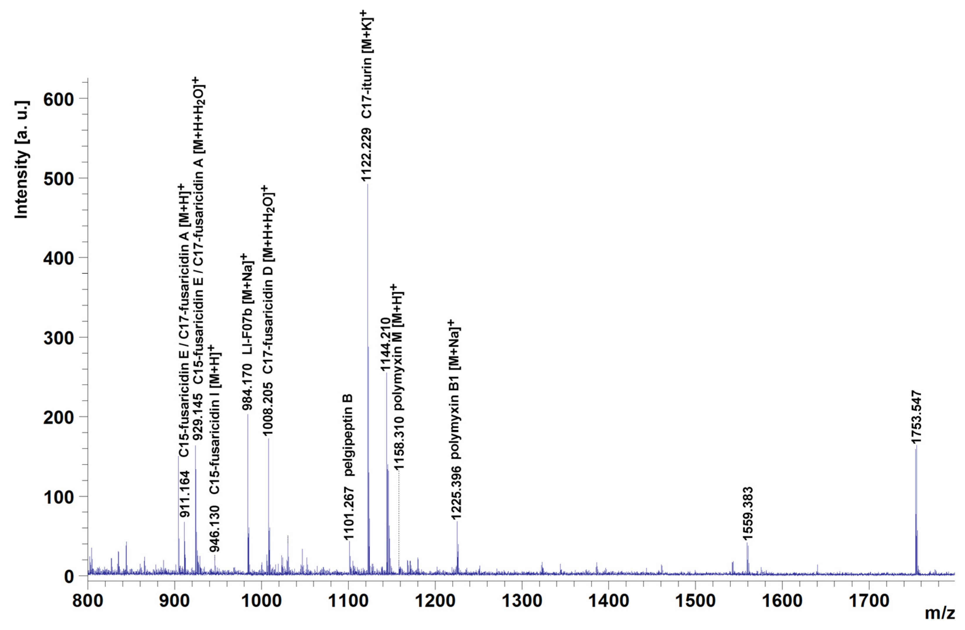
3.6. P. polymyxa ShX301 Produced Multiple Lipopeptides

MALDI-TOF MS detected lipopeptides which appeared in a protonated form [M+H]+ and alkali adducts, such as [M+Na]+ and [M+K]+. Fusaricidins also appeared as open-chain products [M+H+H2O]+ [26,27]. MALDI-TOF MS detected eight distinct clusters of mass peaks (m/z 802–865, m/z 904–946, m/z 984–1052, m/z 1101, m/z 1122, m/z 1144–1225, m/z 1542–1559 and m/z 1753) in the range of m/z 800–1800 from CFCS of P. polymyxa ShX301 (Figure 4). The mass peaks m/z 904–946 and m/z 984–1052 were in the m/z range for fusaricidins [26,27]. The mass peak m/z 1144–1225 was in the m/z range for polymyxins. The mass peak m/z 1101 may correspond to pelgipeptin B [28,29]. The mass peak m/z 1122 was close to m/z 1122.6 [M+K]+ of C17-iturin [30]. The mass peaks m/z 802–865, m/z 1559, and m/z 1753 were not identified.

4. Discussion

Contrary to the hypothesis that polymyxin-resistant Dickeya and Pectobacterium may resist the antagonism from polymyxin-producing strains within the P. polymyxa complex, all the test strains within the P. polymyxa complex, which have broad-spectrum antagonistic activities against fungal phytopathogens [20,25], showed clear antagonistic activities against the polymyxin-resistant D. dadantii strain CZ1501. Moreover, the most effective strain P. polymyxa ShX301 showed clear antagonistic activities against all the tested Dickeya and Pectobacterium strains. The extents of the in vitro inhibition of D. dadantii growth in nutrient agar and the in vivo inhibition of D. dadantii maceration in sweet potato tuber slices are consistent. P. polymyxa ShX301 showed more potent antibacterial activities against Dickeya and Pectobacterium strains on the nutrient-limited minimal medium than on the nutrient-rich medium. Thus, it may inhibit soft rot pathogens in nutrient-limited niches, such as soils, leaf surfaces, and seed tuber surfaces.
CFCS of P. polymyxa ShX301 inhibited D. dadantii growth, indicating that P. polymyxa released antibacterial compounds into the CFCS. The antibacterial compounds in the CFCS appeared to distort and breach D. dadantii cell envelopes, particularly plasma membranes, leading to the release of cell contents, such as nucleic acids and proteins, which were revealed by transmission electron microscopy and spectrophotometry. Disrupting plasma membranes indicates the involvement of amphiphilic lipopeptides in the antibacterial mechanisms of P. polymyxa.
MALDI-TOF-MS revealed that P. polymyxa ShX301 produced fusaricidins, polymyxins, and pelgipeptin-like and iturin-like lipopeptides. Fusaricidins have potent antimicrobial activities against fungi and Gram-positive bacteria but no or weak activity against Gram-negative bacteria [21,31] and thus may not contribute to the cytotoxic action on D. dadantii. Polymyxins consist of up to 30 closely related lipopeptides against Gram-negative bacteria [19], while Dickeya and Pectobacterium have evolved multiple pathways (arnB, eptA, and dlt operons) to resist polymyxins [24]. However, polymyxin P was found to inhibit the growth of Pectobacterium carotovorum (formerly Erwinia carotovora) [32]. Here, although D. dadantii CZ1501 resists 2 μg·mL−1 of polymyxin B, multiple polymyxins produced by P. polymyxa ShX301 may accumulate to a locally high concentration, contributing to the cytotoxic action on the damage of D. dadantii plasma membrane and the inhibition of D. dadantii growth in vitro, in vivo, and in planta. Pelgipeptins produced by Paenibacillus have broad-spectrum activities against Gram-positive and Gram-negative bacteria and fungi, including phytopathogens [28,29,33,34]. Iturins, which are produced by Bacillus and have potent antifungal activity and limited antibacterial activity [21], have also been found to inhibit the growth of phytopathogenic bacteria, including P. carotovorum [35]. The pelgipeptin-like and iturin-like lipopeptides from P. polymyxa ShX301 may also contribute to the cytotoxic action on D. dadantii.
Flagella-mediated motility and biofilm formation facilitate D. dadantii to colonize plant surface, intercellular spaces, and xylem vessels and complete disease cycles [36,37,38,39]. CFCS of P. polymyxa ShX301 inhibited D. dadantii swimming motility and biofilm formation. Fusaricidins and polymyxins from P. polymyxa may not play a major role in cytotoxic action on D. dadantii but may act as biosurfactants to inhibit surface attachment and biofilm formation [40], thus reducing D. dadantii infection and increasing D. dadantii susceptibility to cytotoxic lipopeptides. Multiple lipopeptides produced by P. polymyxa ShX301 play a major role in bactericidal and bacteriostatic actions on the phytopathogens.
From the view of plant-microbe interactions, plants can induce the expression of polymyxin and fusaricidin biosynthesis genes in P. polymyxa [41], while lipopeptides such as fusaricidins can induce plant systemic resistance to fungal and bacterial pathogens [42,43]. Notably, P. polymyxa ShX301 completely eliminated D. dadantii from sweet potato seed tubers at 21 d after the equivalent inoculation of the two bacteria. Bactericidal and bacteriostatic actions and induced plant systemic resistance mediated by multiple lipopeptides may play together to eliminate D. dadantii. The latent infection of seed tubers by Dickeya and Pectobacterium is a major source of the soft rot diseases of potato and sweet potato plants [2,44]. P. polymyxa ShX301 has shown the potential to control soft rot diseases from seed tuber-borne pathogens. Moreover, inoculating P. polymyxa ShX301 alone to sweet potato seed tubers promoted the growth of sweet potato seedlings. Likewise, P. polymyxa ShX301 promoted the growth of cotton seedlings and suppressed the soil-borne fungal pathogen Verticillium dahlia and the Verticillium wilt disease of cotton seedlings [25]. Therefore, P. polymyxa ShX301 is a promising biocontrol agent and plant growth promoter for future application in fields and study of the underlying mechanisms of the broad-spectrum antagonism and mutually beneficial interactions with plants.

5. Conclusions

This study clarifies that the antimicrobial spectrum of polymyxin-producing bacteria within the P. polymyxa complex includes the polymyxin-resistant soft rot Dickeya and Pectobacterium pathogens and strengthens the fact that bacteria within the P. polymyxa complex have high probability of being effective biocontrol agents and plant growth promoters. P. polymyxa ShX301 will be used to clarify the biocontrol mechanism of the P. polymyxa complex against broad-spectrum phytopathogens, including polymyxin-resistant Dickeya and Pectobacterium and to control plant diseases in fields.

Author Contributions

Conceptualization, L.L. and Q.A.; methodology, A.H., M.A.A., J.L. and M.M.I.M.; investigation and analysis, A.H., M.A.A., Y.Y. and M.M.I.M.; resources, L.L., J.L. and Y.J.; writing—original draft preparation, A.H. and M.A.A.; writing—review and editing, L.L. and Q.A.; supervision, B.L. and Q.A.; project administration, L.L., J.L., Y.J. and Y.W.; funding acquisition, L.L., J.L., Y.J., Y.W. and B.L. All authors have read and agreed to the published version of the manuscript.

Funding

This work was supported by the Shanghai Agriculture Applied Technology Development Program (2021-02-08-00-12-F00771), Zhejiang Provincial Key R&D Program of China (2017C02002), Guangxi Provincial Natural Science Foundation of China (2021GXNSFDA196004), and the National Natural Science Foundation of China (32201916), Hangzhou Science and Technology Development Plan Project (202003A05), State Key Laboratory for Managing Biotic and Chemical Threats to the Quality and Safety of Agro-products (2010DS700124-ZZ2014;-KF202101;-KF202205), and Zhejiang Province Key Laboratory of Biology of Crop Pathogens and Insects.

Informed Consent Statement

Not applicable.

Data Availability Statement

Not applicable.

Conflicts of Interest

The authors declare no conflict of interest. The funders had no role in the design of the study; in the collection, analyses, or interpretation of data; in the writing of the manuscript, or in the decision to publish the results.

References

  1. Charkowski, A.O. The changing face of bacterial soft rot diseases. Annu. Rev. Phytopathol. 2018, 56, 269–288. [Google Scholar] [CrossRef]
  2. Ma, B.; Hibbing, M.E.; Kim, H.S.; Reedy, R.M.; Yedidia, I.; Breuer, J.; Breuer, J.; Glasner, J.D.; Perna, N.T.; Kelman, A. Host range and molecular phylogenies of the soft rot enterobacterial genera Pectobacterium and Dickeya. Phytopathology 2007, 97, 1150–1163. [Google Scholar] [CrossRef] [PubMed] [Green Version]
  3. Degefu, Y.; Potrykus, M.; Golanowska, M.; Virtanen, E.; Lojkowska, E. A new clade of Dickeya spp. plays a major role in potato blackleg outbreaks in North Finland. Ann. Appl. Biol. 2013, 162, 231–241. [Google Scholar] [CrossRef]
  4. Van Gijsegem, F.; Toth, I.K.; van der Wolf, J.M. Outlook—Challenges and perspectives for management of diseases caused by Pectobacterium and Dickeya species. In Plant Diseases Caused by Dickeya and Pectobacterium Species; Springer: Cham, Switzerland, 2021; pp. 283–289. [Google Scholar]
  5. Liu, Q.G.; Zhang, Q.; Wei, C.D. Advances in research of rice bacterial foot rot. Sci. Agric. Sin. 2013, 46, 2923–2931. [Google Scholar]
  6. Toth, I.; Van Der Wolf, J.; Saddler, G.; Lojkowska, E.; Hélias, V.; Pirhonen, M.; Tsror, L.; Elphinstone, J. Dickeya species: An emerging problem for potato production in Europe. Plant Pathol. 2011, 60, 385–399. [Google Scholar] [CrossRef]
  7. Pu, X.; Zhou, J.; Lin, B.; Shen, H. First report of bacterial foot rot of rice caused by a Dickeya zeae in China. Plant Dis. 2012, 96, 1818. [Google Scholar] [CrossRef] [PubMed]
  8. Kumar, A.; Hunjan, M.S.; Kaur, H.; Rawal, R.; Kumar, A.; Singh, P. A review on bacterial stalk rot disease of maize caused by Dickeya zeae. J. Appl. Nat. Sci. 2017, 9, 1214–1225. [Google Scholar] [CrossRef] [Green Version]
  9. Huang, L.; Fang, B.; Luo, Z.; Chen, J.; Zhang, X.; Wang, Z. First report of bacterial stem and root rot of sweet potato caused by a Dickeya sp. (Erwinia chrysanthemi) in China. Plant Dis. 2010, 94, 1503. [Google Scholar] [CrossRef]
  10. Stahr, M.N.; Butler, S.; Huerta, A.I.; Ritchie, D.F.; Quesada-Ocampo, L.M. First report of bacterial root rot, caused by Dickeya dadantii, on sweet potato (Ipomoea batatas) in North Carolina. Plant Dis. 2020, 104, 2723. [Google Scholar] [CrossRef]
  11. Liu, Q.; Xiao, W.; Wu, Z.; Li, S.; Yuan, Y.; Li, H. Identification of Dickeya dadantii as a causal agent of banana bacterial sheath rot in China. J. Plant Pathol. 2016, 98, 503–510. [Google Scholar]
  12. Zhang, J.; Shen, H.; Pu, X.; Lin, B.; Hu, J. Identification of Dickeya zeae as a causal agent of bacterial soft rot in banana in China. Plant Dis. 2014, 98, 436–442. [Google Scholar] [CrossRef] [PubMed] [Green Version]
  13. Czajkowski, R. Bacteriophages of soft rot Enterobacteriaceae-a minireview. FEMS Microbiol. Lett. 2016, 363, fnv230. [Google Scholar] [CrossRef] [Green Version]
  14. Des Essarts, Y.R.; Cigna, J.; Quêtu-Laurent, A.; Caron, A.; Munier, E.; Beury-Cirou, A.; Hélias, V.; Faure, D. Biocontrol of the potato blackleg and soft rot diseases caused by Dickeya dianthicola. Appl. Environ. Microbiol. 2016, 82, 268–278. [Google Scholar] [CrossRef] [PubMed] [Green Version]
  15. Hossain, A.; Islam Masum, M.M.; Wu, X.; Abdallah, Y.; Ogunyemi, S.O.; Wang, Y.; Sun, G.; Li, B.; An, Q. Screening of Bacillus strains in biocontrol of pathogen Dickeya dadantii causing stem and root rot disease of sweet potato. Biocontrol Sci. Technol. 2020, 30, 1180–1198. [Google Scholar] [CrossRef]
  16. Grady, E.N.; MacDonald, J.; Liu, L.; Richman, A.; Yuan, Z.C. Current knowledge and perspectives of Paenibacillus: A review. Microb. Cell Factories 2016, 15, 203. [Google Scholar] [CrossRef] [PubMed] [Green Version]
  17. Rybakova, D.; Cernava, T.; Köberl, M.; Liebminger, S.; Etemadi, M.; Berg, G. Endophytes-assisted biocontrol: Novel insights in ecology and the mode of action of Paenibacillus. Plant Soil 2016, 405, 125–140. [Google Scholar] [CrossRef]
  18. Shafi, J.; Tian, H.; Ji, M. Bacillus species as versatile weapons for plant pathogens: A review. Biotechnol. Biotechnol. Equip. 2017, 31, 446–459. [Google Scholar] [CrossRef] [Green Version]
  19. Rabanal, F.; Cajal, Y. Recent advances and perspectives in the design and development of polymyxins. Nat. Prod. Rep. 2017, 34, 886–908. [Google Scholar] [CrossRef]
  20. Ali, M.A.; Lou, Y.; Hafeez, R.; Li, X.; Hossain, A.; Xie, T.; Lin, L.; Li, B.; Yin, Y.; Yan, J.; et al. Functional analysis and genome mining reveal high potential of biocontrol and plant growth promotion in nodule-inhabiting bacteria within Paenibacillus polymyxa complex. Front. Microbiol. 2021, 11, 618601. [Google Scholar] [CrossRef]
  21. Cochrane, S.A.; Vederas, J.C. Lipopeptides from Bacillus and Paenibacillus spp.: A gold mine of antibiotic candidates. Med. Res. Rev. 2016, 36, 4–31. [Google Scholar] [CrossRef]
  22. Needham, B.D.; Trent, M.S. Fortifying the barrier: The impact of lipid A remodelling on bacterial pathogenesis. Nat. Rev. Microbiol. 2013, 11, 467–481. [Google Scholar] [CrossRef]
  23. Perego, M.; Glaser, P.; Minutello, A.; Strauch, M.A.; Leopold, K.; Fischer, W. Incorporation of D-alanine into lipoteichoic acid and wall teichoic acid in Bacillus subtilis. Identification of genes and regulation. J. Biol. Chem. 1995, 270, 15598–15606. [Google Scholar] [CrossRef] [PubMed] [Green Version]
  24. Pandin, C.; Caroff, M.; Condemine, G. Antimicrobial peptide resistance genes in the plant pathogen Dickeya dadantii. Appl. Environ. Microbiol. 2016, 82, 6423–6430. [Google Scholar] [CrossRef] [PubMed] [Green Version]
  25. Zhang, F.; Li, X.L.; Zhu, S.J.; Ojaghian, M.R.; Zhang, J.Z. Biocontrol potential of Paenibacillus polymyxa against Verticillium dahliae infecting cotton plants. Biol. Control 2018, 127, 70–77. [Google Scholar] [CrossRef]
  26. Vater, J.; Herfort, S.; Doellinger, J.; Weydmann, M.; Dietel, K.; Faetke, S.; Lasch, P. Fusaricidins from Paenibacillus polymyxa M-1, a family of lipohexapeptides of unusual complexity—A mass spectrometric study. J. Mass Spectrom. 2017, 52, 7–15. [Google Scholar] [CrossRef]
  27. Vater, J.; Niu, B.; Dietel, K.; Borriss, R. Characterization of novel fusaricidins produced by Paenibacillus polymyxa M1 using MALDI-TOF mass spectrometry. J. Am. Soc. Mass Spectrom. 2015, 26, 1548–1558. [Google Scholar] [CrossRef] [Green Version]
  28. Huang, E.; Yang, X.; Zhang, L.; Moon, S.H.; Yousef, A.E. New Paenibacillus strain produces a family of linear and cyclic antimicrobial lipopeptides: Cyclization is not essential for their antimicrobial activity. FEMS Microbiol. Lett. 2017, 364, fnx049. [Google Scholar] [CrossRef] [Green Version]
  29. Wu, X.C.; Shen, X.B.; Ding, R.; Qian, C.D.; Fang, H.H.; Li, O. Isolation and partial characterization of antibiotics produced by Paenibacillus elgii B69. FEMS Microbiol. Lett. 2010, 310, 32–38. [Google Scholar] [CrossRef] [Green Version]
  30. Vater, J.; Gao, X.; Hitzeroth, G.; Wilde, C.; Franke, P. “Whole cell”-matrix-assisted laser desorption ionization-time of flight-mass spectrometry, an emerging technique for efficient screening of biocombinatorial libraries of natural compounds-present state of research. Comb. Chem. High Throughput Screen. 2003, 6, 557–567. [Google Scholar] [CrossRef]
  31. Bionda, N.; Pitteloud, J.P.; Cudic, P. Cyclic lipodepsipeptides: A new class of antibacterial agents in the battle against resistant bacteria. Future Med. Chem. 2013, 5, 1311–1330. [Google Scholar] [CrossRef] [Green Version]
  32. Niu, B.; Vater, J.; Rueckert, C.; Blom, J.; Lehmann, M.; Ru, J.J.; Chen, X.H.; Wang, Q.; Borriss, R. Polymyxin P is the active principle in suppressing phytopathogenic Erwinia spp. by the biocontrol rhizobacterium Paenibacillus polymyxa M-1. BMC Microbiol. 2013, 13, 137. [Google Scholar] [CrossRef] [PubMed] [Green Version]
  33. Kim, J.; Le, K.D.; Yu, N.H.; Kim, J.I.; Kim, J.C.; Lee, C.W. Structure and antifungal activity of pelgipeptins from Paenibacillus elgii against phytopathogenic fungi. Pestic. Biochem. Physiol. 2020, 163, 154–163. [Google Scholar] [CrossRef] [PubMed]
  34. Le, K.D.; Kim, J.; Yu, N.H.; Kim, B.; Lee, C.W.; Kim, J.C. Biological control of tomato bacterial wilt, kimchi cabbage soft rot, and red pepper bacterial leaf spot using Paenibacillus elgii JCK-5075. Front. Plant Sci. 2020, 11, 775. [Google Scholar] [CrossRef] [PubMed]
  35. Zeriouh, H.; Romero, D.; García-Gutiérrez, L.; Cazorla, F.M.; de Vicente, A.; Pérez-García, A. The iturin-like lipopeptides are essential components in the biological control arsenal of Bacillus subtilis against bacterial diseases of cucurbits. Mol. Plant Microbe Interact. 2011, 24, 1540–1552. [Google Scholar] [CrossRef] [Green Version]
  36. Antúnez-Lamas, M.; Cabrera-Ordonez, E.; Lopez-Solanilla, E.; Raposo, R.; Trelles-Salazar, O.; Rodríguez-Moreno, A.; Rodríguez-Palenzuela, P. Role of motility and chemotaxis in the pathogenesis of Dickeya dadantii 3937 (ex Erwinia chrysanthemi 3937). Microbiology 2009, 155, 434–442. [Google Scholar] [CrossRef] [Green Version]
  37. Jahn, C.E.; Selimi, D.A.; Barak, J.D.; Charkowski, A.O. The Dickeya dadantii biofilm matrix consists of cellulose nanofibres, and is an emergent property dependent upon the type III secretion system and the cellulose synthesis operon. Microbiology 2011, 157, 2733–2744. [Google Scholar] [CrossRef] [Green Version]
  38. Joko, T.; Hirata, H.; Tsuyumu, S. Sugar transporter (MfsX) of the major facilitator superfamily is required for flagella-mediated pathogenesis in Dickeya dadantii 3937. J. Gen. Plant Pathol. 2007, 73, 266–273. [Google Scholar] [CrossRef]
  39. Prigent-Combaret, C.; Zghidi-Abouzid, O.; Effantin, G.; Lejeune, P.; Reverchon, S.; Nasser, W. The nucleoid-associated protein Fis directly modulates the synthesis of cellulose, an essential component of pellicle–biofilms in the phytopathogenic bacterium Dickeya dadantii. Mol. Microbiol. 2012, 86, 172–186. [Google Scholar] [CrossRef] [PubMed]
  40. Quinn, G.A.; Maloy, A.P.; McClean, S.; Carney, B.; Slater, J.W. Lipopeptide biosurfactants from Paenibacillus polymyxa inhibit single and mixed species biofilms. Biofouling 2012, 28, 1151–1166. [Google Scholar] [CrossRef] [PubMed]
  41. Liu, H.; Li, Y.; Ge, K.; Du, B.; Liu, K.; Wang, C.; Ding, Y. Interactional mechanisms of Paenibacillus polymyxa SC2 and pepper (Capsicum annuum L.) suggested by transcriptomics. BMC Microbiol. 2021, 21, 70. [Google Scholar] [CrossRef]
  42. Li, Y.; Chen, S. Fusaricidin produced by Paenibacillus polymyxa WLY78 induces systemic resistance against Fusarium wilt of cucumber. Int. J. Mol. Sci. 2019, 20, 5240. [Google Scholar] [CrossRef] [PubMed] [Green Version]
  43. Timmusk, S.; Wagner, E.G.H. The plant-growth-promoting rhizobacterium Paenibacillus polymyxa induces changes in Arabidopsis thaliana gene expression: A possible connection between biotic and abiotic stress responses. Mol. Plant Microbe Interact. 1999, 12, 951–959. [Google Scholar] [CrossRef] [PubMed] [Green Version]
  44. Tsror, L.; Erlich, O.; Hazanovsky, M.; Ben Daniel, B.; Zig, U.; Lebiush, S. Detection of Dickeya spp. latent infection in potato seed tubers using PCR or ELISA and correlation with disease incidence in commercial field crops under hot-climate conditions. Plant Pathol. 2012, 61, 161–168. [Google Scholar] [CrossRef]
Figure 1. Sweet potato seed tubers and seedlings at 21 d after inoculation with water, Dickeya dadantii CZ1501, Paenibacillus polymyxa ShX301, or both D. dadantii and P. polymyxa. Vertical bars represent the standard errors of mean values (n = 6). Different letters on the bars indicate a significant difference between the treatments at p < 0.05.
Microorganisms 11 00817 g001
Figure 2. Antibacterial activities of cell-free culture supernatant (CFCS) of Paenibacillus polymyxa ShX301 against Dickeya dadantii CZ1501. (A) D. dadantii growth in nutrient broth in microplate wells determined by optical density at 600 nm (OD600). (B) Biofilm formed on the surface of microplate wells by D. dadantii during 24-h incubation determined by crystal violet absorbance at 590 nm (OD590). (C) Diameter of D. dadantii colonies formed on 0.3% (w/v) agar via swimming. Vertical bars represent the standard errors of mean values (n = 6). Different letters on the bars indicate a significant difference between the treatments at p < 0.05.
Microorganisms 11 00817 g002
Figure 3. Transmission electron micrographs show cells of Dickeya dadantii CZ 1501. (A) Control cells with intact cell envelopes and electron-dense cytoplasm. (B,C) Damaged D. dadantii cells with convoluted cell envelopes (arrowheads), breached cell envelopes (arrows), and cleared cytoplasm under the cell-free culture supernatant of Paenibacillus polymyxa ShX301.
Microorganisms 11 00817 g003
Figure 4. MALDI-TOF mass spectrum of cell-free culture supernatant of Paenibacillus polymyxa ShX301 in the range of m/z 800–1800.
Microorganisms 11 00817 g004
Table 1. Bacterial strains used in this study.
OrganismIsolation Source
Paenibacillus polymyxa ShX301Cotton rhizosphere
P. polymyxa ShX302Cotton rhizosphere
P. polymyxa ShX303Cotton rhizosphere
P. polymyxa RP31Black locust nodule
P. peoriae RP20Black locust nodule
P. peoriae RP51Black locust nodule
P. peoriae RP62Black locust nodule
P. peoriae CFCC 1854Dendrolobium triangulare nodule
P. kribbensis CFCC 1865Ormosia semicastrata nodule
Dickeya dadantii CZ1501Sweet potato
D. dadantii NCPPB 898TPelargonium capitatum
D. solani NCPPB 4479Potato
D. dianthicola NCPPB 3534Potato
D. fangzhongdai CGMCC 1.15464TPyrus pyrifolia
D. chrysanthemi NCPPB 402TChrysanthemum
D. oryzae ACCC 61554TRice
Pectobacterium carotovorum CFCC 10814TPotato
Pectobacterium cacticida CFCC 10813TCactus
Table 2. Inhibition of Dickeya dadantii by strains within Paenibacillus polymyxa complex based on in vitro overlay culture assay and in vivo tuber slice assay.
Paenibacillus StrainsIn Vitro Inhibition Rate (%)In Vivo Inhibition Rate (%)
P. polymyxa ShX30153 ± 4 a,*80 ± 2 a,*
P. polymyxa ShX30239 ± 3 bc57 ± 1 c
P. polymyxa ShX30344 ± 3 ab70 ± 1 b
P. polymyxa RP 3142 ± 1 ab67 ± 2 b
P. peoriae RP 5137 ± 5 bc57 ± 2 c
P. peoriae RP 2016 ± 3 e33 ± 2 f
P. peoriae RP 6213 ± 4 e20 ± 1 g
P. peoriae CFCC 185422 ± 3 de38 ± 2 e
P. kribbensis CFCC 186530 ± 2 cd48 ±2 d
* The different letters following the mean value ± standard error in the same column indicate a significant difference between the treatments at p < 0.05.
Table 3. Inhibition of Dickeya and Pectobacterium strains by P. polymyxa ShX301 based on in vitro overlay culture assays on nutrient agar and M9 minimal agar.
Dickeya and PectobacteriumInhibition Rate (%)
Nutrient AgarM9 Agar
D. dadantii CZ150153 ± 3 a,*74 ± 2 b,*
D. dadantii NCPPB 898T50 ± 2 a70 ± 3 b
D. solani NCPPB 447945 ± 2 a65 ± 2 b
D. dianthicola NCPPB 353054 ± 3 a72 ± 2 b
D. fangzhongdai CGMCC 1.15464T48 ± 1 a69 ± 2 b
D. chrysanthemi NCPPB 402T53 ± 1 a72 ± 1 b
D. zeae NCPPB 353146 ± 3 a64 ± 1 b
P. carotovorum CFCC 10814T54 ± 2 a72 ± 3 b
P. cacticida CFCC 10813T52 ± 2 a70 ± 2 b
* The letters a and b following the mean value ± standard error in the same row indicate a significant difference between the treatments at p < 0.05.
Disclaimer/Publisher’s Note: The statements, opinions and data contained in all publications are solely those of the individual author(s) and contributor(s) and not of MDPI and/or the editor(s). MDPI and/or the editor(s) disclaim responsibility for any injury to people or property resulting from any ideas, methods, instructions or products referred to in the content.

