Article

Fungal Pathogens Associated with Aerial Symptoms of Avocado (Persea americana Mill.) in Tenerife (Canary Islands, Spain) Focused on Species of the Family Botryosphaeriaceae

by
David Hernández
1,
Omar García-Pérez
1,
Santiago Perera
2,
Mario A. González-Carracedo
3,4,
Ana Rodríguez-Pérez
4 and
Felipe Siverio
1,5,*
1
Unidad de Protección Vegetal, Instituto Canario de Investigaciones Agrarias, 38270 San Cristóbal de La Laguna, Spain
2
Servicio Técnico de Agricultura y Desarrollo Rural del Cabildo Insular de Tenerife, 38007 Santa Cruz de Tenerife, Spain
3
Instituto Universitario de Enfermedades Tropicales y Salud Pública de Canarias, 38200 San Cristóbal de La Laguna, Spain
4
Departamento de Bioquímica, Microbiología, Biología Celular y Genética, Universidad de La Laguna, 38200 San Cristóbal de La Laguna, Spain
5
Sección de Laboratorio de Sanidad Vegetal, Consejería de Agricultura, Ganadería, Pesca y Aguas del Gobierno de Canarias, 38270 San Cristóbal de La Laguna, Spain
*
Author to whom correspondence should be addressed.
Microorganisms 2023, 11(3), 585; https://doi.org/10.3390/microorganisms11030585
Submission received: 31 December 2022 / Revised: 20 February 2023 / Accepted: 23 February 2023 / Published: 25 February 2023
(This article belongs to the Special Issue Plant Pathogenic Microorganisms: State-of-the-Art Research in Spain)

Abstract

:
Fungi of the family Botryosphaeriaceae are considered responsible for various symptoms in avocado such as dieback, external necrosis of branches and inflorescences, cankers on branches and trunks, or stem-end rot of fruits. In recent years, these problems are becoming more frequent in avocado orchards in the Canary Islands (Spain). This work includes the characterization of fungal species involved in these diseases, which were isolated from avocado crops in Tenerife Island between 2018 and 2022. A total of 158 vegetal samples were collected, from which 297 fungal isolates were culture-isolated. Fifty-two of them were selected according to their morphological features as representative isolates of Botryosphaeriaceae, and their molecular characterization was carried out, sequencing the ITS1-2 region as well as the β-tubulin and the elongation factor 1-alpha genes. Five species of Botryosphaeriaceae were isolated, including Neofusicoccum australe, N. cryptoaustrale/stellenboschiana, N. luteum, N. parvum, and Lasiodiplodia brasiliensis. This is the first time that L. brasiliensis has been associated with avocado dieback and that N. cryptoaustrale/stellenboschiana has been cited in avocado causing symptoms of dieback and stem-end rot. However, it was not possible to assign our isolates unequivocally to N. cryptoaustrale or N. stellenboschiana even additionally using the rpb2 marker for their molecular characterization. Botryosphaeriaceae family seem to be involved in avocado dieback, in the premature fall of fruits during their development in the field and in post-harvest damage in Tenerife, but further studies are needed to clarify the fungal pathogens associated with symptoms in relation to phenological plant growth stages or less frequently observed.

1. Introduction

Avocado (Persea americana Mill., 1768) is one of the most important crops in the Canary Islands, with the production area increasing at 5% per annum over the past decade. Plantations currently occupy about 2300 ha (more than 10% of its dedicated cultivation area in Spain) [1,2]. The avocado crops on the Canary Islands are affected by various diseases, with those caused by fungi of the Botryosphaeriaceae family more frequent over the past years but depending on the environmental conditions. In other avocado-producing regions, species of Botryosphaeriaceae have been associated with cankers and dieback on young and adult avocado plants, as well as with fruit rot (mainly stem-end rot) in post-harvest conditions (Table 1).
Symptoms of dieback are characterized by necrosis and death of twigs and branch-es in the tree canopy, with symptomatic plants exhibiting dry branches, external necrosis in twigs, and wilting leaves and inflorescences [3,4,5,6,7]. Cankers associated with Botryosphaeriaceae species (formerly Dothiorella canker) can occur on branches and trunks, where infections are initiated by spores entering through fresh wounds resulting from pruning, mechanical injury, sunburn or frost damage [8]. Cankers on avocado may exudate reddish-brown sap that turns to a whitish-yellowish powder when dry [8,9,10,11]. The bark appears darkened and friable and can be easily removed in old cankers. Underneath the bark, cankers are reddish-brown in colour and variable in shape, sometimes penetrating into the heartwood and affecting the xylem [9,10]; this may lead to dieback and collapse of branches or the entire tree [5,8,10]. In addition, fruit rot caused by Botryosphaeriaceae species is a common problem in avocado-producing regions (Table 1), representing an important threat to avocado production throughout the word. Symptoms of fruit rot are mostly developed after harvest when fruits begin to ripen. Then dark-coloured spots appear mainly in the insertion area of the peduncle (stem-end rot). As fruit ages, lesions gradually increase in size and become sunken, while decay spreads inside the fruit affecting the flesh that turns brown and watery [6,11,12,13,14]. Botryosphaeriaceae species associated with stem-end rot are present in cankers and living and dead branches and twigs collected from avocado trees [6,7,14], indicating that stem-end infections can be originated from endophytic fungal colonization. However, stem-end rot appears to be mostly originated from infections of peduncle that occur during fruit harvesting [7].
The Botryosphaeriaceae family (order Botryosphaeriales, Ascomycota fungi) includes numerous species. Some of them are morphologically similar, having only been differentiated and described in recent years by combining morphological characterization with phylogenetic analysis [15,16,17]. The species most frequently associated with avocado damage belong to the genera Botryosphaeria, Diplodia, Dothiorella, Lasiodiplodia and, especially, Neofusicoccum (Table 1). Fungal species of the Botryosphaeriaceae family are pathogenic to a wide range of woody hosts. They can also be found in nature as endophytes, behaving as latent or opportunistic pathogens that develop their pathogenicity when their hosts are subjected to stress conditions [18]. However, species of Colletotrichum, Alternaria and other genera, have also been described as causing similar symptoms, which sometimes could be erroneously attributed to fungal species of the Botryosphaeriaceae family [19].
In this work we show results of the studies carried out between 2018 and 2022 in Tenerife (Canary Islands, Spain), in an effort to obtain information about the symptoms attributed to Botryosphaeriaceae species in avocado orchards and post-harvest fruits. Morphological and molecular methods were used to characterize fungal isolates obtained from symptomatic samples, to determine the fungal agents associated with each symptomatology. Furthermore, pathogenicity of Botryosphaeriacerae species was tested on avocado seedlings.

2. Materials and Methods

2.1. Sampling and Fungal Isolation

Between 2018 and 2022, 40 avocado orchards located in Tenerife (Canary Island, Spain), were surveyed for the presence of symptoms in the aerial parts of the trees. Orchards were selected based on information provided by Agricultural Extension Services (Council of Tenerife), or by technical advisors from local agricultural cooperatives who noticed symptoms of dieback of inflorescences, shoots and branches, branch or trunk cankers, or fruit rots. A total of 123 samples of necrotic panicles, shoots and branches, wood samples cut from cankers, or rotten avocado fruits were collected in the surveyed orchards. In addition, necrotic branches (11 samples), which were not located on the distribution maps, were directly provided by several avocado growers. Moreover, samples of postharvest fruits were obtained from two avocado packaging facilities (15 fruits), and from four retail stores (9 fruits). Samples were kept refrigerated at 5 °C until they were processed.
Vegetal fragments were taken with sterile scalpels from the transition zone between healthy and diseased tissue, disinfected with 0.5% sodium hypochlorite for 10 min, placed in 70% ethanol for 30 s, rinsed with sterile distilled water, and left to dry on sterile filter paper in a laminar flow cabinet. Afterwards, small pieces of about 5 mm3 were cut from the disease progression zones with a sterile scalpel, and fragments were plated in potato dextrose agar (PDA; Becton Dickinson, Sparks, MD, USA) plates, supplemented with 500–1000 mg/L Streptomycin (Sigma-Aldrich, St. Louis, MO, USA). Plates were incubated in the dark at 25 °C and examined for fungal growth over a 7-day period. Fungal colonies were isolated by transferring hyphal tips from the edge of the colonies to fresh PDA-Streptomycin (PDAS) plates (one isolate per plate). Isolates were preserved at −20 °C and −80 °C as mycelial plugs (5 mm diameter) in cryotubes containing 1 mL of 30% glycerol, as part of the fungal culture collection maintained at the Instituto Canario de Investigaciones Agrarias (ICIA, Canary Islands, Spain).

2.2. Morphological and Molecular Identification of Fungal Isolates

Isolates were grown on PDA plates and examined for colony colour and growth pattern, mycelial morphology, as well as for production and morphology of reproductive structures. Isolates were grouped according to their morphological characteristics, in order to obtain a first tentative identification at genus level. Based on this preliminary characterization, representative isolates of Botryosphaeriaceae fungi, as well as other fungal species, were selected for molecular identification. Fungal isolates were grown on PDA plates for 7 days at 25 °C, and about 20 mm2 of mycelia was scraped off with a sterile scalpel. Recovered mycelia was grounded in an Eppendorf tube with a micropestle, and DNA was extracted with the EZNA Tissue DNA Purification Kit (Omega BIO-TEK, Norcross, GA, USA) or using a modification of the rapid HotSHOT extraction method of Truett et al. [34] as described in Collado-Romero et al. [35]. Genomic DNA extracts were stored at –20 °C until used.
Molecular identification of Botryosphaeriaceae species was carried out by PCR amplification and sequencing of the ribosomal DNA ITS1-5.8S-ITS2 region (ITS1-2), a region from the translation elongation factor 1-alpha (tef1) gene, and part of the beta-tubulin (tub2) gene, using primers pairs ITS-1/ITS-4 (5′-CTTGGTCATTTAGAGGAAGTAA-3′/5′-TCCTCCGCTTATTGATATGC-3′) [36], EF1-688F/EF1-986R (5′-CGGTCACTTGATCTACAAGTGC-3′/5′-TACTTGAAGGAACCCTTACC-3’) [37], and Bt2a/Bt2b (5′-GGTAACCAAATCGGTGCTGCTTTC-3′/5′-ACCCTCAGTGTAGTGACCCTTGGC-3′) [38], respectively. Additionally, and in order to contribute to the identification of the isolates obtained close to the species N. cryptoaustrale or N. stellenboschiana, PCR amplification and sequencing of their DNA extracts were carried out with the primers RPB2bot6F/RPB2bot7R (5′ GGTAGCGACGTCACTCCC-3′/5′-GGATGGATCTCGCAATGCG-3′) corresponding to the rpb2 region [39]. Molecular identification of non-Botryosphaeriaceae fungal isolates was based only on the ITS1-2 region.
PCR conditions for all amplifications consisted of an initial denaturation step (95 °C, 3 min), followed by 35 cycles of denaturation (95 °C, 30 s), annealing (53 °C, 30 s), and extension (72 °C, 1 min), and one final extension step (72 °C, 10 min). Each 25 µL PCR mix included 2.5 µL of 10× NH4 Buffer), 1.5 µL of 50 mM MgCl2, 0.25 µL of 100 mM dNTPs, 1.0 µL of 10 µM of each primer, 0.2 µL of 5 U/µL Taq DNA polymerase (VWR Life Science, Radnor, PA, USA), and 2.0 µL of DNA extract (substituted by 2.0 µL of H2O in negative control reactions). PCRs were incubated in a Mastercycler Gradient Thermocycler (Eppendorf AG, Hamburg, German). Amplicons were analysed in 1% agarose gels, prepared in 1× TAE (40 mM Acetate, 2 mM EDTA, 40 mM Tris-HCl, pH 8.0), and containing 1× RealSafe Nucleic Acid Staining (Durviz, Valencia, Spain). Gels were photographed under UV light using a UV transilluminator 2000 (Bio-Rad, Hercules, CA, USA), equipped with a Gel Logic 100 Imaging System (Kodak, Rochester, NY, USA). ExoSAP-IT PCR Product Cleanup (Applied Biosystems, Waltham, MA, USA) was used for enzymatic cleanup of PCR products, which were sequenced in both directions, with the same primers used for PCR amplification, at the Servicio de Genómica of the Universidad de La Laguna (Canary Islands, Spain).
DNA sequences were manually inspected using the MEGA 11 package [40], to confirm basecalling quality, and edited to trim the tails. Next, a unique high-quality contig sequence was generated for each marker, after the alignment of forward and reverse reads. The contig sequence for each marker was independently used as query for a nucleotide BLAST search [41], against the NCBI GenBank nucleotide collection database (nr/nt) [42]. Species-level assignment was carried out considering the ten most closely related NCBI results, ordered by preference according to the minimum BLAST E-value. For species-level assignment, a minimum of 98% coverage and 98.5% identity percentage with a reference sequence was established as threshold. When reference sequences associated with different species were found inside the species-level assignment range, priority was established in base to published works. For Botryosphaeriaceae isolates, only those isolates identified as the same species independently using the three markers were considered as correctly characterized at species-level.
Representative isolates of the different Botryosphaeriaceae species on the basis of BLAST searches were subjected to further morphological characterization. Isolates were cultured in PDAS medium, and mycelial plugs (5 mm diameter) were cut after 7 days, seeded in salt-cellulose medium enriched with sugarcane bagasse (SC) [43], and incubated for up to 42 days at 25 °C in the dark. This process was performed in Petri dishes sealed for 14 days with Parafilm M, and also in unsealed plates. Some of these isolates were also cultured in water agar medium amended with autoclaved pine needles (WA, 20 g/L BactoTMAgar; (Becton Dickinson, Sparks, MD, USA)). These plates were incubated at 22 °C, 70% relative humidity and 12-h photoperiod, for 28 days. Plates were checked for pycnidia after 3 days, and then every 7 days. The time that each isolate took to produce pycnidia and conidia was recorded, and photographs of the cultures were taken. Pycnidia production and conidia characteristics were studied under a light microscope (Nikon Eclipse 80i) according to Marques et al. [44], Crous et al. [45], Yang et al. [46] and Phillips et al. [16]. The pycnidia were extracted from the medium and prepared on slides with 60% lactic acid. If the presence of conidia was detected, appearance, length and width were recorded for a total of 50 conidia per isolate.
Phylogenetic trees were constructed independently for Neofusiccocum and Lasiodiplodia genera. ITS1-2, tef1 and tub2 gene sequences of representative isolates obtained in this work (Table 2) were compared with reference sequences downloaded from the NCBI database, including ex-type strains (Tables S1 and S2). Sequence alignments were obtained for each marker separately, using the ClustalW algorithm implemented in the MEGA 11 software [40]. Each alignment was manually inspected to confirm gap positions and trimmed from both ends to obtain a rectangular matrix dataset, prior to the concatenation of the three alignments.
The concatenated alignment for Neofusicoccum species included 29 reference ingroups and sequences from 46 strains isolated in the present study. Botryosphaeria dothidea CBS 115476 strain was included as outgroup. The three loci resulted in 1137 aligned sites. Among them, 43 positions corresponded with gaps, which were excluded from subsequent analysis. In the final dataset (1094 sites), a total of 165 positions were variable, being 78 of them parsimony-informative, and 87 singleton variants. The best substitution model was estimated for each marker independently, using IQ-Tree v.2.1.2 tool [47], at the CIPRES server [48], considering the Bayesian Information Criterion (BIC) value. For the three markers, the best substitution model was GTR + F [49], with BIC values of 2471.26 (ITS1-2), 1905.60 (tef1), and 1814.28 (tub2). For Lasiodiplodia species, the alignment included 24 reference ingroups, one strain from the present study, and Neodeightonia phoenicum CBS 122528 as outgroup. The three markers resulted in 1030 aligned sites. However, 61 positions were excluded since they corresponded with gaps. Therefore, the final dataset contains 969 sites, being 107 variables, 23 parsimony-informative, and 84 singletons variants. The best substitution model, estimated as described above, was GTR + F with BIC values of 1496.25, 1407.46, and 1480.50, for ITS1-2, tef1 and tub2, respectively.
Maximum Likelihood (ML) and Bayesian Inference (BY) approaches were conducted for phylogenetic analyses, using the concatenated alignments. The ML and BY analyses were performed on the CIPRES server, using RAxML-HPC2 v.8.2.12 [50] and MrBayes v.3.2.7 [51], respectively. For the ML phylogeny, parameters were maintained as default, but 1000 bootstrap replicates were included. For BY analyses, the posterior probabilities (PP) were calculated by four Markov Chain Monte Carlo (MCMC) runs. Each chain included 5 × 10⁶ generations, and data were sampled every 100 generations. The first 12,000 calculations were discarded as the burn-in phase for each chain. The ML and BY trees of both genera were plotted using FigTree v.1.4.4 software, and topology of ML and BY trees were manually compared for congruence checking. As both approaches generate almost identical topologies, the BY tree was selected and ML bootstrap values were also included over the branches.

2.3. Pathogenicity Tests

Pathogenicity of representative isolates of the different Botryosphaeriaceae species that were isolated in this study was tested by inoculating 6- to 12-month-old avocado seedlings (cv. Topa-topa or West Indian) grown at room temperature in pots (16.5 cm diameter × 36.5 cm height) with a volume of approximately 5 L, which were watered on demand. Avocado seedlings were inoculated with mycelial plugs (0.5 cm in diameter) of each isolate, previously grown on PDA for 7 days at 25 °C in the dark. Control plants were inoculated with sterile PDA discs without mycelium. Several pathogenicity tests were conducted throughout this study. Briefly, for plant inoculation with L. brasiliensis (isolate B161), N. luteum (isolate B153) and N. parvum (isolate B156), a wound of 0.5 cm in diameter was made with a sterile cork borer on the stem of each plant at 5 cm from the ground (6–8 plants per isolate), in which it was introduced a mycelial plug. The inoculated wounds were sealed with Parafilm M, which was removed after 15 days. Immediately after inoculation, half of the plants were bagged for 10 days with transparent plastic to increase the relative humidity. In subsequent tests, pathogenicity of representative isolates of N. australe (B018) and N. crytoaustrale/stellenboschiana (B043, B050), as well as of additional, representative isolates of N. luteum (B004, B047) and N. parvum (B034, B045, B113), was tested by pruning the seedlings at the top (2–8 plants per isolate), and then a mycelial plug was placed onto the pruning-cut wound and it was sealed with Parafilm M, which was removed after 4 days. Plants inoculated in this way were not bagged. At the end of the tests, pathogenicity was evaluated by examining the plants for external symptoms (dieback necrosis) and by cutting the stems longitudinally to assess necrotic inner lesions developed from the inoculation point. Koch’s postulates were confirmed after the reisolation from pieces of stem cut from the lesion margins as described above for fungal isolation.

3. Results

3.1. Sampling and Fungal Isolation

A total of 158 symptomatic avocado samples were studied in this work, from which 297 fungal strains were isolated. The analysed samples included 123 samples collected from 40 surveyed orchards; 11 samples of necrotic branches that were directly provided by growers; and 24 avocado fruits obtained from packaging facilities or stores. Symptoms usually observed in the aerial part of avocado plants included dieback, necrosis of twigs and inflorescences, or cankers on branches and trunks (Figure 1A–D). In addition, fruit rot was observed in postharvest, and also in developing fruits (Figure 1E,F).
The most frequent symptom in the surveyed orchards was necrosis of branches (mainly diebacks), which was observed in 90.0% of them (Figure S1). Dieback represents a serious problem in new avocado plantations (Figure S1A,B), as necrosis spreads down both inside and outside of the small tender trunk from the apex, causing the necrosis above the graft, and subsequently the rootstock sprout out or die. Necrosis could continue to rot the branches downwards despite pruning them in healthy areas below the advance front of necrosis (Figure S1D–F), causing the collapse and death of the whole plant in the most severe cases (Figure S1C). Another symptom found in adult plantations was panicle dieback (25.0% of orchards), which results in necrosis of inflorescences (Figure S2A–D). Therefore, if necrosis affects a significant proportion of the panicles, avocado production could be greatly reduced. This necrosis also prevented the development of the shoots and could progress downwards, affecting the corresponding branch (Figure S2E–H) that become necrotic form the apex. Usually, the entire tree was not affected and therefore it remained productive in the healthy parts of the tree’s canopy. Cankers on branches and trunks were less frequent in affected plantations of the Canary Islands, as cankers were only observed in 7.5% of surveyed orchards. Sugary exudates could be seen in them, which once dry turns into a whitish-yellowish powder (Figure S3A–C), although avocado plants could produce the exudates for other reasons. In turn, the bark of the cankers could be cracked, dark in colour, or slightly sunken (Figure S3C). Beneath the canker, the inner bark and wood were brown, sometimes with reddish hues, instead of the normal pale colour. When the branch was cut transversely at the level of the canker, a wedge-shaped necrosis could be seen extending into the xylem (Figure S3D). In addition, fruit rot was observed in 12.5% of orchards. Symptoms of rot in developing fruits could appear either constricting the peduncle or directly affecting the fruit, which could detach or remains mummified hanging from the dry peduncle (Figure S3E–H). Regarding fruit rot in harvested avocados, they usually showed symptoms of stem-end rot, but also necrotic spots in other locations on the fruit (Figure S4). In postharvest, dark-coloured spots appeared mainly in the insertion area of the peduncle (stem-end rot), from where they gradually increase in size and can cover the entire surface of the fruit. Generally, the fungus also invaded the pulp (Figure S4), which began to discolour and gave a characteristic unpleasant odour when the fruit was opened for consumption.

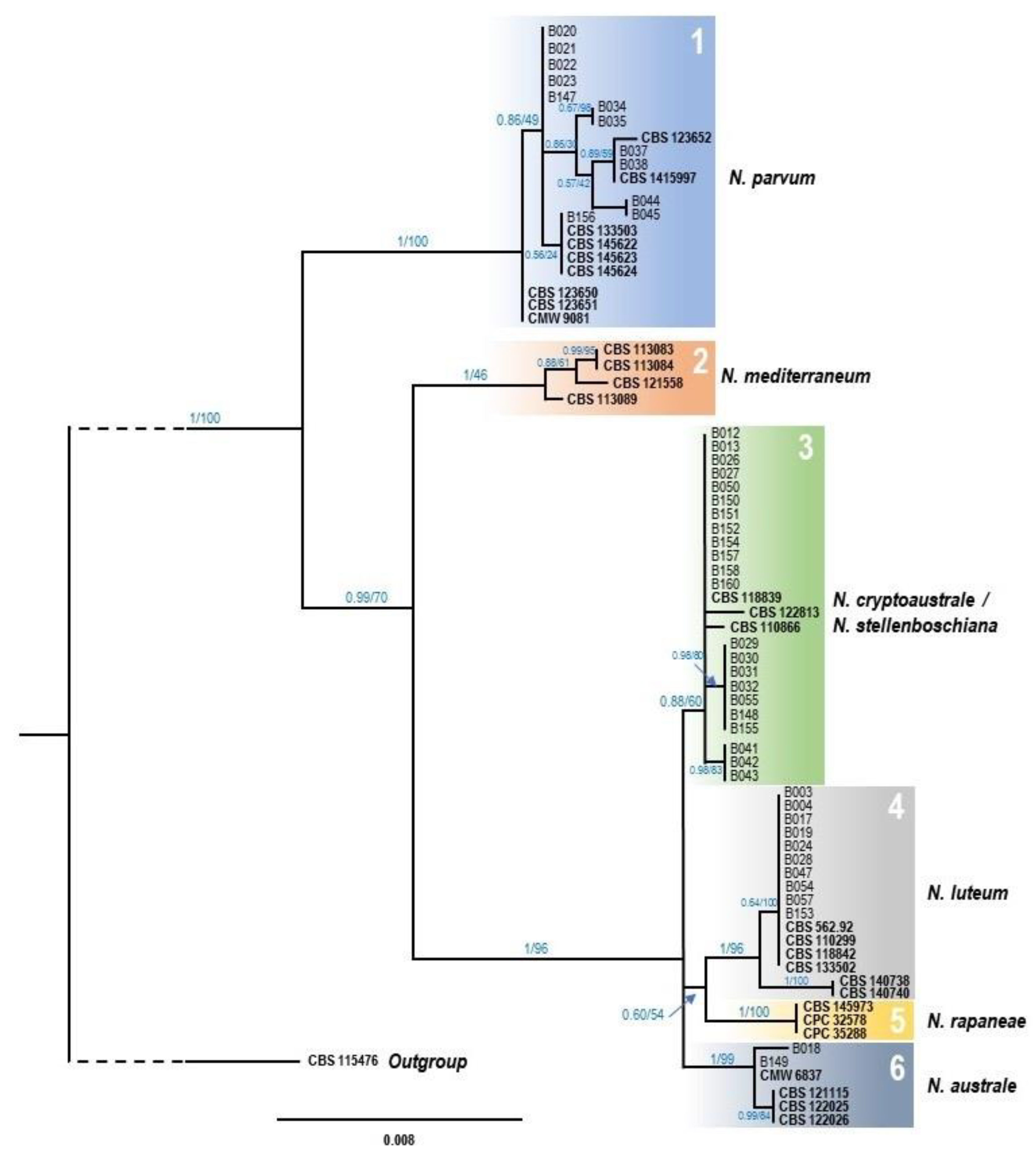
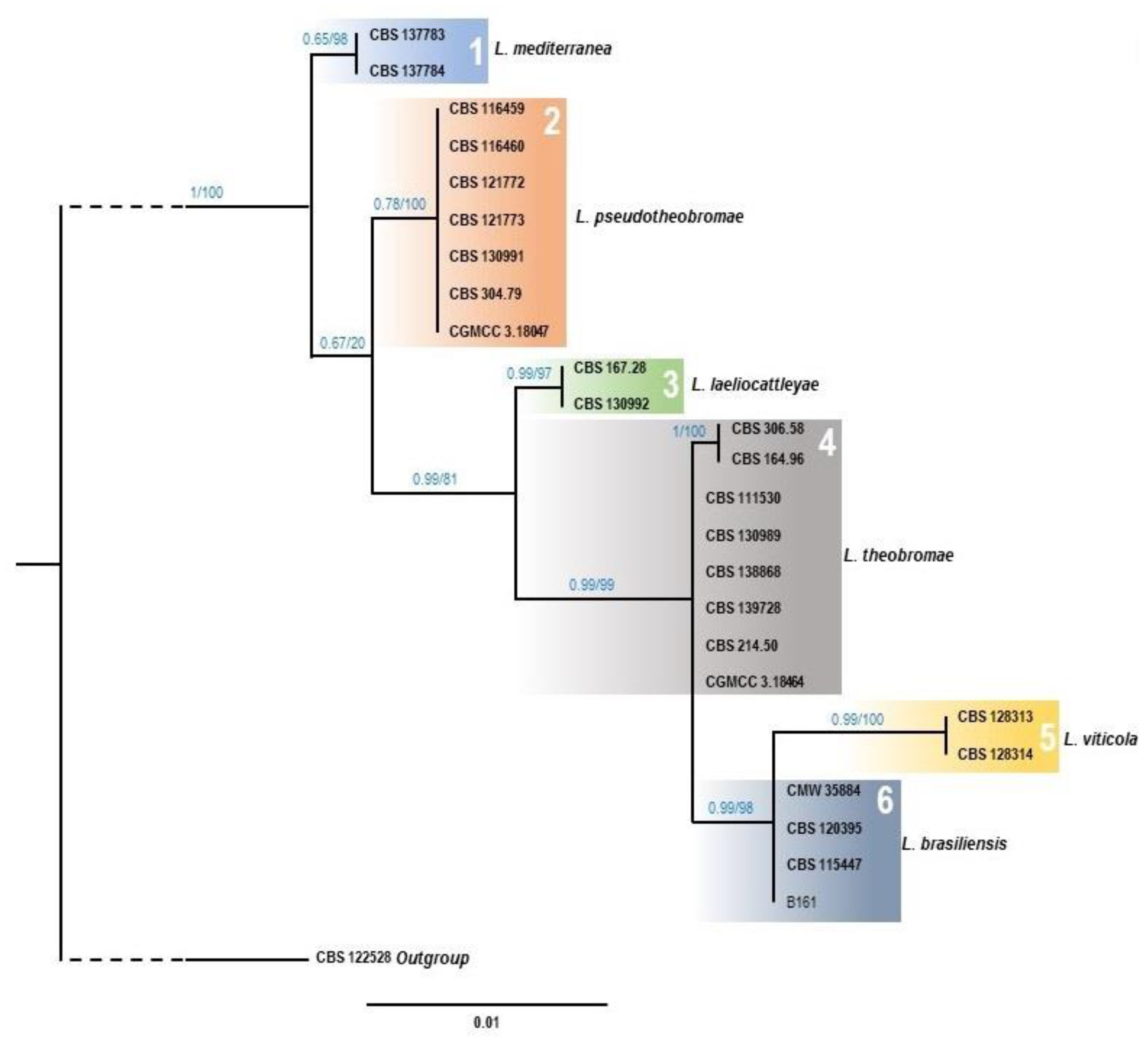
3.2. Morphological and Molecular Identification of Fungal Isolates

After a preliminary morphological characterization, 74 isolates showed morphological and growth features according to the Botryosphaeriaceae family, from which 52 were selected for their molecular identification based on the ITS1-2, tef1 and tub2 genetic markers. PCR amplification of ITS1-2, tef1 and tub2 markers gave products of 500–540 bp, 246–300 bp and 410 bp, respectively, and absence of contamination and non-specific PCR products were confirmed from agarose gels (not shown). Sequences obtained allowed the molecular identification of five different Botryosphaeriaceae species, based on independent BLAST searches with the three sequenced markers. One fungal isolate was identified as Lasiodiplodia brasiliensis, while four species of the Neofusiccocum genus were detected, including N. australe (2 isolates), N. cryptoaustrale/stellenboschiana (22 isolates), N. luteum (10 isolates), and N. parvum (12 isolates). The remaining five isolates were not identified to species level as they could not be recovered from storage. Twenty one out of the 22 isolates identified as N. cryptoaustrale/stellenboschiana with ITS1-2, tef1 and tub2 genomic regions showed for rpb2 the same sequence (OQ401613, OQ401614 or Q401615), which matches all GenBank sequences from N. stellenboschiana and all-but-one N. cryptoaustrale. One isolate (B043) showed a sequence (OQ401617) equivalent to KX464014.1 which corresponds to that of the reference strain CBS 122813 of N. cryptoaustrale. These results were confirmed by repeating the analyses by means of a new DNA extraction using an alternative second procedure, PCR amplification and sequencing.
Colony growth, pycnidia and conidia of representative isolates of the different species of Botryosphaeriaceae identified in this work are shown in Figures S5–S9. All Neofusiccocum isolates generated pycnidia on SC medium with and without Parafilm M, except the isolate B022 of N. parvum, which only produced pycnidia on WA medium (Table 3). No conidia were obtained from N. australe, represented by isolate B018, but isolates of N. cryptoaustrale/stellenboschiana (B026), N. luteum (B003, B017, B024) and N. parvum (B020, B022) produced conidia on SC medium without Parafilm M and/or WA medium. In addition, the isolate B161, identified as L. brasiliensis, produced pycnidia and conidia on PDAS. Mature conidia of L. brasiliensis were dark-walled, one-septate, striate (Figure S9B), whereas conidia of Neofusicoccum species were hyaline and aseptate (Figures S6H, S7H and S8G). The conidia measurements are shown in Table 4, as well as reference measurements previously reported by other authors.
For the multiple alignment of Neofusicocum species, the ML and BY phylogenetic analysis generated identical topologies, showing six statistically well supported clades where the isolates from this study were placed in four of them. The BY tree is presented with PP/ML values at the corresponding branch (Figure 2). The first clade grouped twelve isolates from this study, along with the nine N. parvum reference sequences selected for the analysis, therefore confirming their molecular identification as N. parvum strains. The second clade, which includes the four reference sequences of N. mediterraneum, did not contain any isolate from the present work, suggesting that this species seems to be absent from the avocados sampled in Tenerife island. The third clade includes 22 of the new isolates along with N. cryptoaustrale CBS 122813 reference strain, but also with N. stellenboschiana CBS 110866 and CBS 118839. Interestingly, 12 of the isolates were placed in the main branch with the three sequences mentioned above, but the 10 remaining isolates were ubicated in two sub-clades, the first with seven isolates, and the second with three. The fourth clade corresponded with the N. luteum reference strains where ten isolates from this work were ubicated, all of them in the same group, along with reference strains CBS 562.92, CBS 110299, CBS 118842 and CBS 133502. Neither of the isolates of this study appeared in the fifth clade, corresponding this with N. rapaneae. Finally, the sixth and last clade grouped the two remaining isolates, along with N. australe reference strains.
In the case of Lasiodiplodia species ML and BY trees (Figure 3) showed exactly the same topology, showing six well supported clades corresponding to the species of L. mediterranea, L. pseudotheobromae, L. laeliocattleyae, L. theobromae, L. viticola, and L. brasiliensis. The unique isolate from the present work assigned to Lasiodiplodia genus (B161), was clearly assigned to the clade of L. brasiliensis.

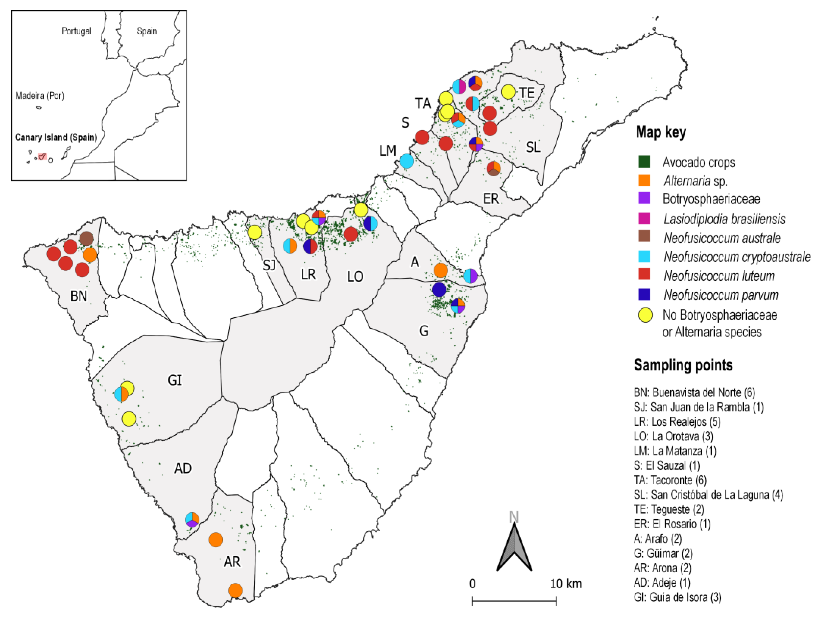
3.3. Symptoms and Distribution of Botryosphaeriaceae Species Associated to Avocado in Tenerife

Some species of the Botryosphaeriaceae family were detected in 26 out of 40 orchards surveyed (Figure 4): N. luteum, 16 orchards; N. cryptoaustrale/stellenboschiana, 11 orchards; N. parvum, 6 orchards; N. australe, 2 orchards; L. brasiliensis, 1 orchard; and 5 orchards with unidentified species of Botryosphaeriaceae. There were also isolated species of Alternaria from 13 orchards (in four of them as the sole fungus, in the other nine along with Botryosphaeriaceae fungi). Other fungal species such as Aureobasidium sp., Cladosporium sp., Colletotrichum sp., Nigrospora sp. and Pestalotiopsis sp. were also isolated in 10 orchards.
Botryosphaeriaceae fungal species were isolated from samples of necrotic branches (mainly diebacks) and fruits collected in the surveyed orchards, with 47.6% and 30.3% of positive results, respectively. However, no Botryosphaeriaceae fungi were isolated from cankers and the isolates obtained from samples showing panicle blight were not species of Botryosphaeriaceae. In addition, N. luteum and N. australe were isolated from samples of necrotic branches that could not be geographically located, with the first species in two samples and the second in one. In samples of postharvest fruits collected in 6 sampling points (2 avocado packaging facilities and 4 retail stores), N. luteum was detected in 3 of them, N. cryptoaustrale/stellenboschiana in 2, and N. parvum and N. australe in 1 sampling point each one.

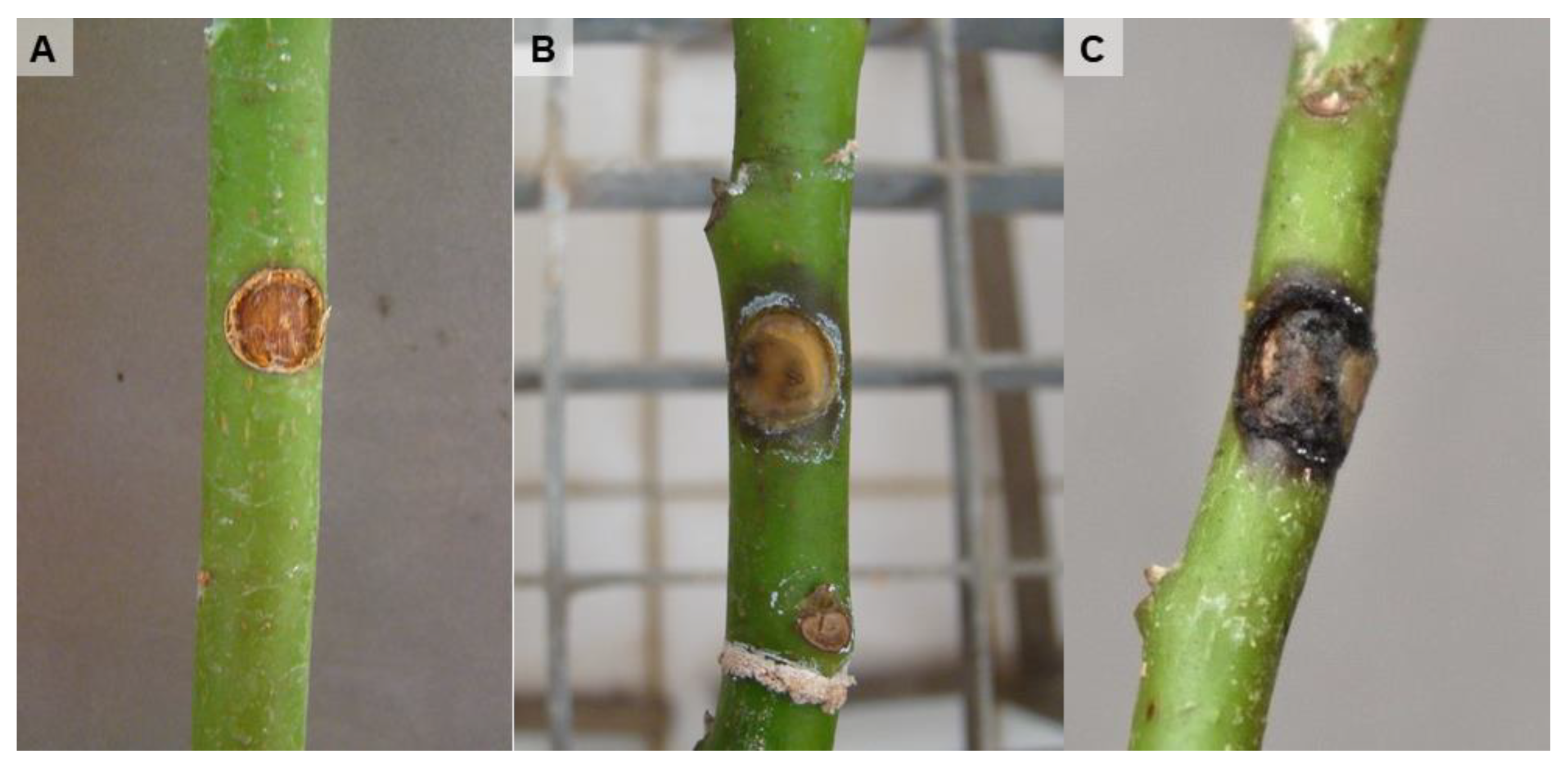
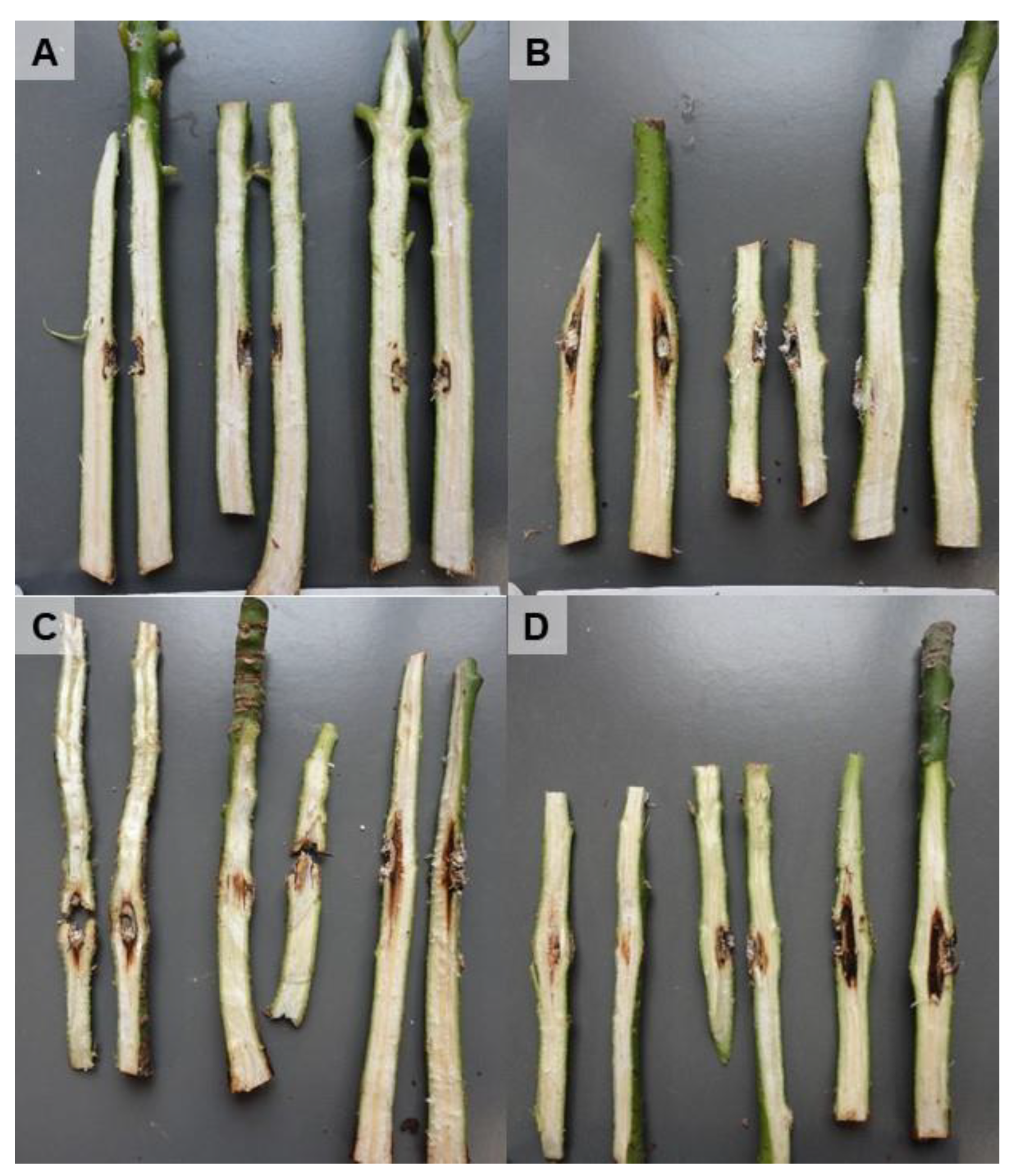
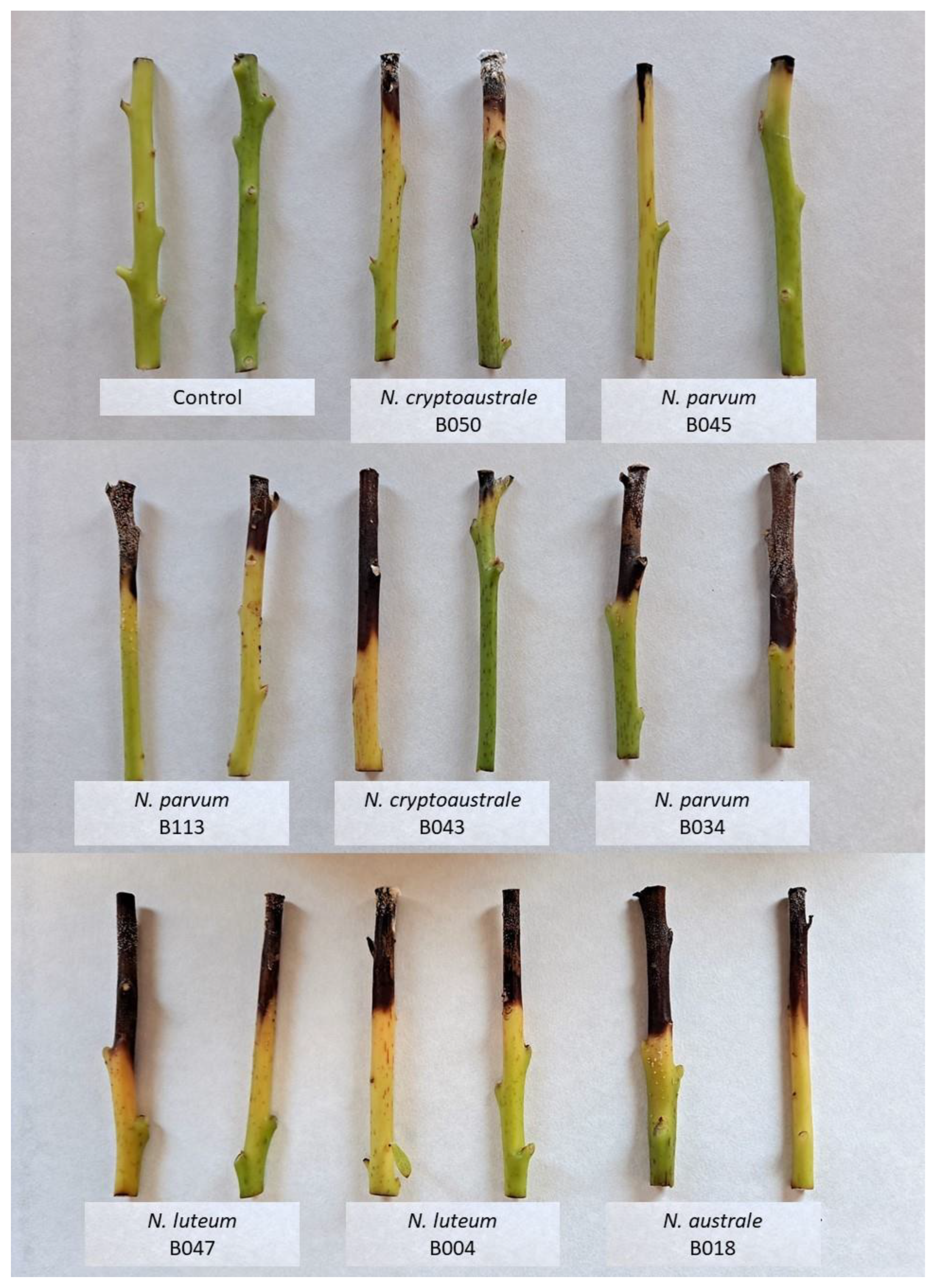
3.4. Pathogenicity Tests

Plants inoculated by inserting a mycelial plug in a wound made in the stem at 5 cm from the ground showed symptoms after 3–6 months being inoculated with L. brasiliensis, N. luteum and N. parvum. Figure 5 shows external stem necrosis produced by L. brasiliensis, with exudates of whitish sugars characteristic of avocado at the inoculation point or in the parts where Parafilm M was applied to fix the inoculum to the stem. All isolates caused internal necrosis that progressed from the inoculation point inside the plants, although irregularly and with variations in its length depending on the inoculated plant (Figure 6). No substantial differences were observed in the internal symptoms between bagged and non-bagged plants. When N. australe, N. cryptoaustrale/stellenboschiana, N. luteum and N. parvum were inoculated by pruning the plants at the top and placing the fungal inoculum onto the pruning-cut wound, external symptoms were clearly visible at 21 days after inoculation. Inoculated plants showed stem discolouration and necrosis that progressed along the outside of the plant stem downwards from the point of inoculation, along with the typical whitish-dry exudate (Figure 7, Table 5), showing the same symptoms of dieback that can be observed in the field. All the inoculated fungi in the different pathogenicity tests were re-isolated from the plant lesions, thus complying with Koch’s postulates.

4. Discussion

This work studied the occurrence of damages to the aerial part of avocado plants on the island of Tenerife and the diversity of the associated fungi. The symptom most frequently found was the necrosis in branches (dieback-type symptoms), which produces losses on plantlets in recent plantations and young trees, but also in adult trees. Panicle blight and fruit drop during its early development stages also caused significant losses in production. Cankers on trunks and branches appeared less frequently on field, although, very occasionally, some orchards may have many affected trees. We determined the presence and diversity of fungi associated with these symptoms using sequence analysis of ITS1-2, with particular interest in species of the Botryosphaeriaceae family, for which additional markers, tef1 and tub2, were also used.
Five species of Botryosphaeriaceae were isolated from symptomatic avocados: Neofusiccocum australe, N. cryptoaustrale/stellenboschiana, N. luteum, N. parvum and Lasiodiplodia brasiliensis, and it is important to highlight that it was frequent to find more than one species of this family in the same orchard. Neofusicoccum australe, N. cryptoaustrale/stellenboschiana, N. luteum and N. parvum have been reported in different countries as causal agents of diseases in avocado plants [5,6,7,10,33,52]. The high frequencies of N. luteum or N. cryptoaustrale/stellenboschiana found in this study does not correspond to what has been described in avocado-growing areas in the south of the Spanish mainland where N. parvum predominates [5]. No relationship was found in our study between the identified fungal species and the geographic location of avocado orchards. In other countries, the various types of symptoms seem to be caused in a general way by different species of the Botryosphaeriaceae family, along with other species of fungi, with the prevalence of one or the other according to countries or regions. Some publications even mention that the incidence and distribution of species varies between areas within the same region [5,10,53].
Moreover, fungi belonging to the Botryosphaeriaceae family have been isolated in the Canary Islands in other crops such as vineyards, almond trees, or mangoes [54,55], and in ornamental plants such as Indian laurels in which they cause significant damage in urban gardens [56]. The presence of Botryosphaeriaceae species infecting other hosts rather than avocado in the Canary Islands could play a role in the epidemiology of the diseases in avocado, as has been recently reported for other woody crops [57,58].
To the best of our knowledge, this study constitutes the first report of N. cryptoaustrale/stellenboschiana infecting avocado plants in Spain and the first time that N. cryptoaustrale/stellenboschiana has been cited in avocado causing symptoms of dieback and stem-end rot, although it had been previously cited causing branch cankers on avocado in Crete (Greece) [52]. However, it was not possible to assign our isolates unequivocally to one of these two species, as reference strains of N. cryptoaustrale and N. stellenboschiana are grouped in a unique clade. The additional analysis of the rpb2 marker for N. cryptoaustrale/stellenboschiana showed that all analysed isolates except one presented a single sequence cited by various authors who have worked with this marker in the two species (the entire sequence fully matches for the two species). In addition, a sequence was obtained for isolate B043 that corresponded to the reference CBS:122813 registered in GenBank as KX464014.1. This sequence shows a difference of 24 snp with the others cited for N. cryptoaustrale and N. stellenboschiana. Therefore, in view of the results obtained it is necessary to clarify the taxonomic situation of both species and determine the origins of the differences, which is beyond the scope of this work. Moreover, results of conidial dimensions obtained for these isolates are similar to the references for both species, and they could not be used to morphologically discriminate them. Similar results were described by other authors who could not differentiate between N. cryptoaustrale and N. stellenboschiana using ITS1-2, tef1 or tub2 markers [59,60].
Neofusicoccum cryptoaustrale/stellenboschiana seems to be quite widespread in avocado in Tenerife, considering the frequency with which it has been found in different locations (11 out of 40 orchards). Neofusicoccum cryptoaustrale has been isolated from branches and leaves of living Eucalyptus trees in South Africa [45], but also from other species of arboreal plants or other geographical locations such as Pistacia spp. [46,61], Olea europea [46] or different mangrove trees [62]. Neofusicoccum stellenboschiana was also recently described in South Africa isolated from Vitis vinifera [46], but also has been cited associated to Arum italicum leaf spot [46], Olea europea [46], Persea americana cankers [52], Prunus spp. [46] and Quercus suber [63]. Our pathogenicity tests carried out with isolates of this species show its ability to develop disease symptoms in avocado seedlings equivalent to those of N. parvum or N. luteum, which makes it a species to consider in the epidemiology of this disease in the Canary Islands.
The unique isolate of the genus Lasiodiplodia obtained here was initially thought to belong to the most frequent species L. theobromae, already cited in avocado in several countries as well as in other plants in the Canary Islands [54,56]. However, its phylogenetic analysis indicates that it belongs to the species L. brasiliensis. This species was described in Brazil in 2014 associated with stem-end rot of papaya [64]. It has also been detected in that country associated to dieback in grapevine [65], postharvest fruit rot of custard apple [66] and cankers and dieback of apple trees [67]. Moreover, L. brasiliensis causes stem-end rot of mango in China [68]. It is closely related to L. viticola, but it differentiates on conidial sizes, which are longer and larger than those described for this last species [69]. Six nucleotides in the ITS1-2 region differentiate L. brasiliensis from L. viticola, with no difference in the nucleotide sequence of tef1. In our work, a single isolate of L. brasiliensis was obtained from an avocado plant with symptoms of dieback in an orchard in which N. cryptoaustrale/stellenboschiana was also detected. The results of the pathogenicity tests pointed out that L. brasiliensis was capable of reproducing symptoms of necrosis in inoculated avocado plants. This is the first time that this species has been reported in avocado and the first time that it has been reported in Spain.
In this study, the number of samples of the main types of symptoms attributed to Botryosphaeriaceae in field conditions was highly unbalanced in favour of branch necrosis (mainly diebacks), with some representation of fruit rot and panicle blight and low representation of cankers on trunks or branches. According to the fungi isolated from these samples, the symptoms of branch necrosis (dieback-type) seem to be caused mainly by species of Botryosphaeriaceae, whereas fungi of other genus were prevalently isolated from panicle blight. No pathogens were consistently isolated from the few samples of cankers that were analysed. No fungal isolates from the Botryosphaeriaceae family were detected, nor were other species of harmful organisms to which cankers could be attributed such as Phytophthora species or bacteria (data not shown). Regarding the necrosis in the peduncles of unripe fruits that come off the trees without completing their development, it was possible to isolate species of Botryosphaeriaceae, Alternaria sp., but also other species. Although the loss of developing fruit is a natural process in the summer, we have to consider that stress, pests and diseases, such as those produced by Botryosphaeriaceae or other fungi, can cause excessive fruit drop.
The same Neofusicoccum species found in avocado orchards (N. australe, N. cryptoaustrale/stellenboschiana, N. luteum and N. parvum) were isolated from post-harvest fruits showing symptoms of stem-end rot or dark spots in other locations on the fruit. In addition, Colletotrichum sp. was isolated only from one sample. It has been described that damages due to the Botryosphaeriaceae family are located in the area of insertion of the peduncle in the fruit (stem-end rot) [12,27,70], while those caused by Colletotrichum species are characterized by the appearance of darkening of the skin of the fruit (anthracnose) [12,27]. In this work, most of Neofusicoccum and Colletotrichum isolates were obtained from branches, suggesting that the fungi that infest the branches of avocado trees in Tenerife may also be the ones that cause damage to the fruit in post-harvest.
Regarding the pathogenicity tests, Koch’s postulates conducted with isolates of Botryosphaeriaceae obtained in this work confirmed their pathogenicity on avocado plants. Most of the inoculation procedures carried out with Botryosphaeriaceae fungi on avocado plants, but also in other woody plants, are mainly based on the use of a fungal mycelial plug, which is placed into a stem wound performed at a fixed distance from the ground and far from the top of the plant [3,6,9]. Using this procedure, inoculated plants show symptoms of necrosis several months after inoculation, and similar results were obtained in our study. However, when plants were inoculated by placing the inoculum onto pruning-cut wounds at the apex, inoculated plants showed symptoms of stem necrosis in less than a month. This procedure, based on wound inoculation at the apical tip region of the plants, has been described by some authors for pathogenicity tests of Botryosphaeriaceae fungi on avocado [4,11] and other woody plants [71], resulting in brown stem lesions 2–4 weeks after inoculation. Therefore, wound inoculation at the apex of the plants seems to reproduce disease symptoms in a short time, suggesting it could facilitate the study of different aspects of Botryosphaeriaceae disease on avocado such as varietal differences, variation in virulence between species and isolates, as well as to evaluate methods for its control.
In Tenerife, similarly to other avocado-growing regions, fungi of the Botryosphaeriaceae family seem to be involved in avocado dieback, in the premature fall of fruits during their development in the field, and in post-harvest damage. However, further research is needed in relation to panicle blight and avocado cankers in Tenerife. Panicle blight or inflorescence dieback occurs frequently and yields in some years are greatly reduced in the worst affected orchards, but little information is available on this symptom in avocado [19]. Fungi belonging to the family Botryosphaeriaceae have been described to cause cankers on avocado and other woody plants. However, no Botryosphaeriaceae fungi or other pathogens were consistently isolated from cankers in this work. The samples were taken from the cankers just in borders inside of it in adult trees without killing the tree, where other saprophytic organisms may have probably settled. In these old cankers, it is probably necessary to take the samples from more internal points at the zone of advancing necrosis. Furthermore, cankers are not frequent in avocado orchards in Tenerife and, for this reason, only a very low number of cankers could be analysed. Therefore, it is necessary to carry out a greater number of isolations, particularly from cankers, as well as from symptoms in relation to phenological growth stages to clarify their association with the fungal pathogens of avocado.

Supplementary Materials

The following supporting information can be downloaded at: https://www.mdpi.com/article/10.3390/microorganisms11030585/s1, Figure S1: Symptoms in aerial parts of young and adult avocado plants; Figure S2: Panicle blight and dieback symptoms; Figure S3: Cankers in blanch and trunks, and rots in developing fruits; Figure S4: Symptoms in post-harvest avocado fruits; Figure S5. Neofusiccocum australe; Figure S6. Neofusiccocum cryptoaustrale/stellenboschiana; Figure S7. Neofusiccocum luteum; Figure S8. Neofusiccocum parvum; Figure S9. Lasiodiplodia brasiliense. Table S1. GenBank and culture collection accession numbers of Neofusicoccum species treated in the phylogenies. Table S2. GenBank and culture collection accession numbers of Lasiodiplodia species treated in the phylogenies.

Author Contributions

Conceptualization, S.P., M.A.G.-C., A.R.-P. and F.S.; methodology, O.G.-P., M.A.G.-C., A.R.-P. and F.S.; software, D.H., M.A.G.-C. and F.S.; validation, D.H., M.A.G.-C., A.R.-P. and F.S.; formal analysis, D.H., O.G.-P., M.A.G.-C. and F.S.; investigation, D.H., O.G.-P. and F.S.; resources, D.H., S.P. and F.S.; data curation, D.H. and F.S.; writing—original draft preparation, D.H., A.R.-P. and F.S.; writing—review and editing, D.H., M.A.G.-C., A.R.-P. and F.S.; visualization, D.H., and F.S.; supervision, A.R.-P. and F.S. project administration, F.S.; funding acquisition, F.S. All authors have read and agreed to the published version of the manuscript.

Funding

This work was funded by a CAIA Project 2017-0001 “Optimization of avocado production systems” of the Ministry of Agriculture, Livestock and Fisheries of the Government of the Canary Islands. David Hernández-Hernández was recipient of a 2021–2025 PhD grant PRE2020-095122 from the Agencia Estatal de Investigación (AEI).

Data Availability Statement

Not applicable.

Acknowledgments

The authors are grateful for the participation of the Agricultural Extension Services of the Technical Service for Agriculture and Rural Development of the Cabildo Insular de Tenerife, which collaborated in carrying out the surveys and in the collection of samples. To all the farmers who participated in the survey and who allowed the collection of samples in their crops. We also want to thank the Plant Health Service of the General Direction of Agriculture of the Government of the Canary Islands for allowing the use of its facilities and equipment.

Conflicts of Interest

The authors declare no conflict of interest.

References

  1. ISTAC Estadística Agraria de Canarias/Series Anuales de Agricultura. Municipios, Islas y Provincias de Canarias. 1999–2021. Available online: http://www.gobiernodecanarias.org/istac/jaxi-istac/tabla.do?uripx=urn:uuid:803af13a-04f6-4789-8e78-48f5fdff64a7&uripub=urn:uuid:ef5f2e5c-e2c4-4c1d-b5ed-c20fe946ce6f%0A%0A (accessed on 30 December 2022).
  2. MAPA Encuesta Sobre Superficies y Rendimientos Cultivos (ESYRCE). Encuesta de Marco de Áreas de España. Available online: https://www.mapa.gob.es/es/estadistica/temas/estadisticas-agrarias/agricultura/esyrce/default.aspx (accessed on 30 December 2022).
  3. Auger, J.; Palma, F.; Pérez, I.; Esterio, M. First Report of Neofusicoccum australe (Botryosphaeria australis), as a Branch Dieback Pathogen of Avocado Trees in Chile. Plant Dis. 2013, 97, 842. [Google Scholar] [CrossRef]
  4. Zea-Bonilla, T.; González-Sánchez, M.A.; Martín-Sánchez, P.M.; Pérez-Jiménez, R.M. Avocado Dieback Caused by Neofusicoccum parvum in the Andalucia Region, Spain. Plant Dis. 2007, 91, 1052. [Google Scholar] [CrossRef] [PubMed]
  5. Arjona-Girona, I.; Ruano-Rosa, D.; López-Herrera, C.J. Identification, Pathogenicity and Distribution of the Causal Agents of Dieback in Avocado Orchards in Spain. Span. J. Agric. Res. 2019, 17, e1003. [Google Scholar] [CrossRef] [Green Version]
  6. Valencia, A.L.; Gil, P.M.; Latorre, B.A.; Rosales, I.M. Characterization and Pathogenicity of Botryosphaeriaceae Species Obtained from Avocado Trees with Branch Canker and Dieback and from Avocado Fruit with Stem End Rot in Chile. Plant Dis. 2019, 103, 996–1005. [Google Scholar] [CrossRef]
  7. Hartill, W.F.T.; Everett, K.R. Inoculum Sources and Infection Pathways of Pathogens Causing Stem-End Rots of ‘Hass’ Avocado (Persea americana). N. Zeal. J. Crop Hortic. Sci. 2002, 30, 249–260. [Google Scholar] [CrossRef]
  8. Twizeyimana, M.; McDonald, V.; Mayorquin, J.S.; Wang, D.H.; Na, F.; Akgül, D.S.; Eskalen, A. Effect of Fungicide Application on the Management of Avocado Branch Canker (Formerly Dothiorella Canker) in California. Plant Dis. 2013, 97, 897–902. [Google Scholar] [CrossRef] [PubMed] [Green Version]
  9. McDonald, V.; Lynch, S.; Eskalen, A. First Report of Neofusicoccum australe, N. luteum, and N. parvum Associated with Avocado Branch Canker in California. Plant Dis. 2009, 93, 967. [Google Scholar] [CrossRef]
  10. McDonald, V.; Eskalen, A. Botryosphaeriaceae Species Associated with Avocado Branch Cankers in California. Plant Dis. 2011, 95, 1465–1473. [Google Scholar] [CrossRef] [Green Version]
  11. Alama, I.; Maldonado, E.; Rodríguez-Gálvez, E. Lasiodiplodia theobromae Affect the Cultivation of Palto (Persea americana) under the Conditions of Piura, Peru. Universalia 2006, 11, 4–13. [Google Scholar]
  12. Hartill, W.F.T. Post-Harvest Diseases of Avocado Fruits in New Zealand. N. Zeal. J. Crop Hortic. Sci. 1991, 19, 297–304. [Google Scholar] [CrossRef]
  13. Darvas, J.; Kotze, J. Fungi Associated with Pre-and Postharvest Diseases of Avocado Fruit at Westfalia Estate, South Africa. Phytophylactica 1987, 19, 83–85. [Google Scholar]
  14. Twizeyimana, M.; Förster, H.; McDonald, V.; Wang, D.H.; Adaskaveg, J.E.; Eskalen, A. Identification and Pathogenicity of Fungal Pathogens Associated with Stem-End Rot of Avocado in California. Plant Dis. 2013, 97, 1580–1584. [Google Scholar] [CrossRef] [PubMed]
  15. Denman, S.; Crous, P.W.; Taylor, J.E.; Kang, J.C.; Pascoe, I.; Wingfield, M.J. An Overview of the Taxonomic History of Botryosphaeria, and a Re-Evaluation of Its Anamorphs Based on Morphology and ITS RDNa Phylogeny. Stud. Mycol. 2000, 2000, 129–140. [Google Scholar]
  16. Phillips, A.J.L.; Alves, A.; Abdollahzadeh, J.; Slippers, B.; Wingfield, M.J.; Groenewald, J.Z.; Crous, P.W. The Botryosphaeriaceae: Genera and Species Known from Culture. Stud. Mycol. 2013, 76, 51–167. [Google Scholar] [CrossRef] [Green Version]
  17. Dissanayake, A.J.; Phillips, A.J.L.; Li, X.H.; Hyde, K.D. Botryosphaeriaceae: Current Status of Genera and Species. Mycosphere 2016, 7, 1001–1073. [Google Scholar] [CrossRef]
  18. Slippers, B.; Wingfield, M.J. Botryosphaeriaceae as Endophytes and Latent Pathogens of Woody Plants: Diversity, Ecology and Impact. Fungal Biol. Rev. 2007, 21, 90–106. [Google Scholar] [CrossRef]
  19. Dann, E.; Prabhakaran, A.; Bransgrove, K. Panicle Blight (Flower Dieback). Available online: https://avocado.org.au/public-articles/ta31v3panicle/ (accessed on 30 December 2022).
  20. Burgess, T.I.; Tan, Y.P.; Garnas, J.; Edwards, J.; Scarlett, K.A.; Shuttleworth, L.A.; Daniel, R.; Dann, E.K.; Parkinson, L.E.; Dinh, Q. Current Status of the Botryosphaeriaceae in Australia. Australas. Plant Pathol. 2019, 48, 35–44. [Google Scholar] [CrossRef]
  21. Peterson, R.A. Susceptibility of Fuerte Avocado Fruit at Various Stages of Growth, to Infection by Anthracnose and Stem End Rot Fungi. Aust. J. Exp. Agric. 1978, 18, 158–160. [Google Scholar] [CrossRef] [Green Version]
  22. Firmino, A.C.; Fischer, I.H.; Tozze Júnior, H.J.; Dias Rosa, D.; Luiz Furtado, E. Identificação de Espécies de Fusicoccum Causadoras de Podridão Em Frutos de Abacate. Summa Phytopathol. 2016, 42, 100–102. [Google Scholar] [CrossRef] [Green Version]
  23. Montealegre, J.R.; Ramírez, M.; Riquelme, D.; Armengol, J.; León, M.; Pérez, L.M. First Report of Neofusicoccum australe in Chile Causing Avocado Stem-End Rot. Plant Dis. 2016, 100, 2532. [Google Scholar] [CrossRef]
  24. Tapia, L.; Larach, A.; Riquelme, N.; Guajardo, J.; Besoain, X. First Report of Neofusicoccum luteum Causing Stem-End Rot Disease on Avocado Fruits in Chile. Plant Dis. 2020, 104, 2027. [Google Scholar] [CrossRef]
  25. Qiu, F.; Xu, G.; Zhou, J.; Zheng, F.Q.; Zheng, L.; Miao, W.G.; Wang, W.L.; Xie, C.P. First Report of Botryosphaeria dothidea Causing Stem-End Rot in Avocado (Persea americana) in China. Plant Dis. 2019, 104, 286. [Google Scholar] [CrossRef]
  26. Ramirez-Gil, J.G. Avocado Wilt Complex Disease, Implications and Management in Colombia. Rev. Fac. Nac. Agron. Colomb. 2018, 71, 8525–8541. [Google Scholar] [CrossRef]
  27. Ramírez-Gil, J.G.; López, J.H.; Henao-Rojas, J.C. Causes of Hass Avocado Fruit Rejection in Preharvest, Harvest, and Packinghouse: Economic Losses and Associated Variables. Agronomy 2019, 10, 8. [Google Scholar] [CrossRef] [Green Version]
  28. Kebede, M.; Belay, A. Fungi Associated With Post-Harvest Avocado Fruit Rot at Jimma Town, Southwestern Ethiopia. J. Plant Pathol. Microbiol. 2019, 10, 2. [Google Scholar] [CrossRef] [Green Version]
  29. Guarnaccia, V.; Aiello, D.; Cirvilleri, G.; Polizzi, G.; Susca, A.; Epifani, F.; Perrone, G. Characterisation of Fungal Pathogens Associated with Stem-End Rot of Avocado Fruit in Italy. Acta Hortic. 2016, 1144, 133–139. [Google Scholar] [CrossRef]
  30. Molina-Gayosso, E.; Silva-Rojas, H.V.; García-Morales, S.; Ávila-Quezada, G. First Report of Black Spots on Avocado Fruit Caused by Neofusicoccum parvum in Mexico. Plant Dis. 2011, 96, 287. [Google Scholar] [CrossRef]
  31. Trakunyingcharoen, T.; Cheewangkoon, R.; To-anun, C. Phylogenetic Study of the Botryosphaeriaceae Species Associated with Avocado and Para Rubber in Thailand. Chiang Mai J. Sci. 2015, 42, 104–116. [Google Scholar]
  32. Ni, H.F.; Liou, R.F.; Hung, T.H.; Chen, R.S.; Yang, H.R. First Report of a Fruit Rot Disease of Avocado Caused by Neofusicoccum mangiferae. Plant Dis. 2009, 93, 760. [Google Scholar] [CrossRef]
  33. Ni, H.; Chuang, M.; Hsu, S.; Lai, S.; Yang, H. Survey of Botryosphaeria spp., Causal Agents of Postharvest Disease of Avocado, in Taiwan. J. Taiwan Agric. Res. 2011, 60, 157–166. [Google Scholar]
  34. Truett, G.E.; Heeger, P.; Mynatt, R.L.; Truett, A.A.; Walker, J.A.; Warman, M.L. Preparation of PCR-Quality Mouse Genomic Dna with Hot Sodium Hydroxide and Tris (HotSHOT). Biotechniques 2000, 29, 52–54. [Google Scholar] [CrossRef] [PubMed]
  35. Collado-Romero, M.; Mercado-Blanco, J.; Olivares-García, C.; Valverde-Corredor, A.; Jiménez-Díaz, R.M. Molecular Variability within and among Verticillium dahliae Vegetative Compatibility Groups Determined by Fluorescent Amplified Fragment Length Polymorphism and Polymerase Chain Reaction Markers. Phytopathology 2006, 96, 485–495. [Google Scholar] [CrossRef] [Green Version]
  36. White, T.J.J.; Bruns, T.D.; Lee, S.B.; Taylor, J.W. Amplification and Direct Sequencing of Fungal Ribosomal RNA Genes for Phylogenetics. PCR Protoc. 1990, 315–322. [Google Scholar] [CrossRef]
  37. Alves, A.; Crous, P.W.; Correia, A.; Phillips, A.J.L. Morphological and Molecular Data Reveal Cryptic Speciation in Lasiodiplodia theobromae. Fungal Divers. 2008, 28, 1–13. [Google Scholar]
  38. Glass, N.L.; Donaldson, G.C. Development of Primer Sets Designed for Use with the PCR to Amplify Conserved Genes from Filamentous Ascomycetes. Appl. Environ. Microbiol. 1995, 61, 1323–1330. [Google Scholar] [CrossRef] [PubMed] [Green Version]
  39. Sakalidis, M.L.; Hardy, G.E.S.J.; Burgess, T.I. Use of the Genealogical Sorting Index (GSI) to Delineate Species Boundaries in the Neofusicoccum Parvum-neofusicoccum Ribis Species Complex. Mol. Phylogenet. Evol. 2011, 60, 333–344. [Google Scholar] [CrossRef] [Green Version]
  40. Tamura, K.; Stecher, G.; Kumar, S. MEGA11: Molecular Evolutionary Genetics Analysis Version 11. Mol. Biol. Evol. 2021, 38, 3022–3027. [Google Scholar] [CrossRef]
  41. Altschul, S.F.; Gish, W.; Miller, W.; Myers, E.W.; Lipman, D.J. Basic Local Alignment Search Tool. J. Mol. Biol. 1990, 215, 403–410. [Google Scholar] [CrossRef]
  42. Sayers, E.W.; Bolton, E.E.; Brister, J.R.; Canese, K.; Chan, J.; Comeau, D.C.; Connor, R.; Funk, K.; Kelly, C.; Kim, S.; et al. Database Resources of the National Center for Biotechnology Information. Nucleic Acids Res. 2022, 50, D20–D26. [Google Scholar] [CrossRef]
  43. Mussi-Dias, V.; Valmir dos Santos, A.; das Graças Machado Freire, M. Pycnidia and Conidia Quantification of Lasiodiplodia Using a Culture Medium Enriched with Sugarcane Bagasse. J. Agric. Res. 2016, 2, 1–16. [Google Scholar]
  44. Marques, M.W.; Lima, N.B.; De Morais, M.A.; Barbosa, M.A.G.; Souza, B.O.; Michereff, S.J.; Phillips, A.J.L.; Câmara, M.P.S. Species of Lasiodiplodia Associated with Mango in Brazil. Fungal Divers. 2013, 61, 181–193. [Google Scholar] [CrossRef]
  45. Crous, P.W.; Wingfield, M.J.; Guarro, J.; Cheewangkoon, R.; van der Bank, M.; Swart, W.J.; Stchigel, A.M.; Cano-Lira, J.F.; Roux, J.; Madrid, H.; et al. Fungal Planet Description Sheets: 154–213. Persoonia-Mol. Phylogeny Evol. Fungi 2013, 31, 188–296. [Google Scholar] [CrossRef] [PubMed]
  46. Yang, T.; Groenewald, J.Z.; Cheewangkoon, R.; Jami, F.; Abdollahzadeh, J.; Lombard, L.; Crous, P.W. Families, Genera, and Species of Botryosphaeriales. Fungal Biol. 2017, 121, 322–346. [Google Scholar] [CrossRef]
  47. Nguyen, L.T.; Schmidt, H.A.; Von Haeseler, A.; Minh, B.Q. IQ-TREE: A Fast and Effective Stochastic Algorithm for Estimating Maximum-Likelihood Phylogenies. Mol. Biol. Evol. 2015, 32, 268–274. [Google Scholar] [CrossRef] [PubMed]
  48. Miller, M.A.; Pfeiffer, W.; Schwartz, T. The CIPRES Science Gateway. In Proceedings of the 1st Conference of the Extreme Science and Engineering Discovery Environment on Bridging from the eXtreme to the Campus and beyond—XSEDE ’12, Chicago, IL, USA, 16–20 July 2012; ACM Press: New York, NY, USA, 2012; p. 1. [Google Scholar]
  49. Tavaré, S. Some Probabilistic and Statistical Problems in the Analysis of DNA Sequences. Am. Math. Soc. Lect. Math. Life Sci. 1986, 17, 57–86. [Google Scholar]
  50. Stamatakis, A. RAxML Version 8: A Tool for Phylogenetic Analysis and Post-Analysis of Large Phylogenies. Bioinformatics 2014, 30, 1312–1313. [Google Scholar] [CrossRef] [PubMed] [Green Version]
  51. Ronquist, F.; Teslenko, M.; Van Der Mark, P.; Ayres, D.L.; Darling, A.; Höhna, S.; Larget, B.; Liu, L.; Suchard, M.A.; Huelsenbeck, J.P. Mrbayes 3.2: Efficient Bayesian Phylogenetic Inference and Model Choice across a Large Model Space. Syst. Biol. 2012, 61, 539–542. [Google Scholar] [CrossRef] [Green Version]
  52. Guarnaccia, V.; Polizzi, G.; Papadantonakis, N.; Gullino, M.L. Neofusicoccum Species Causing Branch Cankers on Avocado in Crete (Greece). J. Plant Pathol. 2020, 102, 1251–1255. [Google Scholar] [CrossRef]
  53. Eskalen, A.; McDonald, V. Geographical Distribution of Botryosphaeriaceae and Phomopsis/Diaporthe Canker Pathogens of Avocado in California. Calif. Avocado Soc. 2010, 93, 87–98. [Google Scholar]
  54. Gallo Llobet, L.; Hernández Hernández, J.M.; del Jaizme Vega, M.C.; Sala Mayato, L. Especies Fúngicas Enontradas En Los Cultivos de Canarias En El Período 1974–1984. In Proceedings of the III Congreso Nacional de Fitopatología; Sociedad Española de Fitopatología, Ed.; The Canary Islands: Tenerife, Spain, 1984; pp. 43–50. [Google Scholar]
  55. Márquez, M.P.; Santana, M.L.; Albalat, A.R.; Armengol, J. Muestreo e Identificación de Hongos Asociados a Las Enfermedades de La Madera de La Vid En Lanzarote (Canarias). Enoviticultura 2020, 65, 16–26. [Google Scholar]
  56. Benito Hernández, P. Botryosphaeriaceae Asociadas al Decaimiento de Ficus microcarpa en Gran Canaria. Master Thesis, Universitat Politècnica de València, Valencia, Spain, 2018. [Google Scholar]
  57. Mojeremane, K.; Lebenya, P.; Plessis, I.L.D.; Van Der Rijst, M.; Mostert, L.; Armengol, J.; Halleen, F. Cross Pathogenicity of Neofusicoccum Australe and Neofusicoccum stellenboschiana on Grapevine and Selected Fruit and Ornamental Trees. Phytopathol. Mediterr. 2020, 59, 581–593. [Google Scholar] [CrossRef]
  58. Sessa, L.; Abreo, E.; Bettucci, L.; Lupo, S. Botryosphaeriaceae Species Associated with Wood Diseases of Stone and Pome Fruits Trees: Symptoms and Virulence across Different Hosts in Uruguay. Eur. J. Plant Pathol. 2016, 146, 519–530. [Google Scholar] [CrossRef]
  59. Van Dyk, M.; Spies, C.F.J.; Mostert, L.; Halleen, F. Survey of Trunk Pathogens in South African Olive Nurseries. Plant Dis. 2021, 105, 1630–1639. [Google Scholar] [CrossRef] [PubMed]
  60. Spies, C.F.J.; Mostert, L.; Carlucci, A.; Moyo, P.; van Jaarsveld, W.J.; du Plessis, I.L.; van Dyk, M.; Halleen, F. Dieback and Decline Pathogens of Olive Trees in South Africa. Persoonia Mol. Phylogeny Evol. Fungi 2020, 45, 196–220. [Google Scholar] [CrossRef] [PubMed]
  61. Linaldeddu, B.T.; Maddau, L.; Franceschini, A.; Alves, A.; Phillips, A.J.L. Botryosphaeriaceae Species Associated with Lentisk Dieback in Italy and Description of Diplodia insularis sp. Nov. Mycosphere 2016, 7, 962–977. [Google Scholar] [CrossRef]
  62. Osorio, J.A.; Crous, C.J.; de Beer, Z.W.; Wingfield, M.J.; Roux, J. Endophytic Botryosphaeriaceae, Including Five New Species, Associated with Mangrove Trees in South Africa. Fungal Biol. 2017, 121, 361–393. [Google Scholar] [CrossRef] [Green Version]
  63. Mahamedi, A.E.; Phillips, A.J.L.; Lopes, A.; Djellid, Y.; Arkam, M.; Eichmeier, A.; Zitouni, A.; Alves, A.; Berraf-Tebbal, A. Diversity, Distribution and Host Association of Botryosphaeriaceae Species Causing Oak Decline across Different Forest Ecosystems in Algeria. Eur. J. Plant Pathol. 2020, 158, 745–765. [Google Scholar] [CrossRef]
  64. Netto, M.S.B.; Assunção, I.P.; Lima, G.S.A.; Marques, M.W.; Lima, W.G.; Monteiro, J.H.A.; de Queiroz Balbino, V.; Michereff, S.J.; Phillips, A.J.L.; Câmara, M.P.S. Species of Lasiodiplodia Associated with Papaya Stem-End Rot in Brazil. Fungal Divers. 2014, 67, 127–141. [Google Scholar] [CrossRef]
  65. Correia, K.C.; Silva, M.A.; de Morais, M.A.; Armengol, J.; Phillips, A.J.L.; Câmara, M.P.S.; Michereff, S.J. Phylogeny, Distribution and Pathogenicity of Lasiodiplodia Species Associated with Dieback of Table Grape in the Main Brazilian Exporting Region. Plant Pathol. 2016, 65, 92–103. [Google Scholar] [CrossRef]
  66. Cardoso, J.E.; Lima, J.S.; Viana, F.M.P.; Ootani, M.A.; Araújo, F.S.A.; Fonseca, W.L.; Lima, C.S.; Martins, M.V.V. First Report of Lasiodiplodia brasiliense Causing Postharvest Fruit Rot of Custard Apple (Annona squamosa) in Brazil. Plant Dis. 2017, 101, 1542. [Google Scholar] [CrossRef]
  67. Martins, M.V.V.; Lima, J.S.; Hawerroth, F.J.; Ootani, M.A.; Araujo, F.S.A.; Cardoso, J.E.; Serrano, L.A.L.; Viana, F.M.P. First Report of Lasiodiplodia brasiliense Causing Disease in Apple Trees in Brazil. Plant Dis. 2018, 102, 1027. [Google Scholar] [CrossRef]
  68. Zhang, H.; Wei, Y.X.; Qi, Y.X.; Pu, J.J.; Liu, X.M. First Report of Lasiodiplodia brasiliense Associated with Stem-End Rot of Mango in China. Plant Dis. 2018, 102, 679. [Google Scholar] [CrossRef]
  69. Urbez-Torres, J.R.; Peduto, F.; Striegler, R.K.; Urrea-Romero, K.E.; Rupe, J.C.; Cartwright, R.D.; Gubler, W.D. Characterization of Fungal Pathogens Associated with Grapevine Trunk Diseases in Arkansas and Missouri. Fungal Divers. 2012, 52, 169–189. [Google Scholar] [CrossRef]
  70. Menge, J.A.; Ploetz, R.C. Diseases of Avocado. In Diseases of Tropical Fruit Crops; CABI: Wallingford, UK, 2003; pp. 35–71. [Google Scholar]
  71. Rodríguez-Gálvez, E.; Hilário, S.; Lopes, A.; Alves, A. Diversity and Pathogenicity of Lasiodiplodia and Neopestalotiopsis Species Associated with Stem Blight and Dieback of Blueberry Plants in Peru. Eur. J. Plant Pathol. 2020, 157, 89–102. [Google Scholar] [CrossRef]
Figure 1. Symptoms usually observed in the aerial part of the avocado plants. (A), panicle blight; (B), branch dieback; (C), trunk canker; (D), collapse of entire young plant; (E), rots in developing fruits; (F), postharvest stem-end rot of fruit.
Microorganisms 11 00585 g001
Figure 2. Phylogenetic tree of Neofusicoccum species resulting from a Bayesian analysis of the combined ITS1-2, tef1 and tub2 sequence alignment. Maximum likelihood bootstrap values and Bayesian posterior probabilities are shown at the nodes. Ex-type strains are indicated in bold font. The tree was rooted to Botryosphaeria dothidea (CBS 115476).
Microorganisms 11 00585 g002
Figure 3. Phylogenetic tree of Lasiodiplodia species resulting from a Bayesian analysis of the combined ITS1-2, tef1 and tub2 sequence alignment. Maximum likelihood bootstrap values and Bayesian posterior probabilities are shown at the nodes. Ex-type strains are indicated in bold font. The tree was rooted to Neodeightonia phoenicum (CBS 122528).
Microorganisms 11 00585 g003
Figure 4. Map showing the sampling points and the species of fungi that were detected in Tenerife Island. Avocado plantations in the Canary Islands are highlighted in green. The map indicates the townships and the abbreviated names of places where samples were collected.
Microorganisms 11 00585 g004
Figure 5. External symptoms in Topa Topa avocado seedlings 3 months after inoculation. Plants were inoculated by inserting a mycelial plug in a wound made in the stem at 5 cm from the ground. (A), negative control (plants inoculated with a fungus-free agar fragment); (B,C), damage caused by isolate B161 of Lasiodiplodia brasiliensis.
Microorganisms 11 00585 g005
Figure 6. Internal symptoms in Topa Topa avocado seedlings 6 months after inoculation. Plants were inoculated by inserting a mycelial plug in a wound made in the stem at 5 cm from the ground. All plants shown in the figure were bagged for 10 d after inoculation. (A), negative control (plants inoculated with a fungus-free agar fragment); plants inoculated with isolates: (B), Neofusicoccum luteum B153; (C), N. parvum B156; and (D), Lasiodiplodia brasiliensis B161.
Microorganisms 11 00585 g006
Figure 7. External symptoms in West Indian avocado seedlings 21 days after inoculation. Plants were pruned at the top, and they were inoculated by placing a mycelial plug onto the pruning-cut wound. Isolates B043, B050: N. cryptoaustrale/stellenboschiana. Negative control consists of plants inoculated with a fungus-free agar fragment.
Microorganisms 11 00585 g007
Table 1. Fungal species isolated from avocado plants or fruits showing symptoms usually attributed to fungi of the Botryosphaeriaceae family.
CountryReferenceSymptomsBotryosphaeriaceae SpeciesNon-Botryosphaeriaceae Species
PlantFruit
Branch or Trunk CankerDiebackStem-End RotBlack SpotsBotryosphaeria dothideaDiplodia mutilaDiplodia pseudoseriataDiplodia seriataDothiorella aromaticaDothiorella dominicanaDothiorella ibericaDothiorella sp.Lasiodiplodia pseudotheobromaeLasiodiplodia theobromaeNeofusiccocum australeNeofusiccocum luteumNeofusiccocum mangiferaeNeofusiccocum mediterraneumNeofusiccocum nonquaesitumNeofusiccocum parvumNeofusiccocum sp.
Australia[20] XXX XX
Australia[12] XXX XX
Australia[21] XX X Cg
Brasil[22] XXX X
Chile[3] X X
Chile[23] X X
Chile[6]XXX XXX X XX XX
Chile[24] XX X
China[25] X X
Colombia[26]XX X
Colombia[27] XX X X Cg, Phs, Pp, Ps
Ethiopia[28] XX Af, An, Cg, Cls, Fs, Pes
Italy[29] X X Cf, Cg,
Mexico[30] X X
New Zealand[12] X X X X Ca, Cg, Phs
New Zealand[7] XX X X X Ca, Cg, Phs
Peru[11]XXX X As, Cos, Fs
South Africa[13] XX X Cg, Cls, Np, Phs, Pms, Pp, Ps
Spain[4] X X
Spain[5]XX XXX X X Cg
Thailandia[31]X XX X
Taiwan[32] X X
Taiwan[33] XXX X X X
USA[9]X X XX X
USA[10]X X X XX XXCos, Fs, Phs
USA[8]X X XX X Phs
USA[14] X X Cg, Phs
Abbreviations: As, Alternaria spp.; Af, Aspergillus flavus; An, Aspergillus niger; Cls, Cladosporium spp.; Ca, Colletotrichum acutatum; Cf, Colletotrichum fructicola; Cg, Colletotrichum gloeosporioides; Cos, Colletotrichum spp.; Fs, Fusarium spp.; Np, Nectria pseudotrichia; Pes, Pestalotiopsis spp.; Phs, Phomosis spp.; Pms, Phoma spp.; Pp, Pseudocercospora purpurea; Ps, Pestalotia spp.
Table 2. GenBank accessions, representative isolates and groups of isolates with same sequences of Neofusicoccum and Lasiodiplodia species treated in the phylogenies.
Genbank Accessions
SpeciesIsolates 1ITStef1tub2
N. australe2B018, B149OP788200OQ236719OQ181384
N. cryptoaustrale/stellenboschiana12B012, B013, B026, B027, B050, B150, B151, B152, B154, B157, B158, B160OQ176234OQ236715OQ181395
7B029, B030, B031, B032, B055, B148, B155OP788375OQ236717OQ181394
3B041, B042, B043OP788376OQ236716OQ181393
N. luteum9B003, B004, B017, B019, B028, B047, B054, B057, B153OP788373OQ236713OQ181386
1B024OP788372OQ236714OQ181387
N. parvum5B020, B021, B022, B023, B147OP788194OQ236708OQ181388
2B034, B035OP788193OQ236709OQ181389
2B037, B038OP788192OQ236710OQ181390
2B044, B045OP788176OQ236711OQ181391
1B156OQ176235OQ236712OQ181392
L. brasiliensis1B161OP788199OQ236718OQ181385
1 Representative isolates submitted to GenBank are in bold.
Table 3. Production of pycnidia/conidia by Neofusicoccum isolates.
SpeciesIsolateApparition of Pycnidia/Conidia (Days)
SC-PSCWA
N. australeB01814/NP21/NP14/NP
N. cryptoaustrale/stellenboschianaB01214/NP14/NPND
B02614/NP14/NP14/21
B03021/NP14/NPND
N. luteumB00321/NP21/4214/21
B01714/NP14/14ND
B02414/NP14/28ND
N. parvumB02028/NP28/4214/NP
B022NP/NPNP/NP21/21
SC-P: salt-cellulose agar medium + sugarcane bagasse sealed first 14 days with Parafilm M; SC: salt-cellulose agar medium + sugarcane bagasse without Parafilm M; WA: water-agar medium + pine needles; ND, not determined; NP, not produced.
Table 4. Conidial dimensions of Lasiodiplodia and Neofusicoccum isolates from this study and comparison with previous reports.
SpeciesIsolatesValues Obtained in This Study References
Length (L) (µm)Width (W) (µm)L/W RatioLength (L) (µm)Width (W) (µm)
L. brasiliensisB16123.49 ± 1.4214.95 ± 1.181.5725.1–27.3 113.3–14.79 1
N. cryptoaustrale/stellenboschianaB02620.44 ± 1.416.02 ± 0.513.4(18–) 20.5–21 (–26.5) 2
(17–) 19–21 (−22) 3
5–6 (–6.5) 2
(4.5–) 5.5–6 3
N. luteumB00318.60 ± 2.725.93 ± 0.603.14(15–) 16.5–22.5 (–24) 44.5–6 (–7.5) 4
B01721.63 ± 1.556.34 ± 0.693.41
B02418.34 ± 1.565.50 ± 0.383.33
N. parvumB02014.97 ± 1.055.70 ± 0.652.63(12–) 13.5–21 (–24) 44–6 (–10) 4
The sizes of the conidia were taken from: 1 L. brasiliensis from Marques et al. [44], 2 N. cryptoaustrale from Crous et al. [45], 3 N. stellenboschiana from Yang et al. [46] and 4 N. luteum and N. parvum from Phillips et al. [16].
Table 5. External symptoms (length of necrotic lesion) caused by Neofusicoccum species in West Indian avocado seedlings 21 days after inoculation in pruning-cut wound.
SpeciesIsolateMean Lesion Length (cm)Standard
Deviation (cm)
Control-0.00.0
N. australeB0184.80.2
N. cryptoaustrale/stellenboschianaB043 13.7/2.53.5/1.3
B0502.40.3
N. luteumB0044.30.3
B0475.11.2
N. parvumB0345.61.2
B0451.40.7
B113 13.4/3.90.8/2.7
1 Pathogenicity tests with isolates B043 and B113 were repeated twice.
Disclaimer/Publisher’s Note: The statements, opinions and data contained in all publications are solely those of the individual author(s) and contributor(s) and not of MDPI and/or the editor(s). MDPI and/or the editor(s) disclaim responsibility for any injury to people or property resulting from any ideas, methods, instructions or products referred to in the content.

