Article

Systematic Survey of Vibrio spp. and Salmonella spp. in Bivalve Shellfish in Apulia Region (Italy): Prevalence and Antimicrobial Resistance

Istituto Zooprofilattico Sperimentale della Puglia e della Basilicata, Via Manfredonia 20, 71121 Foggia, Italy
*
Authors to whom correspondence should be addressed.
Microorganisms 2023, 11(2), 450; https://doi.org/10.3390/microorganisms11020450
Submission received: 14 December 2022 / Revised: 1 February 2023 / Accepted: 7 February 2023 / Published: 10 February 2023
(This article belongs to the Special Issue Seafood-Borne Pathogens)

Abstract

:
The emergence of antimicrobial resistance (AMR) is increasingly common across the globe and aquatic ecosystems could be considered a reservoir of antibiotic-resistant bacteria. This study aimed to determine prevalence and antibiotic susceptibility of the potential pathogenic bacteria Salmonella spp. and Vibrio spp. in bivalve molluscs intended for human consumption, collected over a period of 19 months along the northern coast of Apulia region. The AMR profile was also determined in non-pathogenic Vibrio species, common natural inhabitants of seawater and a useful indicator for the surveillance of AMR in the environment. The current study presents data on the AMR of 5 Salmonella and 126 Vibrio isolates by broth microdilution MIC. Multidrug resistance (MDR) was observed in one S. Typhimurium strain towards sulfamethoxazole, trimethoprim, tetracycline, gentamicin, and ampicillin and in 41.3% of the Vibrio strains, mostly towards sulphonamides, penicillin, and cephems. All Vibrio isolates were sensitive to azithromycin, chloramphenicol, tetracycline, amoxicillin/clavulanic acid, gentamicin, streptomycin, amikacin, and levofloxacin. The AMR phenomenon in the investigated area is not highly worrying but not entirely negligible; therefore, in-depth continuous monitoring is suggested. Results concerning the antibiotic agents without available specific clinical breakpoints could be useful to upgrade the MIC distribution for Vibrio spp. but, also, the establishment of interpretative criteria for environmental species is necessary to obtain a more complete view of this issue.

1. Introduction

The global increasingly spreading antimicrobial resistance (AMR) is such an alarming current issue that many scientific activities have been implemented worldwide to analyse the phenomenon and to conceive effective contrast measures. Several bacterial species are involved in this phenomenon and the zoonotic ones are clearly of greater concern. Resistant bacteria can affect humans directly through consumption of contaminated food or indirectly by transferring mobile genetic elements (e.g., plasmids, transposable elements, super-integron, and integrating conjugative elements genes) for antibiotic resistance to human pathogens [1,2]. Antibiotic-resistant bacteria (ARB) have emerged in the marine environment as a consequence of the excessive use of antibiotics in human, agriculture, and aquaculture systems during the past few decades [3]. Despite the use of antimicrobials in aquaculture being considered a leading cause of development of ARB [1], only five active substances are approved and registered in Italy: amoxicillin, flumequine, oxytetracycline, chlortetracycline, and sulfadiazine-trimethoprim.
Vibrio species could represent an important indicator of the presence of antibiotic resistance in the marine and estuarine ecosystems, as they are natural inhabitants of coastal waters. Approximately 12 Vibrio spp. can cause infections in humans via oral route by ingestion of contaminated water or raw or undercooked contaminated seafood (in particular, Vibrio parahaemolyticus and V. cholerae, but also V. vulnificus, V. fluvialis, V. mimicus, and V. hollisae) or through skin wound exposure to contaminated water, which could result in secondary septicaemia (especially V. vulnificus and, to a lesser extent, V. alginolyticus) [4]. Shellfish can easily harbour pathogenic microorganisms because of their filter-feeding behaviour and Vibrio have been proposed as the most common bacteria responsible for food poisoning after shellfish consumption [2]. V. cholerae is the causative agent of cholera, a severe worldwide disease affecting mainly children <5 years of age and with frequent cases of deaths. V. cholerae strains are classified into more than 200 serogroups on the basis of the chemical composition of the O antigen of lipopolysaccharide (LPS): strains belonging to serogroup O1 and O139 Bengal are responsible for the vast majority of cholera cases, while non-O1 and non-O139 strains can cause sporadic gastrointestinal and extraintestinal infections [4]. The detection of virulence-associated factors is useful to discriminate between pathogenic and non-pathogenic strains: the stn/sto genes encoding the heat-stable enterotoxin are one of the main factors associated with enteropathogenicity in V. cholerae [5]. V. parahaemolyticus is the most common pathogen causing seafood-borne illnesses in many countries by eating raw or undercooked shellfish, and the strains harbouring the tdh (encoding thermostable direct hemolysin) and trh (encoding tdh-related hemolysin) genes are pathogenic in humans [6,7]; moreover, it is able to form biofilm on mussel surface [8]. V. vulnificus is usually considered an opportunistic pathogen affecting mostly immunocompromised individuals, but it is a highly fatal human pathogen, responsible for >95% of seafood-related deaths in the United States. Vibrio alginolyticus causes mainly superficial wound and ear infections, generally cured by an appropriate antibiotic therapy, although rare cases of septicaemia may also occur [4]. However, this species is widespread in the marine environment; therefore, it may represent an essential indicator of environmental antibiotic resistance more than others. V. harveyi is one of the most common species infecting farmed aquatic animals [9,10]; however, sporadic cases of human infections, especially wound infections, have been reported in recent years, also, in the Mediterranean Sea [11,12]. Vibrio spp. is one of the zoonotic agents listed in Annex I to Directive 2003/99/EC [13] to be monitored according to the epidemiological situation. In Italy, vibriosis outbreaks occurred and occur nowadays, as recently reported in the European Union One Health Zoonoses Report 2019 [14], and, also, the Apulia region was historically involved in important cholera epidemics in the past years [15,16]. Therefore, research activities and monitoring of Vibrio spp. are of great interest in the Apulian territory.
Salmonella spp. is another important zoonotic pathogen frequently found in bivalve molluscs and for which the occurrence of multidrug resistance is widely reported also in the areas investigated in the present work [17]. Moreover, Salmonella spp. is one of the bacteria for which the monitoring and reporting of AMR is mandatory and specific guidelines on the antimicrobial susceptibility testing (AST) have been laid down [13,18,19].
In 2015, Member States adopted the Global Action Plan on AMR and Italy issued the National Action Plan on AMR 2022–2025 in September 2022. Following the principles announced in the Global Action Plan on AMR, it is necessary to have a consistent, standardised approach to collecting and reporting resistance data, so that trends and patterns of resistance evaluated at national, regional, and local level should guide targeted policy decisions to contrast the phenomenon [20].
Starting from these assumptions, the present study aimed primarily to investigate the presence of potential pathogenic bacteria belonging to the genera Salmonella and Vibrio in edible bivalve molluscs collected systematically along the northern Apulian coast for subsequent evaluation of the AMR profile of isolates, since antimicrobial resistance surveillance in the marine environment is a crucial aspect to implement effective local antibiotic reduction programs.

2. Materials and Methods
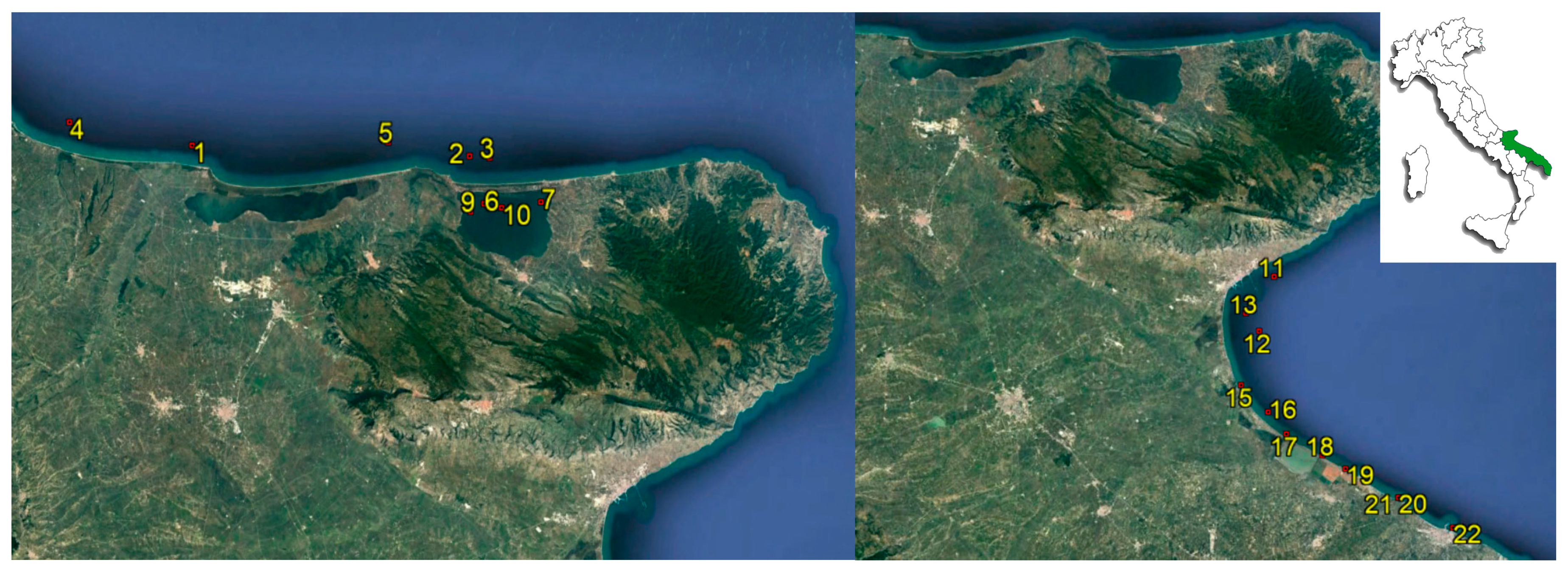
2.1. Sampling

Overall, 296 shellfish samples were collected from March 2021 to October 2022 along the northern Apulian coast (provinces of Foggia and Barletta-Andria-Trani). Most of them (263/296; 88.85%) derived from the implementation of the official classification programme of bivalve mollusc production and harvesting areas with regard to the Commission Implementing Regulation 2019/627 [21]. The geographical distribution of sampling points was established after a sanitary survey in order to choose the location at highest risk of faecal pollution and ensure that analytical results were representative of the area. At least 12 samples were taken from each sampling point over at least a 6-month period, as recommended in the community guide to the principles of good practice for the microbiological classification and monitoring of bivalve mollusc production and relaying areas with regard to Implementing Regulation 2019/627 [22]. The interval between two sampling occasions was approximatively 2 weeks, depending on the weather conditions and the availability of a commercial-size product. It is important to highlight that search for Salmonella is mandatory according to the European legislation [23]; otherwise, Vibrio detection was carried out only for research purposes.
The remaining samples (33/296; 11.15%) consisted of live bivalve molluscs originated from the same investigated areas and sampled for official control in accordance with the Integrated Regional Control Plan of Apulia region; they were collected in purification and dispatch centres or at retail and also tested for the aim of this study.
As regards the specimens collected for the official classification purpose, they consisted of oysters (Crassostrea gigas), mussels (Mytilus galloprovinciallis and Modiolus barbatus), clams (Venus gallina/Chamelea gallina), cockles (Acanthocardia tuberculata), and Japanese carpet shells (Ruditapes philippinarum). They were collected from fixed sampling stations, transported to the laboratory on the same day at temperatures between 4 and 10 °C and processed within 24 h of arrival. Environmental parameters, such as water temperature (°C) and pH, were measured on site during the sampling. The sampling stations are illustrated in Figure 1.
Detailed information about mollusc species and sampling points are reported in Table 1.

2.2. Vibrio Detection and Antimicrobial Susceptibility Testing

Vibrio spp. detection was performed according to the standard ISO 21872–1:2017 [24] by preparing two enrichment broths, which were incubated at 37 ± 1 °C and 41.5 ± 1 °C, respectively, to enhance the recovery of most Vibrio species. All the presumptive Vibrio spp. isolates were identified at species level based on the API® ID 20E and 20NE systems (BioMérieux, Nürtingen, Germany), the halotolerance test with various concentrations of NaCl (0, 6 and 10%), and the MALDI-TOF MS (Bruker Daltonics, Bremen, Germany) procedure by the direct transfer method, as previously described [25]. Moreover, the isolates recognised as V. parahaemolyticus, V. cholerae, and V. vulnificus were confirmed by the conventional PCR method described in Annex C to ISO 21872–1:2017, which considers, also, the detection of V. parahaemolyticus virulence genes (tdh and trh).
The broth microdilution MIC method was carried out on colonies grown on nonselective medium saline nutrient agar with 1% NaCl (SNA) incubated at 36 ± 1 °C overnight, using Sensititre™ Gram Negative GN4F® AST Plate and Sensititre™ NARMS® Gram Negative CMV4AGNF AST Plate (Thermofisher Scientific, Paisley, UK). Sensititre™ Gram Negative GN4F® AST Plate contained the following antimicrobials: amikacin (AMI 8–32 µg/mL), ampicillin (AMP 8–16 µg/mL), ampicillin/sulbactam (A/S2 4/2–16/8 µg/mL), aztreonam (AZT 1–16 µg/mL), cefazolin (FAZ 1–16 µg/mL), cefepime (FEP 4–32 µg/mL), ceftazidime (TAZ 1–16 µg/mL), ceftriaxone (AXO 0.5–32 µg/mL), ciprofloxacin (CIP 0.5–2 µg/mL), doripenem (DOR 0.5–4 µg/mL), ertapenem (ETP 0.25–8 µg/mL), gentamicin (GEN 2–8 µg/mL), imipenem (IMI 0.5–8 µg/mL), levofloxacin (LEVO 1–8 µg/mL), meropenem (MERO 0.5–8 µg/mL), minocycline (MIN 1–8 µg/mL), nitrofurantoin (NIT 32–64 µg/mL), piperacillin (PIP 16–64 µg/mL), piperacillin/tazobactam constant 4 (P/T4 8/4–128/4 µg/mL), tetracycline (TET 4–8 µg/mL), ticarcillin/clavulanic acid constant 2 (TIM2 8/2–64/2 µg/mL), tigecycline (TGC 1–8 µg/mL), tobramycin (TOB 2–8 µg/mL), and trimethoprim/sulfamethoxazole (SXT 2/38–4/76 µg/mL). Sensititre™ NARMS® Gram Negative CMV4AGNF AST contained the following antimicrobials: amoxicillin/clavulanic acid 2:1 ratio (AUG2 1/0.5–32/16 µg/mL), ampicillin (AMP 1–32 µg/mL), azithromycin (AZI 0.25–32 µg/mL), cefoxitin (FOX 0.5–32 µg/mL), ceftriaxone (AXO 0.25–64 µg/mL), chloramphenicol (CHL 2–32 µg/mL), ciprofloxacin (CIP 0.015–4 µg/mL), gentamicin (GEN 0.25–16 µg/mL), meropenem (MERO 0.06–4 µg/mL), nalidixic acid (NAL 0.5–32 µg/mL), streptomycin (STR 2–64 µg/mL), sulfisoxazole (FIS 16–256 µg/mL), tetracycline (TET 4–32 µg/mL), and trimethoprim/sulfamethoxazole (SXT 0.12/2.38–4/76 µg/mL). The antibiotics used in the study were chosen in accordance with Clinical and Laboratory Standards Institute (CLSI) recommendations, including those for the treatment of Vibrio infections. It was decided to use these Sensititre™ plates in order to include most of the antibiotic compounds listed in CLSI M45 guidelines [26], even if some molecules were present in both antimicrobial plates but at different concentrations.
The bacterial inoculum was prepared by dissolving a fresh pure colony in 2.5% NaCl solution using a sterile cotton swab until it achieved the turbidity of the 0.5 McFarland standard. Then, 0.1 mL of this suspension was added to 9.9 mL of cation-adjusted Mueller–Hinton broth (Becton Dickinson, Milan, Italy), the antimicrobial plate wells were inoculated with 50 µL of this suspension, and the plate was incubated aerobically at 36 ± 1 °C for 24 h. Escherichia coli ATCC® 25922 was used as quality control in each batch. The results were interpreted according to CLSI clinical breakpoints specific for Vibrio spp. No CLSI breakpoints were available for the following agents: ceftriaxone, nalidixic acid, streptomycin, tigecycline, ticarcillin/clavulanic acid, nitrofurantoin, doripenem, minocycline, ertapenem, tobramycin, and aztreonam. Hence, results of broth microdilution assays referring to the above-mentioned molecules are reported in the present study without any interpretative criteria. Only for streptomycin, the breakpoints described by other authors were used [27,28].

2.3. Salmonella Detection and Antimicrobial Susceptibility Testing

Salmonella spp. detection and serotyping were performed according to the ISO 6579–1:2017/Amd 1:2020 [29] and ISO/TR 6579–3:2014 [30], respectively. Antimicrobial susceptibility testing was performed on Salmonella strains by broth microdilution using Sensititre™ EUVSEC3® (Termofisher Scientific, Paisley, UK), which contained the compounds specified in the Commission Implementing Decision (EU) 2020/1729: ampicillin (1–32 µg/mL), azithromycin (2–64 µg/mL), amikacin (4–128 µg/mL), gentamicin (0.5–16 µg/mL), tigecycline (0.25–8 µg/mL), ceftazidime (0.25–8 µg/mL), cefotaxime (0.25–4 µg/mL), colistin (1–16 µg/mL), nalidixic acid (4–64 µg/mL), tetracycline (2–32 µg/mL), trimethoprim (0.25–16 µg/mL), sulfamethoxazole (8–512 µg/mL), chloramphenicol (8–64 µg/mL), meropenem (0.03–16 µg/mL), and ciprofloxacin (0.015–8 µg/mL). The quality control of the batch was performed with E. coli ATCC® 25922. The bacterial inoculum was prepared as described for Vibrio spp. but with 0.9% NaCl solution.
The epidemiological cut-off value (ECOFF) indicated in the Commission Implementing Decision (EU) 2020/1729 [18] was used as the interpretation threshold of AMR, except for colistin and tigecycline, for which the values defined for Enterobacteriales in the EUCAST clinical breakpoint table [31] were chosen, whereas the MIC breakpoints stated in CLSI document M100 [32] were used for azithromicyn and sulfamethoxazole (MIC values for sulphonamides). Multidrug resistance (MDR) was defined as nonsusceptibility to at least one agent in three or more antimicrobial categories [33].
Finally, the multiple antibiotic resistance (MAR) index was calculated as the ratio between the number of antibiotics to which a strain was resistant to and the total number of antibiotics used [34].

3. Results

3.1. Salmonella Isolates and Their Antimicrobial Resistance Profile

Among the 296 live bivalve molluscs analysed, only 1.7% of them were contaminated with Salmonella spp. Overall, five strains were isolated and tested for susceptibility to antimicrobial agents: three strains from clams (S. enterica subsp. enterica Kasenyi, S. enterica subsp. enterica Typhimurium, and S. bongori 48:z35:-) and two strains from mussels (S. enterica subsp. enterica Fischerhütte and S. enterica subsp. enterica Typhimurium). All isolates were sensitive to almost all antibiotics, except for S. Typhimurium isolated from Venus gallina, which showed resistance to sulfamethoxazole, trimethoprim, tetracycline, gentamicin, and ampicillin. Instead, S. Kasenyi and S. bongori 48:z35:- were resistant only to sulfamethoxazole.

3.2. Vibrio Isolates and Their Antimicrobial Resistance Profile

In total, 126 Vibrio strains (mostly V. alginolyticus and V. parahaemolyticus) were detected, with 38.2% (113/296) of shellfish samples contaminated with them. The API 20E system and the MALDI-TOF MS identification results were consistent, whereas the API 20NE displayed disputable and unreliable results. Furthermore, it is well known that the biochemical tests are inadequate for an accurate identification of V. harveyi [12,35] and that profiles for V. harveyi are not included in the bioMérieux database; thus, the API 20E and API 20NE systems do not allow this species to be properly identified via Apiweb™ and the identification of V. harveyi in this study was provided by the MALDI-TOF MS system. Vibrio spp. strains have been detected in shellfish collected from all sampling stations, with the exception of point 13. The temperature range for all isolates was of 9.4–29.9 °C, but isolation of potentially enteropathogenic species (V. cholerae, V. parahaemolyticus, and V. vulnificus) occurred when temperature ranged between 19.5 and 29.9 °C. The correlation between source and Vibrio species is shown in Figure 2. V. alginolyticus was the most prevalent species, found mostly in Mytilus galloprovincialis, followed by Venus gallina/Chamelea gallina.
No pathogenic genes were found in V. cholerae and V. parahaemolyticus isolates, except for one V. parahaemolyticus strain harbouring trh gene. The highest MAR index values for each sampling point are reported in Table 2 by specifying Vibrio species, resistance pattern, and source.
More specific details for each isolate about origin, resistance pattern, and MAR index are reported in Supplementary Table S1.
None of the 126 Vibrio isolates showed resistance to AZI, CHL, TET, AUG2, GEN, STR, AMI, or LEVO. Despite the interpretive criteria for Vibrio spp. other than V. cholerae provided in CLSI M45 guidelines [26] being uncertain for azithromycin, one V. harveyi (MIC = 4 µg/mL) could be surely stated as nonsusceptible and belonging to at least the intermediate category. Moreover, one V. parahaemolyticus displayed intermediate resistance to TET, one V. parahaemolyticus and five V. alginolyticus to AUG2, while intermediate resistance to STR was observed in all Vibrio species. The intermediate resistance percentages for each Vibrio species are detailed in Table 3.
High resistance percentages to FIS (57.1%; 72/126) (MIC > 256 µg/mL), AMP (85.7%; 108/126), and FAZ (56.3%; 71/126) were found among all Vibrio species, except for V. vulnificus and V. cholerae, which were sensitive to AMP. Moreover, 38% and 24.6% of isolates were resistant (MIC > 64 µg/mL) and intermediate resistant (MIC = 32–64 µg/mL) to PIP, respectively. Resistance to FOX was expressed only by one V. alginolyticus isolate, although four V. alginolyticus, one V. vulnificus, one V. cholerae, and one V. parahaemolyticus were intermediate resistant. Resistance to MERO, SXT, PIP, P/T4, IMI, TAZ, A/S2, and FEP was observed only in V. alginolyticus strains with low resistance percentages, as results show in Table 4.
Among V. parahaemolyticus strains, 94.1% (16/17) were resistant to FAZ, 76.5% (13/17) to AMP, 70.6% (12/17) to FIS, and 35.3% (6/17) to PIP. Susceptibility to all tested antimicrobials, except for FAZ, was found in the V. parahaemolyticus trh+ strain.
Overall, 41.3% (52/126) of strains displayed MDR, mostly towards sulphonamides, penicillin, and cephems. The most prevalent MDR profile was FIS-AMP-PIP-FAZ, followed by FIS-AMP-FAZ, both of them found primarily in V. alginolyticus and secondarily in V. parahaemolyticus.
The MIC values are shown in Supplementary Table S2, including those relating to the antibiotic agents without available specific CLSI breakpoints.

4. Discussion

Overall, few shellfish samples (5/296) have been found to be contaminated with Salmonella spp. in the current study and only one S. Typhimurium strain showed multidrug resistance. Despite the low prevalence of Salmonella, the detection of pathogenic serovars raises concerns about consumer health, especially given the recent finding of S. Typhimurium strains of human origin, collected from the same region and resistant mainly to ampicillin, tetracycline, azithromycin, and sulfamethoxazole [17], which are the same antimicrobials depicted in our multidrug-resistant strain, except for azithromycin replaced with gentamicin. Giacometti et al. [36] carried out a similar investigation on 102 Salmonella isolates from bivalve molluscs and water samples collected during the official monitoring programme in the area of the province of Ferrara (northwestern area of the Adriatic Sea) between 2001 and 2017. The most common resistances observed by the authors were to streptomycin (58.8%), ampicillin (52%), and tetracycline (45.1%), whereas 44.12% of isolates were MDR. Thus, it seems to suggest that resistance to these antimicrobial classes (penicillin, aminoglycoside, and tetracycline) is widely spread in the Adriatic Sea, in accordance with the AMR profile exhibited by our S. Typhimurium strain. However, any comparison with our results is difficult due to the long period of sampling (7 years), the type of samples (molluscs and water), and the method used to evaluate the AMR (agar disk diffusion). It is easy to understand that the AMR patterns vary according to the geographical origin of the samples; indeed, results concerning 27 Salmonella strains isolated by Lozano-Leon et al. [37] from mussel samples harvested in Galicia are completely different: all isolates showed MDR and were resistant to cefuroxime and cefuroxime/axetil; the majority of them expressed resistance to cefoxitin and gentamicin and some resistance was observed towards ampicillin, amikacin, cephalothin, and tobramycin, although the antimicrobial susceptibility was performed only on some strains.
As regards Vibrio spp., three different identification techniques were performed simultaneously in the present work to obtain a more reliable result, given that differentiation of closely related Vibrio species can be difficult. Although MALDI TOF MS is considered a valid tool, its discriminative power depends on the fullness of the reference library, which mainly contains clinically relevant species; thus, the reference library could be increased by inserting environmental bacterial isolates, especially marine bacteria, to make the MALDI TOF MS result more consistent. The API 20E and MALDI TOF MS systems gave the same results, except for the V. harveyi species because of the lack in the bioMérieux database. Therefore, the MALDI TOF identification of this Vibrio species was considered reliable and might perhaps be supported by biomolecular analysis, such as whole genome sequencing, which offers a great identification accuracy. Vibrio detection occurred during the entire investigation period but it is worth noting that potentially enteropathogenic species were isolated when water temperature was above 19.5 °C. It is well known that occurrence and densities of V. parahaemolyticus in molluscs are positively correlated to water temperatures [38] and outbreaks occur mainly during the warmer months in temperate zones [39]. However, the increase in surface temperature in coastal European seas in recent years has been linked to outbreaks caused by Vibrio cholerae nonO1-nonO139, V. parahaemolyticus, and V. vulnificus in several European countries [40]. Among potentially enteropathogenic species, V. parahaemolyticus was the most frequent one, isolated mainly from Mytilus galloprovincialis, but the only strain harbouring trh gene, hence proving to be harmful to humans, was isolated from Venus gallina o Chamelea gallina. V. cholerae isolation from Acanthocardia tuberculata cockles is a rare finding and certainly noteworthy given the scarcity of studies concerning this shellfish, mostly carried out in Morocco. For example, Boutaib et al. [41] found Salmonella spp. and non-pathogenic Vibrio parahaemolyticus in seven and five Acanthocardia tuberculata samples, respectively, but no results concerned V. cholerae, since this species was not included in the study.
In the current survey, the majority of Vibrio strains displayed resistance to ampicillin, sulfisoxazole, and cefazolin. Resistance to ampicillin was prevalent, as in previous studies [1,2,42,43].
If we compare our results with similar studies previously conducted in Italy, data seem to be quite varied. For instance, in the study carried out by Ottaviani et al. in 2001 [44] on several Vibrio species isolated from seafood, all strains exhibited susceptibility to imipenem, meropenem, chloramphenicol, and tetracycline, except 58% of V. alginolyticus, which were resistant to tetracycline; more than 90% of isolates showed susceptibility to oxolinic acid, cefotaxime, flumequine, doxycycline, and trimethoprim–sulphamethoxazole and more than 80% to nalidixic acid and ciprofloxacin. Moreover, resistance to streptomycin and lincomycin was found in more than 90% of isolates and many strains of V. alginolyticus, V. harveyi, V. vulnificus, and V. parahaemolyticus were resistant to penicillin, carbenicillin, ampicillin, cephalothin, and kanamycin, while V. alginolyticus and V. parahaemolyticus strains were resistant to rifampicin. A later investigation of antimicrobial resistance in V. parahaemolyticus from indigenous bivalves collected from harvesting areas along Italian coasts of the south Adriatic Sea, the central Tyrrhenian Sea, and the central Adriatic Sea reported that all isolates were resistant to ampicillin and amoxicillin but no resistances were observed to chloramphenicol, tetracycline, oxytetracycline, doxycycline, and trimethoprim/sulfamethoxazole [45]. Some strains exhibited resistance to cefotaxime (24.1%), cefalothin (43.7%), cefalexin (67.8%), colistin sulphate (13.8%), erythromycin (20.7%), and streptomycin (32.2%), whereas very low resistance percentages were found towards polymyxin B, nalidixic acid, oxolinic acid, nitrofurantoin, ciprofloxacin, kanamycin, and neomycin. Lopatek et al. [28] investigated the antimicrobial susceptibility of V. parahaemolyticus strains isolated from different species of raw shellfish and marine fish originated from various countries. They found that the majority of the strains were resistant to ampicillin and streptomycin and were recovered mainly from Italian samples; resistance to gentamicin was found in 12.5% of the strains, isolated from Italian, Dutch, and Norwegian samples, whereas only one strain was resistant to ciprofloxacin, isolated from Italian clams.
It should be noted that the majority of the studies on AMR in Vibrio spp. performed the agar disk diffusion method; thus, few works can be properly compared with our results.
In the current study, the V. vulnificus strain exhibited a poor resistance pattern, showing resistance only to cefazolin and an intermediate resistance to cefoxitin. However, Baker-Austin et al. [46] found numerous coastal and, also, septicaemia isolates in the USA resistant to antibiotics routinely prescribed for V. vulnificus infections, such as doxycycline, tetracycline, aminoglycosides, and cephalosporins, thus suggesting the importance of continued monitoring. In contrast, Bier et al. [27] reported that most antimicrobial agents recommended for treatment of V. vulnificus and V. cholerae non-O1/non-O139 infections were effective in vitro; likewise, in our study, both V. vulnificus and V. cholerae isolates did not display any worrisome resistance.
Banerjee and Farber [42] characterised 1021 Vibrio strains isolated from molluscs harvested in Canada between 2006 and 2012 and found that only 4.9% of them were sensitive to all tested drugs, while the antibiotics contributing the most to AMR were ampicillin, cephalothin, erythromycin, kanamycin, and streptomycin, although a declining trend in the frequency of MDR/AMR Vibrio spp. was registered until 2012.
Recently, Chahouri et al. [47] conducted a similar microbiological investigation in the Agadir Bay (Morocco), performing the search and AMR characterisation of Vibrio and Salmonella strains from mussels, sediment, and water samples. In accordance with our results, they isolated Vibrio strains at a high frequency, while a low percentage was noted for Salmonella. The eight Salmonella isolates showed resistance to ampicillin (100%), chloramphenicol (87.5%), and amoxicillin/clavulanic acid (62.5%) but were sensitive to all the other antibiotics used. Vibrio strains were mainly resistant to ampicillin (57.7%), cephalothin (62%), amikacin (60.6%), and, to a lesser extent, to ciprofloxacin (26.8%). Moreover, the authors perfectly agree with us when they highlight the importance of environmental survey to properly assess the microbiological quality in aquatic ecosystems.
In our study, almost all Vibrio strains exhibited MAR index values ranging from 0.064 to 0.193, below the arbitrary value of 0.2, indicating low-risk contamination sites [34], with the only exception of one V. alginolyticus isolate, which was resistant to seven antibiotics (MAR = 0.225). Since indices between 0.20 and 0.25 are in a range of ambiguity [34] and no Vibrio strains displayed MAR indices above 0.25, it could be stated that isolates originated from a low-risk environment where antibiotics are not regularly used perhaps. Moreover, resistance was observed mostly towards unusual antimicrobial compounds, such as ampicillin, piperacillin, sulfisoxazole, and the first-generation cephalosporin cefazolin, while few strains exhibited resistance to trimethoprim/sulfamethoxazole, piperacillin/tazobactam, and ampicillin/sulbactam (five, two, and one V. alginolyticus, respectively), proving that the antimicrobial combination therapy is still quite effective. In conclusion, the level of AMR in the tested Salmonella and above all Vibrio strains, which represent a consistent bacterial population in the marine environment, seems to indicate a poor diffusion of this phenomenon in the investigated area, suggesting that the actual condition is not highly worrying but perhaps promising, especially in view of the strategies currently implemented for the prudent use of antimicrobials in both human medicine and the zootechnical sector. However, our findings are not negligible, as some V. alginolyticus isolates exhibited resistance towards the critically important antimicrobials for human medicine. The epidemiological value of our study is of great relevance given the methodical approach used for monitoring: the survey was conducted over a period of 19 months and in a rather large area particularly devoted to shellfish farming and shellfish harvesting. Thus, the large amount of systematic data collected give an overview of the current scenario in one of the most representative regions for the Italian shellfish production sector [48].
The present study, like other previous ones, suggests the opportunity to implement a national/European programme to monitor the prevalence and distribution of antimicrobial resistance pattern in several not only pathogenic, but also environmental bacteria. Special consideration should be given to bacteria isolated from seafood which are generally eaten raw or undercooked and could represent a great threat to human health by transferring mobile genetic elements for antibiotic resistance to human pathogens. With regard to the latter aspect, it is worth mentioning, for example, the isolation of a V. parahaemolyticus strain carrying the blaNDM-1 gene from seafood and displaying in vitro carbapenemase activity but not phenotypical resistance [49]; this is a certainly highly worrisome finding, which could lead to therapeutic failure; thus, further research should be encouraged. Investigation of AMR in microorganisms within a specific area is necessary to formulate effective antibiotic reduction programmes. Indeed, although antibiotic resistance is an ancient and natural phenomenon, it is generally recognised that it occurs mainly in bacteria inhabiting the gastrointestinal tract of subjects receiving antibiotics and that distribution of antibiotic resistance genes (ARGs) in the aquatic ecosystem reflects mostly the faecal contamination by ARB. Furthermore, antibiotic pollution contributes to promoting the emergence and maintenance of ARGs and ARB in a delimited area [50]. Hence, insightful information on trends of AMR distribution in site-specific microbial populations is fundamental to better understand the local use and abuse of antibiotics and realise appropriate corrective measures. In this perspective, it would be appropriate to fix MIC breakpoints, also, for environmental Vibrio species and our results could be useful to upgrade the MIC distribution for Vibrio spp. relating to the antibiotic agents without available specific CLSI breakpoints.

Supplementary Materials

The following supporting information can be downloaded at: https://www.mdpi.com/article/10.3390/microorganisms11020450/s1, Table S1. Information on Vibrio isolates: origin, resistance profile and MAR index. Table S2: Results for the broth microdilution assays of Vibrio isolates.

Author Contributions

Conceptualisation, M.E.M., E.G.; methodology, M.E.M., A.A.; formal analysis, M.E.M.; investigation A.A., A.D. (Adelia Donatiello), A.D. (Antonella Didonna), S.F., G.O., F.C., L.P., V.R., A.M.D., R.C.; resources, C.P., L.D.; data curation, A.A.; writing—original draft preparation, M.E.M.; writing—review and editing, M.E.M., A.A., P.D.T.; supervision, E.G. All authors have read and agreed to the published version of the manuscript.

Funding

This research received no external funding.

Institutional Review Board Statement

Not applicable.

Informed Consent Statement

Not applicable.

Data Availability Statement

Not applicable.

Acknowledgments

Selicato Patrizia for technical support.

Conflicts of Interest

The authors declare no conflict of interest.

References

  1. Shakerian, A.; Barton, M.D.; Akinbowale, O.L.; Khamesipour, F. Antimicrobial resistance profile and resistance genes of Vibrio species isolated from giant freshwater prawn (Macrobrachium Rosenbergii) raised in Iran. J. Hellenic. Vet. Med. Soc. 2018, 68, 79–88. [Google Scholar] [CrossRef]
  2. Sudha, S.; Mridula, C.; Silvester, R.; Hatha, A.A.M. Prevalence and antibiotic resistance of pathogenic Vibrios in shellfishes from Cochin market. Indian J. Geo-Mar. Sci. 2014, 43, 815–824. [Google Scholar]
  3. Cabello, F.C.; Godfrey, H.P.; Tomova, A.; Ivanova, L.; Dölz, H.; Millanao, A.; Buschmann, A.H. Antimicrobial use in aquaculture re-examined: Its relevance to antimicrobial resistance and to animal and human health. Environ. Microbiol. 2013, 15, 1917–1942. [Google Scholar] [CrossRef] [PubMed]
  4. Baker-Austin, C.; Oliver, J.D.; Alam, M.; Ali, A.; Waldor, M.K.; Quadri, F.; Martinez-Urtaza, J. Vibrio spp. infections. Nat. Rev. Dis. Primers 2018, 4, 1–19. [Google Scholar] [CrossRef]
  5. Rivera, I.N.; Chun, J.; Huq, A.; Sack, R.B.; Colwell, R.R. Genotypes associated with virulence in environmental isolates of Vibrio cholerae. Appl. Environ. Microbiol. 2001, 67, 2421–2429. [Google Scholar] [CrossRef]
  6. Dutta, D.; Kaushik, A.; Kumar, D.; Bag, S. Foodborne pathogenic Vibrios: Antimicrobial resistance. Front. Microbiol. 2021, 30, 638331. [Google Scholar] [CrossRef]
  7. Mok, J.S.; Ryu, A.; Kwon, J.Y.; Kim, B.; Park, K. Distribution of Vibrio species isolated from bivalves and bivalve culture environments along the Gyeongnam coast in Korea: Virulence and antimicrobial resistance of Vibrio parahaemolyticus isolates. Food Control 2019, 106, 106697. [Google Scholar] [CrossRef]
  8. Ashrafudoulla, M.; Mizan, M.F.R.; Park, H.; Byun, K.H.; Lee, N.; Park, S.H.; Ha, S.D. Genetic relationship, virulence factors, drug resistance profile and biofilm formation ability of Vibrio parahaemolyticus isolated from mussel. Front. Microbiol. 2019, 10, 513. [Google Scholar] [CrossRef]
  9. Ina-Salwany, M.Y.; Al-Saari, N.; Mohamad, A.; Mursidi, F.A.; Mohd-Aris, A.; Amal, M.n.A.; Kasai, H.; Mino, S.; Sawabe, T.; Zamri-Saad, M. Vibriosis in fish: A review on disease development and prevention. J. Aquat. Anim. Health 2019, 31, 3–22. [Google Scholar] [CrossRef]
  10. Labella, A.; Gennari, M.; Ghidini, V.; Trento, I.; Manfrin, A.; Borrego, J.J.; Lleo, M.M. High incidence of antibiotic multi-resistant bacteria in coastal areas dedicated to fish farming. Mar. Pollut. Bull. 2013, 70, 197–203. [Google Scholar] [CrossRef]
  11. Brehm, T.T.; Berneking, L.; Rohde, H.; Chistner, M.; Schlickewei, C.; Sena Martins, M.; Schmiedel, S. Wound infection with Vibrio harveyi following a traumatic leg amputation after a motorboat propeller injury in Mallorca, Spain: A case report and review of literature. BMC Infect. Dis. 2020, 20, 104. [Google Scholar] [CrossRef] [PubMed] [Green Version]
  12. Montánchez, I.; Kaberdin, V.R. Vibrio harveyi: A brief survey of general characteristics and recent epidemiological traits associated with climate change. Mar. Environ. Res. 2020, 154, 104850. [Google Scholar] [CrossRef]
  13. Directive 2003/99/EC of the European Parliament and of the Council of 17 November 2003 on the Monitoring of Zoonoses and Zoonotic Agents, Amending Council Decision 90/424/EEC and Repealing Council Directive 92/117/EEC. 2003. Available online: http://data.europa.eu/eli/dir/2003/99/2013-07-01 (accessed on 6 February 2023).
  14. European Food Safety Authority and European Centre for Disease Prevention and Control. The European Union One Health 2019 Zoonoses Report. EFSA J. 2021, 19, e06406. [Google Scholar]
  15. Maggi, P.; Carbonara, S.; Fico, C.; Santantonio, T.; Romanelli, C.; Sforza, E.; Pastore, G. Epidemiological, clinical and therapeutic evaluation of the Italian cholera epidemic in 1994. Eur. J. Epidemiol. 1997, 13, 95–97. [Google Scholar] [CrossRef] [PubMed]
  16. Rizzo, G.; Barbuti, S.; Leogrande, G.; Jatta, E. Osservazioni sulla diagnosi batteriologica di colera e sulle caratteristiche degli stipiti isolati nella epidemia pugliese dell’estate 1973 [Studies on the bacteriological diagnosis of cholera and on the characteristics of isolated strains in the Apulia epidemic during the summer of 1973]. Ann. Sclavo 1975, 17, 441–448. [Google Scholar]
  17. Alessiani, A.; Goffredo, E.; Mancini, M.; Occhiochiuso, G.; Faleo, S.; Didonna, A.; Fischetto, R.; Suglia, F.; De Vito, D.; Stallone, A.; et al. Evaluation of antimicrobial resistance in Salmonella strains isolated from food, animal and human samples between 2017 and 2021 in southern Italy. Microorganisms 2022, 10, 812. [Google Scholar] [CrossRef]
  18. Commission Implementing Decision (EU) 2020/1729 of 17 November 2020 on the monitoring and reporting of antimicrobial resistance in zoonotic and commensal bacteria and repealing Implementing Decision 2013/652/EU, (2020). Available online: http://data.europa.eu/eli/dec_impl/2020/1729/oj (accessed on 6 February 2023).
  19. Regulation (EC) No 2160/2003 of the European Parliament and of the Council of 17 November 2003 on the control of salmonella and other specified food-borne zoonotic agents, (2003). Available online: http://data.europa.eu/eli/reg/2003/2160/2021-04-21 (accessed on 6 February 2023).
  20. World Health Organization. 2021 TrACSS Country Report on the Implementation of National Action Plan on Antimicrobial Resistance (AMR). Available online: https://cdn.who.int/media/docs/default-source/antimicrobial-resistance/amr-spc-npm/tracss/tracss-2021-italy.pdf?sfvrsn=10e5e354_4&download=true (accessed on 9 December 2022).
  21. Commission Implementing Regulation (EU) 2019/627 of 15 March 2019 laying down uniform practical arrangements for the performance of official controls on products of animal origin intended for human consumption in accordance with Regulation (EU) 2017/625 of the European Parliament and of the Council and amending Commission Regulation (EC) No 2074/2005 as regards official controls, (2019). Available online: http://data.europa.eu/eli/reg_impl/2019/627/2021-10-14 (accessed on 6 February 2023).
  22. European Union Reference Laboratory for Monitoring of Marine Biotoxins. Community Guide to the Principles of Good Practice for the Microbiological Classification and Monitoring of Bivalve Mollusc Production and Relaying Areas with Regard to Implementing Regulation 2019/627. Issue 4. 2021. Available online: https://www.aesan.gob.es/en/CRLMB/docs/docs/procedimientos/Micro_Control_Guide_DEC_2021.pdf (accessed on 9 December 2022).
  23. Commission Regulation (EC) No 2073/2005 of 15 November 2005 on microbiological criteria for foodstuffs, (2005). Available online: http://data.europa.eu/eli/reg/2005/2073/2020-03-08 (accessed on 6 February 2023).
  24. ISO 21872–1:2017; Microbiology of the Food Chain—Horizontal Method for the Determination of Vibrio spp.—Part 1: Detection of Potentially Enteropathogenic Vibrio Parahaemolyticus, Vibrio Cholerae and Vibrio vulnificus. International Organization for Standardization: Geneva, Switzerland, 2017.
  25. Mancini, M.E.; Beverelli, M.; Donatiello, A.; Didonna, A.; Dattoli, L.; Faleo, S.; Occhiochiuso, G.; Galante, D.; Rondinone, V.; Del Sambro, L.; et al. Isolation and characterization of Yersinia enterocolitica from foods in Apulia and Basilicata regions (Italy) by conventional and modern methods. PLoS ONE 2022, 17, e0268706. [Google Scholar] [CrossRef]
  26. Clinical and Laboratory Standards Institute (CLSI). Methods for Antimicrobial Dilution and Disk Susceptibility Testing of Infrequently Isolated or Fastidious Bacteria, 3rd ed.; CLSI guideline M45; Clinical and Laboratory Standards Institute: Wayne, AR, USA, 2015; Available online: https://goums.ac.ir/files/deputy_treat/md_labs_ef39a/files/CLSI-M45ed3e-2018.pdf (accessed on 9 December 2022).
  27. Bier, N.; Schwartz, K.; Guerra, B.; Strauch, E. Survey on antimicrobial resistance patterns in Vibrio vulnificus and Vibrio cholerae non-O1/non-O139 in Germany reveals carbapenemase-producing Vibrio cholerae in coastal waters. Front. Microbiol. 2015, 6, 1179. [Google Scholar] [CrossRef]
  28. Lopatek, M.; Wieczorek, K.; Osek, J. Antimicrobial resistance, virulence factors, and genetic profiles of Vibrio parahaemolyticus from seafood. Appl. Environ. Microbiol. 2018, 84, e00537-18. [Google Scholar] [CrossRef]
  29. ISO 6579–1:2017/Amd 1:2020; Microbiology of the Food Chain—Horizontal Method for the Detection, Enumeration and Serotyping of Salmonella—Part 1: Detection of Salmonella spp.—Amendment 1: Broader Range of Incubation Temperatures, Amendment to the Status of Annex D, and Correction of the Composition of MSRV and SC. International Organization for Standardization: Geneva, Switzerland, 2020.
  30. ISO/TR 6579–3:2014; Microbiology of the Food Chain—Horizontal Method for the Detection, Enumeration and Serotyping of Salmonella—Part 3: Guidelines for Serotyping of Salmonella spp. International Organization for Standardization: Geneva, Switzerland, 2014.
  31. European Committee on Antimicrobial Susceptibility Testing (EUCAST). Breakpoint Tables for Interpretation of MICs and Zone Diameters. Version 12.0. Available online: https://www.eucast.org/fileadmin/src/media/PDFs/EUCAST_files/Breakpoint_tables/v_12.0_Breakpoint_Tables.pdf (accessed on 9 December 2022).
  32. Clinical and Laboratory Standards Institute (CLSI). Performance Standards for Antimicrobial Susceptibility Testing. 32nd ed. In CLSI Supplement M100; Clinical and Laboratory Standards Institute: Wayne, AR, USA, 2022; Available online: http://em100.edaptivedocs.net/GetDoc.aspx?doc=CLSI%20M100%20ED31:2021&scope=user (accessed on 9 December 2022).
  33. Magiorakos, A.P.; Srinivasan, A.; Carey, R.B.; Carmeli, Y.; Falagas, M.E.; Giske, C.G.; Harbarth, S.; Hindler, J.F.; Kahlmeter, G.; Olsson-Liljequist, B.; et al. Multidrug-resistant, extensively drug-resistant and pandrug-resistant bacteria: An international expert proposal for interim standard definitions for acquired resistance. Clin. Microbiol. Infect. 2012, 18, 268–281. [Google Scholar] [CrossRef]
  34. Krumperman, P.H. Multiple antibiotic resistance indexing of Escherichia coli to identify high-risk sources of fecal contamination of foods. Appl. Environ. Microbiol. 1983, 46, 165–170. [Google Scholar] [CrossRef] [PubMed]
  35. Stalin, N.; Srinivasan, P. Molecular characterization of antibiotic resistant Vibrio harveyi isolated from shrimp aquaculture environment in the south east coast of India. Microb. Pathog. 2016, 97, 110–118. [Google Scholar] [CrossRef] [PubMed]
  36. Giacometti, F.; Pezzi, A.; Galletti, G.; Tamba, T.; Merialdi, G.; Piva, S.; Serraino, A.; Rubini, S. Antimicrobial resistance patterns in Salmonella enterica subsp. enterica and Escherichia coli isolated from bivalve molluscs and marine environment. Food Control 2021, 121, 107590. [Google Scholar]
  37. Lozano-León, A.; García-Omil, C.; Rodríguez-Souto, R.R.; Lamas, A.; Garrido-Maestu, A. An evaluation of the pathogenic potential, and the antimicrobial resistance, of Salmonella strains isolated from mussels. Microorganisms 2022, 10, 126. [Google Scholar] [CrossRef]
  38. Shen, X.; Cai, Y.; Liu, C.; Liu, W.; Hui, Y.; Su, Y.C. Effect of temperature on uptake and survival of Vibrio parahaemolyticus in oysters (Crassostrea plicatula). Int. J. Food Microbiol. 2009, 136, 129–132. [Google Scholar] [CrossRef]
  39. Nair, G.B.; Ramamurthy, T.; Bhattacharya, S.K.; Dutta, B.; Takeda, Y.; Sack, D.A. Global dissemination of Vibrio parahaemolyticus serotype O3:K6 and its serovariants. Clin. Microbiol. Rev. 2007, 20, 39–48. [Google Scholar] [CrossRef]
  40. Le Roux, F.; Wegner, K.M.; Baker-Austin, C.; Vezzulli, L.; Osorio, C.R.; Amaro, C.; Ritchie, J.M.; Defoirdt, T.; Destoumieux-Garzón, D.; Blokesch, M.; et al. The emergence of Vibrio pathogens in Europe: Ecology, evolution, and pathogenesis (Paris, 11–12th March 2015). Front. Microbiol. 2015, 6, 830. [Google Scholar]
  41. Boutaib, R.; Marhraoui, M.; Oulad Abdellah, M.K.; Bouchrif, B. Comparative study on faecal contamination and occurrence of Salmonella spp. and Vibrio parahaemolyticus in two species of shellfish in Morocco. Open Environ. Sci. 2011, 5, 30–37. [Google Scholar] [CrossRef]
  42. Banerjee, S.K.; Farber, J.M. Trend and pattern of antimicrobial resistance in molluscan Vibrio species sourced to Canadian estuaries. Antimicrob. Agents Chemother. 2018, 62, 00799-18. [Google Scholar] [CrossRef]
  43. Håkonsholm, F.; Lunestad, B.T.; Aguirre Sánchez, J.R.; Martinez-Urtaza, J.; Marathe, N.P.; Svanevik, C.S. Vibrios from the Norwegian marine environment: Characterization of associated antibiotic resistance and virulence genes. Microbiologyopen 2020, 9, e1093. [Google Scholar] [CrossRef]
  44. Ottaviani, D.; Bacchiocchi, I.; Masini, L.; Leoni, F.; Carraturo, A.; Giammarioli, M.; Sbaraglia, G. Antimicrobial susceptibility of potentially pathogenic halophilic vibrios isolated from seafood. Int. J. Antimicrob. Agents 2001, 18, 135–140. [Google Scholar] [CrossRef] [PubMed]
  45. Ottaviani, D.; Leoni, F.; Talevi, G.; Masini, L.; Santarelli, S.; Rocchegiani, E.; Susini, F.; Montagna, C.; Monno, R.; D’Annibale, L.; et al. Extensive investigation of antimicrobial resistance in Vibrio parahaemolyticus from shellfish and clinical sources, Italy. Int. J. Antimicrob. Agents 2013, 42, 191–193. [Google Scholar] [CrossRef] [PubMed]
  46. Baker-Austin, C.; McArthur, J.V.; Lindell, A.H.; Wright, M.S.; Tuckfield, R.C.; Gooch, J.; Warner, L.; Oliver, J.; Stepanauskas, R. Multi-site analysis reveals widespread antibiotic resistance in the marine pathogen Vibrio vulnificus. Microb. Ecol. 2009, 57, 151–159. [Google Scholar] [CrossRef] [PubMed]
  47. Chahouri, A.; Radouane, n.; Yacoubi, B.; Moukrim, A.; Banaoui, A. Microbiological assessment of marine and estuarine ecosystems using fecal indicator bacteria, Salmonella, Vibrio and antibiotic resistance pattern. Mar. Pollut. Bull. 2022, 180, 113824. [Google Scholar] [CrossRef]
  48. Istituto Superiore per la Protezione e la Ricerca Ambientale (ISPRA). Stato Dell’ambiente 84/2019. Available online: https://www.isprambiente.gov.it/public_files/annuario-2018.pdf (accessed on 9 December 2022).
  49. Briet, A.; Helsens, N.; Delannoy, S.; Debuiche, S.; Brisabois, A.; Midelet, G.; Granier, S.A. NDM-1-producing Vibrio parahaemolyticus isolated from imported seafood. J. Antimicrob. Chemother. 2018, 73, 2578–2579. [Google Scholar] [CrossRef]
  50. Haenni, M.; Dagot, C.; Chesneau, O.; Bibbal, D.; Labanowski, J.; Vialette, M.; Bouchard, D.; Martin-Laurent, F.; Calsat, L.; Nazaret, S.; et al. Environmental contamination in a high-income country (France) by antibiotics, antibiotic-resistant bacteria, and antibiotic resistance genes: Status and possible causes. Environ. Int. 2022, 159, 107047. [Google Scholar] [CrossRef]
Figure 1. Sampling station locations.
Microorganisms 11 00450 g001
Figure 2. The percentages of Vibrio species isolated from each shellfish.
Microorganisms 11 00450 g002
Table 1. Number and type of samples for each sampling point.
Sampling StationAreaShellfish Speciesn. Samples
1Northern coast of GarganoMytilus galloprovincialis12
2Northern coast of GarganoMytilus galloprovincialis13
3Northern coast of GarganoCrassostrea gigas13
4Northern coast of GarganoMytilus galloprovincialis13
5Northern coast of GarganoMytilus galloprovincialis15
6Varano lakeRuditapes philippinarum6
Mytilus galloprovincialis14
7Varano lakeCrassostrea gigas14
9Varano lakeMytilus galloprovincialis13
10Varano lakeMytilus galloprovincialis14
11Southern coast of GarganoCrassostrea gigas6
Mytilus galloprovincialis11
12Southern coast of GarganoMytilus galloprovincialis14
13Southern coast of GarganoModiolus barbatus7
15Southern coast of GarganoAcanthocardia tuberculata8
Modiolus barbatus1
16Southern coast of GarganoAcanthocardia tuberculata8
17Southern coast of GarganoAcanthocardia tuberculata8
18Coastline of BAT 1 Province Venus gallina/Chamelea gallina17
19Coastline of BAT 1 ProvinceVenus gallina/Chamelea gallina15
20Coastline of BAT 1 ProvinceVenus gallina/Chamelea gallina17
21Coastline of BAT 1 ProvinceVenus gallina/Chamelea gallina12
22Coastline of BAT 1 ProvinceVenus gallina/Chamelea gallina12
1 BAT: Barletta-Andria-Trani.
Table 2. The highest values of multiple antibiotic resistance (MAR) index of Vibrio isolates.
Sampling StationMatrixVibrio SpeciesResistance PatternMAR Index
1Mytilus galloprovincialisV. alginolyticusMERO, FIS, AMP, P/T4, PIP, FEP0.193
2Mytilus galloprovincialisV. alginolyticusFIS, SXT, AMP, PIP, FAZ0.161
3Crassostrea gigasV. alginolyticusFIS, AMP, P/T4, PIP, FAZ, TAZ, FEP0.225
4Mytilus galloprovincialisV. alginolyticusFIS, AMP, PIP, FAZ0.129
V. harveyiFIS, AMP, PIP, FAZ0.129
5Mytilus galloprovincialisV. alginolyticusFIS, AMP, PIP, FAZ0.129
V. alginolyticusFIS, AMP, PIP, FAZ0.129
6Mytilus galloprovincialisV. alginolyticusFIS, AMP, PIP, FAZ0.129
V. parahaemolyticusFIS, AMP, PIP, FAZ0.129
Ruditapes philippinarumV. alginolyticusFIS, AMP, PIP, FAZ0.129
V. alginolyticusFIS, AMP, PIP, FAZ0.129
7Crassostrea gigasV. harveyiAMP, PIP0.064
9Mytilus galloprovincialisV. parahaemolyticusFIS, AMP, PIP, FAZ0.129
10Mytilus galloprovincialisV. alginolyticusFIS, AMP, PIP, FAZ0.129
V. alginolyticusFIS, AMP, PIP, FAZ0.129
V. parahaemolyticusFIS, AMP, PIP, FAZ0.129
11Mytilus galloprovincialisV. vulnificusFIS, FAZ0.064
12Mytilus galloprovincialisV. alginolyticusFIS, AMP, FAZ0.096
V. alginolyticusFIS, AMP, FAZ0.096
15Acanthocardia tuberculataV. alginolyticusFIS, AMP, FAZ0.096
16Acanthocardia tuberculataV. parahaemolyticusFIS, AMP, FAZ0.096
17Acanthocardia tuberculataV. alginolyticusAMP, PIP, FAZ0.096
18Venus gallina/ Chamelea gallinaV. alginolyticusFIS, AMP, PIP, FAZ0.129
19Venus gallina/ Chamelea gallinaV. alginolyticusFIS, AMP, PIP, FAZ0.129
20Venus gallina/ Chamelea gallinaV. alginolyticusFIS, AMP, PIP, FAZ0.129
21Venus gallina/ Chamelea gallinaV. alginolyticusFIS, AMP, PIP, FAZ0.129
22Venus gallina/ Chamelea gallinaV. harveyiFIS, AMP, PIP, FAZ0.129
V. parahaemolyticusFIS, AMP, PIP, FAZ0.129
V. alginolyticusFIS, AMP, PIP, FAZ0.129
Table 3. Intermediate profile to single antimicrobial agents in percentage. The antimicrobial agents for which no intermediate resistance was found are not shown.
Vibrio SpeciesnAntimicrobial Agents
FOXAZITETAUG2STRP/T4PIPFAZTAZA/S2
V. alginolyticus904.4 5.63.31.126.7301.16.7
V. parahaemolyticus175.9 5.95.95.9 11.8
V. harveyi17 5.9 17.6 29.411.8
V. vulnificus1100 100
V. cholerae1100 100
n: number of isolates.
Table 4. Resistance percentages to single antimicrobial agents. The antimicrobial agents for which no resistance was found are not shown.
Vibrio SpeciesnAntimicrobial Agents
FOXMEROFISSXTAMPP/T4IMIPIPFAZTAZA/S2FEP
V. alginolyticus901.11.161.15.693.32.21.142.255.61.11.13.3
V. parahaemolyticus17 70.6 76.5 35.394.1
V. harveyi17 17.6 64.7 23.517.6
V. vulnificus1 100 100
V. cholerae1 100 100
n: number of isolates.
Disclaimer/Publisher’s Note: The statements, opinions and data contained in all publications are solely those of the individual author(s) and contributor(s) and not of MDPI and/or the editor(s). MDPI and/or the editor(s) disclaim responsibility for any injury to people or property resulting from any ideas, methods, instructions or products referred to in the content.

