
Biosynthesis of Metal and Metal Oxide Nanoparticles Using Microbial Cultures: Mechanisms, Antimicrobial Activity and Applications to Cultural Heritage
 , , , and
Abstract
:1. Introduction
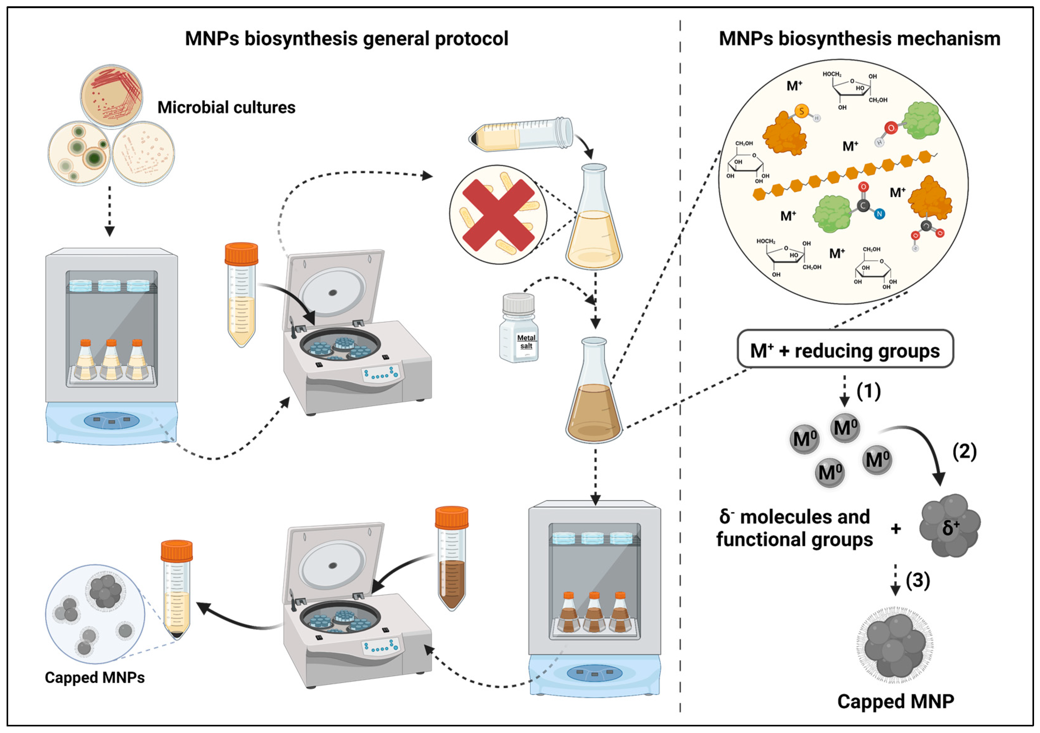
2. Metal Nanoparticles Synthesis Using Biological Extracts
2.1. Techniques Employed in the Study of Metal Nanoparticles’ Biosynthesis Mechanisms
2.2. Biosynthesis Mechanisms—State of the Art
2.3. Evidence-Based Proposed Biosynthesis Mechanisms
3. Recent Studies on the Biosynthesis of Metal Nanoparticles Using Microorganisms

| Metal * | Microbial Genera | NPs Size (nm) | Precursor | Antimicrobial Activity Studies | Toxicity Studies | Main Properties | Ref. |
|---|---|---|---|---|---|---|---|
| Molds | |||||||
| Ag | Anamorphous | 10 to 70 | AgNO3 | Yes | Yes | Antimicrobial | [136] |
| Ag | Aspergillus | 1 to 50 | AgNO3 | Yes | No | Antimicrobial | [137] |
| Ag | Aspergillus | 3 to 28 | AgNO3 | Yes | Yes | Antimicrobial Photocatalytic Acaricidal | [138] |
| Ag | Aspergillus | 5 to 37 | AgNO3 | Yes | No | Antimicrobial | [139] |
| Ag | Aspergillus | 15 to 35 | AgNO3 | No | No | NS | [140] |
| Ag | Aspergillus | 7 to 23 | AgNO3 | Yes | Yes | Antimicrobial Cytotoxicity | [141] |
| Ag | Aspergillus | 13 to 49 | AgNO3 | Yes | Yes | Antimicrobial Cytotoxicity | [142] |
| Ag | Aspergillus | 1 to 21 | AgNO3 | Yes | Yes | Antimicrobial Cytotoxicity | [143] |
| Ag | Aspergillus | 3.5 to 28.2 | AgNO3 | Yes | No | Antiamoebic | [144] |
| Ag | Aspergillus | ~100 a,b | AgNO3 | Yes | Yes | Antimicrobial Cytotoxicity | [145] |
| Ag | Aspergillus | 1 to 10.5 | AgNO3 | Yes | No | Antimicrobial | [66] |
| Ag | Aspergillus | 2 to 13 | No | Yes | Mosquitocidal | [146] | |
| Ag | Aspergillus | 56 a,b | Yes | No | Antimicrobial | [147] | |
| Ag | Aspergillus | 20 to 60 | AgNO3 | Yes | No | Antimicrobial Antioxidant Photocatalytic | [60] |
| Ag | Aspergillus | 10 to 100 | AgNO3 | Yes | Yes | Antimicrobial Cytotoxicity | [148] |
| Ag | Aspergillus | 1 to 15 | AgNO3 | Yes | No | Antimicrobial Antioxidant | [149] |
| Ag | Botryodiplodia | 66.8 to 111.2 | AgNO3 | No | Yes | Cytotoxicity | [150] |
| Ag | Eurotium | 15 to 20 | AgNO3 | Yes | No | Antimicrobial | [151] |
| Ag | Fusarium | ~40 c | AgNO3 | Yes | No | Antimicrobial Photocatalytic | [152] |
| Ag | Fusarium | 2 to 20 | AgNO3 | Yes | No | Antimicrobial | [153] |
| Ag | Humicola | 15 to 40 | AgNO3 Na2SO3 | No | Yes | Antiparasitic Cytotoxicity | [154] |
| Ag | Letendraea | 33.8 a | AgNO3 | No | Yes | Photocatalytic Antialgal | [155] |
| Ag | Letendraea | 8 to 56 | AgNO3 | Yes | No | Antimicrobial Antioxidant Photocatalytic | [156] |
| Ag | Neopestalotiopsis | 4.8 to 20.7 | AgNO3 | Yes | Yes | Antimicrobial Antibiofilm | [157] |
| Ag | Penicillium | 2 to 20 | AgNO3 | Yes | No | Antimicrobial | [158] |
| Ag | Penicillium | 18 to 60 | AgNO3 | Yes | No | Antimicrobial | [159] |
| Ag | Penicillium | 48.2 a,b | AgNO3 | Yes | No | Antimicrobial Antibiofilm | [160] |
| Ag | Penicillium | 60 to 80 | AgNO3 | Yes | No | Antimicrobial | [161] |
| Ag | Phomopsis | 5 to 60 | AgNO3 | Yes | No | Antimicrobial | [162] |
| Ag | Talaromyces | 5 to 30 | AgNO3 | Yes | Yes | Antimicrobial Cytotoxicity Larvicidal | [163] |
| Ag | Trichoderma | 10 to 70 | AgNO3 | Yes | Yes | Antimicrobial Antibiofilm Antioxidant Cytotoxicity | [164] |
| Ag | Trichoderma | 5 to 35 | AgNO3 | Yes | No | Antimicrobial | [165] |
| Ag | Trichoderma | 5 to 50 | AgNO3 | Yes | No | Antimicrobial | [166] |
| Ag | Trichoderma | 15 to 25 | AgNO3 | No | No | NS | [167] |
| Au | Aspergillus | 37 to 62 | HAuCl4 | Yes | No | Antimicrobial | [139] |
| Au | Aspergillus | 20 to 50 | AuCl3 | No | No | NS | [140] |
| Au | Aspergillus | 30 to 40 | AuCl3 | Yes | No | Antimicrobial Antibiofilm | [168] |
| Au | Aspergillus | 7 to 15 | HAuCl4 | No | Yes | Photocatalytic Cytotoxicity | [169] |
| Au | Fusarium | 22 to 30 | HAuCl4 | Yes | No | Antimicrobial | [170] |
| Au | Trichoderma | 8 to 30 | HAuCl4 | Yes | Yes | Antimicrobial Antibiofilm Antioxidant Cytotoxicity | [164] |
| Au | Trichoderma | 1 to 24 | HAuCl4 | No | No | Photocatalytic | [171] |
| Cu | Aspergillus | 9 to 25 | CuSO4 | No | No | NS | [172] |
| Cu | Penicillium | 10.5 to 59.7 | Cu(CH3COO)2 | Yes | No | Antimicrobial Antibiofilm | [173] |
| Cu | Trichoderma | 1.3 to 30 | CuSO4 | Yes | Yes | Antimicrobial Cytotoxicity | [174] |
| Fe | Aspergillus | 6.0 to 36.0 | FeCl3 | No | No | Photocatalytic Detoxification | [175] |
| Fe | Aspergillus | 32.7 to 47.6 | FeSO4 | Yes | No | Antimicrobial Photocatalytic | [176] |
| Fe | Aspergillus | 73.1 a | Fe(NO3)3 | No | No | Detoxification | [177] |
| Fe | Penicillium | 15 to 66 | FeCl3 | No | No | Photocatalytic | [178] |
| Mg | Aspergillus | 20.0 to 86.0 | Mg(NO3)2 | No | No | Photocatalytic Detoxification | [175] |
| Mg | Aspergillus | 30 to 85 | Mg(NO3)2 | Yes | Yes | Photocatalytic Detoxification | [179] |
| Mg | Aspergillus | 8 to 38 | Mg(NO3)2 | Yes | No | Antimicrobial Photocatalytic Detoxification | [180] |
| Mg | Penicillium | 7 to 40 | Mg(NO3)2 | Yes | No | Antimicrobial Mosquitocidal | [181] |
| Mg | Rhizopus | 8.0 to 47.5 | Mg(NO3)2 | Yes | No | Antimicrobial Mosquitocidal Photocatalytic Detoxification | [182] |
| Pt | Penicillium | 2 to 25 | H2PtCl6 | Yes | Yes | Antimicrobial Cytotoxicity | [183] |
| V | Fusarium | 10 to 20 | NH4VO3 | Yes | Yes | Antimicrobial Cytotoxicity | [184] |
| Zn | Aspergillus | 10 to 45 | Zn(CH3CO2)2 | Yes | Yes | Antimicrobial UV protection | [185] |
| Zn | Aspergillus | 80 to 100 a | Zn(CH3CO2)2 | Yes | No | Antimicrobial Photocatalytic Antibiofilm | [186] |
| Zn | Cochliobolus | 2 to 9 | Zn(CH3CO2)2 | No | No | Photocatalytic | [187] |
| Zn | Cochliobolus | 2 to 6 | Zn(CH3CO2)2 | No | No | NS | [188] |
| Zn | Penicillium | 9 to 35 | Zn(CH3CO2)2 | Yes | No | Antimicrobial Antibiofilm | [173] |
| Yeasts | |||||||
| Ag | Candida | 2.7 d | AgNO3 | Yes | No | Antimicrobial | [189] |
| Ag | Pichia | 4 to 12 | AgNO3 | Yes | Yes | Antimicrobial Antioxidant Cytotoxicity Photocatalytic | [190] |
| Ag | Pichia | 20 to 30 | AgNO3 | Yes | Yes | Antimicrobial Anti-inflammatory Cytotoxicity | [191] |
| Ag | Saccharomyces | 11 to 25 | AgNO3 | Yes | No | Antimicrobial | [192] |
| Ag | Saccharomyces | 7.3 d | AgNO3 | Yes | No | Antimicrobial | [189] |
| Ag | Saccharomyces | 12 to 21 | AgNO3 | Yes | Yes | Antimicrobial Anti-inflammatory Cytotoxicity | [191] |
| Ag | Yarrowia | 50 a | AgNO3 | No | No | Antimicrobial | [193] |
| Au | Magnusiomyces | 20 to 30 | HauCl4 | No | No | Photocatalytic | [194] |
| Pt | Rhodotorula | 2.83 a | H2PtCl6 | Yes | No | Antimicrobial Antioxidant | [195] |
| Metal * | Microbial Genera | NPs Size (nm) | Precursor | Antimicrobial Activity Studies | Toxicity Studies | Main Properties | Ref. |
|---|---|---|---|---|---|---|---|
| Microalgae | |||||||
| Ag | Chlorella | 5.3 a,c | AgNO3 | Yes | No | Antimicrobial | [189] |
| Ag | Chlorella | 10 to 20 b | AgNO3 | Yes | No | Antimicrobial | [199] |
| Ag | Lyngbya | 10 to 20 b | AgNO3 | Yes | No | Antimicrobial | [199] |
| Ag | Oocystis | 10 to 20 b | AgNO3 | Yes | No | Antimicrobial | [199] |
| Ag | Parachlorella | 12 a | AgNO3 | No | No | NS | [200] |
| Ag | Spirogyra | 50 to 114 | AgNO3 | Yes | No | Antimicrobial Insecticidal Antioxidant | [201] |
| Ag | Spirulina | 9.0 b,c | AgNO3 | Yes | No | Antimicrobial | [189] |
| Fe | Spirulina | <10 | FeCl3 | No | No | Photocatalytic | [202] |
| Ti | Phaeodactylum | 50 to 130 | Ti(OH)2 | Yes | Yes | Cytotoxicity Antimicrobial | [203] |
4. Antimicrobial Activity of Metal Nanoparticles
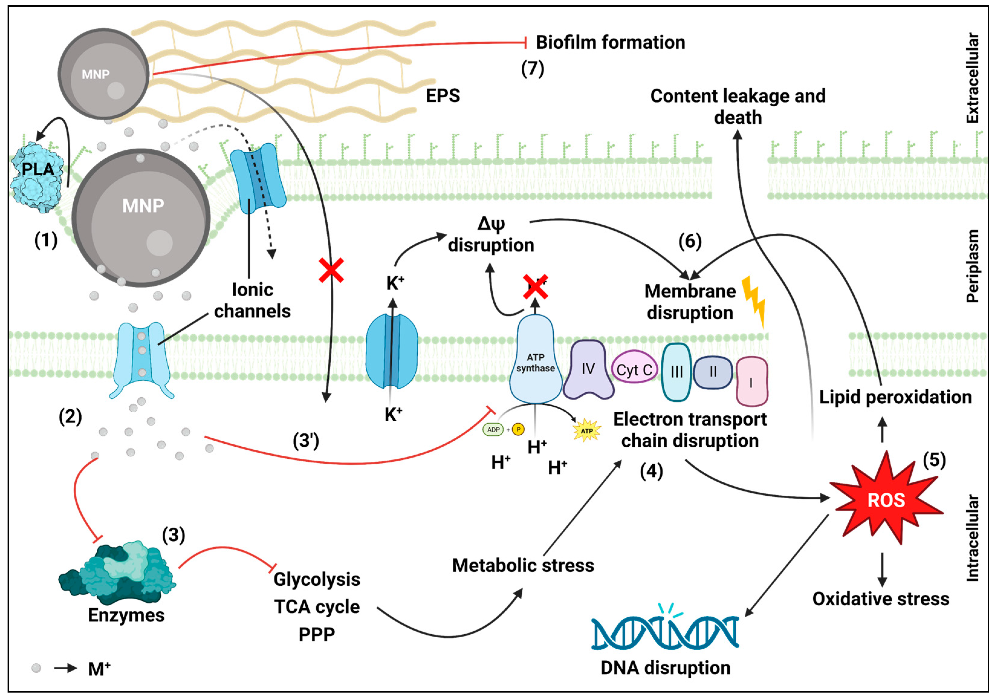
4.1. Antimicrobial Mechanisms—State of the Art
4.2. Evidence-Based Proposed Antimicrobial Mechanisms
5. Potential Use of Metal Nanoparticles in Cultural Heritage Conservation
5.1. Metal Nanoparticles Application on Materials Used in Cultural Heritage
5.1.1. Stone
5.1.2. Paper
5.1.3. Textile
5.1.4. Wood
5.2. Metal Nanoparticles’ Antimicrobial Activity against Microorganisms Collected from Contaminated Cultural Heritage Materials
6. Closing Remarks and Future Research Guidelines
Author Contributions
Funding
Data Availability Statement
Acknowledgments
Conflicts of Interest
References
- The British Museum Drinking-Cup | British Museum. Available online: https://www.britishmuseum.org/collection/object/H_1958-1202-1 (accessed on 20 December 2022).
- Barber, D.J.; Freestone, I.C. An Investigation of the Origin of the Colour of the Lycurgus Cup by Analytical Transmission Electron Microscopy. Archaeometry 1990, 32, 33–45. [Google Scholar] [CrossRef]
- Hulla, J.; Sahu, S.; Hayes, A. Nanotechnology: History and Future. Hum. Exp. Toxicol. 2015, 34, 1318–1321. [Google Scholar] [CrossRef] [PubMed]
- National Nanotechnology Initiative What Is Nanotechnology? Available online: https://www.nano.gov/nanotech-101/what/definition (accessed on 20 December 2022).
- Khan, Y.; Sadia, H.; Ali Shah, S.Z.; Khan, M.N.; Shah, A.A.; Ullah, N.; Ullah, M.F.; Bibi, H.; Bafakeeh, O.T.; Khedher, N.B.; et al. Classification, Synthetic, and Characterization Approaches to Nanoparticles, and Their Applications in Various Fields of Nanotechnology: A Review. Catalysts 2022, 12, 1386. [Google Scholar] [CrossRef]
- Khan, S.A. Metal Nanoparticles Toxicity: Role of Physicochemical Aspects. In Metal Nanoparticles for Drug Delivery and Diagnostic Applications; Elsevier: Amsterdam, The Netherlands, 2020; pp. 1–11. ISBN 978-0-12-816960-5. [Google Scholar]
- Yaqoob, A.A.; Ahmad, H.; Parveen, T.; Ahmad, A.; Oves, M.; Ismail, I.M.I.; Qari, H.A.; Umar, K.; Mohamad Ibrahim, M.N. Recent Advances in Metal Decorated Nanomaterials and Their Various Biological Applications: A Review. Front. Chem. 2020, 8, 341. [Google Scholar] [CrossRef]
- Khan, I.; Saeed, K.; Khan, I. Nanoparticles: Properties, Applications and Toxicities. Arab. J. Chem. 2019, 12, 908–931. [Google Scholar] [CrossRef]
- Wang, L.; Hu, C.; Shao, L. The Antimicrobial Activity of Nanoparticles: Present Situation and Prospects for the Future. Int. J. Nanomed. 2017, 12, 1227–1249. [Google Scholar] [CrossRef]
- Rudramurthy, G.; Swamy, M.; Sinniah, U.; Ghasemzadeh, A. Nanoparticles: Alternatives Against Drug-Resistant Pathogenic Microbes. Molecules 2016, 21, 836. [Google Scholar] [CrossRef] [PubMed]
- Nanowerk Nanowerk Catalog. Available online: https://www.nanowerk.com/nanocatalog/Nanoparticles/14/list/productasc/order/1/page (accessed on 20 December 2022).
- Khandel, P.; Yadaw, R.K.; Soni, D.K.; Kanwar, L.; Shahi, S.K. Biogenesis of Metal Nanoparticles and Their Pharmacological Applications: Present Status and Application Prospects. J. Nanostructure Chem. 2018, 8, 217–254. [Google Scholar] [CrossRef]
- Singh, P.; Kim, Y.-J.; Zhang, D.; Yang, D.-C. Biological Synthesis of Nanoparticles from Plants and Microorganisms. Trends Biotechnol. 2016, 34, 588–599. [Google Scholar] [CrossRef]
- Singh, J.; Dutta, T.; Kim, K.-H.; Rawat, M.; Samddar, P.; Kumar, P. ‘Green’ Synthesis of Metals and Their Oxide Nanoparticles: Applications for Environmental Remediation. J. Nanobiotechnol. 2018, 16, 84. [Google Scholar] [CrossRef]
- UNESCO UNESCO World Heritage Center—World Heritage. Available online: https://whc.unesco.org/en/about/ (accessed on 20 December 2022).
- Cappitelli, F.; Cattò, C.; Villa, F. The Control of Cultural Heritage Microbial Deterioration. Microorganisms 2020, 8, 1542. [Google Scholar] [CrossRef] [PubMed]
- Abdel-Maksoud, G.; Abdel-Nasser, M.; Sultan, M.H.; Eid, A.M.; Alotaibi, S.H.; Hassan, S.E.-D.; Fouda, A. Fungal Biodeterioration of a Historical Manuscript Dating Back to the 14th Century: An Insight into Various Fungal Strains and Their Enzymatic Activities. Life 2022, 12, 1821. [Google Scholar] [CrossRef] [PubMed]
- Fouda, A.; Abdel-Nasser, M.; Khalil, A.M.A.; Hassan, S.E.-D.; Abdel-Maksoud, G. Investigate the Role of Fungal Communities Associated with a Historical Manuscript from the 17th Century in Biodegradation. Npj Mater. Degrad. 2022, 6, 88. [Google Scholar] [CrossRef]
- David, M.E.; Ion, R.-M.; Grigorescu, R.M.; Iancu, L.; Andrei, E.R. Nanomaterials Used in Conservation and Restoration of Cultural Heritage: An Up-to-Date Overview. Materials 2020, 13, 2064. [Google Scholar] [CrossRef]
- Franco-Castillo, I.; Hierro, L.; de la Fuente, J.M.; Seral-Ascaso, A.; Mitchell, S.G. Perspectives for Antimicrobial Nanomaterials in Cultural Heritage Conservation. Chem 2021, 7, 629–669. [Google Scholar] [CrossRef]
- Reyes-Estebanez, M.; Ortega-Morales, B.O.; Chan-Bacab, M.; Granados-Echegoyen, C.; Camacho-Chab, J.C.; Pereañez-Sacarias, J.E.; Gaylarde, C. Antimicrobial Engineered Nanoparticles in the Built Cultural Heritage Context and Their Ecotoxicological Impact on Animals and Plants: A Brief Review. Herit. Sci. 2018, 6, 52. [Google Scholar] [CrossRef]
- Chang, H.; Kim, B.H.; Lim, S.G.; Baek, H.; Park, J.; Hyeon, T. Role of the Precursor Composition in the Synthesis of Metal Ferrite Nanoparticles. Inorg. Chem. 2021, 60, 4261–4268. [Google Scholar] [CrossRef]
- Jeevanandam, J.; Chan, Y.S.; Danquah, M.K. Biosynthesis of Metal and Metal Oxide Nanoparticles. ChemBioEng Rev. 2016, 3, 55–67. [Google Scholar] [CrossRef]
- Zikalala, N.; Matshetshe, K.; Parani, S.; Oluwafemi, O.S. Biosynthesis Protocols for Colloidal Metal Oxide Nanoparticles. Nano-Struct. Nano-Objects 2018, 16, 288–299. [Google Scholar] [CrossRef]
- Salem, S.S.; Fouda, A. Green Synthesis of Metallic Nanoparticles and Their Prospective Biotechnological Applications: An Overview. Biol. Trace Elem. Res. 2021, 199, 344–370. [Google Scholar] [CrossRef]
- Huq, M.A.; Ashrafudoulla, M.; Rahman, M.M.; Balusamy, S.R.; Akter, S. Green Synthesis and Potential Antibacterial Applications of Bioactive Silver Nanoparticles: A Review. Polymers 2022, 14, 742. [Google Scholar] [CrossRef] [PubMed]
- Zhan, G.; Huang, J.; Lin, L.; Lin, W.; Emmanuel, K.; Li, Q. Synthesis of Gold Nanoparticles by Cacumen Platycladi Leaf Extract and Its Simulated Solution: Toward the Plant-Mediated Biosynthetic Mechanism. J. Nanoparticle Res. 2011, 13, 4957–4968. [Google Scholar] [CrossRef]
- Mittal, A.K.; Bhaumik, J.; Kumar, S.; Banerjee, U.C. Biosynthesis of Silver Nanoparticles: Elucidation of Prospective Mechanism and Therapeutic Potential. J. Colloid Interface Sci. 2014, 415, 39–47. [Google Scholar] [CrossRef]
- Liu, Y.; Jin, X.; Chen, Z. The Formation of Iron Nanoparticles by Eucalyptus Leaf Extract and Used to Remove Cr(VI). Sci. Total Environ. 2018, 627, 470–479. [Google Scholar] [CrossRef] [PubMed]
- Yang, B.; Qi, F.; Tan, J.; Yu, T.; Qu, C. Study of Green Synthesis of Ultrasmall Gold Nanoparticles Using Citrus Sinensis Peel. Appl. Sci. Switz. 2019, 9, 2423. [Google Scholar] [CrossRef]
- Buazar, F.; Sweidi, S.; Badri, M.; Kroushawi, F. Biofabrication of Highly Pure Copper Oxide Nanoparticles Using Wheat Seed Extract and Their Catalytic Activity: A Mechanistic Approach. Green Process. Synth. 2019, 8, 691–702. [Google Scholar] [CrossRef]
- Bandeira, M.; Possan, A.L.; Pavin, S.S.; Raota, C.S.; Vebber, M.C.; Giovanela, M.; Roesch-Ely, M.; Devine, D.M.; Crespo, J.S. Mechanism of Formation, Characterization and Cytotoxicity of Green Synthesized Zinc Oxide Nanoparticles Obtained from Ilex Paraguariensis Leaves Extract. Nano-Struct. Nano-Objects 2020, 24, 100532. [Google Scholar] [CrossRef]
- Biswal, A.K.; Misra, P.K. Biosynthesis and Characterization of Silver Nanoparticles for Prospective Application in Food Packaging and Biomedical Fields. Mater. Chem. Phys. 2020, 250, 123014. [Google Scholar] [CrossRef]
- Pradeep, M.; Kruszka, D.; Kachlicki, P.; Mondal, D.; Franklin, G. Uncovering the Phytochemical Basis and the Mechanism of Plant Extract-Mediated Eco-Friendly Synthesis of Silver Nanoparticles Using Ultra-Performance Liquid Chromatography Coupled with a Photodiode Array and High-Resolution Mass Spectrometry. ACS Sustain. Chem. Eng. 2022, 10, 562–571. [Google Scholar] [CrossRef]
- Mukherjee, P.; Roy, M.; Mandal, B.P.; Dey, G.K.; Mukherjee, P.K.; Ghatak, J.; Tyagi, A.K.; Kale, S.P. Green Synthesis of Highly Stabilized Nanocrystalline Silver Particles by a Non-Pathogenic and Agriculturally Important Fungus T. Asperellum. Nanotechnol. 2008, 19, 075103. [Google Scholar] [CrossRef]
- Vaidyanathan, R.; Gopalram, S.; Kalishwaralal, K.; Deepak, V.; Pandian, S.R.K.; Gurunathan, S. Enhanced Silver Nanoparticle Synthesis by Optimization of Nitrate Reductase Activity. Colloids Surf. B Biointerfaces 2010, 75, 335–341. [Google Scholar] [CrossRef] [PubMed]
- Moteshafi, H.; Mousavi, S.M.; Shojaosadati, S.A. The Possible Mechanisms Involved in Nanoparticles Biosynthesis. J. Ind. Eng. Chem. 2012, 18, 2046–2050. [Google Scholar] [CrossRef]
- Maliszewska, I.; Juraszek, A.; Bielska, K. Green Synthesis and Characterization of Silver Nanoparticles Using Ascomycota Fungi Penicillium Nalgiovense AJ12. J. Clust. Sci. 2014, 25, 989–1004. [Google Scholar] [CrossRef]
- Singh, D.K.; Kumar, J.; Sharma, V.K.; Verma, S.K.; Singh, A.; Kumari, P.; Kharwar, R.N. Mycosynthesis of Bactericidal Silver and Polymorphic Gold Nanoparticles: Physicochemical Variation Effects and Mechanism. Nanomedicine 2018, 13, 191–207. [Google Scholar] [CrossRef]
- Ma, L.; Lv, S.; Tang, J.; Liu, J.; Li, W.; Deng, J.; Deng, Y.; Du, J.; Liu, X.; Zeng, X. Study on Bioactive Molecules Involved in Extracellular Biosynthesis of Silver Nanoparticles by Penicillium Aculeatum Su1. Mater. Express 2019, 9, 475–483. [Google Scholar] [CrossRef]
- Wanarska, E.; Maliszewska, I. The Possible Mechanism of the Formation of Silver Nanoparticles by Penicillium Cyclopium. Bioorganic Chem. 2019, 93, 102803. [Google Scholar] [CrossRef] [PubMed]
- Krishnan, S.; Jayakumar, D.; Madhyastha, H.; Chadha, A. The Complexity of Microbial Metal Nanoparticle Synthesis: A Study of Candida Parapsilosis ATCC 7330 Mediated Gold Nanoparticles Formation. BioNanoScience 2021, 11, 336–344. [Google Scholar] [CrossRef]
- Roy, M.; Mukherjee, P.; Mandal, B.P.; Sharma, R.K.; Tyagi, A.K.; Kale, S.P. Biomimetic Synthesis of Nanocrystalline Silver Sol Using Cysteine: Stability Aspects and Antibacterial Activities. RSC Adv. 2012, 2, 6496–6503. [Google Scholar] [CrossRef]
- Guo, Q.; Guo, Q.; Yuan, J.; Zeng, J. Biosynthesis of Gold Nanoparticles Using a Kind of Flavonol: Dihydromyricetin. Colloids Surf. Physicochem. Eng. Asp. 2014, 441, 127–132. [Google Scholar] [CrossRef]
- Kim, H.-S.; Seo, Y.S.; Kim, K.; Han, J.W.; Park, Y.; Cho, S. Concentration Effect of Reducing Agents on Green Synthesis of Gold Nanoparticles: Size, Morphology, and Growth Mechanism. Nanoscale Res. Lett. 2016, 11, 1–9. [Google Scholar] [CrossRef] [Green Version]
- Xiang, S.; Ma, X.; Shi, H.; Ma, T.; Tian, C.; Chen, Y.; Chen, H.; Chen, X.; Luo, K.; Cai, L.; et al. Green Synthesis of an Alginate-Coated Silver Nanoparticle Shows High Antifungal Activity by Enhancing Its Cell Membrane Penetrating Ability. ACS Appl. Bio Mater. 2019, 2, 4087–4096. [Google Scholar] [CrossRef] [PubMed]
- Haiss, W.; Thanh, N.T.K.; Aveyard, J.; Fernig, D.G. Determination of Size and Concentration of Gold Nanoparticles from UV-Vis Spectra. Anal. Chem. 2007, 79, 4215–4221. [Google Scholar] [CrossRef] [PubMed]
- Lim, H.S.; Yeu, J.E.; Hong, S.P.; Kang, M.S. Characterization of Antibacterial Cell-Free Supernatant from Oral Care Probiotic Weissella Cibaria, CMU. Molecules 2018, 23, 1984. [Google Scholar] [CrossRef]
- Fuochi, V.; Coniglio, M.A.; Laghi, L.; Rescifina, A.; Caruso, M.; Stivala, A.; Furneri, P.M. Metabolic Characterization of Supernatants Produced by Lactobacillus spp. With in Vitro Anti-Legionella Activity. Front. Microbiol. 2019, 10, 1403. [Google Scholar] [CrossRef] [PubMed]
- Assal, N.; Rennie, B.; Walrond, L.; Cyr, T.; Rohonczy, L.; Lin, M. Proteome Characterization of the Culture Supernatant of Mycobacterium Bovis in Different Growth Stages. Biochem. Biophys. Rep. 2021, 28, 101154. [Google Scholar] [CrossRef]
- Haji, S.H.; Ali, F.A.; Aka, S.T.H. Synergistic Antibacterial Activity of Silver Nanoparticles Biosynthesized by Carbapenem-Resistant Gram-Negative Bacilli. Sci. Rep. 2022, 12, 1–13. [Google Scholar] [CrossRef]
- Raguvaran, K.; Kalpana, M.; Manimegalai, T.; Maheswaran, R. Insecticidal, Not-Target Organism Activity of Synthesized Silver Nanoparticles Using Actinokineospora Fastidiosa. Biocatal. Agric. Biotechnol. 2021, 38, 102197. [Google Scholar] [CrossRef]
- Guerrero, D.S.; Bertani, R.P.; Ledesma, A.; Frías, M.D.L.A.; Romero, C.M.; Dávila Costa, J.S. Silver Nanoparticles Synthesized by the Heavy Metal Resistant Strain Amycolatopsis Tucumanensis and Its Application in Controlling Red Strip Disease in Sugarcane. Heliyon 2022, 8, e09472. [Google Scholar] [CrossRef]
- Huq, M.A.; Akter, S. Characterization and Genome Analysis of Arthrobacter bangladeshi sp. Nov., Applied for the Green Synthesis of Silver Nanoparticles and Their Antibacterial Efficacy against Drug-Resistant Human Pathogens. Pharmaceutics 2021, 13, 1691. [Google Scholar] [CrossRef]
- Ahmed, T.; Noman, M.; Shahid, M.; Niazi, M.B.K.; Hussain, S.; Manzoor, N.; Wang, X.; Li, B. Green Synthesis of Silver Nanoparticles Transformed Synthetic Textile Dye into Less Toxic Intermediate Molecules through LC-MS Analysis and Treated the Actual Wastewater. Environ. Res. 2020, 191, 110142. [Google Scholar] [CrossRef]
- Ahmed, T.; Shahid, M.; Noman, M.; Niazi, M.B.K.; Mahmood, F.; Manzoor, I.; Zhang, Y.; Li, B.; Yang, Y.; Yan, C.; et al. Silver Nanoparticles Synthesized by Using Bacillus Cereus SZT1 Ameliorated the Damage of Bacterial Leaf Blight Pathogen in Rice. Pathogens 2020, 9, 160. [Google Scholar] [CrossRef] [PubMed]
- Muthulakshmi, K.; Uma, C. Antimicrobial Activity of Bacillus Subtilis Silver Nanoparticles. Front. Biosci.—Elite 2019, 11, 89–101. [Google Scholar] [CrossRef]
- Samuel, M.S.; Jose, S.; Selvarajan, E.; Mathimani, T.; Pugazhendhi, A. Biosynthesized Silver Nanoparticles Using Bacillus Amyloliquefaciens; Application for Cytotoxicity Effect on A549 Cell Line and Photocatalytic Degradation of p-Nitrophenol. J. Photochem. Photobiol. B 2020, 202, 111642. [Google Scholar] [CrossRef]
- Dharmaraj, D.; Krishnamoorthy, M.; Rajendran, K.; Karuppiah, K.; Annamalai, J.; Durairaj, K.R.; Santhiyagu, P.; Ethiraj, K. Antibacterial and Cytotoxicity Activities of Biosynthesized Silver Oxide (Ag2O) Nanoparticles Using Bacillus Paramycoides. J. Drug Deliv. Sci. Technol. 2021, 61, 102111. [Google Scholar] [CrossRef]
- Aguirre, D.P.R.; Loyola, E.F.; Salcido, N.M.D.L.F.; Sifuentes, L.R.; Moreno, A.R.; Marszalek, J.E. Comparative Antibacterial Potential of Silver Nanoparticles Prepared via Chemical and Biological Synthesis. Arab. J. Chem. 2020, 13, 8662–8670. [Google Scholar] [CrossRef]
- Kordzangeneh, H.; Jookar Kashi, F. A New Bacillus paralicheniformis sp. Tmas-01 as Bioreactor for Synthesis of Ag/AgCl Composite–Different Effects of Biological and Rodamin B Dye Decolorization, Anticancer, Genotoxic Activity. Arch. Microbiol. 2022, 204, 1–15. [Google Scholar] [CrossRef] [PubMed]
- El-Bendary, M.A.; Afifi, S.S.; Moharam, M.E.; Abo El-Ola, S.M.; Salama, A.; Omara, E.A.; Shaheen, M.N.F.; Hamed, A.A.; Gawdat, N.A. Biosynthesis of Silver Nanoparticles Using Isolated Bacillus Subtilis: Characterization, Antimicrobial Activity, Cytotoxicity, and Their Performance as Antimicrobial Agent for Textile Materials. Prep. Biochem. Biotechnol. 2020, 51, 54–68. [Google Scholar] [CrossRef]
- Alfryyan, N.; Kordy, M.G.M.; Abdel-Gabbar, M.; Soliman, H.A.; Shaban, M. Characterization of the Biosynthesized Intracellular and Extracellular Plasmonic Silver Nanoparticles Using Bacillus Cereus and Their Catalytic Reduction of Methylene Blue. Sci. Rep. 2022, 12, 1–14. [Google Scholar] [CrossRef]
- Alsamhary, K.I. Eco-Friendly Synthesis of Silver Nanoparticles by Bacillus Subtilis and Their Antibacterial Activity. Saudi J. Biol. Sci. 2020, 27, 2185–2191. [Google Scholar] [CrossRef]
- Gajera, H.P.; Hirpara, D.G.; Bhadani, R.V.; Golakiya, B.A. Green Synthesis and Characterization of Nanosilver Derived from Extracellular Metabolites of Potent Bacillus Subtilis for Antifungal and Eco-Friendly Action against Phytopathogen. BioMetals 2022, 35, 479–497. [Google Scholar] [CrossRef]
- Hieu, H.N.; Trang, D.T.H.; Hien, V.T.T.; Nghia, N.V.; Lam, N.T.; Nguyen, T.M.D. Microorganism-Mediated Green Synthesis of Silver Nanoparticles Using Aspergillus Niger and Bacillus Megaterium. Dig. J. Nanomater. Biostructures 2022, 17, 359–367. [Google Scholar] [CrossRef]
- El-Bendary, M.A.; Moharam, M.E.; Abdelraof, M.; Allam, M.A.; Roshdy, A.M.; Shaheen, M.N.F.; Elmahdy, E.M.; Elkomy, G.M. Multi-Bioactive Silver Nanoparticles Synthesized Using Mosquitocidal Bacilli and Their Characterization. Arch. Microbiol. 2020, 202, 63–75. [Google Scholar] [CrossRef] [PubMed]
- Alsharif, S.M.; Salem, S.S.; Abdel-Rahman, M.A.; Fouda, A.; Eid, A.M.; El-Din Hassan, S.; Awad, M.A.; Mohamed, A.A. Multifunctional Properties of Spherical Silver Nanoparticles Fabricated by Different Microbial Taxa. Heliyon 2020, 6, e03943. [Google Scholar] [CrossRef]
- Ibrahim, S.; Ahmad, Z.; Manzoor, M.Z.; Mujahid, M.; Faheem, Z.; Adnan, A. Optimization for Biogenic Microbial Synthesis of Silver Nanoparticles through Response Surface Methodology, Characterization, Their Antimicrobial, Antioxidant, and Catalytic Potential. Sci. Rep. 2021, 11, 1–18. [Google Scholar] [CrossRef]
- Wang, X.; Lee, S.-Y.; Akter, S.; Huq, M.A. Probiotic-Mediated Biosynthesis of Silver Nanoparticles and Their Antibacterial Applications against Pathogenic Strains of Escherichia coli O157:H7. Polymers 2022, 14, 1834. [Google Scholar] [CrossRef]
- Mondal, A.H.; Yadav, D.; Ali, A.; Khan, N.; Jin, J.O.; Haq, Q.M.R. Anti-Bacterial and Anti-Candidal Activity of Silver Nanoparticles Biosynthesized Using Citrobacter spp. Ms5 Culture Supernatant. Biomolecules 2020, 10, 944. [Google Scholar] [CrossRef] [PubMed]
- Xiao, A.; Wang, B.; Zhu, L.; Jiang, L. Production of Extracellular Silver Nanoparticles by Radiation-Resistant Deinococcus Wulumuqiensis R12 and Its Mechanism Perspective. Process. Biochem. 2021, 100, 217–223. [Google Scholar] [CrossRef]
- Hanna, A.L.; Hamouda, H.M.; Goda, H.A.; Sadik, M.W.; Moghanm, F.S.; Ghoneim, A.M.; Alenezi, M.A.; Alnomasy, S.F.; Alam, P.; Elsayed, T.R. Biosynthesis and Characterization of Silver Nanoparticles Produced by Phormidium Ambiguum and Desertifilum Tharense Cyanobacteria. Bioinorg. Chem. Appl. 2022, 2022, 1–14. [Google Scholar] [CrossRef] [PubMed]
- Ashraf, N.; Ahmad, F.; Jing Jie, C.; Tuo Di, Z.; Feng-Zhu, Z.; Yin, D.-C. Optimization of Enterobacter Cloacae Mediated Synthesis of Extracellular Silver Nanoparticles by Response Surface Methodology and Their Characterization. Part. Sci. Technol. 2020, 38, 931–943. [Google Scholar] [CrossRef]
- Shanshoury, A.E.R.E.; Sabae, S.Z.; Shouny, W.A.E.; Shady, A.M.A.; Badr, H.M. Extracellular Biosynthesis of Silver Nanoparticles Using Aquatic Bacterial Isolate and Its Antibacterial and Antioxidant Potentials. Egypt. J. Aquat. Biol. Fish. 2020, 24, 183–201. [Google Scholar] [CrossRef]
- Bigdeli, R.; Shahnazari, M.; Panahnejad, E.; Cohan, R.A.; Dashbolaghi, A.; Asgary, V. Cytotoxic and Apoptotic Properties of Silver Chloride Nanoparticles Synthesized Using Escherichia coli Cell-Free Supernatant on Human Breast Cancer MCF 7 Cell Line. Artif. Cells Nanomed. Biotechnol. 2019, 47, 1603–1609. [Google Scholar] [CrossRef]
- El-Dein, M.M.N.; Baka, Z.A.M.; Abou-Dobara, M.I.; El-Sayed, A.K.A.; El-Zahed, M.M. Extracellular Biosynthesis, Optimization, Characterization and Antimicrobial Potential of Escherichia coli D8 Silver Nanoparticles. J. Microbiol. Biotechnol. Food Sci. 2021, 10, 648–656. [Google Scholar] [CrossRef]
- Huo, Y.; Han, Y.X.; Singh, P.; Kang, J.P.; Pu, J.Y.; Piao, C.H.; Yang, D.C. Antimicrobial, Antioxidant, and Anticancer Potentials of AgCl Nanoparticles Biosynthesized by Flavobacterium Panacis. Appl. Phys. A 2021, 127, 1–10. [Google Scholar] [CrossRef]
- Sandhya, S.V.; Vijayan, K.K. Biogenesis of Silver Nanoparticles by Marine Bacteria labrenzia sp. Mab 26 Associated with Isochrysis Galbana. Curr. Sci. 2020, 119, 1830–1833. [Google Scholar] [CrossRef]
- Rajoka, M.S.R.; Mehwish, H.M.; Zhang, H.; Ashraf, M.; Fang, H.; Zeng, X.; Wu, Y.; Khurshid, M.; Zhao, L.; He, Z. Antibacterial and Antioxidant Activity of Exopolysaccharide Mediated Silver Nanoparticle Synthesized by Lactobacillus Brevis Isolated from Chinese Koumiss. Colloids Surf. B Biointerfaces 2020, 186, 110734. [Google Scholar] [CrossRef] [PubMed]
- Naseer, Q.A.; Xue, X.; Wang, X.; Dang, S.; Din, S.U.; Kalsoom; Jamil, J. Synthesis of Silver Nanoparticles Using Lactobacillus Bulgaricus and Assessment of Their Antibacterial Potential [Síntese de Nanopartículas de Prata Usando Lactobacillus Bulgaricus e Avaliação de Seu Potencial Antibacteriano]. Braz. J. Biol. 2022, 82, e232434. [Google Scholar] [CrossRef]
- Abdelmoneim, H.M.; Taha, T.H.; Elnouby, M.S.; AbuShady, H.M. Extracellular Biosynthesis, OVAT/Statistical Optimization, and Characterization of Silver Nanoparticles (AgNPs) Using Leclercia Adecarboxylata THHM and Its Antimicrobial Activity. Microb. Cell Factories 2022, 21, 1–24. [Google Scholar] [CrossRef]
- Huq, M.A. Biogenic Silver Nanoparticles Synthesized by Lysinibacillus Xylanilyticus MAHUQ-40 to Control Antibiotic-Resistant Human Pathogens Vibrio Parahaemolyticus and Salmonella Typhimurium. Front. Bioeng. Biotechnol. 2020, 8, 597502. [Google Scholar] [CrossRef]
- El-Bendary, M.A.; Abdelraof, M.; Moharam, M.E.; Elmahdy, E.M.; Allam, M.A. Potential of Silver Nanoparticles Synthesized Using Low Active Mosquitocidal Lysinibacillus Sphaericus as Novel Antimicrobial Agents. Prep. Biochem. Biotechnol. 2021, 51, 926–935. [Google Scholar] [CrossRef]
- Huq, M.A.; Akter, S. Biosynthesis, Characterization and Antibacterial Application of Novel Silver Nanoparticles against Drug Resistant Pathogenic Klebsiella Pneumoniae and Salmonella Enteritidis. Molecules 2021, 26, 5996. [Google Scholar] [CrossRef]
- Mohamed, A.M.H.A.; Sorokin, V.V.; Skladnev, D.A.; Shevlyagina, N.V.; Zhukhovitsky, V.G.; Pshenichnikova, A.B. Biosynthesis of Silver Nanoparticles by Methylophilus Quaylei, Characterization and Its Impact on Established Biofilms. BioNanoScience 2020, 10, 885–898. [Google Scholar] [CrossRef]
- Husain, S.; Verma, S.K.; Hemlata; Azam, M.; Sardar, M.; Haq, Q.M.R.; Fatma, T. Antibacterial Efficacy of Facile Cyanobacterial Silver Nanoparticles Inferred by Antioxidant Mechanism. Mater. Sci. Eng. C 2021, 122, 111888. [Google Scholar] [CrossRef] [PubMed]
- Huq, M.A.; Akter, S. Bacterial Mediated Rapid and Facile Synthesis of Silver Nanoparticles and Their Antimicrobial Efficacy against Pathogenic Microorganisms. Materials 2021, 14, 2615. [Google Scholar] [CrossRef] [PubMed]
- Wypij, M.; Jędrzejewski, T.; Trzcińska-Wencel, J.; Ostrowski, M.; Rai, M.; Golińska, P. Green Synthesized Silver Nanoparticles: Antibacterial and Anticancer Activities, Biocompatibility, and Analyses of Surface-Attached Proteins. Front. Microbiol. 2021, 12, 632505. [Google Scholar] [CrossRef]
- Huq, M.A. Green Synthesis of Silver Nanoparticles Using Pseudoduganella Eburnea MAHUQ-39 and Their Antimicrobial Mechanisms Investigation against Drug Resistant Human Pathogens. Int. J. Mol. Sci. 2020, 21, 1510. [Google Scholar] [CrossRef]
- Jimoh, A.A.; Akpeji, B.H.; Azeez, S.O.; Ayipo, Y.O.; Abdulsalam, Z.A.; Adebayo, Z.F.; Ajao, A.T.; Zakariyah, A.T.; Elemike, E.E. Biosynthesis of Ag and TiO2 Nanoparticles and the Evaluation of Their Antibacterial Activities. Inorg. Chem. Commun. 2022, 141, 109503. [Google Scholar] [CrossRef]
- Salem, S.S.; Ali, O.M.; Reyad, A.M.; Abd-Elsalam, K.A.; Hashem, A.H. Pseudomonas indica-Mediated Silver Nanoparticles: Antifungal and Antioxidant Biogenic Tool for Suppressing Mucormycosis Fungi. J. Fungi 2022, 8, 126. [Google Scholar] [CrossRef] [PubMed]
- Mohammed, A.B.A.; Elhamid, M.M.A.; Khalil, M.K.M.; Ali, A.S.; Abbas, R.N. The potential activity of biosynthesized silver nanoparticles of Pseudomonas aeruginosa as an antibacterial agent against multidrug-resistant isolates from intensive care unit and anticancer agent. Environ. Sci. Eur. 2022, 34, 1–15. [Google Scholar] [CrossRef]
- Mondal, A.H.; Yadav, D.; Mitra, S.; Mukhopadhyay, K. Biosynthesis of Silver Nanoparticles Using Culture Supernatant of Shewanella sp. ARY1 and Their Antibacterial Activity. Int. J. Nanomed. 2020, 15, 8295–8310. [Google Scholar] [CrossRef]
- Singh, P.; Pandit, S.; Mokkapati, V.; Garnæs, J.; Mijakovic, I. A Sustainable Approach for the Green Synthesis of Silver Nanoparticles from Solibacillus isronensis sp. and Their Application in Biofilm Inhibition. Molecules 2020, 25, 2783. [Google Scholar] [CrossRef] [PubMed]
- Mishra, S.; Yang, X.; Singh, H.B. Evidence for positive response of soil bacterial community structure and functions to biosynthesized silver nanoparticles: An approach to conquer nanotoxicity? J. Environ. Manag. 2019, 253, 109584. [Google Scholar] [CrossRef] [PubMed]
- Fouda, A.; Hassan, S.E.-D.; Abdo, A.M.; El-Gamal, M.S. Antimicrobial, Antioxidant and Larvicidal Activities of Spherical Silver Nanoparticles Synthesized by Endophytic streptomyces spp. Biol. Trace Elem. Res. 2020, 195, 707–724. [Google Scholar] [CrossRef] [PubMed]
- Mechouche, M.S.; Merouane, F.; Messaad, C.E.H.; Golzadeh, N.; Vasseghian, Y.; Berkani, M. Biosynthesis, characterization, and evaluation of antibacterial and photocatalytic methylene blue dye degradation activities of silver nanoparticles from Streptomyces tuirus strain. Environ. Res. 2021, 204, 112360. [Google Scholar] [CrossRef]
- Wypij, M.; Świecimska, M.; Dahm, H.; Rai, M.; Golinska, P. Controllable biosynthesis of silver nanoparticles using actinobacterial strains. Green Process. Synth. 2018, 8, 207–214. [Google Scholar] [CrossRef]
- Marathe, K.; Naik, J.; Maheshwari, V. Biogenic Synthesis of Silver Nanoparticles Using Streptomyces spp. and their Antifungal Activity Against Fusarium verticillioides. J. Clust. Sci. 2020, 32, 1299–1309. [Google Scholar] [CrossRef]
- Elnady, A.; Sorour, N.M.; Abbas, R.N. Characterization, cytotoxicity, and genotoxicity properties of novel biomediated nanosized-silver by Egyptian Streptomyces roseolus for safe antimicrobial applications. World J. Microbiol. Biotechnol. 2022, 38, 1–17. [Google Scholar] [CrossRef]
- Pallavi, S.S.; Rudayni, H.A.; Bepari, A.; Niazi, S.K.; Nayaka, S. Green synthesis of Silver nanoparticles using Streptomyces hirsutus strain SNPGA-8 and their characterization, antimicrobial activity, and anticancer activity against human lung carcinoma cell line A549. Saudi J. Biol. Sci. 2021, 29, 228–238. [Google Scholar] [CrossRef]
- Raguvaran, K.; Kalpana, M.; Manimegalai, T.; Maheswaran, R. Larvicidal, antibacterial, antibiofilm, and anti-quorum sensing activities of silver nanoparticles biosynthesized from Streptomyces sclerotialus culture filtrate. Mater. Lett. 2022, 316, 132000. [Google Scholar] [CrossRef]
- Murugaiah, H.; Teh, C.L.; Loh, K.C.; Yahya, A.R.M.; Noh, N.A.M.; Abu Bakar, N.H.H.; Kernain, D.; Hashim, R.; Bustami, Y. Study of Antibacterial and Anticancer Properties of bioAgNPs Synthesized Using Streptomyces sp. PBD-311B and the Application of bioAgNP-CNC/Alg as an Antibacterial Hydrogel Film against P. aeruginosa USM-AR2 and MRSA. Molecules 2021, 26, 6414. [Google Scholar] [CrossRef]
- Akter, S.; Lee, S.-Y.; Siddiqi, M.Z.; Balusamy, S.R.; Ashrafudoulla; Rupa, E.J.; Huq, A. Ecofriendly Synthesis of Silver Nanoparticles by Terrabacter humi sp. nov. and Their Antibacterial Application against Antibiotic-Resistant Pathogens. Int. J. Mol. Sci. 2020, 21, 9746. [Google Scholar] [CrossRef]
- Bharti, S.; Mukherji, S.; Mukherji, S. Extracellular synthesis of silver nanoparticles by Thiosphaera pantotropha and evaluation of their antibacterial and cytotoxic effects. 3 Biotech. 2020, 10, 1–12. [Google Scholar] [CrossRef] [PubMed]
- Zamanpour, N.; Esmaeily, A.M.; Mashreghi, M.; Shahnavaz, B.; Sharifmoghadam, M.R.; Kompany, A. Application of a marine luminescent Vibrio sp. B4L for biosynthesis of silver nanoparticles with unique characteristics, biochemical properties, antibacterial and antibiofilm activities. Bioorganic Chem. 2021, 114, 105102. [Google Scholar] [CrossRef] [PubMed]
- Kabiri, F.; Aghaei, S.S.; Pourbabaee, A.A.; Soleimani, M.; Movahhed, T.K. Antibiofilm and cytotoxic potential of extracellular biosynthesized gold nanoparticles using actinobacteria Amycolatopsis sp. KMN. Prep. Biochem. Biotechnol. 2022, 55, 618–635. [Google Scholar] [CrossRef] [PubMed]
- Camas, M.; Celik, F.; Camas, A.S.; Ozalp, H.B. Biosynthesis of gold nanoparticles using marine bacteria and Box–Behnken design optimization. Part. Sci. Technol. 2018, 37, 31–38. [Google Scholar] [CrossRef]
- Kang, M.-G.; Khan, F.; Jo, D.-M.; Oh, D.; Tabassum, N.; Kim, Y.-M. Antibiofilm and Antivirulence Activities of Gold and Zinc Oxide Nanoparticles Synthesized from Kimchi-Isolated Leuconostoc sp. Strain C2. Antibiotics 2022, 11, 1524. [Google Scholar] [CrossRef]
- Manivasagan, P.; Alam, M.S.; Kang, K.-H.; Kwak, M.; Kim, S.-K. Extracellular synthesis of gold bionanoparticles by Nocardiopsis sp. and evaluation of its antimicrobial, antioxidant and cytotoxic activities. Bioprocess Biosyst. Eng. 2015, 38, 1167–1177. [Google Scholar] [CrossRef]
- Patil, M.P.; Kang, M.-J.; Niyonizigiye, I.; Singh, A.; Kim, J.-O.; Seo, Y.B.; Kim, G.-D. Extracellular synthesis of gold nanoparticles using the marine bacterium Paracoccus haeundaensis BC74171T and evaluation of their antioxidant activity and antiproliferative effect on normal and cancer cell lines. Colloids Surfaces B Biointerfaces 2019, 183, 110455. [Google Scholar] [CrossRef]
- Elshaer, S.L.; Shaaban, M.I. Inhibition of Quorum Sensing and Virulence Factors of Pseudomonas aeruginosa by Biologically Synthesized Gold and Selenium Nanoparticles. Antibiotics 2021, 10, 1461. [Google Scholar] [CrossRef]
- John, M.; Nagoth, J.; Zannotti, M.; Giovannetti, R.; Mancini, A.; Ramasamy, K.; Miceli, C.; Pucciarelli, S. Biogenic Synthesis of Copper Nanoparticles Using Bacterial Strains Isolated from an Antarctic Consortium Associated to a Psychrophilic Marine Ciliate: Characterization and Potential Application as Antimicrobial Agents. Mar. Drugs 2021, 19, 263. [Google Scholar] [CrossRef]
- Joshi, M.H.; Patil, A.A.; Chaudhary, S.; Kale, R.D. Microbial synthesis of CuNPs using Brevundimonas diminuta strain and its antibacterial activity. Adv. Nat. Sci. Nanosci. Nanotechnol. 2020, 11, 015008. [Google Scholar] [CrossRef]
- Noman, M.; Shahid, M.; Ahmed, T.; Tahir, M.; Naqqash, T.; Muhammad, S.; Song, F.; Abid, H.M.A.; Aslam, Z. Green copper nanoparticles from a native Klebsiella pneumoniae strain alleviated oxidative stress impairment of wheat plants by reducing the chromium bioavailability and increasing the growth. Ecotoxicol. Environ. Saf. 2020, 192, 110303. [Google Scholar] [CrossRef] [PubMed]
- Kouhkan, M.; Ahangar, P.; Babaganjeh, L.A.; Allahyari-Devin, M. Biosynthesis of Copper Oxide Nanoparticles Using Lactobacillus casei Subsp. Casei and its Anticancer and Antibacterial Activities. Curr. Nanosci. 2020, 16, 101–111. [Google Scholar] [CrossRef]
- Li, C.-X.; Huang, R.-T.; Shi, X.-Y. Biosynthesis of Cu nanoparticles supported on carbon nanotubes and its catalytic performance under different test conditions. J. Chem. Technol. Biotechnol. 2020, 95, 1511–1518. [Google Scholar] [CrossRef]
- El-Sherbiny, G.M.; Kalaba, M.H.; Sharaf, M.H.; Moghannem, S.A.; Radwan, A.A.; Askar, A.A.; Ismail, M.K.A.; El-Hawary, A.S.; Abushiba, M.A. Biogenic synthesis of CuO-NPs as nanotherapeutics approaches to overcome multidrug-resistant Staphylococcus aureus (MDRSA). Artif. Cells Nanomed. Biotechnol. 2022, 50, 260–274. [Google Scholar] [CrossRef] [PubMed]
- Bukhari, S.I.; Hamed, M.M.; Al-Agamy, M.H.; Gazwi, H.S.S.; Radwan, H.H.; Youssif, A.M. Biosynthesis of Copper Oxide Nanoparticles Using Streptomyces MHM38 and Its Biological Applications. J. Nanomater. 2021, 2021, 1–16. [Google Scholar] [CrossRef]
- Sharma, R.A.; Jagtap, P.K. Scrutinizing The Applications Of Mangrove Actinomycetes Mediated Biosynthesized Copper Nanoparticles. J. Water Environ. Nanotechnol. 2022, 7, 407–427. [Google Scholar] [CrossRef]
- Khan, S.; Akhtar, N.; Rehman, S.U.; Shujah, S.; Rha, E.S.; Jamil, M. Biosynthesized Iron Oxide Nanoparticles (Fe3O4 NPs) Mitigate Arsenic Toxicity in Rice Seedlings. Toxics 2020, 9, 2. [Google Scholar] [CrossRef]
- Daneshvar, M.; Hosseini, M.R. From the iron boring scraps to superparamagnetic nanoparticles through an aerobic biological route. J. Hazard. Mater. 2018, 357, 393–400. [Google Scholar] [CrossRef] [PubMed]
- Rajeswaran, S.; Thirugnanasambandan, S.S.; Dewangan, N.K.; Moorthy, R.K.; Kandasamy, S.; Vilwanathan, R. Multifarious Pharmacological Applications of Green Routed Eco-Friendly Iron Nanoparticles Synthesized by Streptomyces sp. (SRT12). Biol. Trace Element Res. 2019, 194, 273–283. [Google Scholar] [CrossRef]
- Al-Kordy, H.M.H.; Sabry, S.A.; Mabrouk, M.E.M. Statistical optimization of experimental parameters for extracellular synthesis of zinc oxide nanoparticles by a novel haloalaliphilic Alkalibacillus sp.W7. Sci. Rep. 2021, 11, 1–14. [Google Scholar] [CrossRef]
- El-Belely, E.F.; Farag, M.M.S.; Said, H.A.; Amin, A.S.; Azab, E.; Gobouri, A.A.; Fouda, A. Green Synthesis of Zinc Oxide Nanoparticles (ZnO-NPs) Using Arthrospira platensis (Class: Cyanophyceae) and Evaluation of their Biomedical Activities. Nanomaterials 2021, 11, 95. [Google Scholar] [CrossRef]
- Dharmaraj, D.; Krishnamoorthy, M.; Rajendran, K.; Karuppiah, K.; Jeyaraman, R.; Ethiraj, K. Protein Leakage Induced Marine Antibiofouling Activity of Biosynthesized Zinc Oxide Nanoparticles. J. Clust. Sci. 2020, 32, 643–650. [Google Scholar] [CrossRef]
- El-Ghwas, D.E. Short Communication: Characterization and biological synthesis of zinc oxide nanoparticles by new strain of Bacillus foraminis. Biodiversitas. J. Biol. Divers. 2021, 23. [Google Scholar] [CrossRef]
- Sabir, S.; Zahoor, M.A.; Waseem, M.; Siddique, M.H.; Shafique, M.; Imran, M.; Hayat, S.; Malik, I.R.; Muzammil, S. Biosynthesis of ZnO Nanoparticles Using Bacillus Subtilis: Characterization and Nutritive Significance for Promoting Plant Growth in Zea mays L. Dose-Response 2020, 18, 1559325820958911. [Google Scholar] [CrossRef]
- El-Rab, S.M.F.G.; Abo-Amer, A.E.; Asiri, A.M. Biogenic Synthesis of ZnO Nanoparticles and Its Potential Use as Antimicrobial Agent Against Multidrug-Resistant Pathogens. Curr. Microbiol. 2020, 77, 1767–1779. [Google Scholar] [CrossRef]
- Yusof, H.M.; Rahman, N.A.; Mohamad, R.; Zaidan, U.H.; Samsudin, A. Antibacterial Potential of Biosynthesized Zinc Oxide Nanoparticles against Poultry-Associated Foodborne Pathogens: An In Vitro Study. Animals 2021, 11, 2093. [Google Scholar] [CrossRef]
- Ogunyemi, S.O.; Zhang, M.; Abdallah, Y.; Ahmed, T.; Qiu, W.; Ali, A.; Yan, C.; Yang, Y.; Chen, J.; Li, B. The Bio-Synthesis of Three Metal Oxide Nanoparticles (ZnO, MnO2, and MgO) and Their Antibacterial Activity Against the Bacterial Leaf Blight Pathogen. Front. Microbiol. 2020, 11, 588326. [Google Scholar] [CrossRef] [PubMed]
- Rameshbabu, D.; Sarojini, K.; Sanjivkumar, M.; Ramasubburayan, R.; Prakash, S.; Punitha, M.J.; Immanuel, G. Investigation on characterization, antifouling and cytotoxic properties of zinc oxide nanoparticles biosynthesized by a mangrove-associated actinobacterium Streptomyces olivaceus (MSU3). Arch. Microbiol. 2022, 204, 1–15. [Google Scholar] [CrossRef] [PubMed]
- Salem, N.F.A.; Abouelkheir, S.S.; Yousif, A.M.; Meneses-Brassea, B.P.; Sabry, S.A.; Ghozlan, H.A.; El-Gendy, A.A. Large scale production of superparamagnetic iron oxide nanoparticles by the haloarchaeon Halobiforma sp. N1 and their potential in localized hyperthermia cancer therapy. Nanotechnology 2021, 32, 09LT01. [Google Scholar] [CrossRef]
- Korshed, P.; Li, L.; Liu, Z.; Mironov, A.; Wang, T. Size-dependent antibacterial activity for laser-generated silver nanoparticles. J. Interdiscip. Nanomed. 2019, 4, 24–33. [Google Scholar] [CrossRef] [Green Version]
- Echavarría, J.O.; Vanegas, N.A.G.; Orozco, C.P.O. Chitosan/carboxymethyl cellulose wound dressings supplemented with biologically synthesized silver nanoparticles from the ligninolytic fungus Anamorphous Bjerkandera sp. R1. Heliyon 2022, 8, e10258. [Google Scholar] [CrossRef] [PubMed]
- Ratvijitvech, T.; Na Pombejra, S. Antibacterial Efficiency of Microporous Hypercrosslinked Polymer Conjugated with Biosynthesized Silver Nanoparticles from Aspergillus niger. Mater. Today Commun. 2021, 28, 102617. [Google Scholar] [CrossRef]
- Fouda, A.; Awad, M.A.; Al-Faifi, Z.E.; Gad, M.E.; Al-Khalaf, A.A.; Yahya, R.; Hamza, M.F. Aspergillus flavus-Mediated Green Synthesis of Silver Nanoparticles and Evaluation of Their Antibacterial, Anti-Candida, Acaricides, and Photocatalytic Activities. Catalysts 2022, 12, 462. [Google Scholar] [CrossRef]
- Sheikh, H.; Awad, M.F. Biogenesis of nanoparticles with inhibitory effects on aflatoxin B1 production by Aspergillus flavus. Electron. J. Biotechnol. 2022, 60, 26–35. [Google Scholar] [CrossRef]
- Abdelaziz, M.; Shalabi, A.; Radwan, A.A.; Khaled, E.; Hassan, R.Y.A. Biosynthesis and Bio-sensing Applications of Silver and Gold Metal Nanoparticles. Egypt. J. Chem. 2021, 64, 1057–1063. [Google Scholar] [CrossRef]
- Lotfy, W.A.; Alkersh, B.M.; Sabry, S.A.; Ghozlan, H.A. Biosynthesis of Silver Nanoparticles by Aspergillus terreus: Characterization, Optimization, and Biological Activities. Front. Bioeng. Biotechnol. 2021, 9, 633468. [Google Scholar] [CrossRef]
- Othman, A.M.; Elsayed, M.A.; Al-Balakocy, N.G.; Hassan, M.M.; Elshafei, A.M. Biosynthesized silver nanoparticles by Aspergillus terreus NRRL265 for imparting durable antimicrobial finishing to polyester cotton blended fabrics: Statistical optimization, characterization, and antitumor activity evaluation. Biocatal. Agric. Biotechnol. 2021, 31, 101908. [Google Scholar] [CrossRef]
- Wang, D.; Xue, B.; Wang, L.; Zhang, Y.; Liu, L.; Zhou, Y. Fungus-mediated green synthesis of nano-silver using Aspergillus sydowii and its antifungal/antiproliferative activities. Sci. Rep. 2021, 11, 1–9. [Google Scholar] [CrossRef]
- Farrag, H.M.M.; Mostafa, F.A.A.M.; Mohamed, M.E.; Huseein, E.A.M. Green biosynthesis of silver nanoparticles by Aspergillus niger and its antiamoebic effect against Allovahlkampfia spelaea trophozoite and cyst. Exp. Parasitol. 2020, 219, 108031. [Google Scholar] [CrossRef]
- Elshafei, A.M.; Othman, A.M.; Elsayed, M.A.; Al-Balakocy, N.G.; Hassan, M.M. Green synthesis of silver nanoparticles using Aspergillus oryzae NRRL447 exogenous proteins: Optimization via central composite design, characterization and biological applications. Environ. Nanotechnol. Monit. Manag. 2021, 16, 100553. [Google Scholar] [CrossRef]
- Awad, M.A.; Eid, A.M.; Elsheikh, T.M.Y.; Al-Faifi, Z.E.; Saad, N.; Sultan, M.H.; Selim, S.; Al-Khalaf, A.A.; Fouda, A. Mycosynthesis, Characterization, and Mosquitocidal Activity of Silver Nanoparticles Fabricated by Aspergillus niger Strain. J. Fungi 2022, 8, 396. [Google Scholar] [CrossRef] [PubMed]
- El-Mekkawy, R.M.; Almanaa, T.N.; Yassin, M.A.; Rabie, G.; Saleh, N. Silver Nanoparticles (AgNPs) Biosynthesized by Aspergillus flavus KF946095; their Characterization and Antibacterial Activity. J. Pure Appl. Microbiol. 2021, 15, 105–113. [Google Scholar] [CrossRef]
- El-Sherbiny, G.M.; Lila, M.K.; Shetaia, Y.M.; Elswify, M.M.; Mohamed, S.S. Antimicrobial Activity of Biosynthesised Silver Nanoparticles against multidrug-Resistant Microbes Isolated from Cancer Patients with Bacteraemia and Candidaemia. Indian J. Med. Microbiol. 2020, 38, 371–378. [Google Scholar] [CrossRef]
- Mistry, H.; Thakor, R.; Patil, C.; Trivedi, J.; Bariya, H. Biogenically proficient synthesis and characterization of silver nanoparticles employing marine procured fungi Aspergillus brunneoviolaceus along with their antibacterial and antioxidative potency. Biotechnol. Lett. 2020, 43, 307–316. [Google Scholar] [CrossRef]
- Janakiraman, V.; Govindarajan, K.; Magesh, C.R. Biosynthesis of Silver Nanoparticles from Endophytic Fungi, and its Cytotoxic Activity. Bionanoscience 2019, 9, 573–579. [Google Scholar] [CrossRef]
- Lin, P.; Wang, F.-Q.; Li, C.-T.; Yan, Z.-F. An Enhancement of Antibacterial Activity and Synergistic Effect of Biosynthesized Silver Nanoparticles by Eurotium cristatum with Various Antibiotics. Biotechnol. Bioprocess Eng. 2020, 25, 450–458. [Google Scholar] [CrossRef]
- Gupta, K.; Chundawat, T.S.; Malek, N.A.N.N. Antibacterial, Antifungal, Photocatalytic Activities and Seed Germination Effect of Mycosynthesized Silver Nanoparticles using Fusarium oxysporum. Biointerface Res. Appl. Chem. 2021, 11, 12082–12091. [Google Scholar] [CrossRef]
- Rodríguez-Serrano, C.; Guzmán-Moreno, J.; Ángeles-Chávez, C.; Rodríguez-González, V.; Ortega-Sigala, J.J.; Ramírez-Santoyo, R.M.; Vidales-Rodríguez, L.E. Biosynthesis of silver nanoparticles by Fusarium scirpi and its potential as antimicrobial agent against uropathogenic Escherichia coli biofilms. PLoS ONE 2020, 15, e0230275. [Google Scholar] [CrossRef] [PubMed]
- Syed, A.; Al Saedi, M.H.; Bahkali, A.H.; Elgorban, A.M.; Kharat, M.; Pai, K.; Ghodake, G.; Ahmad, A. Biological synthesis of α-Ag2S composite nanoparticles using the fungus Humicola sp. and its biomedical applications. J. Drug Deliv. Sci. Technol. 2021, 66, 102770. [Google Scholar] [CrossRef]
- Qiao, Z.; Guo, P.; Yang, D.; Pei, Z.; Wang, M.; Liu, J.; Wang, Q. Evaluation of acute toxicity response to the algae Chlorella pyrenoidosa of biosynthetic silver nanoparticles catalysts. Environ. Sci. Pollut. Res. 2022. [Google Scholar] [CrossRef]
- Qiao, Z.-P.; Wang, M.-Y.; Liu, J.-F.; Wang, Q.-Z. Green synthesis of silver nanoparticles using a novel endophytic fungus Letendraea sp. WZ07: Characterization and evaluation of antioxidant, antibacterial and catalytic activities (3-in-1 system). Inorg. Chem. Commun. 2022, 138, 109301. [Google Scholar] [CrossRef]
- Kahraman, T.; Korcan, S.E.; Liman, R.; Ciğerci, H.; Acikbas, Y.; Konuk, M.; Akkuş, G.U. Synthesis, Characterization, and Optimization of Green Silver Nanoparticles Using Neopestalotiopsis clavispora and Evaluation of Its Antibacterial, Antibiofilm, and Genotoxic Effects. EuroBiotech. J. 2021, 5, 109–122. [Google Scholar] [CrossRef]
- Yassin, M.A.; Elgorban, A.M.; El-Samawaty, A.E.-R.M.; Almunqedhi, B.M. Biosynthesis of silver nanoparticles using Penicillium verrucosum and analysis of their antifungal activity. Saudi J. Biol. Sci. 2021, 28, 2123–2127. [Google Scholar] [CrossRef]
- Soliman, A.M.; Abdel-Latif, W.; Shehata, I.H.; Fouda, A.; Abdo, A.M.; Ahmed, Y.M. Green Approach to Overcome the Resistance Pattern of Candida spp. Using Biosynthesized Silver Nanoparticles Fabricated by Penicillium chrysogenum FBiol. Trace Element Res. 2020, 199, 800–811. [Google Scholar] [CrossRef] [PubMed]
- Barabadi, H.; Mohammadzadeh, A.; Vahidi, H.; Rashedi, M.; Saravanan, M.; Talank, N.; Alizadeh, A. Penicillium chrysogenum-Derived Silver Nanoparticles: Exploration of Their Antibacterial and Biofilm Inhibitory Activity Against the Standard and Pathogenic Acinetobacter baumannii Compared to Tetracycline. J. Clust. Sci. 2021, 33, 1929–1942. [Google Scholar] [CrossRef]
- Feroze, N.; Arshad, B.; Younas, M.; Afridi, M.I.; Saqib, S.; Ayaz, A. Fungal mediated synthesis of silver nanoparticles and evaluation of antibacterial activity. Microsc. Res. Tech. 2019, 83, 72–80. [Google Scholar] [CrossRef] [PubMed]
- Gond, S.K.; Mishra, A.; Verma, S.K.; Sharma, V.K.; Kharwar, R.N. Synthesis and Characterization of Antimicrobial Silver Nanoparticles by an Endophytic Fungus Isolated from Nyctanthes arbor-tristis. Proc. Natl. Acad. Sci. India Sect. B Boil. Sci. 2019, 90, 641–645. [Google Scholar] [CrossRef]
- Mohanta, Y.K.; Nayak, D.; Mishra, A.K.; Chakrabartty, I.; Ray, M.K.; Mohanta, T.K.; Tayung, K.; Rajaganesh, R.; Vasanthakumaran, M.; Muthupandian, S.; et al. Green Synthesis of Endolichenic Fungi Functionalized Silver Nanoparticles: The Role in Antimicrobial, Anti-Cancer, and Mosquitocidal Activities. Int. J. Mol. Sci. 2022, 23, 10626. [Google Scholar] [CrossRef]
- Soliman, M.K.Y.; Salem, S.S.; Abu-Elghait, M.; Azab, M.S. Biosynthesis of Silver and Gold Nanoparticles and Their Efficacy Towards Antibacterial, Antibiofilm, Cytotoxicity, and Antioxidant Activities. Appl. Biochem. Biotechnol. 2022, 195, 1158–1183. [Google Scholar] [CrossRef]
- Qu, M.; Yao, W.; Cui, X.; Xia, R.; Qin, L.; Liu, X. Biosynthesis of silver nanoparticles (AgNPs) employing Trichoderma strains to control empty-gut disease of oak silkworm (Antheraea pernyi). Mater. Today Commun. 2021, 28, 102619. [Google Scholar] [CrossRef]
- Tomah, A.; Alamer, I.; Li, B.; Zhang, J.-Z. Mycosynthesis of Silver Nanoparticles Using Screened Trichoderma Isolates and Their Antifungal Activity against Sclerotinia sclerotiorum. Nanomaterials 2020, 10, 1955. [Google Scholar] [CrossRef] [PubMed]
- Gemishev, O.T.; I Panayotova, M.; Mintcheva, N.N.; Djerahov, L.P.; Tyuliev, G.T.; Gicheva, G.D. A green approach for silver nanoparticles preparation by cell-free extract from Trichoderma reesei fungi and their characterization. Mater. Res. Express 2019, 6, 095040. [Google Scholar] [CrossRef]
- James, C.; Kumar, G. Biosynthesis of gold nanoparticles and inhibition of various stages of bacterial biofilms formed by drug-resistant Aeromonas hydrophila, Escherichia coli, and Klebsiella pneumoniae. Arch. Microbiol. 2022, 204, 1–14. [Google Scholar] [CrossRef] [PubMed]
- Abu-Tahon, M.A.; Ghareib, M.; Abdallah, W.E. Environmentally benign rapid biosynthesis of extracellular gold nanoparticles using Aspergillus flavus and their cytotoxic and catalytic activities. Process. Biochem. 2020, 95, 1–11. [Google Scholar] [CrossRef]
- Naimi-Shamel, N.; Pourali, P.; Dolatabadi, S. Green synthesis of gold nanoparticles using Fusarium oxysporum and antibacterial activity of its tetracycline conjugant. J. Mycol. Méd. 2019, 29, 7–13. [Google Scholar] [CrossRef] [PubMed]
- Qu, Y.; Li, X.; Lian, S.; Dai, C.; Jv, Z.; Zhao, B.; Zhou, H. Biosynthesis of gold nanoparticles using fungus Trichoderma sp. WL-Go and their catalysis in degradation of aromatic pollutants. IET Nanobiotechnol. 2018, 13, 12–17. [Google Scholar] [CrossRef]
- Mousa, A.M.; Aziz, O.A.A.; Al-Hagar, O.E.; Gizawy, M.A.; Allan, K.F.; Attallah, M.F. Biosynthetic new composite material containing CuO nanoparticles produced by Aspergillus terreus for 47Sc separation of cancer theranostics application from irradiated Ca target. Appl. Radiat. Isot. 2020, 166, 109389. [Google Scholar] [CrossRef]
- Mohamed, A.A.; Abu-Elghait, M.; Ahmed, N.E.; Salem, S.S. Eco-friendly Mycogenic Synthesis of ZnO and CuO Nanoparticles for In Vitro Antibacterial, Antibiofilm, and Antifungal Applications. Biol. Trace Element Res. 2020, 199, 2788–2799. [Google Scholar] [CrossRef]
- Garcia-Marin, L.E.; Juarez-Moreno, K.; Vilchis-Nestor, A.R.; Castro-Longoria, E. Highly Antifungal Activity of Biosynthesized Copper Oxide Nanoparticles against Candida albicans. Nanomaterials 2022, 12, 3856. [Google Scholar] [CrossRef]
- Fouda, A.; Hassan, S.E.-D.; Abdel-Rahman, M.A.; Farag, M.M.; Shehal-Deen, A.; Mohamed, A.A.; Alsharif, S.M.; Saied, E.; Moghanim, S.A.; Azab, M.S. Catalytic degradation of wastewater from the textile and tannery industries by green synthesized hematite (α-Fe2O3) and magnesium oxide (MgO) nanoparticles. Curr. Res. Biotechnol. 2021, 3, 29–41. [Google Scholar] [CrossRef]
- Abdullah, N.H. Optimization of magnetic nano-iron production by Aspergillus flavipes MN956655.1 using response surface methodology and evaluation of their dye decolorizing and antifungal activities. Sci. Rep. 2022, 12, 1–18. [Google Scholar] [CrossRef]
- Mahanty, S.; Chatterjee, S.; Ghosh, S.; Tudu, P.; Gaine, T.; Bakshi, M.; Das, S.; Das, P.; Bhattacharyya, S.; Bandyopadhyay, S.; et al. Synergistic approach towards the sustainable management of heavy metals in wastewater using mycosynthesized iron oxide nanoparticles: Biofabrication, adsorptive dynamics and chemometric modeling study. J. Water Process. Eng. 2020, 37, 101426. [Google Scholar] [CrossRef]
- Fouda, A.; Hassan, S.E.-D.; Saied, E.; Azab, M.S. An eco-friendly approach to textile and tannery wastewater treatment using maghemite nanoparticles (γ-Fe2O3-NPs) fabricated by Penicillium expansum strain (K-w). J. Environ. Chem. Eng. 2020, 9, 104693. [Google Scholar] [CrossRef]
- Fouda, A.; Hassan, S.E.-D.; Saied, E.; Hamza, M.F. Photocatalytic degradation of real textile and tannery effluent using biosynthesized magnesium oxide nanoparticles (MgO-NPs), heavy metal adsorption, phytotoxicity, and antimicrobial activity. J. Environ. Chem. Eng. 2021, 9, 105346. [Google Scholar] [CrossRef]
- Saied, E.; Eid, A.M.; Hassan, S.E.-D.; Salem, S.S.; Radwan, A.A.; Halawa, M.; Saleh, F.M.; Saad, H.A.; Saied, E.M.; Fouda, A. The Catalytic Activity of Biosynthesized Magnesium Oxide Nanoparticles (MgO-NPs) for Inhibiting the Growth of Pathogenic Microbes, Tanning Effluent Treatment, and Chromium Ion Removal. Catalysts 2021, 11, 821. [Google Scholar] [CrossRef]
- Fouda, A.; Awad, M.A.; Eid, A.M.; Saied, E.; Barghoth, M.G.; Hamza, M.F.; Abdelbary, S.; Hassan, S.E.-D. An Eco-Friendly Approach to the Control of Pathogenic Microbes and Anopheles stephensi Malarial Vector Using Magnesium Oxide Nanoparticles (Mg-NPs) Fabricated by Penicillium chrysogenum. Int. J. Mol. Sci. 2021, 22, 5096. [Google Scholar] [CrossRef] [PubMed]
- Hassan, S.E.-D.; Fouda, A.; Saied, E.; Farag, M.M.S.; Eid, A.M.; Barghoth, M.G.; Awad, M.A.; Hamza, M.F. Rhizopus oryzae-Mediated Green Synthesis of Magnesium Oxide Nanoparticles (MgO-NPs): A Promising Tool for Antimicrobial, Mosquitocidal Action, and Tanning Effluent Treatment. J. Fungi 2021, 7, 372. [Google Scholar] [CrossRef]
- Gholami-Shabani, M.; Sotoodehnejadnematalahi, F.; Shams-Ghahfarokhi, M.; Eslamifar, A.; Razzaghi-Abyaneh, M. Platinum Nanoparticles as Potent Anticancer and Antimicrobial Agent: Green Synthesis, Physical Characterization, and In-Vitro Biological Activity. J. Clust. Sci. 2022. [Google Scholar] [CrossRef]
- Gholami-Shabani, M.; Sotoodehnejadnematalahi, F.; Shams-Ghahfarokhi, M.; Eslamifar, A.; Razzaghi-Abyaneh, M. Mycosynthesis and Physicochemical Characterization of Vanadium Oxide Nanoparticles Using the Cell-Free Filtrate of Fusarium oxysporum and Evaluation of Their Cytotoxic and Antifungal Activities. J. Nanomater. 2021, 2021, 1–12. [Google Scholar] [CrossRef]
- Fouda, A.; Hassan, S.E.-D.; Salem, S.S.; Shaheen, T.I. In-Vitro cytotoxicity, antibacterial, and UV protection properties of the biosynthesized Zinc oxide nanoparticles for medical textile applications. Microb. Pathog. 2018, 125, 252–261. [Google Scholar] [CrossRef]
- Kumar, R.; Vinoth, S.; Baskar, V.; Arun, M.; Gurusaravanan, P. Synthesis of zinc oxide nanoparticles mediated by Dictyota dichotoma endophytic fungi and its photocatalytic degradation of fast green dye and antibacterial applications. S. Afr. J. Bot. 2022, 151, 337–344. [Google Scholar] [CrossRef]
- Kadam, V.V.; Shanmugam, S.D.; Ettiyappan, J.P.; Balakrishnan, R.M. Photocatalytic degradation of p-nitrophenol using biologically synthesized ZnO nanoparticles. Environ. Sci. Pollut. Res. 2020, 28, 12119–12130. [Google Scholar] [CrossRef] [PubMed]
- Kadam, V.V.; Ettiyappan, J.P.; Balakrishnan, R.M. Mechanistic insight into the endophytic fungus mediated synthesis of protein capped ZnO nanoparticles. Mater. Sci. Eng. B 2019, 243, 214–221. [Google Scholar] [CrossRef]
- Ananthi, V.; Prakash, G.S.; Rasu, K.M.; Gangadevi, K.; Boobalan, T.; Raja, R.; Anand, K.; Sudhakar, M.; Chuturgoon, A.; Arun, A. Comparison of integrated sustainable biodiesel and antibacterial nano silver production by microalgal and yeast isolates. J. Photochem. Photobiol. B Biol. 2018, 186, 232–242. [Google Scholar] [CrossRef]
- Eze, F.N.; Nwabor, O.F. Valorization of Pichia spent medium via one-pot synthesis of biocompatible silver nanoparticles with potent antioxidant, antimicrobial, tyrosinase inhibitory and reusable catalytic activities. Mater. Sci. Eng. C 2020, 115, 111104. [Google Scholar] [CrossRef] [PubMed]
- Ammar, H.A.; El Aty, A.A.A.; El Awdan, S.A. Extracellular myco-synthesis of nano-silver using the fermentable yeasts Pichia kudriavzeviiHA-NY2 and Saccharomyces uvarumHA-NY3, and their effective biomedical applications. Bioprocess Biosyst. Eng. 2021, 44, 841–854. [Google Scholar] [CrossRef] [PubMed]
- Kthiri, A.; Hamimed, S.; Othmani, A.; Landoulsi, A.; O’Sullivan, S.; Sheehan, D. Novel static magnetic field effects on green chemistry biosynthesis of silver nanoparticles in Saccharomyces cerevisiae. Sci. Rep. 2021, 11, 1–9. [Google Scholar] [CrossRef]
- Katharine, S.D.; Tabitha, A.; Praveen Kumar, E.; Aadhil, J.M.M.; Saxena, M.; Radha, P. Sustainable Biosynthesis of Silver Nanoparticles and Their Application to Recover “Single Cell Oil” from Yarrowia lipolytica for Biodiesel Synthesis. Bionanoscience 2022, 12, 890–900. [Google Scholar] [CrossRef]
- Qu, Y.; You, S.; Zhang, X.; Pei, X.; Shen, W.; Li, Z.; Li, S.; Zhang, Z. Biosynthesis of gold nanoparticles using cell-free extracts of Magnusiomyces ingens LH-F1 for nitrophenols reduction. Bioprocess Biosyst. Eng. 2017, 41, 359–367. [Google Scholar] [CrossRef]
- Eramabadi, P.; Masoudi, M.; Makhdoumi, A.; Mashreghi, M. Microbial cell lysate supernatant (CLS) alteration impact on platinum nanoparticles fabrication, characterization, antioxidant and antibacterial activity. Mater. Sci. Eng. C 2020, 117, 111292. [Google Scholar] [CrossRef]
- Zhang, X.-F.; Liu, Z.-G.; Shen, W.; Gurunathan, S. Silver Nanoparticles: Synthesis, Characterization, Properties, Applications, and Therapeutic Approaches. Int. J. Mol. Sci. 2016, 17, 1534. [Google Scholar] [CrossRef] [PubMed]
- Ijaz, I.; Gilani, E.; Nazir, A.; Bukhari, A. Detail review on chemical, physical and green synthesis, classification, characterizations and applications of nanoparticles. Green Chem. Lett. Rev. 2020, 13, 59–81. [Google Scholar] [CrossRef]
- Luciano, K.; Wang, X.; Liu, Y.; Eyler, G.; Qin, Z.; Xia, X. Noble Metal Nanoparticles for Point-of-Care Testing: Recent Advancements and Social Impacts. Bioengineering 2022, 9, 666. [Google Scholar] [CrossRef] [PubMed]
- Kashyap, M.; Samadhiya, K.; Ghosh, A.; Anand, V.; Shirage, P.M.; Bala, K. Screening of microalgae for biosynthesis and optimization of Ag/AgCl nano hybrids having antibacterial effect. RSC Adv. 2019, 9, 25583–25591. [Google Scholar] [CrossRef]
- Mražíková, A.; Velgosová, O.; Kavuličová, J.; Matvija, M.; Čižmárová, E.; Willner, J. Characteristics of Silver Nanoparticles in Different PH Values. Arch. Metall. Mater. 2018, 63, 993–998. [Google Scholar] [CrossRef]
- Abdullah; Al-Radadi, N.S.; Hussain, T.; Faisal, S.; Shah, S.A.R. Novel biosynthesis, characterization and bio-catalytic potential of green algae (Spirogyra hyalina) mediated silver nanomaterials. Saudi J. Biol. Sci. 2021, 29, 411–419. [Google Scholar] [CrossRef]
- Shalaby, S.M.; Madkour, F.F.; El-Kassas, H.Y.; Mohamed, A.A.; Elgarahy, A.M. Green synthesis of recyclable iron oxide nanoparticles using Spirulina platensis microalgae for adsorptive removal of cationic and anionic dyes. Environ. Sci. Pollut. Res. 2021, 28, 65549–65572. [Google Scholar] [CrossRef]
- Caliskan, G.; Mutaf, T.; Agba, H.C.; Elibol, M. Green Synthesis and Characterization of Titanium Nanoparticles Using Microalga, Phaeodactylum tricornutum. Geomicrobiol. J. 2021, 39, 83–96. [Google Scholar] [CrossRef]
- León-Buitimea, A.; Garza-Cárdenas, C.R.; Garza-Cervantes, J.A.; Lerma-Escalera, J.A.; Morones-Ramírez, J.R. The Demand for New Antibiotics: Antimicrobial Peptides, Nanoparticles, and Combinatorial Therapies as Future Strategies in Antibacterial Agent Design. Front. Microbiol. 2020, 11, 1669. [Google Scholar] [CrossRef]
- Kadiyala, U.; Turali-Emre, E.S.; Bahng, J.H.; Kotov, N.A.; VanEpps, J.S. Unexpected insights into antibacterial activity of zinc oxide nanoparticles against methicillin resistantStaphylococcus aureus(MRSA). Nanoscale 2018, 10, 4927–4939. [Google Scholar] [CrossRef]
- Bondarenko, O.M.; Sihtmäe, M.; Kuzmičiova, J.; Ragelienė, L.; Kahru, A.; Daugelavičius, R. Plasma membrane is the target of rapid antibacterial action of silver nanoparticles in Escherichia coli and Pseudomonas aeruginosa. Int. J. Nanomed. 2018, 13, 6779–6790. [Google Scholar] [CrossRef] [PubMed]
- Alhajjar, R.K.; Roche, K.M.; Techtmann, S.M. Comparative Analysis of the Mechanism of Resistance to Silver Nanoparticles and the Biocide 2,2-Dibromo-3-Nitrilopropionamide. Antimicrob. Agents Chemother. 2022, 66, e0203121. [Google Scholar] [CrossRef]
- Zhu, X.; Wang, J.; Cai, L.; Wu, Y.; Ji, M.; Jiang, H.; Chen, J. Dissection of the antibacterial mechanism of zinc oxide nanoparticles with manipulable nanoscale morphologies. J. Hazard. Mater. 2022, 430, 128436. [Google Scholar] [CrossRef]
- Theofilou, S.; Antoniou, C.; Potamiti, L.; Hadjisavvas, A.; Panayiotidis, M.; Savva, P.; Costa, C.; Fotopoulos, V. Immobilized Ag-nanoparticles (iNPs) for environmental applications: Elucidation of immobilized silver-induced inhibition mechanism of Escherichia coli. J. Environ. Chem. Eng. 2021, 9, 106001. [Google Scholar] [CrossRef]
- Du, M.; Zhao, W.; Ma, R.; Xu, H.; Zhu, Y.; Shan, C.; Liu, K.; Zhuang, J.; Jiao, Z. Visible-light-driven photocatalytic inactivation of S. aureus in aqueous environment by hydrophilic zinc oxide (ZnO) nanoparticles based on the interfacial electron transfer in S. aureus/ZnO composites. J. Hazard. Mater. 2021, 418, 126013. [Google Scholar] [CrossRef] [PubMed]
- Hsu, I.-L.; Yeh, F.H.; Chin, Y.-C.; Cheung, C.I.; Chia, Z.C.; Yang, L.-X.; Chen, Y.-J.; Cheng, T.-Y.; Wu, S.-P.; Tsai, P.-J.; et al. Multiplex antibacterial processes and risk in resistant phenotype by high oxidation-state nanoparticles: New killing process and mechanism investigations. Chem. Eng. J. 2020, 409, 128266. [Google Scholar] [CrossRef]
- Zheng, K.; Setyawati, M.I.; Leong, D.T.; Xie, J. Observing antimicrobial process with traceable gold nanoclusters. Nano Res. 2020, 14, 1026–1033. [Google Scholar] [CrossRef]
- Ji, H.; Zhou, S.; Fu, Y.; Wang, Y.; Mi, J.; Lu, T.; Wang, X.; Lü, C. Size-controllable preparation and antibacterial mechanism of thermo-responsive copolymer-stabilized silver nanoparticles with high antimicrobial activity. Mater. Sci. Eng. C 2020, 110, 110735. [Google Scholar] [CrossRef]
- Shen, T.; Wang, Q.; Li, C.; Zhou, B.; Li, Y.; Liu, Y. Transcriptome sequencing analysis reveals silver nanoparticles antifungal molecular mechanism of the soil fungi Fusarium solani species complex. J. Hazard. Mater. 2020, 388, 122063. [Google Scholar] [CrossRef]
- Theofilou, S.P.; Constantinou, B.K.; Chatziiona, V.K.; Pantelidou, N.; Plyastsov, S.; Kapnisis, K.; Savva, P.G.; Meshkovsky, I.; Anayiotos, A.; Costa, C.N. New insights into the antimicrobial treatment of water on Ag-supported solids. J. Chem. Technol. Biotechnol. 2018, 94, 1134–1143. [Google Scholar] [CrossRef]
- Lv, Q.; Zhang, B.; Xing, X.; Zhao, Y.; Cai, R.; Wang, W.; Gu, Q. Biosynthesis of copper nanoparticles using Shewanella loihica PV-4 with antibacterial activity: Novel approach and mechanisms investigation. J. Hazard. Mater. 2018, 347, 141–149. [Google Scholar] [CrossRef] [PubMed]
- Yan, X.; Bin He, B.; Liu, L.; Qu, G.; Shi, J.; Hu, L.; Jiang, G. Antibacterial mechanism of silver nanoparticles in Pseudomonas aeruginosa: Proteomics approach. Metallomics 2018, 10, 557–564. [Google Scholar] [CrossRef] [PubMed]
- Deng, C.-H.; Gong, J.-L.; Zeng, G.-M.; Zhang, P.; Song, B.; Zhang, X.-G.; Liu, H.-Y.; Huan, S.-Y. Graphene sponge decorated with copper nanoparticles as a novel bactericidal filter for inactivation of Escherichia coli. Chemosphere 2017, 184, 347–357. [Google Scholar] [CrossRef] [PubMed]
- Singh, M. Elucidation of biogenic silver nanoparticles susceptibility towards Escherichia coli: An investigation on the antimicrobial mechanism. IET Nanobiotechnol. 2016, 10, 276–280. [Google Scholar] [CrossRef]
- Karimi, F.; Dabbagh, S.; Alizadeh, S.; Rostamnia, S. Evaluation of AgClNPs@SBA-15/IL nanoparticle-induced oxidative stress and DNA mutation in Escherichia coli. Appl. Microbiol. Biotechnol. 2016, 100, 7161–7170. [Google Scholar] [CrossRef]
- He, Y.; Ingudam, S.; Reed, S.; Gehring, A.; Strobaugh, T.P.; Irwin, P. Study on the mechanism of antibacterial action of magnesium oxide nanoparticles against foodborne pathogens. J. Nanobiotechnol. 2016, 14, 1–9. [Google Scholar] [CrossRef]
- Soni, D.; Bafana, A.; Gandhi, D.; Sivanesan, S.; Pandey, R.A. Stress response ofPseudomonasspecies to silver nanoparticles at the molecular level. Environ. Toxicol. Chem. 2014, 33, 2126–2132. [Google Scholar] [CrossRef]
- Xiu, Z.; Liu, Y.; Mathieu, J.; Wang, J.; Zhu, D.; Alvarez, P.J. Elucidating the genetic basis for Escherichia coli defense against silver toxicity using mutant arrays. Environ. Toxicol. Chem. 2014, 33, 993–997. [Google Scholar] [CrossRef]
- Kora, A.J.; Arunachalam, J. Assessment of antibacterial activity of silver nanoparticles on Pseudomonas aeruginosa and its mechanism of action. World J. Microbiol. Biotechnol. 2010, 27, 1209–1216. [Google Scholar] [CrossRef]
- Elbasuney, S.; El-Sayyad, S.G. Silver Nanoparticles Coated Medical Fiber Synthesized by Surface Engineering with Bio-Inspired Mussel Powered Polydopamine: An Investigated Antimicrobial Potential with Bacterial Membrane Leakage Reaction Mechanism. Microb. Pathog. 2022, 169, 105680. [Google Scholar] [CrossRef]
- Gabrielyan, L.; Hovhannisyan, A.; Gevorgyan, V.; Ananyan, M.; Trchounian, A. Antibacterial effects of iron oxide (Fe3O4) nanoparticles: Distinguishing concentration-dependent effects with different bacterial cells growth and membrane-associated mechanisms. Appl. Microbiol. Biotechnol. 2019, 103, 2773–2782. [Google Scholar] [CrossRef] [PubMed]
- Estevez, M.B.; Mitchell, S.G.; Faccio, R.; Alborés, S. Biogenic silver nanoparticles: Understanding the antimicrobial mechanism using Confocal Raman Microscopy. Mater. Res. Express 2019, 6, 1250f5. [Google Scholar] [CrossRef]
- Shen, W.; Li, P.; Feng, H.; Ge, Y.; Liu, Z.; Feng, L. The bactericidal mechanism of action against Staphylococcus aureus for AgO nanoparticles. Mater. Sci. Eng. C 2017, 75, 610–619. [Google Scholar] [CrossRef] [PubMed]
- Jin, J.-C.; Wu, X.-J.; Xu, J.; Wang, B.-B.; Jiang, F.-L.; Liu, Y. Ultrasmall silver nanoclusters: Highly efficient antibacterial activity and their mechanisms. Biomater. Sci. 2016, 5, 247–257. [Google Scholar] [CrossRef] [PubMed]
- Jiang, Y.; Zhang, L.; Wen, D.; Ding, Y. Role of physical and chemical interactions in the antibacterial behavior of ZnO nanoparticles against E. coli. Mater. Sci. Eng. C 2016, 69, 1361–1366. [Google Scholar] [CrossRef] [PubMed]
- Sadiq, I.M.; Chandrasekaran, N.; Mukherjee, A. Studies on Effect of TiO2 Nanoparticles on Growth and Membrane Permeability of Escherichia coli, Pseudomonas aeruginosa, and Bacillus subtilis. Curr. Nanosci. 2010, 6, 381–387. [Google Scholar] [CrossRef]
- Li, W.-R.; Xie, X.-B.; Shi, Q.-S.; Zeng, H.-Y.; Ou-Yang, Y.-S.; Chen, Y.-B. Antibacterial activity and mechanism of silver nanoparticles on Escherichia coli. Appl. Microbiol. Biotechnol. 2009, 85, 1115–1122. [Google Scholar] [CrossRef]
- Ge, C.; Huang, M.; Huang, D.; Dang, F.; Huang, Y.; Ahmad, H.A.; Zhu, C.; Chen, N.; Wu, S.; Zhou, D. Effect of metal cations on antimicrobial activity and compartmentalization of silver in Shewanella oneidensis MR-1 upon exposure to silver ions. Sci. Total. Environ. 2022, 838, 156401. [Google Scholar] [CrossRef]
- Yang, Y.; Chen, X.; Zhang, N.; Sun, B.; Wang, K.; Zhang, Y.; Zhu, L. Self-defense mechanisms of microorganisms from the antimicrobial effect of silver nanoparticles: Highlight the role of extracellular polymeric substances. Water Res. 2022, 218, 118452. [Google Scholar] [CrossRef]
- Wang, H.; Wang, M.; Xu, X.; Gao, P.; Xu, Z.; Zhang, Q.; Li, H.; Yan, A.; Kao, R.Y.-T.; Sun, H. Multi-target mode of action of silver against Staphylococcus aureus endows it with capability to combat antibiotic resistance. Nat. Commun. 2021, 12, 1–16. [Google Scholar] [CrossRef]
- Kang, F.; Alvarez, P.J.; Zhu, D. Microbial Extracellular Polymeric Substances Reduce Ag+ to Silver Nanoparticles and Antagonize Bactericidal Activity. Environ. Sci. Technol. 2013, 48, 316–322. [Google Scholar] [CrossRef] [PubMed]
- Ostermeyer, A.-K.; Mumuper, C.K.; Semprini, L.; Radniecki, T. Influence of Bovine Serum Albumin and Alginate on Silver Nanoparticle Dissolution and Toxicity to Nitrosomonas europaea. Environ. Sci. Technol. 2013, 47, 14403–14410. [Google Scholar] [CrossRef] [PubMed]
- Mosselhy, D.A.; El-Aziz, M.A.; Hanna, M.; Ahmed, M.A.; Husien, M.M.; Feng, Q. Comparative synthesis and antimicrobial action of silver nanoparticles and silver nitrate. J. Nanoparticle Res. 2015, 17, 1–10. [Google Scholar] [CrossRef]
- Park, G.; Amaris, Z.N.; Eiken, M.K.; Baumgartner, K.; Johnston, K.A.; Williams, M.A.; Marckwordt, J.G.; Millstone, J.E.; Splan, K.E.; Wheeler, K.E. Emerging investigator series: Characterization of silver and silver nanoparticle interactions with zinc finger peptides. Environ. Sci. Nano 2019, 6, 2367–2378. [Google Scholar] [CrossRef] [PubMed]
- Agnihotri, S.; Mukherji, S.; Mukherji, S. Immobilized silver nanoparticles enhance contact killing and show highest efficacy: Elucidation of the mechanism of bactericidal action of silver. Nanoscale 2013, 5, 7328–7340. [Google Scholar] [CrossRef] [PubMed]
- Wang, L.; He, H.; Zhang, C.; Sun, L.; Liu, S.; Wang, S. Antimicrobial activity of silver loaded MnO2 nanomaterials with different crystal phases against Escherichia coli. J. Environ. Sci. 2016, 41, 112–120. [Google Scholar] [CrossRef]
- Bawskar, M.; Deshmukh, S.; Bansod, S.; Gade, A.; Rai, M. Comparative analysis of biosynthesised and chemosynthesised silver nanoparticles with special reference to their antibacterial activity against pathogens. IET Nanobiotechnol. 2015, 9, 107–113. [Google Scholar] [CrossRef]
- Oliveira, M.J.A.; Otubo, L.; Pires, A.; Brambilla, R.F.; Carvalho, A.C.; Santos, P.S.; Neto, A.O.; Vasquez, P. Silver Nanopar-ticles-Based Hydrogels Synthetized by Ionizing Radiation for Cleaning of Tangible Cultural Heritage Surfaces. Radiat. Phys. Chem. 2022, 199, 110345. [Google Scholar] [CrossRef]
- Speziale, A.; González-Sánchez, J.F.; Taşcı, B.; Pastor, A.; Sánchez, L.; Fernández-Acevedo, C.; Oroz-Mateo, T.; Salazar, C.; Navarro-Blasco, I.; Fernández, J.M.; et al. Development of Multifunctional Coatings for Protecting Stones and Lime Mortars of the Architectural Heritage. Int. J. Arch. Herit. 2020, 14, 1008–1029. [Google Scholar] [CrossRef]
- Gherardi, F.; Turyanska, L.; Ferrari, E.; Weston, N.; Fay, M.; Colston, B. Immobilized Enzymes on Gold Nanoparticles: From Enhanced Stability to Cleaning of Heritage Textiles. ACS Appl. Bio Mater. 2019, 2, 5136–5143. [Google Scholar] [CrossRef]
- Hefni, Y.K. Hydrophobic Zinc Oxide Nanocomposites for Consolidation and Protection of Quartzite Sculptures: A Case Study. J. Nano Res. 2020, 63, 64–75. [Google Scholar] [CrossRef]
- Aldoasri, M.A.; Darwish, S.S.; Adam, M.A.; Elmarzugi, N.A.; Ahmed, S.M. Protecting of Marble Stone Facades of Historic Buildings Using Multifunctional TiO2 Nanocoatings. Sustainability 2017, 9, 2002. [Google Scholar] [CrossRef]
- Sassoni, E.; D’Amen, E.; Roveri, N.; Scherer, G.W.; Franzoni, E. Durable Self-Cleaning Coatings for Architectural Surfaces by Incorporation of TiO2 Nano-Particles into Hydroxyapatite Films. Materials 2018, 11, 177. [Google Scholar] [CrossRef] [PubMed]
- Oliva, R.; Salvini, A.; Di Giulio, G.; Capozzoli, L.; Fioravanti, M.; Giordano, C.; Perito, B. TiO2-Oligoaldaramide nanocomposites as efficient core-shell systems for wood preservation. J. Appl. Polym. Sci. 2015, 132, 42047. [Google Scholar] [CrossRef]
- Girginova, P.I.; Galacho, C.; Veiga, R.; Silva, A.S.; Candeias, A. Study of mechanical properties of alkaline earth hydroxide nanoconsolidants for lime mortars. Constr. Build. Mater. 2019, 236, 117520. [Google Scholar] [CrossRef]
- Becerra, J.; Zaderenko, A.; Ortiz, R.; Karapanagiotis, I.; Ortiz, P. Comparison of the performance of a novel nanolime doped with ZnO quantum dots with common consolidants for historical carbonate stone buildings. Appl. Clay Sci. 2020, 195, 105732. [Google Scholar] [CrossRef]
- Rong, Y.; Yang, J.; Huang, S.; Li, Y. Barium Hydroxide Nanoparticle–Phosphoric Acid System for Desalination and Consolidation of Tomb Murals. Crystals 2022, 12, 1171. [Google Scholar] [CrossRef]
- Xie, L.; Chen, X.; Zhang, B.; Hu, Y. Preliminary assessment of the efficacy of nano–MgO–based dispersion for the consolidation of artificial weathered sandstone. Archaeometry 2021, 64, 997–1012. [Google Scholar] [CrossRef]
- Giorgi, R.; Ambrosi, M.; Toccafondi, N.; Baglioni, P. Nanoparticles for Cultural Heritage Conservation: Calcium and Barium Hydroxide Nanoparticles for Wall Painting Consolidation. Chem. A Eur. J. 2010, 16, 9374–9382. [Google Scholar] [CrossRef] [PubMed]
- Giorgi, R.; Bozzi, C.; Dei, L.; Gabbiani, C.; Ninham, A.B.W.; Baglioni, P. Nanoparticles of Mg(OH)2: Synthesis and Application to Paper Conservation. Langmuir 2005, 21, 8495–8501. [Google Scholar] [CrossRef]
- Saoud, K.M.; Ibala, I.; Ladki, D.E.; Ezzeldeen, O.; Saeed, S. Microwave Assisted Preparation of Calcium Hydroxide and Barium Hydroxide Nanoparticles and Their Application for Conservation of Cultural Heritage. In Proceedings of the Digital Heritage; Progress in Cultural Heritage: Documentation, Preservation, and Protection: 5th International Conference, Limassol, Cyprus, 3–8 November 2014; Lecture Notes in Computer Science. Volume 8740. [Google Scholar]
- De la Rosa-García, S.C.; Fuentes, A.F.; Gómez-Cornelio, S.; Zagada-Domínguez, U.; Quintana, P. Structural characterization of antifungal CaZn2(OH)6·2H2O nanoparticles obtained via mechanochemical processing. J. Mater. Sci. 2018, 53, 13758–13768. [Google Scholar] [CrossRef]
- Mu, B.; Ying, X.; Petropoulos, E.; He, S. Preparation of AgCl/ZnO nano-composite for effective antimicrobial protection of stone-made building elements. Mater. Lett. 2020, 285, 129143. [Google Scholar] [CrossRef]
- Bellissima, F.; Bonini, M.; Giorgi, R.; Baglioni, P.; Barresi, G.; Mastromei, G.; Perito, B. Antibacterial activity of silver nanoparticles grafted on stone surface. Environ. Sci. Pollut. Res. 2013, 21, 13278–13286. [Google Scholar] [CrossRef] [PubMed]
- Li, Q.; Hu, Y.; Zhang, B. Hydrophilic ZnO Nanoparticle-Based Antimicrobial Coatings for Sandstone Heritage Conservation. ACS Appl. Nano Mater. 2021, 4, 13908–13918. [Google Scholar] [CrossRef]
- Carrillo-González, R.; Martínez-Gómez, M.A.; González-Chávez, M.D.C.A.; Hernández, J.C.M. Inhibition of microorganisms involved in deterioration of an archaeological site by silver nanoparticles produced by a green synthesis method. Sci. Total. Environ. 2016, 565, 872–881. [Google Scholar] [CrossRef] [PubMed]
- Sierra-Fernandez, A.; De la Rosa-García, S.C.; Gomez-Villalba, L.S.; Gómez-Cornelio, S.; Rabanal, M.E.; Fort, R.; Quintana, P. Synthesis, Photocatalytic, and Antifungal Properties of MgO, ZnO and Zn/Mg Oxide Nanoparticles for the Protection of Calcareous Stone Heritage. ACS Appl. Mater. Interfaces 2017, 9, 24873–24886. [Google Scholar] [CrossRef]
- Becerra, J.; Ortiz, P.; Zaderenko, A.P.; Karapanagiotis, I. Assessment of nanoparticles/nanocomposites to inhibit micro-algal fouling on limestone façades. Build. Res. Inf. 2019, 48, 180–190. [Google Scholar] [CrossRef]
- Fouda, A.; Abdel-Maksoud, G.; Abdel-Rahman, M.A.; Salem, S.S.; Hassan, S.E.-D.; El-Sadany, M.A.-H. Eco-friendly approach utilizing green synthesized nanoparticles for paper conservation against microbes involved in biodeterioration of archaeological manuscript. Int. Biodeterior. Biodegrad. 2019, 142, 160–169. [Google Scholar] [CrossRef]
- Castillo, I.F.; Guillén, E.G.; De La Fuente, J.M.; Silva, F.; Mitchell, S.G. Preventing fungal growth on heritage paper with antifungal and cellulase inhibiting magnesium oxide nanoparticles. J. Mater. Chem. B 2019, 7, 6412–6419. [Google Scholar] [CrossRef]
- Castillo, I.F.; De Matteis, L.; Marquina, C.; Guillén, E.G.; de la Fuente, J.M.; Mitchell, S.G. Protection of 18th century paper using antimicrobial nano-magnesium oxide. Int. Biodeterior. Biodegrad. 2018, 141, 79–86. [Google Scholar] [CrossRef]
- Pietrzak, K.; Puchalski, M.; Otlewska, A.; Wrzosek, H.; Guiamet, P.; Piotrowska, M.; Gutarowska, B. Microbial diversity of pre-Columbian archaeological textiles and the effect of silver nanoparticles misting disinfection. J. Cult. Herit. 2017, 23, 138–147. [Google Scholar] [CrossRef]
- Eskani, I.N.; Astuti, W.; Farida; Haerudin, A.; Setiawan, J.; Lestari, D.W.; Isnaini; Widayatno, T. Antibacterial activities of synthesised Zno nanoparticles applied on reactive dyed batik fabrics. J. Text. Inst. 2021, 113, 430–439. [Google Scholar] [CrossRef]
- De Filpo, G.; Palermo, A.M.; Rachiele, F.; Nicoletta, F.P. Preventing fungal growth in wood by titanium dioxide nanoparticles. Int. Biodeterior. Biodegrad. 2013, 85, 217–222. [Google Scholar] [CrossRef]
- Gutarowska, B.; Skora, J.; Zduniak, K.; Rembisz, D. Analysis of the sensitivity of microorganisms contaminating museums and archives to silver nanoparticles. Int. Biodeterior. Biodegrad. 2012, 68, 7–17. [Google Scholar] [CrossRef]
- Gambino, M.; Ahmed, M.A.-A.A.; Villa, F.; Cappitelli, F. Zinc oxide nanoparticles hinder fungal biofilm development in an ancient Egyptian tomb. Int. Biodeterior. Biodegrad. 2017, 122, 92–99. [Google Scholar] [CrossRef]
- Fouda, A.; Abdel-Maksoud, G.; Abdel-Rahman, M.A.; Eid, A.M.; Barghoth, M.G.; El-Sadany, M.A.-H. Monitoring the effect of biosynthesized nanoparticles against biodeterioration of cellulose-based materials by Aspergillus niger. Cellulose 2019, 26, 6583–6597. [Google Scholar] [CrossRef]
- Lázaro-Mass, S.; De la Rosa-García, S.; García-Solis, C.; Reyes-Trujeque, J.; Soria-Castro, M.; Fuentes, A.F.; Quintana, P.; Gómez-Cornelio, S. Controlling growth of phototrophic biofilms on limestone using CaZn2(OH)6·2H2O and ZnO nanoparticles. J. Chem. Technol. Biotechnol. 2022. [Google Scholar] [CrossRef]
- Abdel-Maksoud, G.; Gaballah, S.; Youssef, A.M.; Eid, A.M.; Sultan, M.H.; Fouda, A. Eco-friendly approach for control of fungal deterioration of archaeological skeleton dated back to the Greco-Roman period. J. Cult. Herit. 2023, 59, 38–48. [Google Scholar] [CrossRef]
- Mikhailova, E.O. Silver Nanoparticles: Mechanism of Action and Probable Bio-Application. J. Funct. Biomater. 2020, 11, 84. [Google Scholar] [CrossRef]


| Metal * | Microbial Genera | NPs Size (nm) | Precursor | Antimicrobial Activity Studies | Toxicity Studies | Main Properties | Ref. |
|---|---|---|---|---|---|---|---|
| Bacteria | |||||||
| Ag | Acinetobacter | 11 to 9 | AgNO3 | Yes | No | Antimicrobial | [51] |
| Ag | Actinokineospora | 19 to 41 | AgNO3 | No | Yes | Mosquitocidal | [52] |
| Ag | Amycolatopsis | 35 c | AgNO3 | Yes | No | Antimicrobial | [53] |
| Ag | Arthrobacter | 12 to 50 | AgNO3 | Yes | No | Antimicrobial | [54] |
| Ag | Bacillus | 11 to 39 | AgNO3 | No | Yes | Photocatalytic | [55] |
| Ag | Bacillus | 18 to 39 | AgNO3 | Yes | Yes | Antimicrobial | [56] |
| Ag | Bacillus | 10 to 20 | AgNO3 | Yes | No | NS | [57] |
| Ag | Bacillus | 20 to 40 | AgNO3 | No | Yes | Photocatalytic Cytotoxicity | [58] |
| Ag | Bacillus | 25 to 70 | AgNO3 | Yes | Yes | Antimicrobial Antibiofilm Cytotoxicity | [59] |
| Ag | Bacillus | 20 to 60 | AgNO3 | Yes | No | Antimicrobial Antioxidant Photocatalytic | [60] |
| Ag | Bacillus | 10 to 30 | AgNO3 | Yes | Yes | Photocatalytic Cytotoxicity Antimicrobial Antibiofilm | [61] |
| Ag | Bacillus | 1.5 to 8.8 | AgNO3 | Yes | Yes | Antimicrobial Cytotoxicity | [62] |
| Ag | Bacillus | 1.8 to 12.4 | AgNO3 | No | No | Photocatalytic | [63] |
| Ag | Bacillus | 3 to 20 | AgNO3 | Yes | No | Antimicrobial | [64] |
| Ag | Bacillus | 65 to 70 | AgNO3 | Yes | No | Antimicrobial | [65] |
| Ag | Bacillus | 3 to 15 | AgNO3 | Yes | No | Antimicrobial | [66] |
| Ag | Bacillus | 10 to 33 | AgNO3 | Yes | Yes | Antimicrobial Mosquitocidal | [67] |
| Ag | Bacillus | 6 to 50 | AgNO3 | Yes | Yes | Antimicrobial Cytotoxicity Larvicidal | [68] |
| Ag | Bacillus | 5 to 7.1 | AgNO3 | Yes | No | Antimicrobial Antioxidant Photocatalytic | [69] |
| Ag | Bacillus | 13 to 50 | AgNO3 | Yes | No | Antimicrobial | [70] |
| Ag | Citrobacter | 5 to 15 | AgNO3 | Yes | No | Antimicrobial | [71] |
| Ag | Deinococcus | 5 to 16 | AgNO3 | No | No | NS | [72] |
| Ag | Desertifilum | 6.2 to 11.4 | AgNO3 | Yes | No | Antimicrobial Antioxidant | [73] |
| Ag | Enterobacter | 15 to 46 | AgNO3 | No | No | NS | [74] |
| Ag | Enterococcus | 10 to 16 | AgNO3 | Yes | No | Antimicrobial Antioxidant | [75] |
| Ag | Escherichia | 10 to 16.7 | AgNO3 | No | Yes | Cytotoxicity | [76] |
| Ag | Escherichia | 6 to 17 | AgNO3 | Yes | No | Antimicrobial | [77] |
| Ag | Flavobacterium | 10 to 24 | AgNO3 | Yes | Yes | Antimicrobial Antioxidant Cytotoxicity | [78] |
| Ag | Labrenzia | 14.0 to 37.0 | AgNO3 | No | No | NS | [79] |
| Ag | Lactobacillus | 30 to 100 | AgNO3 | Yes | Yes | Antimicrobial Antioxidant Cytotoxicity | [80] |
| Ag | Lactobacillus | 31 to 100 | AgNO3 | Yes | No | Antimicrobial | [81] |
| Ag | Leclercia | 18 to 39 | AgNO3 | Yes | No | Antimicrobial Photocatalytic Antibiofilm | [82] |
| Ag | Lysinibacillus | 8 to 30 | AgNO3 | Yes | No | Antimicrobial | [83] |
| Ag | Lysinibacillus | 14 to 21 | AgNO3 | Yes | Yes | Antimicrobial | [84] |
| Ag | Massilia | 15 to 55 | AgNO3 | Yes | No | Antimicrobial | [85] |
| Ag | Methylophilus | 38.9 a | AgNO3/ [Ag(NH3)2]NO3 | Yes | No | Antibiofilm | [86] |
| Ag | Nostoc | 6 to 45 | AgNO3 | Yes | No | Antioxidant Antimicrobial | [87] |
| Ag | Paenarthrobacter | 13 to 27 | AgNO3 | Yes | No | Antimicrobial | [88] |
| Ag | Phormidium | 6.5 to 12.2 | AgNO3 | Yes | No | Antimicrobial Antioxidant | [73] |
| Ag | Pilimelia | 3 to 36 | AgNO3 | Yes | Yes | Antimicrobial Cytotoxicity | [89] |
| Ag | Pseudoduganella | 8 to 24 | AgNO3 | Yes | No | Antimicrobial | [90] |
| Ag | Pseudomonas | 7.27 a | AgNO3 | Yes | No | Antimicrobial | [91] |
| Ag | Pseudomonas | 2.4 to 53.5 | AgNO3 | Yes | Yes | Antimicrobial Antioxidant | [92] |
| Ag | Pseudomonas | 11 to 25 | AgNO3 | Yes | Yes | Antimicrobial Cytotoxicity | [93] |
| Ag | Rhizopus | 6 to 40 | AgNO3 | Yes | Yes | Antimicrobial Cytotoxicity Larvicidal | [68] |
| Ag | Shewanella | 19 to 73 | AgNO3 | Yes | Yes | Antimicrobial Cytotoxicity | [94] |
| Ag | Solibacillus | 70 to 130 | AgNO3 | Yes | No | Antimicrobial Antibiofilm | [95] |
| Ag | Sphingobium | 7 to 22 | AgNO3 | Yes | No | Antimicrobial | [85] |
| Ag | Stenotrophomonas | 5 to 30 | AgNO3 | No | Yes | Phytotoxicity | [96] |
| Ag | Streptomyces | 11 to 62 | AgNO3 | Yes | No | Antimicrobial Antioxidant Larvicidal | [97] |
| Ag | Streptomyces | 64 a,b | AgNO3 | Yes | No | Antimicrobial Photocatalytic | [98] |
| Ag | Streptomyces | 13.9 to 35.1 a | AgNO3 | No | No | NS | [99] |
| Ag | Streptomyces | 16.4 a | AgNO3 | Yes | No | Antimicrobial | [100] |
| Ag | Streptomyces | 5 to 22 | AgNO3 | Yes | Yes | Antimicrobial | [101] |
| Ag | Streptomyces | 19.0 to 32.1 | AgNO3 | Yes | Yes | Antimicrobial Cytotoxicity | [102] |
| Ag | Streptomyces | 11 to 30 | AgNO3 | Yes | No | Antimicrobial Antibiofilm Larvicidal | [103] |
| Ag | Streptomyces | 6 to 30 | AgNO3 | Yes | Yes | Antimicrobial Cytotoxicity Larvicidal | [68] |
| Ag | Streptomyces | 40 to 100 | AgNO3 | Yes | Yes | Antimicrobial Cytotoxicity | [104] |
| Ag | Terrabacter | 6 to 24 | AgNO3 | Yes | No | Antimicrobial | [105] |
| Ag | Thiosphaera | 5 to 51 | AgNO3 | Yes | Yes | Cytotoxicity Antimicrobial | [106] |
| Ag | Vibrio | 32.7 to 107.2 b | AgNO3 | Yes | No | Antimicrobial Antibiofilm | [107] |
| Au | Amycolatopsis | 44.4 a,b | HAuCl4 | Yes | Yes | Antimicrobial Antibiofilm Antioxidant Cytotoxicity | [108] |
| Au | Citricoccus | 25 to 65 | HAuCl4 | No | No | NS | [109] |
| Au | Leuconostoc | 47.77 a,b | HAuCl4 | Yes | No | Antimicrobial Antibiofilm | [110] |
| Au | Nocardiopsis | 7 to 15 | HAuCl4 | Yes | Yes | Antimicrobial Antioxidant Cytotoxicity | [111] |
| Au | Paracoccus | 20.93 a | HAuCl4 | No | Yes | Antioxidant Cytotoxicity | [112] |
| Au | Streptomyces | 12.2 a | HAuCl4 | Yes | No | Antimicrobial Antibiofilm | [113] |
| Cu | Bacillus | 10 to 70 | CuSO4 | Yes | No | Antimicrobial | [114] |
| Cu | Brevundimonas | 20 to 80 | CuCl2 | Yes | No | Antimicrobial | [115] |
| Cu | Brevundimonas | 10 to 70 | CuSO4 | Yes | No | Antimicrobial | [114] |
| Cu | Klebsiella | 19 to 47 | CuSO4 | No | Yes | NS | [116] |
| Cu | Lactobacillus | 30 to 75 | CuSO4 | Yes | Yes | Antimicrobial Cytotoxicity | [117] |
| Cu | Marinomonas | 10 to 70 | CuSO4 | Yes | No | Antimicrobial | [114] |
| Cu | Pseudomonas | 10 to 70 | CuSO4 | Yes | No | Antimicrobial | [114] |
| Cu | Rhodococcus | 10 to 70 | CuSO4 | Yes | No | Antimicrobial | [114] |
| Cu | Shewanella | 4 to 10 | CuCl2 | No | No | Photocatalytic | [118] |
| Cu | Streptomyces | 1.5 to 8.5 | CuSO4 | Yes | Yes | Antimicrobial | [119] |
| Cu | Streptomyces | 1.7 to 13.5 | CuSO4 | Yes | No | Antimicrobial Antioxidant | [120] |
| Cu | Streptomyces | 13 to 35 | CuSO4 | Yes | No | Antimicrobial Photocatalytic Antibiofilm | [121] |
| Fe | Bacillus | 60 to 80 | Fe2O3 | No | Yes | Antioxidant | [122] |
| Fe | Bacillus | 98.17 b | FeCl2/FeCl3 | No | No | Photocatalytic | [123] |
| Fe | Bacillus | 53.5 b | FeCl2/FeCl3 | No | No | Photocatalytic | [123] |
| Fe | Bacillus | 37.4 b | FeCl2/FeCl3 | No | No | Photocatalytic | [123] |
| Fe | Streptomyces | 65.0 to 86.7 | FeCl2/FeCl3 | Yes | Yes | Antioxidant Antimicrobial Cytotoxicity | [124] |
| Ti | Pseudomonas | 6.83 a | Ti(OBu)4 | Yes | No | Antimicrobial | [91] |
| Zn | Alkalibacillus | 1 to 30 | ZnSO4 | No | No | NS | [125] |
| Zn | Arthrospira | 30 to 55 | Zn(CH3COO)2 | Yes | Yes | Antimicrobial Cytotoxicity | [126] |
| Zn | Bacillus | 22 to 59 | Zn(NO3)2 | Yes | No | Antimicrobial | [127] |
| Zn | Bacillus | 35 to 90 | Zn(NO3)2 | Yes | No | Antimicrobial Antibiofilm | [127] |
| Zn | Bacillus | 16 to 25 | ZnSO4 | No | No | NS | [128] |
| Zn | Bacillus | 16 to 20 | Zn(NO3)2 | No | Yes | Phytotoxicity | [129] |
| Zn | Escherichia | 6 to 19 | Zn(NO3)2 | Yes | No | Antimicrobial | [130] |
| Zn | Lactobacillus | 30 a | Zn(NO3)2 | Yes | No | Antimicrobial | [131] |
| Zn | Paenibacillus | 56 to 110 | ZnO | Yes | No | Antimicrobial | [132] |
| Zn | Streptomyces | 37.9 a | Zn(CH3COO)2 | Yes | Yes | Antimicrobial | [133] |
| Archaea | |||||||
| Fe | Halobiforma | 25 a | FeSO4 | No | No | NS | [134] |
| Material | Metal Nanoparticles | Properties | References |
|---|---|---|---|
| Stone | Ag | Antimicrobial | [261] |
| Ag (i) | Antimicrobial | [259] | |
| Ag; Cu; ZnO; TiO2 | Antimicrobial | [263] | |
| AgCl-ZnO (nc) | Antimicrobial | [258] | |
| MgO; ZnO | Antimicrobial | [262] | |
| ZnO (i) | Antimicrobial | [260] | |
| ZnO; CaZn2(OH)6 | Anti-phototrophic | [273] | |
| Ba(OH)2 | Consolidation | [252] | |
| MgO | Consolidation | [253] | |
| ZnO | Consolidation | [251] | |
| ZnO (nc) | Hydrophobicity | [246] | |
| TiO2 | Photocatalytic | [247] | |
| TiO2 (i) | Photocatalytic | [248] | |
| TiO2-ZnO (nc) | Photocatalytic Hydrophobicity Self-cleaning | [244] | |
| Wall paintings | Ba(OH)2 | Consolidation | [254] |
| Mg(OH)2 | Consolidation | [250] | |
| Paper | Ag; ZnO | Antimicrobial | [264] |
| Ag, ZnO | Antimicrobial | [272] | |
| MgO | Antimicrobial | [266] | |
| MgO | Antimicrobial | [265] | |
| Ag (i) | Cleaning Deacidification | [243] | |
| Mg(OH)2 | Deacidification | [255] | |
| Ba(OH)2 | Deacidification Consolidation | [256] | |
| Canvas | Ag (i) | Cleaning | [243] |
| Textile | Ag | Antimicrobial | [267] |
| ZnO | Antimicrobial | [268] | |
| Au | Cleaning Glue removal | [245] | |
| Wood | TiO2 | Antimicrobial | [269] |
| TiO2 (nc) | Antifungal Photocatalytic | [249] |
Disclaimer/Publisher’s Note: The statements, opinions and data contained in all publications are solely those of the individual author(s) and contributor(s) and not of MDPI and/or the editor(s). MDPI and/or the editor(s) disclaim responsibility for any injury to people or property resulting from any ideas, methods, instructions or products referred to in the content. |
© 2023 by the authors. Licensee MDPI, Basel, Switzerland. This article is an open access article distributed under the terms and conditions of the Creative Commons Attribution (CC BY) license (https://creativecommons.org/licenses/by/4.0/).
Share and Cite
Carrapiço, A.; Martins, M.R.; Caldeira, A.T.; Mirão, J.; Dias, L. Biosynthesis of Metal and Metal Oxide Nanoparticles Using Microbial Cultures: Mechanisms, Antimicrobial Activity and Applications to Cultural Heritage. Microorganisms 2023, 11, 378. https://doi.org/10.3390/microorganisms11020378
Carrapiço A, Martins MR, Caldeira AT, Mirão J, Dias L. Biosynthesis of Metal and Metal Oxide Nanoparticles Using Microbial Cultures: Mechanisms, Antimicrobial Activity and Applications to Cultural Heritage. Microorganisms. 2023; 11(2):378. https://doi.org/10.3390/microorganisms11020378
Chicago/Turabian StyleCarrapiço, António, Maria Rosário Martins, Ana Teresa Caldeira, José Mirão, and Luís Dias. 2023. "Biosynthesis of Metal and Metal Oxide Nanoparticles Using Microbial Cultures: Mechanisms, Antimicrobial Activity and Applications to Cultural Heritage" Microorganisms 11, no. 2: 378. https://doi.org/10.3390/microorganisms11020378