Article

Molecular Detection and Genotyping of Theileria spp. in Deer (Cervidae) in Korea

1
College of Veterinary Medicine, Kyungpook National University, Daegu 41566, Daegu, Republic of Korea
2
Wild Animal Rescue Center, Andong 36605, Gyeongbuk, Republic of Korea
3
Veterinary Epidemiology Division, Animal and Plant Quarantine Agency, Gimcheon 39660, Gyeongbuk, Republic of Korea
4
College of Veterinary Medicine, Chungbuk National University, Cheongju 28644, Chungbuk, Republic of Korea
5
Department of Veterinary Parasitology, Khalsa College of Veterinary & Animal Sciences, Amritsar 143002, Punjab, India
6
Department of Veterinary Nursing Science, Yeungjin University, Daegu 41527, Daegu, Republic of Korea
7
Cardiovascular Research Institute, Kyungpook National University, Daegu 41944, Daegu, Republic of Korea
*
Author to whom correspondence should be addressed.
Microorganisms 2023, 11(11), 2740; https://doi.org/10.3390/microorganisms11112740
Submission received: 12 September 2023 / Revised: 27 October 2023 / Accepted: 6 November 2023 / Published: 9 November 2023
(This article belongs to the Special Issue Ticks and Tick-Borne Diseases in Animals)

Abstract

:
Major clinical symptoms of Theileria infection include fever, anemia, anorexia, jaundice, and decreased milk production. Although several studies have been conducted on tick-borne pathogens, including Theileria in Korea, only a few have focused on Theileria infection in deer, including the Korean water deer. Blood samples from 160 deer were collected and subjected to DNA extraction and polymerase chain reaction (PCR). Next, PCR-positive samples were sequenced and analyzed by constructing a phylogenetic tree. The results showed that the overall infection rate of Theileria was 8.1% (13/160). Infection rates of 100% were observed in the northern and southern regions. However, the study’s limitation was its small sample size, wherein five and one samples were analyzed from the northern and southern regions, respectively. The central region exhibited the lowest infection rate of 2.9% (4/140). Infection rates also differed based on seasons, with the highest (18.4%, 9/49) being observed in spring, followed by that in summer (8.9%, 4/45). However, no infection was observed during autumn and winter. A phylogenetic analysis indicated that the PCR-positive samples contained Theileria luwenshuni, which usually infects small ruminants, such as goats and sheep.

1. Introduction

Theileria (next to Babesia or like Babesia) belongs to the Piroplasmorida group and is one of the two primary genera, along with Babesia, responsible for affecting domestic and wild animals [1]. Piroplasmosis is generally caused by parasites belonging to the Babesia and Theileria genera that infect humans, livestock, and wild animals, and it causes economic losses to agriculture. It can particularly affect livestock, meat, milk production, and the global livestock industry, including trade [2,3]. Ticks transmit the pathogens or etiologic agents that cause piroplasmosis. Some tick species can transmit several species of piroplasm; moreover, some piroplasm species can be transmitted by various phylogenetically related tick species [2]. The life cycle of most ticks is as follows: The first stage is an egg, which subsequently develops into a larva, a nymph, and an adult male or female. Engorged females spawn in concealed places such as under stones, lumps of dirt, cracks in walls, and gaps in the ground and wood because, unlike other parasites, ticks cannot directly lay eggs on the host. Mature females suck the bodily fluids of the host and then drop to the ground to lay eggs. During their lifespan of two to three years, females lay thousands of eggs, usually in spring, before dying. Ticks are one of the most significant blood-sucking arthropods and the most critical parasites worldwide, affecting both humans and animals. They can transmit various pathogens, and tick bites significantly increase the risk of developing diseases [4,5]. While feeding on a host with a blood-borne infection, ticks can ingest the pathogens along with the host’s blood. These pathogens can then survive and multiply within the tick’s body. Consequently, ticks can transmit the acquired pathogen to new hosts while feeding on them.
Theileria and Babesia are protozoan parasites that infect erythrocytes in vertebrate animals, including mammals. Although they share some similarities in their life cycles, there are several key differences between these parasites. When ticks, a vector, bite their animal hosts, they secrete infectious sporozoites along with their saliva, thereby infecting erythrocytes [6,7]. Sporozoites develop in erythrocytes into trophozoites and form merozoites. Merozoites destroy infected erythrocytes and reinfect other erythrocytes [6]. Some merozoites develop into gametocytes, which can develop into gametes when fed by ticks. Gametes fuse together to form zygotes, which then develop into kinetes, a motile state that can leave from the intestine of vector ticks [6]. The subsequent stages represent the differences between Babesia and Theileria [7]. Theileria sporozoites do not directly infect erythrocytes; instead, they penetrate lymphocytes (or macrophages) and develop into schizonts. Merozoites released from these schizonts enter erythrocytes, where they grow into nonpigment-forming piroplasms, proliferate through budding into four daughter cells, and form tetrads, which are often in the shape of a Maltese cross. Babesia parasites do not form pigments within the parasitic cells, which distinguishes them from the genera Plasmodium and Haemoproteus. Babesia directly infect erythrocytes, but they first infect lymphocytes (or monocytes, depending upon the Theileria sp.) and develop into schizonts, which release the erythrocyte-infective form of the parasite.
The traditional classification of Theileria and Babesia spp. is based on differences in their morphology, host specificity, and mode of transmission by a tick vector [8]. Although piroplasms were distinguished based on differences in their life cycle (e.g., transstadial transmission only occurs in Theileria, and they mainly reproduce in the lymphocytes), this classification method had limitations as it led to ambiguous groupings. These limitations have been addressed through the development of molecular methods such as 18S rRNA gene analysis for the phylogenetic analysis of Theileria and Babesia [9]. Molecular phylogenetic research based on the 18S rRNA gene sequence has facilitated the determination of the lineage of piroplasmids based on DNA sequence homology analyses. This method furnishes comprehensive biological information about the species of Babesia and Theileria and provided a basis for the recognition of phenotypes [10].
Theileria is an obligate hemoprotozoan parasite and a tick-borne pathogen. It is usually transmitted by hard ticks and causes theileriosis, which affects productivity and causes severe mortality in livestock husbandry (especially in cattle and horses) [11,12]. There was relatively little interest in ticks or Theileria in ruminants; however, piroplasmosis and infection by Theileria in small ruminants such as goats and sheep has recently garnered considerable interest in many countries [8]. The genus Theileria comprises several species, including those that infect wildlife and exhibit host specificity. Many studies on theileriosis have reported the presence of T. parva and T. annulata in cattle, T. ovis and T. lestoquardi in small ruminants such as sheep and goats, T. annae in foxes, T. equi in horses, and T. youngi in woodrat (Neotoma fuscipes) [8,13,14,15,16].
Theileria spp. can be divided into transforming and nontransforming groups based on their ability to transform the environment of host leukocytes into a state suitable for the proliferation of the parasites. The transforming group includes T. parva, T. annulata, T. taurotragi, and T. lestoquardi, whereas the nontransforming group includes T. mutans, T. cervi, and T. velifera. Although species belonging to the nontransforming group cannot induce proliferation, they can damage livestock [17,18].
Possible clinical signs and pathological manifestations in animals infected with Theileria include fever, chronic anemia, anorexia, jaundice, pyrexia, decreased milk production, weight loss, lymph node swelling, and leukopenia [19,20,21]. Diagnosis is usually made based on clinical signs and microscopic examination. Microscopic examination requires lymph node biopsy. On Giemsa or Wright staining, macroschizonts can appear round, oval, or pear-shaped. Serological methods, such as the immunofluorescent antibody test, can also be used, but they are only effective for the identification of the infection in recovered animals. The most sensitive and specific diagnostic method is polymerase chain reaction (PCR) [22,23]. Treatment for theileriosis has not yet been established; however, buparvaquone is considered a promising drug [24].
Studies on the detection and identification of Theileria spp. have been conducted in many countries. In South Africa, a study was conducted on the detection of Theileria spp. in dogs [21] and domestic ruminants in Ethiopia [25]. A survey on piroplasmosis in wildlife (Babesia and Theileria affecting free-ranging ungulates and carnivores) was conducted in Italy [26], and another survey was conducted in Great Britain on the detection of T. luwenshuni in sheep [27]. Dutch and Chinese researchers have also studied the characteristics of T. buffeli and T. orientalis [23]. Recently, studies from North America (Canada, the USA, and Mexico) on babesiosis and theileriosis have reviewed the pathogenesis, diagnosis, and epidemiology in humans, livestock, and wild animals [28]. In Mexico, a report was published regarding the molecular detection of T. equi in horses [29]. In Spain, a study was conducted on the prevalence of Theileria in roe deer (Capreolus capreolus) [30]. In Brazil, the occurrence of Theileria spp. in brown brocket deer (Mazama gouazoubira) and marsh deer (Blastocerus dichotomus) has been reported [31]. A review was conducted in New Zealand on Theileria in cattle from a disease-monitoring and epidemiological perspective [32,33]. In Australia, a study on the molecular-based detection and characterization of Babesia and Theileria in hard ticks was conducted [34]. Studies from Asia have reported theileriosis in cattle and small ruminants in India and Pakistan and the detection of T. luwenshuni in goats in Myanmar [35,36,37].
Several studies have also been conducted in the neighboring countries of Korea, such as China and Japan. Phylogenetic analyses of the genetic diversity [18], prevalence, and molecular characterization of Theileria spp. in sika deer were conducted in Japan [38]. In China, studies on Theileria infection using molecular methods for the diagnosis of piroplasms (including Theileria) in sika deer [39] and on the prevalence of Theileria in goats, sheep, and cattle [40,41] have been conducted.
Several studies have been published on the infection of Theileria in Korea since the 20th century. Although most studies were restricted to cattle, some assessed the status of Theileria in ticks [11,42]. A study investigated Theileria spp. in ticks in Korea and evaluated the potential risk to wildlife and livestock [11]. In wildlife, studies have been conducted on tick-borne pathogens (including Theileria) in Korean water deer (Hydropotes inermis argyropus) [43,44,45]. The infection rate of Theileria in dairy cattle (Holstein cattle) based on the grazing period depending on the season [46] and the detection of T. equi through a phylogenetic analysis of equine piroplasms [16] have been studied.
Korean water deer are one of the most widely distributed wild animals in Korea and have been reported to serve as a significant reservoir for various tick-borne pathogens (including Theileria) that can infect livestock [43,44]. As only a few studies have focused on the infection of Theileria in Korean water deer with a limited number of samples [43,44], the infection pattern and genotypic characteristics of Theileria in deer including Korean water deer in a relatively larger sample size was investigated.

2. Materials and Methods

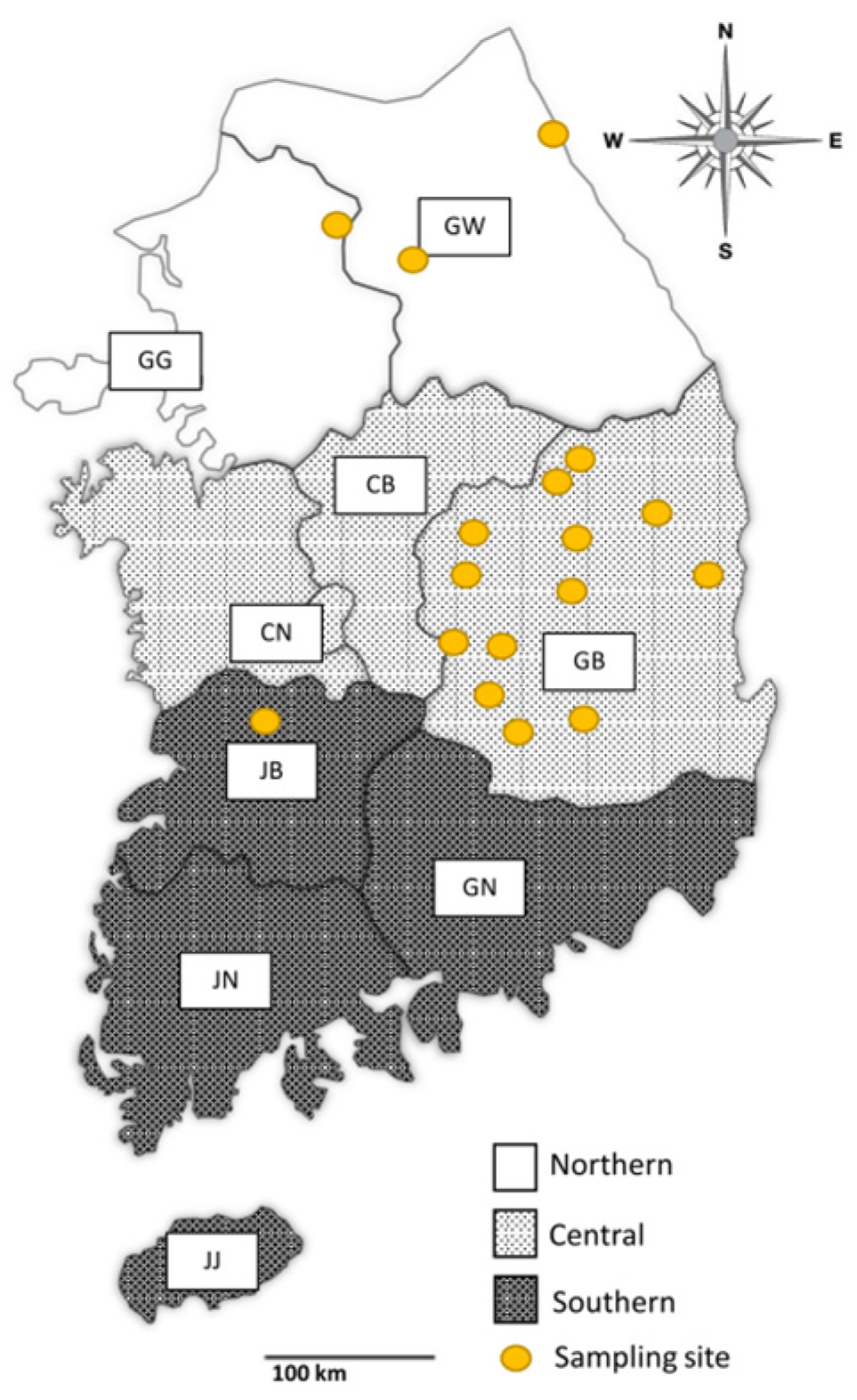
2.1. Blood Sample Collection

Blood samples of 160 deer were collected between December 2014 and February 2023 from 16 cities in Korea. All blood samples were taken from the bodies of deer that died in a road accident. All blood samples were collected in separate tubes and stored at 4 °C before DNA extraction. DNA was extracted within 1–2 days after sample collection.
Data regarding species, region, and season were recorded for each sample whenever possible. The study area was categorized into three groups according to the provincial boundaries: northern (Gangwon (GW) and Gyeonggi (GG) provinces), central (Chungbuk (CB), Chungnam (CN), and Gyeongbuk (GB) provinces), and southern (Jeonbuk (JB), Jeonnam (JN), Gyeongnam (GN), and Jeju (JJ) provinces) regions (Figure 1). Samples with unclear information were labeled “unknown”. Although individual information was labeled at the time of sample collection, there were cases where the label was removed or inaccurate information was recorded in the process of being transferred for autopsy and specimen collection. These cases were classified as unknown samples.

2.2. DNA Extraction and PCR Assay

DNA was extracted using the DNeasy® Blood & Tissue Kit (Qiagen, Hilden, Germany) according to the manufacturer’s instructions. The quality and quantity of DNA were estimated using the Infinite® 200 PRO NanoQuant plate reader (Tecan, Mannedorf, Switzerland). The extracted DNA was stored at −20 °C until it was subjected to PCR.
The PCR premix was prepared using AccuPower® HotStart PCR Premix (Bioneer, Daejon, Korea). The lyophilized premix comprised 1 μL of each PCR primer, 2 μL of DNA template, and 16 μL of distilled water. For the first round of PCR, which was performed to identify the samples positive for piroplasm, the piroplasm 18S rRNA-specific primers BT-F1 (5′-GGT TGA TCC TGC CAG TAG T-3′) and BT-R2 (5′-TTG CGA CCA TAC TCC CCC CA-3′) as well as BT-F3 (5′-TGG GGG GAG TAT GGT CGC AAG-3′) and BT-R3 (5′-CTC CTT CCT TTA AGT GAT AAG-3′) sets were used as previously described [16]. The PCR process included an initial denaturation step of 10 min at 94 °C; 30 cycles of 30 s at 95 °C, 30 s at 65 °C, and 45 s at 72 °C (for BT-F3/BT-R3 primers, 30 s at 61 °C and 30 s at 72 °C); and a post-elongation of 10 min at 72 °C. The expected amplicon sizes were 1024 and 644 bp, respectively. The next round of PCR was performed to distinguish 18S rRNA of Theileria and Babesia. Tsp-F (5′-GTT ATA AAT CGC AAG GAA GTT TAA GGC-3′) and Tsp-R (5′-GTG TAC AAA GGG CAG GGA CGT A-3′) were used as forward and reverse primers, respectively, to identify Theileria [46]. The steps of the PCR reaction included an initial denaturation step of 5 min at 95 °C; 40 cycles of 30 s at 95 °C, 30 s at 61 °C, and 30 s at 72 °C; and a post-elongation of 5 min at 72 °C. The expected amplicon size was 239 bp. Following the amplification of Theileria 18S rRNA, electrophoresis was performed on a 1% agarose gel for 30 min at 130 V, and ethidium bromide was used for staining. Gel Doc™ EZ Imager (Bio-Rad, Pleasanton, CA, USA) was used to obtain images and to confirm the positive bands.

2.3. Statistical and Phylogenetic Analysis

The χ2 test was performed using SPSS version 26.0 (IBM Corporation, Armonk, NY, USA) to test statistical significance. p values of <0.05 were considered statistically significant. Of the 160 blood samples, 13 PCR-positive samples were sent to the Macrogen co. (Daejeon, Korea) for bidirectional nucleotide sequencing. These were aligned with the other sequences of Theileria obtained from the GenBank database using BioEdit 7.2.5 and MEGA7 software (Pennsylvania State University, State College, PA, USA). The obtained sequences were compared with other sequences using NCBI Web BLAST (available at: https://blast.ncbi.nlm.nih.gov/Blast.cgi, accessed on 30 August 2023). A phylogenetic tree was constructed, and a phylogenetic analysis was performed using the maximum likelihood method with 1000 bootstrap replicates.

3. Results

3.1. Region-Wise Infection Rates of Theileria

Among the 160 deer blood samples, 1, 5, and 140 were collected from the southern, northern, and central regions, respectively; the origins of the remaining 14 samples were unknown. In comparison with the central region, which showed an infection rate of 2.9% (4/140), other regions exhibited higher infection rates. An infection rate of 100% was observed in the northern and southern regions despite the small sample size (Table 1). Infection rates significantly differed among the regions (p < 0.001).

3.2. Seasonal Variations in Theileria Infection Rates

The highest Theileria infection rate of 18.4% (9/49) was observed in spring, followed by summer, with an infection rate of 8.9% (4/45). No infection rate was detected in the remaining two seasons (Table 2). Infection rates significantly differed among seasons (p = 0.014).

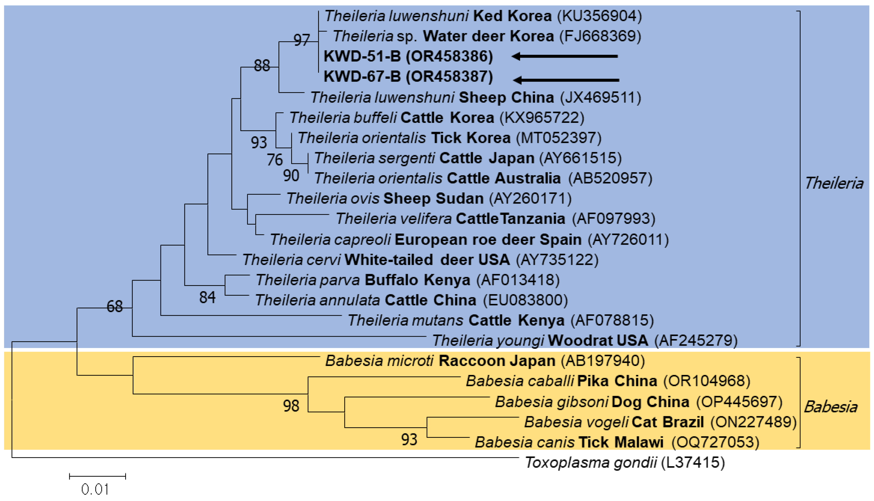
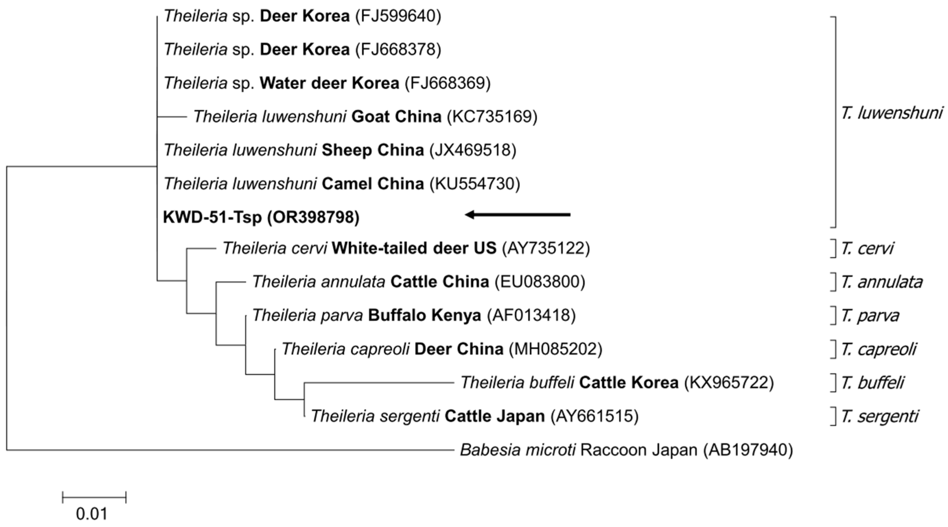
3.3. Sequencing and Phylogenetic Analysis

In the first step, 13 positive samples were confirmed by performing PCR to screen piroplasm-positive samples, and sequencing of these samples yielded two sequences. Next, the sequencing of positive samples was performed to distinguish the 18S rRNA of Babesia and Theileria, which yielded only one sequence. All these sequences were submitted to GenBank (accession numbers OR458386, OR458387, and OR398798). These samples were used for the phylogenetic analysis. A phylogenetic tree was constructed using these sequences and other sequences chosen from the GenBank database. A phylogenetic tree was constructed based on the sequences obtained using piroplasm universal primers and those obtained from Babesia genera, such as B. microti, B. gibsoni, and B. canis, and Theileria genera, such as T. sergenti, T. orientalis, and T. cervi; these sequences were analyzed together. The obtained piroplasm sequences were confirmed to belong to Theileria (Figure 2). Sequences obtained using Theileria-specific primers were grouped along with T. luwenshuni in the phylogenetic tree and were identified as T. luwenshuni. Other sequences used for data analysis included T. orientalis, T. cervi, T. annulata, T. parva, T. capreoli, T. buffeli, and T. sergenti (Figure 3).

4. Discussion

Several studies on tick-borne diseases have been conducted worldwide because of their pathological and zoonotic significance [45,47]. In addition, climatic changes and decreased environment-related diversity have increased the infection rate of tick-borne diseases [43,48]. Piroplasmorida, which includes Babesia and Theileria, is a class containing major pathogens that cause tick-borne diseases [1]. Infection rates of Babesia and/or Theileria were 89.7% (156/174) in roe deer in Spain [30], 58.3% (7/12) in brown brocket deer (Mazama gouazoubira) and marsh deer (Blastocerus dichotomus) in Brazil [31], and 35.3% (24/68) in sika deer (Cervus nippon) in China [39]. In addition, the infection rate of theileriosis in other animals has been studied. The infection rates were 4.7% (12/257) in wild boar, 22.2% (8/36) in Alpine chamois, and 1.0% (2/205) in red foxes in Italy [26]; 80.0% (80/100) in cattle, 93.8% (150/160) in sheep, and 1.9% (5/265) in goat, with 235/525 (44.8%) of the ruminants testing positive for Theileria in Ethiopia [25]; and 6.2% (82/1329) in dogs in South Africa [21]. In Korea, as in other countries, ticks are ubiquitous. Several studies on tick-borne diseases have been conducted in cattle, dogs, and horses [43]. The rate of infection was 1.2% (9/737) in cattle [49], 3.1% (16/510) in dogs [50], and 0.9% (2/224) in horses [16].
In this study, DNA was extracted from the blood samples of 160 deer. After DNA extraction, PCR and electrophoresis were performed. Based on the obtained results, phylogenetic and statistical analyses were performed. The overall infection rate of Theileria in Korean water deer was 8.1% (13/160) in this study. The infection rates in Korean water deer from other studies varied from 80.0% (8/10) in the Jeonbuk Province [43] to 77.8% (14/18) in the southern areas [51] and 72.2% (13/18) in the Chungbuk Province [45]. However, Theileria infection was not detected in 28 Korean water deer in Jeonbuk Province [44]. This difference could be attributed to factors such as the experimental environment, sample size, season, or area of sample collection. Furthermore, a large-scale study is warranted because small sample sizes included in previous studies render their results unreliable.
To examine the effect of region on the infection rate, Korea was divided into three regions: northern, central, and southern. When information on a region was unclear or inadequate, the region was marked as “unknown”. The highest infection rate of 100% was observed in the northern and southern regions. The central and “unknown” regions demonstrated an infection rate of 2.9% (4/140) and 21.4% (3/14), respectively. Infection rates were significantly different among the studied regions (p = 0.001). Nevertheless, the validity of these results was limited because the samples were concentrated in the central region, especially the Gyeongbuk Province.
In terms of season, spring demonstrated the highest infection rate of 18.4% (9/49), followed by summer (8.9%, 4/45). The infection rates in all seasons were significantly different (p = 0.014). However, this comparison has limited validity owing to the limited number of samples collected in autumn and winter. The findings of this study were similar to those of previous studies conducted in Korea. For example, the previously reported infection rate was 5.1% (9/178) in summer [49], 4.9% (8/163) in summer [50], and 2.5% (5/202) in spring. Additionally, infection rates of 1.7% (2/120) in winter and spring and no infection rate in summer and autumn have also been recorded [51]. In New Zealand, the Theileria infection rate was higher in spring and autumn [32,33]. High infection rates of 19.6% (20/102) and 45.4% (45/99) were reported in summer and autumn in India, respectively [35]. The infection rate in the spring (8%, 8/63) and summer (19.6%, 18/92) seasons was higher than that in winter (2.1%, 1/48) in Pakistan [36]. Overall, higher infection rates were recorded in spring and summer than those in the other seasons, indicating that wet conditions facilitated Theileria infection. However, as no positive samples were collected during autumn and winter in this study, this aspect remains unclear.
To date, several species of Theileria have been confirmed to infect small ruminants, including T. ovis, T. separate, and T. recondite, along with T. luwenshuni, T. lestoquardi, and T. uilenbergi, which are highly pathogenic to sheep and goats [52]. The Theileria spp. identified in Korea include T. luwenshuni, T. ovis, T. orientalis, and T. equi [8,29,48,49]. T. equi has been confirmed in horses in Gyeonggi province (0.9%, 2/224) [16]; T. orientalis was identified in cattle and the ticks infesting them [49]; T. luwenshuni and T. ovis were reported in Korean water deer and deer ked (Lipoptena fortisetosa) [52]; and T. luwenshuni was identified in ticks collected from Siberian roe deer (Capreolus pygargus), native Korean goats (Capra hircus coreanae), and horses [53].
No species other than T. luwenshuni could be identified in this study. T. luwenshuni is one of the most common pathogenic species that infect small ruminants such as sheep, goats, and deer [11]. T. luwenshuni was highly prevalent in roe deer on Jeju Island and detected in the blood samples of all 23 deer (100%) and 8 tick pools (34.8%) [54]. In the mid- and southwestern regions of Korea, 541 tick pools were tested, of which 211 (39.0%) were positive for Theileria [11]. Interestingly, the ticks belonging to the Haemaphysalis spp. were found to be most infected with T. luwenshuni, with H. longicornis being the most commonly infected tick with an incidence rate of 92.0% (161/175), followed by H. flava at 5.1% (9/175) [11]. H. longicornis is the most commonly found tick in Korea and has been detected in Jeju Island, far from the mainland [53]. Therefore, it is a vital natural vector for T. luwenshuni [11]. Additionally, H. longicornis is a vector for tick-borne pathogens and can transmit diseases to humans and animals [53]. Although this study did not directly identify ticks infesting deer and tick-borne pathogens (Theileria spp.), they can be identified in the blood samples of wild deer. Therefore, further research is needed regarding ticks occurring in deer that are presumed to transmit this pathogen.
The phylogenetic tree of piroplasms confirmed that they can be broadly classified into two groups: the Theileria spp., and Babesia spp. groups. In the phylogenetic tree of Theileria, B. microti was set as an outgroup, and several different Theileria sequences were analyzed. Based on the two phylogenetic trees, it could be confirmed that the sequences obtained in this study may commonly be identified as those related to T. luwenshuni, which mainly infects goats, sheep, and deer [27,37]. As only a short length of sequence was obtained using 216 bp amplicon, it is necessary to analyze the full-length sequencing to identify species in the future. In this study, the two sequences identified in the phylogenetic tree of the piroplasms, OR458386 and OR458387, were 100% and 99.89% similar to the sequences identified in deer ked in Korea, respectively. The sequence identified in the Theileria phylogenetic tree (OR398798) showed 100% similarity with the sequences identified from deer (FJ599640), sheep (JX469518), and camel (KU554730), although it was shorter than the sequence of piroplasms.

5. Conclusions

The prevention of Theileria infection is gaining urgency because it infects most livestock such as goats, sheep, and cattle. The overall Theileria infection rate in this study was 8.1% (13/160). Significant differences in infection rates among the various regions and seasons (p < 0.001 and p = 0.014, respectively) were observed. Phylogenetic analysis showed that Theileria-positive blood samples contained T. luwenshuni, which mainly infects deer, sheep, and goats.
The primary importance of this study is that it is one of the few studies that focuses on Theileria infection in deer, including Korean water deer, with a large sample size. However, biased sample collection based on the region and shortage of classification standards due to characteristics of wild animals may compromise the validity of these results. Therefore, further studies focusing on livestock and other Theileria spp. are warranted. In addition, it is also necessary to perform an analysis of Theileria species in ticks infesting wild deer, which were not included in this study. Because enzootic stability/instability cannot be excluded in livestock theileriosis, differences in prevalence may appear in various ways. Therefore, additional comprehensive study considering the life cycle of Theileria should be conducted to confirm the prevalence between the host and the pathogen, to prevent the spread of tick-borne pathogens, and to prepare for the prevention of infections in wild animals and nearby livestock farms.

Author Contributions

Conceptualization, D.K. and C.U.C.; methodology, C.U.C. and H.L.; validation, M.-G.S., S.-H.L., K.-T.K. and K.N.; formal analysis, C.U.C. and H.L.; investigation, C.U.C., H.L., M.-G.S., S.-H.L. and K.-T.K.; resources, J.-S.S. and D.H.B.; data curation, M.H.R. and O.-D.K.; writing—original draft preparation, C.U.C. and H.L.; writing—review and editing, H.L., M.-G.S., S.-H.L., K.-T.K., K.N., J.-S.S., D.H.B., M.H.R. and O.-D.K.; visualization, M.H.R. and O.-D.K.; supervision, D.K.; funding acquisition, D.K. All authors have read and agreed to the published version of the manuscript.

Funding

This research was supported by a grant (Grant No. NRF-2016R1D1A1B02015366) from the Basic Science Research Program through the National Research Foundation, Republic of Korea (NRF).

Data Availability Statement

Data supporting the conclusions of this article are included within the article. The newly generated sequences were submitted to the GenBank database under the accession numbers OR458386-OR458387 and OR398798. The datasets used and/or analyzed during the present study are available from the corresponding author upon reasonable request.

Conflicts of Interest

The authors declare no conflict of interest.

References

  1. Mans, B.J.; Pienaar, R.; Latif, A.A. A review of Theileria diagnostics and epidemiology. Int. J. Parasitol. Parasites Wildl. 2015, 4, 104–118. [Google Scholar] [CrossRef] [PubMed]
  2. Abdallah, M.O.; Niu, Q.; Yang, J.; Hassan, M.A.; Yu, P.; Guan, G.; Chen, Z.; Liu, G.; Luo, J.; Yin, H. Identification of 12 piroplasms infecting ten tick species in China using reverse line blot hybridization. J. Parasitol. 2017, 103, 221–227. [Google Scholar] [CrossRef] [PubMed]
  3. Iori, A.; Gabrielli, S.; Calderini, P.; Moretti, A.; Pietrobelli, M.; Tampieri, M.P.; Galuppi, R.; Cancrini, G. Tick reservoirs for piroplasms in central and northern Italy. Vet. Parasitol. 2010, 170, 291–296. [Google Scholar] [CrossRef]
  4. Estrada-Peña, A. Ticks as vectors: Taxonomy, biology and ecology. Rev. Sci. Tech. 2015, 34, 53–65. [Google Scholar] [CrossRef]
  5. Anderson, J.F.; Magnarelli, L.A. Biology of ticks. Infect. Dis. Clin. N. Am. 2008, 22, 195–215. [Google Scholar] [CrossRef] [PubMed]
  6. Fanelli, A. A historical review of Babesia spp. associated with deer in Europe: Babesia divergens/Babesia divergens-like, Babesia capreoli, Babesia venatorum, Babesia cf. odocoilei. Vet. Parasitol. 2021, 294, 109433. [Google Scholar] [CrossRef]
  7. Uilenberg, G. Babesia–a historical overview. Vet. Parasitol. 2006, 138, 3–10. [Google Scholar] [CrossRef]
  8. Ahmed, J.S.; Luo, J.; Schnittger, L.; Seitzer, U.; Jongejan, F.; Yin, H. Phylogenetic position of small-ruminant infecting piroplasms. Ann. N. Y. Acad. Sci. 2006, 1081, 498–504. [Google Scholar] [CrossRef]
  9. Allsopp, M.T.; Allsopp, B.A. Molecular sequence evidence for the reclassification of some Babesia species. Ann. N. Y. Acad. Sci. 2006, 1081, 509–517. [Google Scholar] [CrossRef]
  10. Schnittger, L.; Rodriguez, A.E.; Florin-Christensen, M.; Morrison, D.A. Babesia: A world emerging. Infect. Genet. Evol. 2012, 12, 1788–1809. [Google Scholar] [CrossRef]
  11. Alkathiri, B.; Ahn, K.S.; Lee, H.S.; Cho, Y.S.; Youn, S.Y.; Seo, M.G.; Kwak, D.; Shin, S.S.; Lee, S.H. Molecular epidemiology of species in ticks and its potential threat to livestock in the Republic of Korea. Acta Trop. 2023, 238, 106780. [Google Scholar] [CrossRef] [PubMed]
  12. Bishop, R.; Musoke, A.; Morzaria, S.; Gardner, M.; Nene, V. Theileria: Intracellular protozoan parasites of wild and domestic ruminants transmitted by ixodid ticks. Parasitology 2004, 129 (Suppl. S1), S271–S283. [Google Scholar] [CrossRef] [PubMed]
  13. Camacho, A.T.; Pallas, E.; Gestal, J.J.; Guitián, F.J.; Olmeda, A.S.; Goethert, H.K.; Telford, S.R. Infection of dogs in north-west Spain with a Babesia microti-like agent. Vet. Rec. 2001, 149, 552–555. [Google Scholar] [CrossRef] [PubMed]
  14. Kjemtrup, A.M.; Robinson, T.; Conrad, P.A. Description and epidemiology of Theileria youngi n. sp. from a northern Californian dusky-footed woodrat (Neotoma fuscipes) population. J. Parasitol. 2001, 87, 373–378. [Google Scholar] [CrossRef]
  15. Morrison, W.I. The aetiology, pathogenesis and control of theileriosis in domestic animals. Rev. Sci. Tech. 2015, 34, 599–611. [Google Scholar] [CrossRef]
  16. Seo, M.G.; Yun, S.H.; Choi, S.K.; Cho, G.J.; Park, Y.S.; Cho, K.H.; Kwon, O.D.; Kwak, D. Molecular and phylogenetic analysis of equine piroplasms in the Republic of Korea. Res. Vet. Sci. 2013, 94, 579–583. [Google Scholar] [CrossRef]
  17. Dobbelaere, D.A.; Küenzi, P. The strategies of the parasite: A new twist in host-pathogen interactions. Curr. Opin. Immunol. 2004, 16, 524–530. [Google Scholar] [CrossRef]
  18. Sivakumar, T.; Hayashida, K.; Sugimoto, C.; Yokoyama, N. Evolution and genetic diversity of Theileria. Infect. Genet. Evol. 2014, 27, 250–263. [Google Scholar] [CrossRef]
  19. Baneth, G.; Florin-Christensen, M.; Cardoso, L.; Schnittger, L. Reclassification of Theileria annae as Babesia vulpes sp. nov. Parasit. Vectors 2015, 8, 207. [Google Scholar] [CrossRef]
  20. Jacobson, L.S.; Clark, I.A. The pathophysiology of canine babesiosis: New approaches to an old puzzle. J. S. Afr. Vet. Assoc. 1994, 65, 134–145. [Google Scholar]
  21. Matjila, P.T.; Leisewitz, A.L.; Oosthuizen, M.C.; Jongejan, F.; Penzhorn, B.L. Detection of a Theileria species in dogs in South Africa. Vet. Parasitol. 2008, 157, 34–40. [Google Scholar] [CrossRef] [PubMed]
  22. D’Oliveira, C.; van der Weide, M.; Habela, M.A.; Jacquiet, P.; Jongejan, F. Detection of Theileria annulata in blood samples of carrier cattle by PCR. J. Clin. Microbiol. 1995, 33, 2665–2669. [Google Scholar] [CrossRef] [PubMed]
  23. Gubbels, M.J.; Hong, Y.; van der Weide, M.; Qi, B.; Nijman, I.J.; Guangyuan, L.; Jongejan, F. Molecular characterisation of the Theileria buffeli/orientalis group. Int. J. Parasitol. 2000, 30, 943–952. [Google Scholar] [CrossRef] [PubMed]
  24. Mhadhbi, M.; Naouach, A.; Boumiza, A.; Chaabani, M.F.; BenAbderazzak, S.; Darghouth, M.A. In vivo evidence for the resistance of Theileria annulata to buparvaquone. Vet. Parasitol. 2010, 169, 241–247. [Google Scholar] [CrossRef] [PubMed]
  25. Gebrekidan, H.; Hailu, A.; Kassahun, A.; Rohoušová, I.; Maia, C.; Talmi-Frank, D.; Warburg, A.; Baneth, G. Theileria infection in domestic ruminants in northern Ethiopia. Vet. Parasitol. 2014, 200, 31–38. [Google Scholar] [CrossRef]
  26. Zanet, S.; Trisciuoglio, A.; Bottero, E.; de Mera, I.G.; Gortazar, C.; Carpignano, M.G.; Ferroglio, E. Piroplasmosis in wildlife: Babesia and Theileria affecting free-ranging ungulates and carnivores in the Italian Alps. Parasit. Vectors 2014, 7, 70. [Google Scholar] [CrossRef]
  27. Phipps, L.P.; Hernández-Triana, L.M.; Goharriz, H.; Welchman, D.; Johnson, N. Detection of Theileria luwenshuni in sheep from Great Britain. Parasit. Vectors 2016, 9, 203. [Google Scholar] [CrossRef]
  28. Almazán, C.; Scimeca, R.C.; Reichard, M.V.; Mosqueda, J. Babesiosis and Theileriosis in North America. Pathogens 2022, 11, 168. [Google Scholar] [CrossRef]
  29. Romero-Salas, D.; Solis-Cortés, M.; Zazueta-Islas, H.M.; Flores-Vásquez, F.; Cruz-Romero, A.; Aguilar-Domínguez, M.; Salguero-Romero, J.L.; de León, A.P.; Fernández-Figueroa, E.A.; Lammoglia-Villagómez, M.Á.; et al. Molecular detection of Theileria equi in horses from Veracruz, Mexico. Ticks Tick Borne Dis. 2021, 12, 101671. [Google Scholar] [CrossRef]
  30. Remesar, S.; Díaz, P.; Prieto, A.; Markina, F.; Díaz Cao, J.M.; López-Lorenzo, G.; Fernández, G.; López, C.M.; Panadero, R.; Díez-Baños, P.; et al. Prevalence and distribution of Babesia and Theileria species in roe deer from Spain. Int. J. Parasitol. Parasites Wildl. 2019, 9, 195–201. [Google Scholar] [CrossRef]
  31. Da Silveira, J.A.; Rabelo, E.M.; Ribeiro, M.F. Detection of Theileria and Babesia in brown brocket deer (Mazama gouazoubira) and marsh deer (Blastocerus dichotomus) in the state of Minas Gerais, Brazil. Vet. Parasitol. 2011, 177, 61–66. [Google Scholar] [CrossRef] [PubMed]
  32. Lawrence, K.; Gedye, K.; McFadden, A.; Pulford, D.; Heath, A.; Pomroy, W. Review of the New Zealand Theileria orientalis Ikeda type epidemic and epidemiological research since 2012. Pathogens 2021, 10, 1346. [Google Scholar] [CrossRef] [PubMed]
  33. McFadden, A.M.; Vink, D.; Pulford, D.J.; Lawrence, K.; Gias, E.; Heath, A.C.; McFadden, C.B.; Bingham, P. Monitoring an epidemic of Theileria-associated bovine anaemia (Ikeda) in cattle herds in New Zealand. Prev. Vet. Med. 2016, 125, 31–37. [Google Scholar] [CrossRef] [PubMed]
  34. Storey-Lewis, B.; Mitrovic, A.; McParland, B. Molecular detection and characterisation of Babesia and Theileria in Australian hard ticks. Ticks Tick Borne Dis. 2018, 9, 471–478. [Google Scholar] [CrossRef]
  35. Kohli, S.; Atheya, U.K.; Thapliyal, A. Prevalence of theileriosis in cross-bred cattle: Its detection through blood smear examination and polymerase chain reaction in Dehradun district, Uttarakhand, India. Vet. World 2014, 7, 168–171. [Google Scholar] [CrossRef]
  36. Naz, S.; Maqbool, A.; Ahmed, S.; Ashraf, K.; Ahmed, N.; Saeed, K.; Latif, M.; Iqbal, J.; Ali, Z.; Shafi, K.; et al. Prevalence of theileriosis in small ruminants in Lahore-Pakistan. J. Vet. Anim. Sci. 2011, 2, 16–20. [Google Scholar]
  37. Bawm, S.; Kakisaka, K.; Thu, M.J.; Chel, H.M.; Oo, Y.M.N.; Soe, N.C.; Win, S.Y.; Htun, L.L.; Win, M.M.; Suzuki, H.; et al. First molecular detection of Theileria luwenshuni from goats in Myanmar. Parasitol. Res. 2018, 117, 3361–3364. [Google Scholar] [CrossRef]
  38. Lee, S.H.; Moumouni, P.F.A.; Galon, E.M.; Vudriko, P.; Liu, M.; Benedicto, B.; Tumwebaze, M.A.; Boldbaatar, D.; Umemiya-Shirafuji, R.; Fukumoto, S.; et al. Differential diagnosis and molecular characterization of Theileria spp. in sika deer (Cervus nippon) in Hokkaido, Japan. Parasitol. Int. 2019, 70, 23–26. [Google Scholar] [CrossRef]
  39. Liu, J.; Yang, J.; Guan, G.; Liu, A.; Wang, B.; Luo, J.; Yin, H. Molecular detection and identification of piroplasms in sika deer (Cervus nippon) from Jilin Province, China. Parasit. Vectors 2016, 9, 156. [Google Scholar] [CrossRef]
  40. Ge, Y.; Pan, W.; Yin, H. Prevalence of Theileria infections in goats and sheep in southeastern China. Vet. Parasitol. 2012, 186, 466–469. [Google Scholar] [CrossRef]
  41. Chen, Y.; Chen, Y.Y.; Liu, G.; Lyu, C.; Hu, Y.; An, Q.; Qiu, H.Y.; Zhao, Q.; Wang, C.R. Prevalence of Theileria in cattle in China: A systematic review and meta-analysis. Microb. Pathog. 2022, 162, 105369. [Google Scholar] [CrossRef] [PubMed]
  42. Jang, D.H. Epizootiological study of theileriasis in Korea. Korean J. Parasitol. 1974, 12, 14–20. [Google Scholar] [CrossRef] [PubMed]
  43. Seong, G.; Han, Y.J.; Oh, S.S.; Chae, J.S.; Yu, D.H.; Park, J.; Park, B.K.; Yoo, J.G.; Choi, K.S. Detection of tick-borne pathogens in the Korean Water Deer (Hydropotes inermis argyropus) from Jeonbuk Province, Korea. Korean J. Parasitol. 2015, 53, 653–659. [Google Scholar] [CrossRef] [PubMed]
  44. Shin, S.U.; Park, Y.J.; Ryu, J.H.; Jang, D.H.; Hwang, S.; Cho, H.C.; Park, J.; Han, J.I.; Choi, K.S. Identification of zoonotic tick-borne pathogens from Korean water deer (Hydropotes inermis argyropus). Vector Borne Zoonotic Dis. 2020, 20, 745–754. [Google Scholar] [CrossRef]
  45. Han, J.I.; Jang, H.J.; Lee, S.J.; Na, K.J. High prevalence of Theileria sp. in wild Chinese water deer (Hydropotes inermis argyropus) in South Korea. Vet. Parasitol. 2009, 164, 311–314. [Google Scholar] [CrossRef]
  46. Choi, K.S.; Yu, D.H.; Chae, J.S.; Park, B.K.; Yoo, J.G.; Park, J. Seasonal changes in hemograms and Theileria orientalis infection rates among Holstein cattle pastured in the mountains in the Republic of Korea. Prev. Vet. Med. 2016, 127, 77–83. [Google Scholar] [CrossRef]
  47. Ahmed, J.S.; Glass, E.J.; Salih, D.A.; Seitzer, U. Innate immunity to tropical theileriosis. Innate Immun. 2008, 14, 5–12. [Google Scholar] [CrossRef]
  48. Dantas-Torres, F.; Chomel, B.B.; Otranto, D. Ticks and tick-borne disease: A One Health perspective. Trends Parasitol. 2012, 28, 437–446. [Google Scholar] [CrossRef]
  49. Kwak, D.; Seo, M.G. Genetic diversity of bovine hemoprotozoa in South Korea. Pathogens 2020, 9, 768. [Google Scholar] [CrossRef]
  50. Seo, M.G.; Kwon, O.D.; Kwak, D. Molecular detection and phylogenetic analysis of canine tick-borne pathogens from Korea. Ticks Tick Borne Dis. 2020, 11, 101357. [Google Scholar] [CrossRef]
  51. Han, Y.J.; Park, J.; Lee, Y.S.; Chae, J.S.; Yu, D.H.; Park, B.K.; Kim, H.C.; Choi, K.S. Molecular identification of selected tick-borne pathogens in wild deer and raccoon dogs from the Republic of Korea. Vet. Parasitol. Reg. Stud. Rep. 2017, 7, 25–31. [Google Scholar] [CrossRef] [PubMed]
  52. Lee, S.H.; Kim, K.T.; Kwon, O.D.; Ock, Y.; Kim, T.; Choi, D.; Kwak, D. Novel Detection of Coxiella spp., Theileria luwenshuni, and T. ovis Endosymbionts in Deer Keds (Lipoptena fortisetosa). PLoS ONE 2016, 11, e0156727. [Google Scholar] [CrossRef] [PubMed]
  53. Seo, M.G.; Kwon, O.D.; Kwak, D. Diversity and genotypic analysis of tick-borne pathogens carried by ticks infesting horses in Korea. Med. Vet. Entomol. 2021, 35, 213–218. [Google Scholar] [CrossRef] [PubMed]
  54. Moon, K.; Lee, S.; Choi, C.; Kim, S.; Kang, C.; Lee, K.; Yun, Y. Investigation of Theileria sp. from ticks and roe deer (Capreolus pygargus) in Jeju Island. J. Vet. Clin. 2014, 31, 6–10. [Google Scholar] [CrossRef]
Figure 1. Map representing the regions from which samples were collected. Based on the provincial areas, the country was categorized into three regions: northern (Gangwon (GW) and Gyeonggi (GG) provinces), central (Chungbuk (CB), Chungnam (CN), and Gyeongbuk (GB) provinces), and southern (Jeonbuk (JB), Jeonnam (JN), Gyeongnam (GN), and Jeju (JJ) provinces) regions. Yellow spots indicate the sampling site.
Microorganisms 11 02740 g001
Figure 2. Genera corresponding to the piroplasm 18S rRNA (including Theileria and Babesia) were analyzed by constructing a phylogenetic tree, and the acquired sequences belonged to Theileria. The sequences obtained in this study are indicated using black arrows.
Microorganisms 11 02740 g002
Figure 3. Phylogenetic tree of the Theileria 18S rRNA. The sequence obtained in this study is indicated using a black arrow.
Microorganisms 11 02740 g003
Table 1. Region-wise infection rates of Theileria.
RegionNo. TestedNo. Positive (%)p Value
Northern55 (100)<0.001
Central1404 (2.9)
Southern11 (100)
Unknown143 (21.4)
Total16013 (8.1)
Table 2. Theileria infection rates based on season.
SeasonNo. TestedNo. Positive (%)p Value
Spring499 (18.4)0.014
Summer454 (8.9)
Autumn260
Winter330
Unknown70
Total16013 (8.1)
Disclaimer/Publisher’s Note: The statements, opinions and data contained in all publications are solely those of the individual author(s) and contributor(s) and not of MDPI and/or the editor(s). MDPI and/or the editor(s) disclaim responsibility for any injury to people or property resulting from any ideas, methods, instructions or products referred to in the content.

