
Rickettsial Infection in the COVID-19 Era: The Correlation between the Detection of Rickettsia aeschlimannii in Ticks and Storytelling Photography of a Presumable Human Rickettsiosis Case


Abstract
:1. Introduction
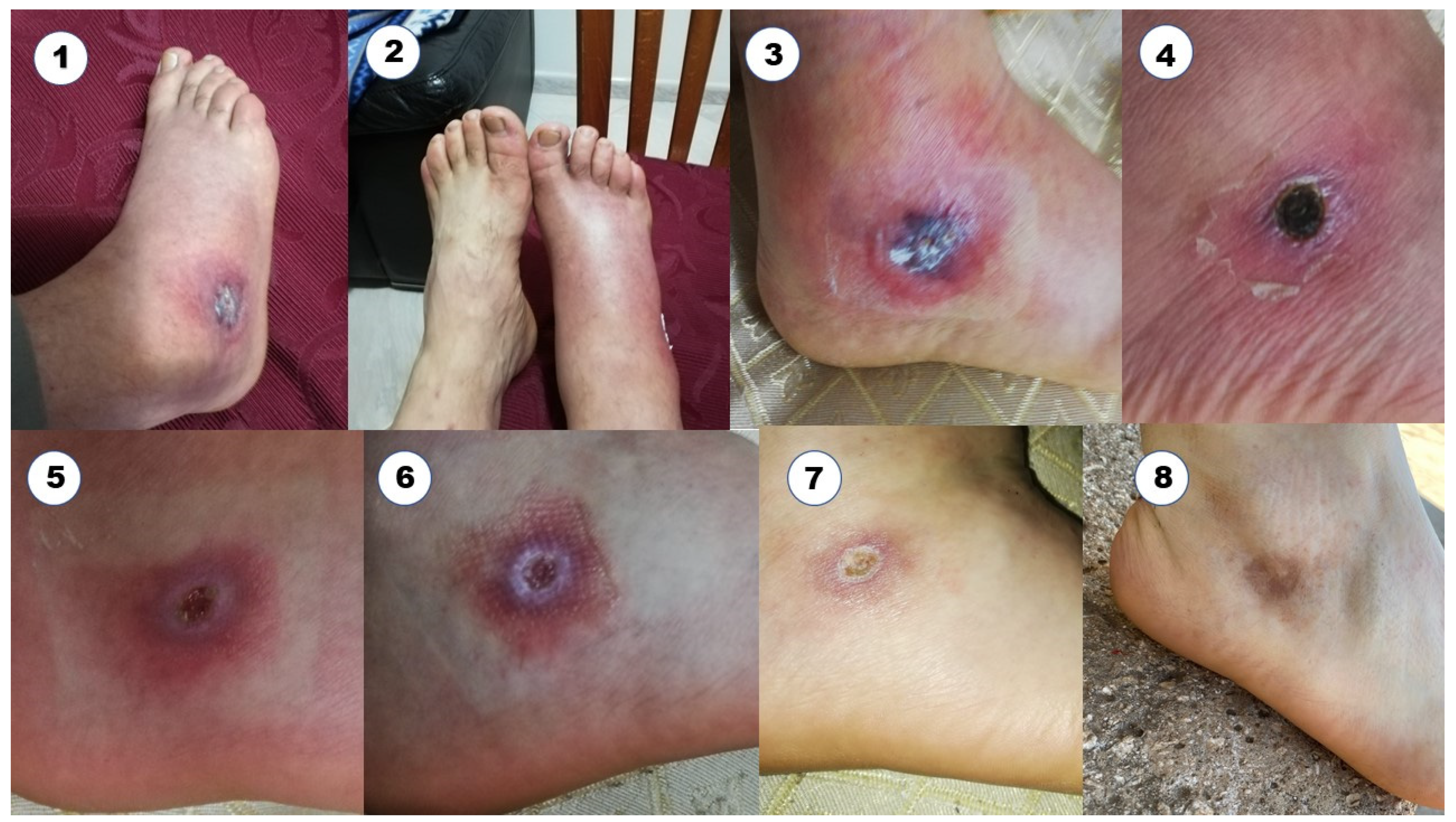
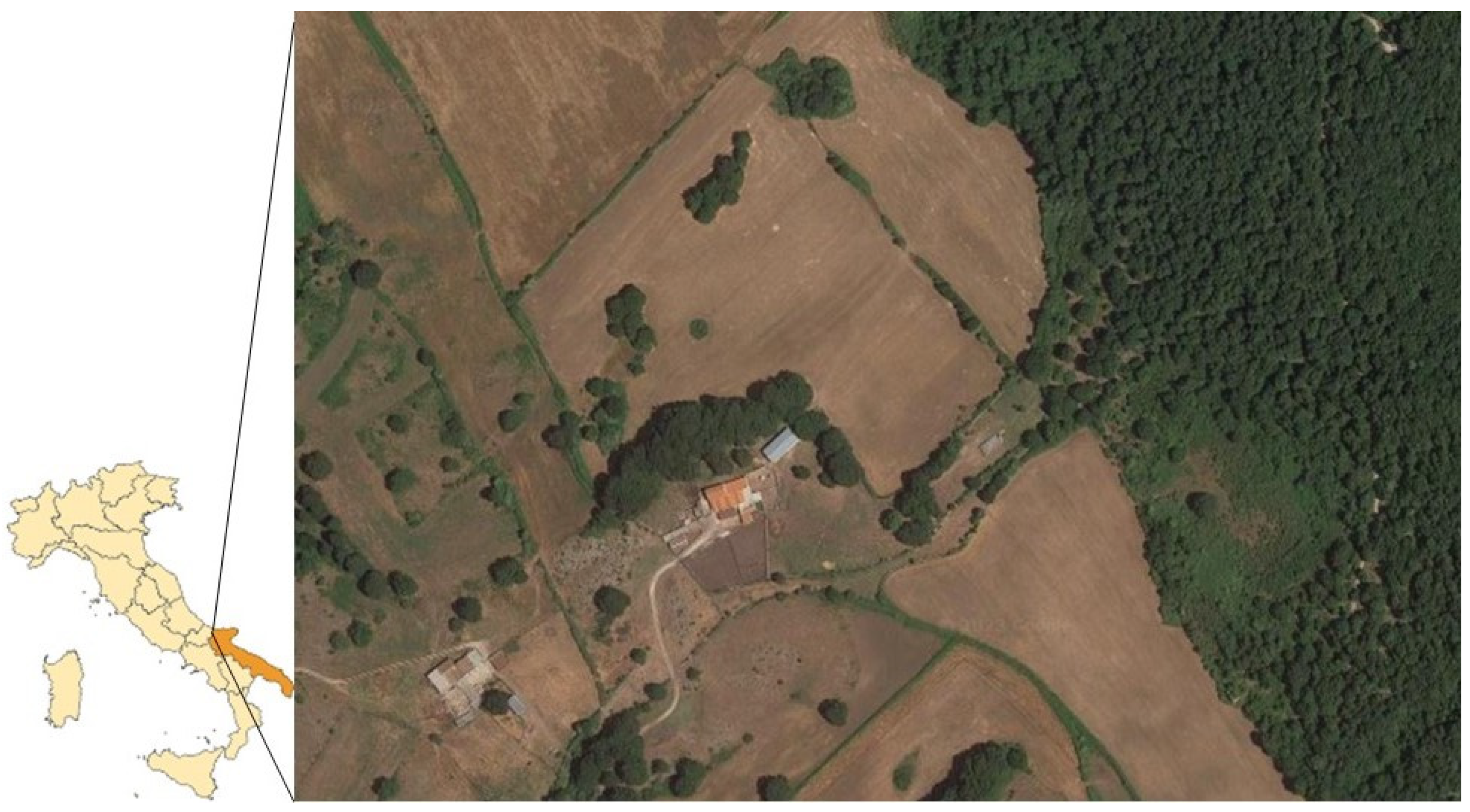
2. The Study
3. Discussion and Conclusions
Author Contributions
Funding
Institutional Review Board Statement
Informed Consent Statement
Data Availability Statement
Conflicts of Interest
References
- Germanakis, A.; Chochlakis, D.; Angelakis, E.; Tselentis, Y.; Psaroulaki, A. Rickettsia aeschlimannii infection in a man, Greece. Emerg. Infect. Dis. 2013, 19, 1176–1177. [Google Scholar] [CrossRef] [PubMed]
- Socolovschi, C.; Parola, P.; Raoult, D. Tick-borne Spotted Fever Rickettsioses. In Hunter’s Tropical Medicine and Emerging Infectious Disease, 9th ed.; Magill, A.J., Hill, D.R., Solomon, T., Ryan, E.T., Eds.; W.B. Saunders: Philadelphia, PA, USA, 2013; pp. 546–552. [Google Scholar]
- Tosoni, A.; Mirijello, A.; Ciervo, A.; Mancini, F.; Rezza, G.; Damiano, F.; Cauda, R.; Gasbarrini, A.; Addolorato, G.; Internal Medicine Sepsis Study Group. Human Rickettsia aeschlimannii infection: First case with acute hepatitis and review of the literature. Eur. Rev. Med. Pharmacol. Sci. 2016, 20, 2630–2633. [Google Scholar] [PubMed]
- Znazen, A.; Rolain, J.; Hammami, N.; Hammami, A.; Ben Jemaa, M.; Raoult, D. Rickettsia felis Infection, Tunisia. Emerg. Infect. Dis. 2006, 12, 138–140. [Google Scholar] [CrossRef] [PubMed]
- Beati, L.; Meskini, M.; Thiers, B.; Raoult, D. Rickettsia aeschlimannii sp. nov., a new spotted fever group rickettsia associated with Hyalomma marginatum ticks. Int. J. Syst. Bacteriol. 1997, 47, 548–554. [Google Scholar] [CrossRef]
- Raoult, D.; Fournier, P.E.; Abboud, P.; Caron, F. First documented human Rickettsia aeschlimannii infection. Emerg. Infect. Dis. 2002, 8, 748–749. [Google Scholar] [CrossRef]
- Oteo, J.A.; Portillo, A. Tick-borne rickettsioses in Europe. Ticks Tick-Borne Dis. 2012, 3, 271–278. [Google Scholar] [CrossRef]
- Igolkina, Y.; Rar, V.; Krasnova, E.; Filimonova, E.; Tikunov, A.; Epikhina, T.; Tikunova, T. Occurrence and clinical manifestations of tick-borne rickettsioses in Western Siberia: First Russian cases of Rickettsia aeschlimannii and Rickettsia slovaca infections. Ticks Tick-Borne Dis. 2022, 13, 101927. [Google Scholar] [CrossRef]
- Guccione, C.; Colomba, C.; Tolomeo, M.; Trizzino, M.; Iaria, C.; Cascio, A. Rickettsiales in Italy. Pathogens 2021, 10, 181. [Google Scholar] [CrossRef]
- Blanda, V.; Torina, A.; La Russa, F.; D’Agostino, R.; Randazzo, K.; Scimeca, S.; Giudice, E.; Caracappa, S.; Cascio, A.; de la Fuente, J. A retrospective study of the characterization of Rickettsia species in ticks collected from humans. Ticks Tick-Borne Dis. 2017, 8, 610–614. [Google Scholar] [CrossRef]
- Chisu, V.; Zobba, R.; Foxi, C.; Pisu, D.; Masala, G.; Alberti, A. Molecular detection and groEL typing of Rickettsia aeschlimannii in Sardinian ticks. Parasitol. Res. 2016, 115, 3323–3328. [Google Scholar] [CrossRef]
- Battisti, E.; Urach, K.; Hodžić, A.; Fusani, L.; Hufnagl, P.; Felsberger, G.; Ferroglio, E.; Duscher, G.G. Zoonotic Pathogens in Ticks from Migratory Birds, Italy. Emerg. Infect. Dis. 2020, 26, 2986–2988. [Google Scholar] [CrossRef]
- Manilla, G. Acari Ixodida, Fauna d’Italia, 3rd ed.; Calderini: Bologna, Italy, 1998; Volume 36. [Google Scholar]
- Webb, L.; Mitchell, C.; Malloy, D.C.; Dasch, G.A.; Azad, A.F. Detection of murine typhus infection in fleas by using the polymerase chain reaction. J. Clin. Microbiol. 1990, 28, 530–534. [Google Scholar] [CrossRef] [PubMed]
- Roux, V.; Fournier, P.E.; Raoult, D. Differentiation of spotted fever group rickettsiae by sequencing and analysis of restriction fragment length polymorphism of PCR- amplified DNA of the gene encoding the protein rOmpA. J. Clin. Microbiol. 1996, 34, 2058–2065. [Google Scholar] [CrossRef] [PubMed]
- Apanaskevich, A.D.; Horak, I.G. The genus Hyalomma Koch, 1844: V. re-evaluation of the taxonomic rank of taxa comprising the H. (Euhyalomma) marginatum koch complex of species (Acari: Ixodidae) with redescription of all parasitic stages and notes on biology. Int. J. Acarol. 2008, 34, 13–42. [Google Scholar] [CrossRef]
- Sobrero, L.; Manilla, G. Aggiornamenti Sulle Zecche d’Italia; Bonifica: Bastogi, Italy, 1988; p. 111. [Google Scholar]
- Toma, L.; Mancini, F.; Di Luca, M.; Cecere, J.G.; Bianchi, R.; Khoury, C.; Quarchioni, E.; Manzia, F.; Rezza, G.; Ciervo, A. Detection of microbial agents in ticks collected from migratory birds in central Italy. Vector Borne Zoonotic Dis. 2014, 14, 199–205. [Google Scholar] [CrossRef]
- Mancuso, E.; Di Domenico, M.; Di Gialleonardo, L.; Menegon, M.; Toma, L.; Di Luca, M.; Casale, F.; Di Donato, G.; D’Onofrio, L.; De Rosa, A.; et al. Tick Species Diversity and Molecular Identification of Spotted Fever Group Rickettsiae Collected from Migratory Birds Arriving from Africa. Microorganisms 2023, 11, 2036. [Google Scholar] [CrossRef]
- Hoffman, T.; Carra, L.G.; Öhagen, P.; Fransson, T.; Barboutis, C.; Piacentini, D.; Figuerola, J.; Kiat, Y.; Onrubia, A.; Jaenson, T.G.T.; et al. Association between guilds of birds in the African-Western Palaearctic region and the tick species Hyalomma rufipes, one of the main vectors of Crimean-Congo hemorrhagic fever virus. One Health 2021, 13, 100349. [Google Scholar] [CrossRef]
- Estrada-Peña, A.; D’Amico, G.; Fernández-Ruiz, N. Modelling the potential spread of Hyalomma marginatum ticks in Europe by migratory birds. Int. J. Parasitol. 2021, 51, 1–11. [Google Scholar] [CrossRef]
- Parola, P.; Paddock, C.D.; Raoult, D. Tick-borne rickettsioses around the world: Emerging diseases challenging old concepts. Clin. Microbiol. Rev. 2005, 18, 719–756. [Google Scholar] [CrossRef]
- Keysary, A.; Eremeeva, M.E.; Leitner, M.; Din, A.B.; Wikswo, M.E.; Mumcuoglu, K.Y.; Inbar, M.; Wallach, A.D.; Shanas, U.; King, R.; et al. Spotted fever group rickettsiae in ticks collected from wild animals in Israel. Am. J. Trop. Med. Hyg. 2011, 85, 919–923. [Google Scholar] [CrossRef]
- Abdel-Shafy, S.; Allam, N.A.; Mediannikov, O.; Parola, P.; Raoult, D. Molecular detection of spotted fever group rickettsiae associated with ixodid ticks in Egypt. Vector Borne Zoonotic Dis. 2012, 12, 346–359. [Google Scholar] [CrossRef]
- Gargili, A.; Palomar, A.M.; Midilli, K.; Portillo, A.; Kar, S.; Oteo, J.A. Rickettsia species in ticks removed from humans in Istanbul, Turkey. Vector Borne Zoonotic Dis. 2012, 12, 938–941. [Google Scholar] [CrossRef] [PubMed]
- Abdelbaset, A.E.; Kwak, M.L.; Nonaka, N.; Nakao, R. Human-biting ticks and zoonotic tick-borne pathogens in North Africa: Diversity, distribution, and trans-Mediterranean public health challenges. One Health 2023, 16, 100547. [Google Scholar] [CrossRef] [PubMed]
- Raele, D.A.; Pugliese, N.; Galante, D.; Cafiero, M.A. Loop-mediated isothermal amplification in the field: Reality beyond the potentiality. Turk. J. Vet. Anim. Sci. 2019, 43, 173–175. [Google Scholar] [CrossRef]
- Raele, D.A.; Galante, D.; Pugliese, N.; Salandra, G.; Cafiero, M.A. Spotted fever group rickettsiae associated with ixodid ticks in wild environment in Southern Italy. Microbiologyopen 2018, 7, e00527. [Google Scholar] [CrossRef]
- Koczwarska, J.; Pawełczyk, A.; Dunaj-Małyszko, J.; Polaczyk, J.; Welc-Falęciak, R. Rickettsia species in Dermacentor reticulatus ticks feeding on human skin and clinical manifesta-tions of tick-borne infections after tick bite. Sci. Rep. 2023, 13, 9930. [Google Scholar] [CrossRef]
- Yang, M.; Jia, Y.; Dong, Z.; Zhang, Y.; Xie, S.; Liu, Q.; Wang, Y. Rickettsia aeschlimannii Infection in a Woman from Xingjiang, Northwestern China. Vector Borne Zoonotic Dis. 2022, 22, 55–57. [Google Scholar]
- Wächter, M.; Wölfel, S.; Pfeffer, M.; Dobler, G.; Kohn, B.; Moritz, A.; Pachnicke, S.; Silaghi, C. Serological differentiation of antibodies against Rickettsia helvetica, R. raoultii, R. slovaca, R. monacensis and R. felis in dogs from Germany by a micro-immunofluorescent antibody test. Parasit. Vectors 2015, 8, 126. [Google Scholar] [CrossRef]
- Stewart, A.G.; Stewart, A.G.A. An Update on the Laboratory Diagnosis of Rickettsia spp. Infection. Pathogens 2021, 10, 1319. [Google Scholar] [CrossRef]
- Grassi, L.; Drigo, M.; Zelená, H.; Pasotto, D.; Cassini, R.; Mondin, A.; Franzo, G.; Tucciarone, C.M.; Ossola, M.; Vidorin, E.; et al. Wild ungulates as sentinels of flaviviruses and tick-borne zoonotic pathogen circulation: An Italian perspective. BMC Vet. Res. 2023, 19, 155. [Google Scholar] [CrossRef]
- Mittal, A.; Elias, M.L.; Schwartz, R.A.; Kapila, R. Recognition and treatment of devastating vasculopathic systemic disorders: Coronavirus disease 2019 and rickettsioses. Dermatol. Ther. 2021, 34, e14984. [Google Scholar] [CrossRef] [PubMed]


Disclaimer/Publisher’s Note: The statements, opinions and data contained in all publications are solely those of the individual author(s) and contributor(s) and not of MDPI and/or the editor(s). MDPI and/or the editor(s) disclaim responsibility for any injury to people or property resulting from any ideas, methods, instructions or products referred to in the content. |
© 2023 by the authors. Licensee MDPI, Basel, Switzerland. This article is an open access article distributed under the terms and conditions of the Creative Commons Attribution (CC BY) license (https://creativecommons.org/licenses/by/4.0/).
Share and Cite
Raele, D.A.; Cafiero, M.A. Rickettsial Infection in the COVID-19 Era: The Correlation between the Detection of Rickettsia aeschlimannii in Ticks and Storytelling Photography of a Presumable Human Rickettsiosis Case. Microorganisms 2023, 11, 2645. https://doi.org/10.3390/microorganisms11112645
Raele DA, Cafiero MA. Rickettsial Infection in the COVID-19 Era: The Correlation between the Detection of Rickettsia aeschlimannii in Ticks and Storytelling Photography of a Presumable Human Rickettsiosis Case. Microorganisms. 2023; 11(11):2645. https://doi.org/10.3390/microorganisms11112645
Chicago/Turabian StyleRaele, Donato Antonio, and Maria Assunta Cafiero. 2023. "Rickettsial Infection in the COVID-19 Era: The Correlation between the Detection of Rickettsia aeschlimannii in Ticks and Storytelling Photography of a Presumable Human Rickettsiosis Case" Microorganisms 11, no. 11: 2645. https://doi.org/10.3390/microorganisms11112645