Article

Laboratory Risk Assessment of Three Entomopathogenic Fungi Used for Pest Control toward Social Bee Pollinators

by
Mariana O. G. Leite
1,2,*,
Denise A. Alves
1,
Antoine Lecocq
2,
José Bruno Malaquias
3,
Italo Delalibera, Jr.
1 and
Annette B. Jensen
2
1
Department of Entomology and Acarology, “Luiz de Queiroz” College of Agriculture, University of São Paulo, Avenida Pádua Dias 11, Piracicaba 13418-900, SP, Brazil
2
Department of Plant and Environmental Science, University of Copenhagen, Thorvaldsensvej 40, 1871 Frederiksberg, Denmark
3
Department of Biostatistics, Institute of Biosciences, São Paulo State University, Rua Prof. Dr. Antônio Celso Wagner Zanin 250, Botucatu 18618-689, SP, Brazil
*
Author to whom correspondence should be addressed.
Microorganisms 2022, 10(9), 1800; https://doi.org/10.3390/microorganisms10091800
Submission received: 20 July 2022 / Revised: 28 August 2022 / Accepted: 30 August 2022 / Published: 7 September 2022
(This article belongs to the Special Issue Microorganisms in Pollinators: Interactions with Other Factors 2.0)

Abstract

:
The use of fungal-based biopesticides to reduce pest damage and protect crop quality is often considered a low-risk control strategy. Nevertheless, risk assessment of mycopesticides is still needed since pests and beneficial insects, such as pollinators, co-exist in the same agroecosystem where mass use of this strategy occurs. In this context, we evaluated the effect of five concentrations of three commercial entomopathogenic fungi, Beauveria bassiana, Metarhizium anisopliae, and Cordyceps fumosorosea, by direct contact and ingestion, on the tropical stingless bees Scaptotrigona depilis and Tetragonisca angustula, temperate bee species, the honey bee Apis mellifera, and the bumble bee Bombus terrestris, at the individual level. Furthermore, we studied the potential of two infection routes, either by direct contact or ingestion. In general, all three fungi caused considerable mortalities in the four bee species, which differed in their response to the different fungal species. Scaptotrigona depilis and B. terrestris were more susceptible to B. bassiana than the other fungi when exposed topically, and B. terrestris and A. mellifera were more susceptible to M. anisopliae when exposed orally. Interestingly, increased positive concentration responses were not observed for all fungal species and application methods. For example, B. terrestris mortalities were similar at the lowest and highest fungal concentrations for both exposure methods. This study demonstrates that under laboratory conditions, the three fungal species can potentially reduce the survival of social bees at the individual level. However, further colony and field studies are needed to elucidate the susceptibility of these fungi towards social bees to fully assess the ecological risks.

1. Introduction

In recent decades, the use of natural biocides has increased as an eco-friendly alternative to chemical pest control in agricultural production [1,2]. Most commercialized products are based on hypocrealean entomopathogenic fungi (EF) and play a key role in integrated pest management programs (IPM) and organic farming [3]. Currently, EF comprises a significant slice of important markets in Brazil, the USA, and Europe [2,4,5]. The inundative application is the most used strategy [6,7], with a massive release of fungal conidia on crops, such as coffee [8], citrus [9], blueberries [10], and tomato [11]. These crops are known to use high levels of EF for pest control, but at the same time, they rely on wild and managed pollinators to improve yield and/or quality [12].
It is essential to understand the possible interactions between the bioagents and pollinators, which so far there is a substantial knowledge gap [13,14]. Under favorable environmental conditions, hypocrealean EFs are considered generalists as they can infect and multiply on a broad spectrum of insect hosts [15]. The primary infection route is through the cuticle when insects are directly exposed to fungal conidia [16]. However, infections can also occur orally or through other body openings [17]. Since high amounts of EF are applied in crop fields, non-target insects can be directly exposed to fungal spores during the application or indirectly exposed when in contact with contaminated leaves, soil, or during foraging activity for nectar and pollen collection [18,19,20].
Among the primary crop pollinators, bees have a prominent role. Of more than 20,000 described species worldwide, a small fraction of them is managed for crop pollination, such as the Western honey bee, some bumble bees, and stingless bee species [21,22]. Given the pivotal role of these social bees in agroecosystems [23] and the increasing use of EF for pest control, risk assessment of EF’s impact on bees is crucial for ensuring more sustainable agricultural practices. In order to minimize the potential environmental risks associated with EF, the Food and Agriculture Organization (FAO) created the International Standards for Phytosanitary Measures No. 3, including the need to carry out risk assessment studies for non-target organisms [24]. Yet, most evaluations of the effect of biopesticides focus on the honey bee Apis mellifera [25,26,27,28,29,30], while bumble bees [11,31], stingless bees [32,33], and solitary bees [34] have received much less attention [35,36].
In such a complex model as the agroecosystem, where EF interact with the target organisms but also with the pollinators, it is critical to understand the responses of multiple bee species to the same strategy of biocontrol. Based on their capacity to infect a wide range of insect hosts by different routes, we hypothesize that EF could potentially harm social bees. The assay was performed at the individual level as it is the most standardized process for biopesticides risk assessments [35,36] and due to the foragers being directly exposed to EF when foraging. More specifically, at laboratory conditions, our study aims are (a) to evaluate the individual direct effect of three of the most commercialized fungal-based biopesticides, Beauveria bassiana, Metarhizium anisopliae, and Cordyceps fumosorosea, on the survival of four social bees, native from tropical (the stingless bees Scaptotrigona depilis and Tetragonisca angustula) and temperate regions (the honey bee A. mellifera, and the bumble bee B. terrestris), (b) at a range of five concentrations, as recommended doses of EF application in crop fields [9,10] and (c) by topical and oral exposure.

2. Materials and Methods

2.1. Stingless Bees

The study was carried out between August and December 2019, using S. depilis and T. angustula colonies. The colonies (five colonies for each species) were maintained in free-foraging wooden nest boxes in an outdoor meliponary shelter at the Department of Entomology and Acarology of the “Luiz de Queiroz” College of Agriculture (ESALQ) at the University of São Paulo (USP), Piracicaba, Brazil. Before the bioassay began, each colony was checked visually for the absence of diseases or pests.
For S. depilis, we sampled brood combs with mature pupae and placed them in a wooden box in an incubator (28 ± 1 °C, 70 ± 5 % RH, 0:24 L:D), allowing us to collect all newly emerged workers and controlling the age [37]. Daily, the newly emerged workers were moved to a wooden box with syrup (1:1 w/v, organic sugar: water) ad libitum and maintained in the same conditions from 12 to 17 days, when they were fully melanized. For the bioassay, five 12–17-day old workers were then carefully transferred with a soft tweezer to a plastic cage (2 cm high, 15 cm diameter) lined with a paper filter, for a total of 36 plastic cages (3 EF × 2 methods of application × 6 concentrations) per colony.
For T. angustula, we collected pollen foragers returning to their colonies between 7:00 and 10:00 h. Subsequently, foragers were chilled for a few seconds at 5 °C to immobilize them and transferred to a wooden box maintained in the same conditions mentioned for S. depilis. For the bioassays, eight workers were transferred with a soft tweezer to a plastic cage (2 cm height × 15 cm diameter) lined with filter paper, for a total of 36 plastic cages (3 EF × 2 methods of application × 6 concentrations) per colony. Since the T. angustula broods are very delicate, we used foragers instead.

2.2. Honey Bees and Bumble Bees

The experiments with A. mellifera and B. terrestris were carried out from May to August 2020 in the Department of Plant and Environmental Science at the University of Copenhagen (KU), Copenhagen, Denmark. For A. mellifera, combs containing mature worker pupae were collected from five hives of the experimental apiary on the campus and maintained in an incubator at 30 ± 1 °C, 70 ± 5% RH, and 0:24 L:D, until the emergence of bees. The newly emerged workers were moved with a soft tweezer to a plastic cage (12.5 cm height × 10 cm diameter) lined with filter paper and supplied with sugar solution (1:1 w/v, organic sugar: water) ad libitum. For the bioassays, ten 4-day-old workers were transferred to a new plastic cage (12.5 cm height × 10 cm diameter) lined with filter paper, repeated for a total of 36 plastic cages (3 EF × 2 methods of application × 6 concentrations) per colony.
For B. terrestris, five colonies were purchased from EWH Bioproduction, Tappernøje, Denmark, and kept in standard laboratory conditions (22 ± 2 °C and 65% RH). They were weekly fed with irradiated sterilized honey bee pollen and sugar solution (1:1 w/v). Each nest was opened inside a dark room under red light to prevent bees from flying off. For the bioassays, five workers were caught with 25 cm long tweezers and put into a plastic cage (12.5 cm height × 10 cm diameter) lined with filter paper, repeated for a total of 36 plastic cages (3 EF × 2 methods of application × 6 concentrations) per colony.

2.3. Fungal Material

The fungi M. anisopliae E9 (Ma), B. bassiana PL63 (Bb), and C. fumosorosea 1296 (Cf), maintained at –80 °C, were provided by the Collection of Entomopathogenic Microorganisms of the Laboratory of Pathology and Microbial Control of Insects, in the Department of Entomology and Acarology, ESALQ-USP. Conidia were produced on Potato Dextrose Agar (PDA, Difco®, Piracicaba, Brazil). They were harvested from each fungus by scraping the surface of the agar plates with a glass rod and rinsing it in glass tubes with 10 mL sterile distilled water containing 0.05% Tween 80. The glass tubes were sealed and vortexed for 1 min to produce a homogenous conidial suspension. A serial dilution (4×) of the conidial suspension was prepared to determine the concentration. From the lowest suspension, 180 µL was pipetted on a Neubauer hemocytometer and adjusted to 0 (control, C0), 5 × 10⁵ conidia mL−1 (C1), 1 × 10⁶ conidia mL−1 (C2), 5 × 10⁶ conidia mL−1 (C3), 1 × 10⁷ conidia mL−1 (C4), 5 × 10⁷ conidia mL−1 (C5) in sterile distilled water. All conidial suspensions were maintained at 4 °C for no longer than 24 h before use.

2.4. Fungal Exposure Bioassay

To test the susceptibility of four social bees to B. bassiana, M. anisopliae, and C. fumosorosea, we used five concentrations of each fungus by both topical and oral exposure. Both exposure methods have been reported as methods for bioproduct risk assessments [35].
For topical exposure, 1 µL of the conidia suspension was applied to the pronotum area of each worker, which was held for 10 s to allow the drop to spread. Due to the differences in body sizes across species, the 1 µL drop represented a different dose/area for each bee species, but each worker got the same dose. Workers were then held in a plastic cage (five S. depilis workers/cage; eight T. angustula workers/cage; ten A. mellifera workers/cage; five B. terrestris workers/cage) at 22 ± 2 °C and 65% RH and provided with sugar solution (1:1 w/v) ad libitum.
For oral exposure, stingless bee workers were individualized in 3 cm glass Petri dishes containing an open reservoir filled with 200 µL of the fungi solution mixed with sugar (1:1 w/v) which assured ad libitum consumption for 24 h. After 24 hours, workers of each stingless bee species were gently moved with a soft tweezer to a 15 cm plastic cage lined with filter paper and containing sugar solution (1:1 w/v) ad libitum. Each cage had eight T. angustula workers or five S. depilis workers.
For honey bees and bumble bees, workers were kept in cages with a plastic tube filled with 1 mL of the conidia suspension mixed with sugar solution (1:1 w/v)—workers had free access to the reservoir through a small hole of 0.5 mm drilled in the lid, as described by [38]. After 24 h, the plastic tube reservoir was substituted with sugar solution (1:1 w/v) ad libitum. Each cage had ten A. mellifera workers and five B. terrestris.
For the topical application, the fungal dose was kept controlled at 1 µL/worker, whereas for the oral exposure the precise dose could not be controlled since the fungus-sugar mix was offered freely to the bees. In this case, it was assumed that the fungal dose ingested by each worker varied according to their body size. All assays were carried out for 7 days, and the mortality rate was evaluated daily. The dead bodies were surface sterilized with 1× sodium hypochlorite, 1× 70% ethanol, and 3× distilled water and put in a humid chamber, individually, in a 60 × 15 mm plastic plate lined with a moistened cotton wool, to verify fungal conidiogenesis [39]. The dead bees were incubated at 25 ± 2 °C, 65% RH, 0:24 L:D, and mycosis was evaluated 2 to 7 days after fungal exposure. The fungal sporulation and consequently mortality by the fungus was confirmed by the presence of white, green, or light purple colored conidia for B. bassiana, M. anisopliae, and C. fumosorosea, respectively. We made five replicates for all the fungi treatments, and the number of replicates was the same for both methods of application and the four bee species.

2.5. Statistical Analysis

The effects of the entomopathogenic fungi on workers’ survival were assessed using Weibull regression survival model. The multiple comparisons of survival curves and the pairwise comparisons between group levels with corrections for multiple testing were performed with R packages survminer [40] and survival [41]. Corrected mortality was assessed using a Bayesian model estimation [42]. The comparisons of mortality curves were performed with Multicomp package [43]. Data of EF concentration were transformed by log10(×) and then fitted to a generalized linear model (GLM) with binomial distribution considering overdispersion and a logit link function. Fixed effects attributed to fungal isolates and concentrations in the model were assessed for significance with F-tests. In all bioassays, mortality was recorded and monitored daily for seven days after the fungal application. Mortality due to the fungal treatment was confirmed and expressed as mycosis (fungal outgrowth) level. Data of concentration mycosis correlation were transformed by log10(×) and then fitted to a generalized linear model (GLM) with binomial distribution considering overdispersion and a logit link function. The comparisons of mycosis curves were performed with R Multicomp package [43]. Fixed effects attributed to fungal isolates and concentrations in the model were assessed for significance with F-tests. All models chosen here to fit these datasets were carefully selected based on their goodness-of-fit, using residual plots and half normal plots [44].

3. Results

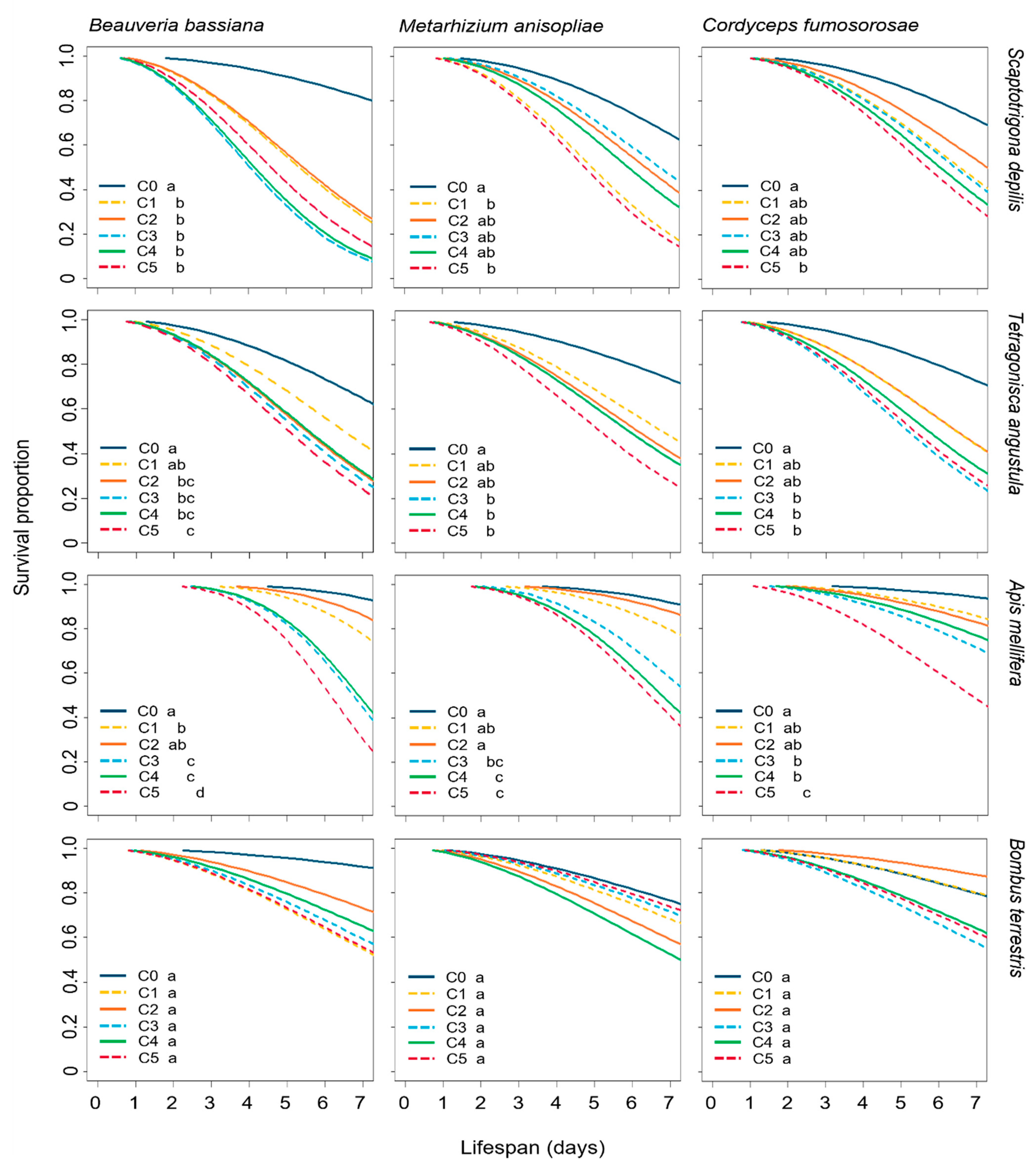
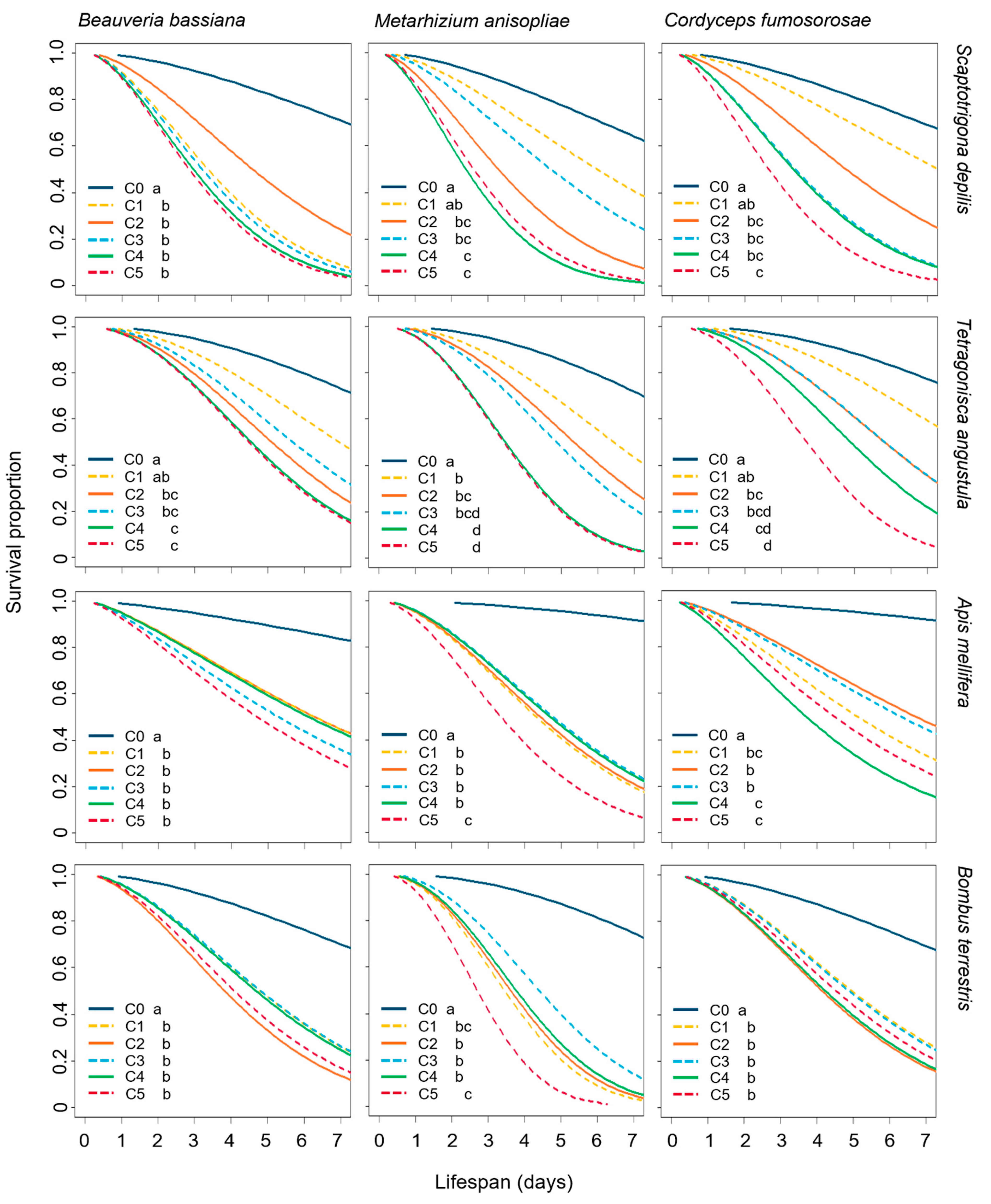
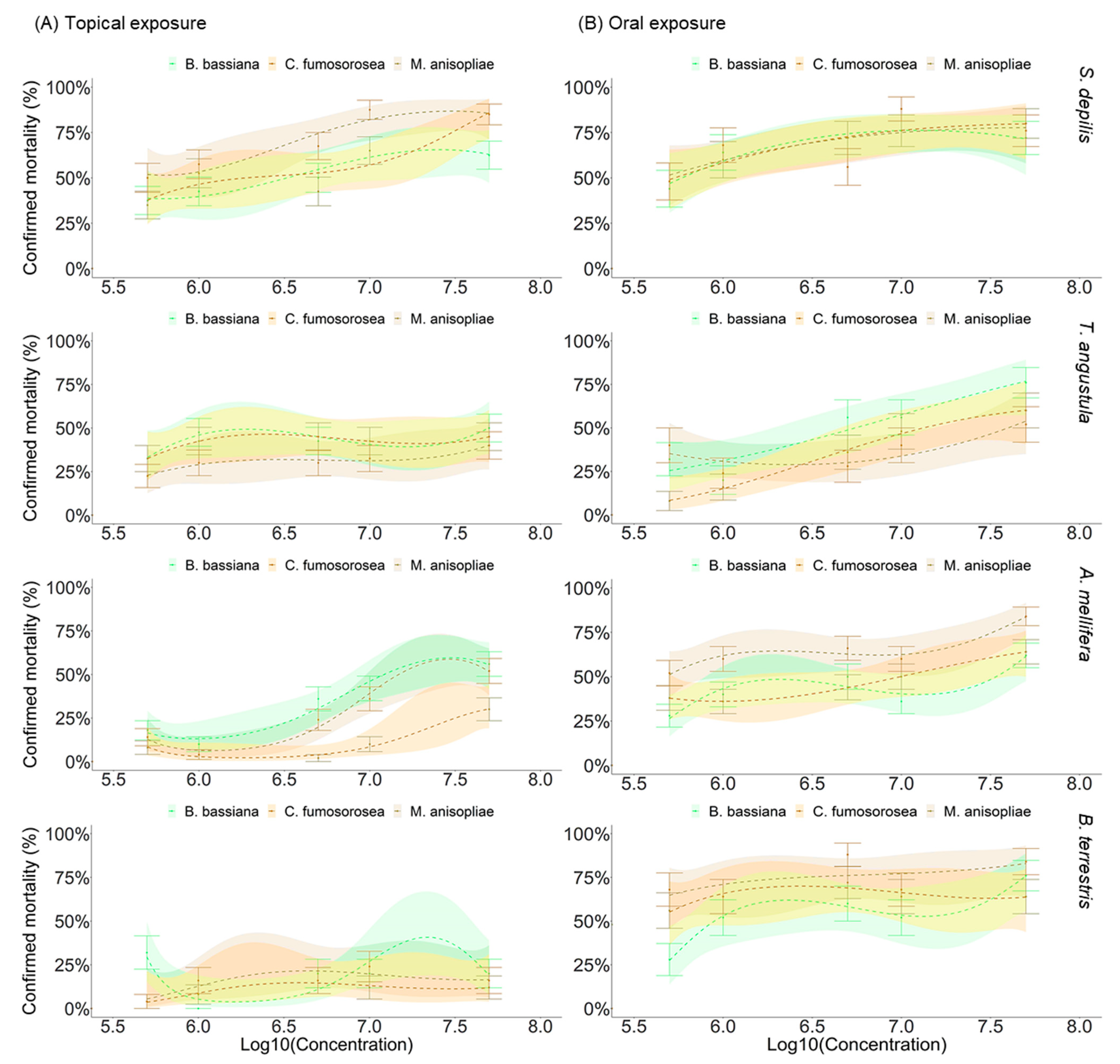
3.1. Effect of EF on the Survival of Bees

The survival effect from the interaction between EF, bee species, and method of exposure was significant (X2 = 18.01, df = 6, p = 0.0062). With regards to the different levels of susceptibility for each fungus among the bee species, we found that the fungus B. bassiana highly affected S. depilis survival when topically administrated (X2 = 23.291, df = 3, p < 0.0001). Yet, when B. bassiana was orally administrated, it decreased the survival of S. depilis, T. angustula, and B. terrestris (X2 = 9.959, df = 3, p = 0.0189). The fungus C. fumosorosea reduced both stingless bees and A. mellifera lifespan when topically applied (X2 = 16.672, df = 3, p = 0.0008), while B. terrestris was highly affected when C. fumosorosea was orally administrated (X2 = 17.949, df = 3, p = 0.0004). The EF M. anisopliae only significantly affected the T. angustula bees when topically applied (X2 = 18.732, df = 3, p = 0.0003), but when orally administrated, it affected the survival of S. depilis, T. angustula, and B. terrestris (X2 = 12.889, df = 3, p = 0.001) (Table 1).
Bee survival was significantly reduced after the exposure to the three EF and both application methods, except for B. terrestris treated by topical exposure, where the fungus did not considerably reduce bumble bee survival compared to the untreated control (Figure 1 and Figure 2).
For the stingless bee S. depilis, the workers had their survival significantly reduced when topically ( = 32.4, df= 5, p < 0.0001) and orally ( = 20.2, df = 5, p = 0.0001) exposed to B. bassiana by all the concentrations. Topically administrated M. anisopliae ( = 20.0, df = 5, p = 0.006) reduced survival with concentrations C1 (p = 0.0065) and C5 (p = 0.0018) meanwhile C. fumosorosea ( = 2.3, df = 5, p = 0.046) affected S. depilis survival with the highest C5 (p = 0.049) (Figure 1). Topically administrated B. bassiana was the most virulent (0.76 ± 0.43, p = 0.0002) to S. depilis workers (Figure S1). The orally administrated M. anisopliae ( = 47.8, df = 5, p < 0.0001) and C. fumosorosea ( = 43.6, df = 5, p < 0.0001) affected the survival from the concentration C2 (Ma; C2: p = 0.0001; C3: p = 0.0182; C4: p < 0.0001; C5: p < 0.0001) and (Cf; C2: p = 0.0094; C3: p = 0.0007; C4: p = 0.0006; C5, p < 0.0001; Figure 1).
The T. angustula workers had their survival significantly reduced when topically (Bb: = 17.6, df = 5, p = 0.0002; Ma: = 20.5, df = 5, p = 0.0003; Cf: = 20.5, df = 5, p = 0.0006) and orally exposed to EF (Bb: = 34.0, df = 5, p < 0.001; Ma: = 74.2, df = 5, p < 0.001; Cf: = 62.4, df = 5, p < 0.001) (Figure 1 and Figure 2).
After topical exposure, the survival of A. mellifera workers was also affected by B. bassiana ( = 27.46, df = 5, p < 0.0001), M. anisopliae ( = 3.931, df = 5, p < 0.0001) and C. fumosorosea ( = 0.532, df = 5, p < 0.0001) at the three highest concentrations (Figure 1). When the three EF were orally administrated, they reduced the workers’ survival at all the concentrations (Bb: = 32.1, df = 5, p < 0.0001; Ma: = 76.5, df = 5, p < 0.0001; Cf: = 56.8, df = 5, p < 0.0001) (Figure 2). By ingestion, M. anisopliae and B. bassiana were the most virulent EF for A. mellifera workers (Ma: 0.52 ± 0.50; Bb: 0.58 ± 0.49, p = 0.0054; Figure S1).
The survival curves of B. terrrestris workers topically treated with EF were similar to the controls (Bb: = 7.3, df = 5, p = 0.2; Ma: = 5, df = 5, p = 0.4; Cf: = 9.1, df = 5, p = 0.1; Figure 1). However, the survival was drastically reduced when workers were fed with B. bassiana ( = 14.5, df = 5, p < 0.0001), M. anisopliae ( = 57.1, df = 5, p < 0.0001) and C. fumosorosea ( = 17.9, df = 5, p < 0.0001). When topically administrated, B. bassiana was the most virulent EF for B. terrestris workers (0.34 ± 0.47, p = 0.0311), although M. anisopliae was the most virulent when orally offered (0.92 ± 0.27, p = 0.0006; Figure S1).

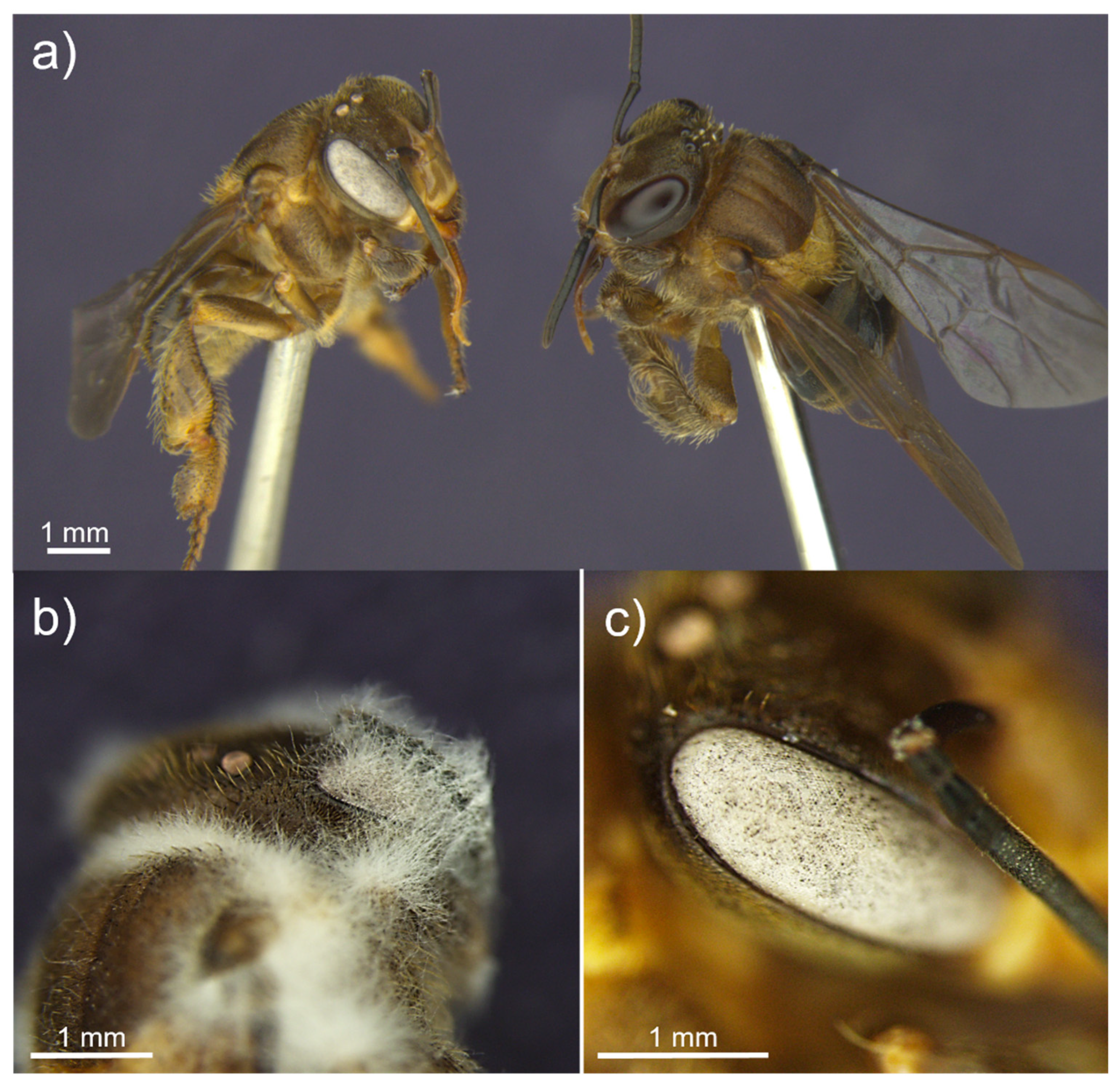
3.2. Sporulation of Entomopathgenic Fungi on Dead Bees

The method of fungal application had a significant effect on the fungal sporulation capacity on dead S. depilis (p = 0.0042), T. angustula (p = 0.02), A. mellifera (p = 0.0028), and B. terrestris (p < 0.0001) workers. In general, there was fungus outgrowth on dead corpses (Figure 3). Although the proportion of sporulated bees varied considerably among the EF, all three EF showed lower outgrowth proportion by topical application than by oral infection, especially at lower concentrations.
The controls did not present sporulation. There was a significantly higher proportion of sporulation in S. depilis workers when orally infected with B. bassiana, M. anisopliae, and C. fumosorosea (p < 0.0001) and when topically treated with M. anisopliae (p = 0.0172) and C. fumosorosea (p = 0.0011) when compared to the controls. Yet, sporulation had a marginally significant effect when B. bassiana was topically applied to workers (p = 0.0504; Figure 3). The fungal outgrowth on T. angustula and A. mellifera workers was significant for the three EF by both exposure methods (p < 0.01). However, T. angustula topically exposed by EFs showed a lower variation between concentrations, and A. mellifera-exposure showed an increased positive concentration response. The fungal sporulation on B. terrestris indicated that M. anisopliae (p = 0.133) and C. fumosorosea (p = 0.566), when topically applied, did not have any significant fungal outgrowth on workers, but it did have when B. bassiana was used (p = 0.0116). When these three fungi were ingested, sporulation was significantly higher than in the controls (p < 0.0001).

4. Discussion

Our study assesses the impact of entomopathogenic fungi on four social bee species at the individual level, at different concentrations, and under exposure methods. The effects of the fungal agents depended on the dose administered and the bee species for both exposure methods. The results revealed that all three entomopathogenic fungi, B. bassiana, M. anisopliae, and C. fumosorosea, significantly decreased T. angustula, S. depilis, and A. mellifera survival at different concentrations by both infection methods and lowered B. terrestris survival after oral exposure.
The stingless bees tended to be more affected by the three fungi. Both stingless bee species had a 50% decrease in survival when topically exposed, depending on the concentration, from the fourth to fifth day post-application. The fungus B. bassiana was most virulent to S. depilis followed by M. anisopliae and C. fumosorosea, whereas all three EF had a similar high dose response on T. angustula (Figure S1). In a similar study, which tested the same three EF species, but different isolates directly applied at a very high concentration (109 conidia mL−1), M. anisopliae affected T. angustula, Melipona beecheii, and S. mexicana workers’ mortality (94%, 53%, and 38.9%, respectively). On the other hand, B. bassiana and Cordyceps fumosorosea (previously known as Isaria fumosorosea) caused less than 30% mortality for all three species [32]. Melipona scutellaris has also shown to be somewhat susceptible to B. bassiana, with mortality over 56% when topically exposed to 1 × 10⁵ conidia mL−1 [33]. However, different isolates of B. bassiana and M. anisopliae, when directly applied in a high concentration (109 conidia mL−1), did not cause significant mortality in Meliponula ferruginea [45].
For A. mellifera and B. terrestris, the three EF species affected the worker’s survival, depending on the route of infection. When orally exposed, all EF species caused a significant effect on A. mellifera and B. terrestris survival, while topical exposure did not significantly affect B. terrestris. The three EF reduced A. mellifera workers’ survival by 50% from the sixth day after application when topically exposed, and from the third to fourth day when orally exposed. When Africanized A. mellifera workers were directly sprayed or orally fed with other isolates of B. bassiana and M. anisopliae (109 conidia mL−1), both reduced workers’ survival, with a faster response when bees were sprayed than orally fed [46]. Both EF also caused A. mellifera mortality above 50% after the fifth day by direct and oral exposure [28]. In this study, the EF B. bassiana and M. anisopliae, at 107 conidia mL−1, caused more than 50% mortality. Topical exposure of several B. bassiana and M. anisopliae strains at 107 conidia mL−1 on A. mellifera workers resulted in mortalities from 40% to 100% [47]. Significantly, deaths were seen in B. terrestris adults when topically exposed to 108 CFU mL−1 of B. bassiana- and C. fumososrosea-based products [48] and to B. terrestris, B. lucorum, and B. lapidarius when exposed to 108 conidia mL−1 of M. anisopliae [49]. The EF B. bassiana affected the lifespan of B. terrestris workers, decreasing by up to 4 days at 18 °C and by 13 days at 28 °C, by applying conidia over the whole worker body, without a specific concentration [50]. When newly emerged B. terrestris workers were exposed orally and topically to B. bassiana-based products, both routes caused mortality. However, contrary to our results, the topical method killed 92% of the workers meanwhile the oral killed less than 30% [51].
Regarding risk assessment of fungal-based products on social bees, we still face a lack of substantial knowledge about its lethal (and sub-lethal) effects. Since most of the published studies only focus on the Western honey bee, the potential effects of EF on non-Apis managed and wild species, which are an important and untapped group of crop pollinators, remain largely unexplored [36]. In this sense, understanding the potential effects that the same EF might cause on different bee species is essential to properly develop the regulation and use of biopesticides for pest management. Here, we show that the bee species responded differently to the EF and it might be due to their morphological traits, which can interfere with the effectiveness of a fungal infection. Our data show that the stingless bees were more affected by the entomopathogenic fungi when in direct contact than the honey bees and bumble bees. Due to their small body size, the same drop size of the fungal suspension resulted in a higher dose per total body area of the stingless bees than for the honey bees and bumble bees, even though the number of conidia/drops was the same. Additionally, the two studied stingless bee species are less hairy than A. mellifera and B. terrestris. During the infection process, EF conidia interact with their environment by electrostatic properties [52]. Conidial surfaces have a net negative charge that attracts them toward positively charged surfaces [53]. Since the bees have branched hairs [54] that have electrostatic forces [55], the conidia could be less likely to adhere to the cuticle, being attached to the hair and thus, not able to get in contact with the cuticle directly. Therefore, less hairy bees, like many stingless bee species, could be more susceptible to contact with fungal conidia and consequently suffer more from the infectious process when topically exposed.
Another critical point is the inter- and intra-specific variation in fungal virulence. Different isolates of EF species were used in the aforementioned studies, so the variable results might reflect different virulence of the fungal strains used, as it is known that virulence traits might vary within a single fungal species [56]. The mechanisms that led to different outcomes of EF virulence on social bee species and routes of infection were not examined in this study, but some possible speculations are suggested hereafter.
Regarding the EF’s virulence and dose used, the infection route showed to play a significant role in workers’ mortality. The cuticle represents the first point of contact and barrier between the fungus and the insect; however, it is known that the fungi can infect through other paths [17,57,58]. Indeed, the EF M. anisopliae caused higher mortality when orally offered, as seen in the study by [28] on Africanized honey bees. Furthermore, the EF M. anisopliae produces specific mucilage and adhesive proteins, increasing the facility to penetrate any part of the workers’ body [59,60], including the buccal parts. For example, the buccal cavity is a known site for M. anisopliae conidia to adhere, germinate on, and penetrate the sheep blowfly Lucilia cuprina [61], the pine weevil Hylobius pales [62], and the desert locusts Schistocerca gregaria [63]. Studies examining the adhesion of B. bassiana to surface substrata showed direct binding of conidia to hydrophobic surfaces [64], like most insect cuticles, which present a hydrophobic barrier rich in lipids [53,57]. Additionally, B. bassiana produces secondary metabolites acting as immunosuppressants, facilitating contact infection, such as beauvericin, bassianolide, oosporein, tenellin, bassiantin, and beauverolides [16].
Our results also showed that the different exposure methods affected the EF mortality differently in social bees, especially B. terrestris. When B. bassiana and M. anisopliae were orally offered to Africanized honey bees, with a dose of 108 conidia mL−1, it caused more significant mortalities (90% and 84%, respectively), compared to when topically applied (84% and 26%) [65]. While the conidia have to activate all the germination and infection pathways through cuticular layers by topical application, oral exposure may take a shortcut for the infection. When bees ingest the fungal suspension, the conidia get in direct contact with the mouthparts, which are softer and with multiple intersegmental parts more susceptible to fungal entrance [17,66]. The higher mortality for oral exposure in our bioassays might also be due to bee body size, especially for B. terrestris. As B. terrestris are our largest study species (19–22 mm length), followed by A. mellifera (12–15 mm), S. depilis (6 mm), and T. angustula (4–5 mm), they were probably capable of consuming a higher volume of the fungal solution compared to honey bees and stingless bees, and thus, a higher amount of conidia over 24 h. Moreover, social bees display prophylactic behaviors against pathogens, such as allogrooming, whereby co-workers clean each other [67]. This behavior could have caused the dispersion of the fungi among worker bodies or even ingestion while they were cleaning each other [68], increasing mortality rates. An important point to be highlighted is that oral infection is commonly used to define mycosis through ingestion, but with no definition of whether this infection process occurred in the mouthparts or the intestinal tract [62].
Interestingly, we visually detected a cue of the entomopathogenic fungal infection on the workers before any external development. Entomopathogenic fungi infect and multiply within the insect hosts as hyphae, and after the host dies, the fungus becomes visible by hyphal growth and subsequent sporulation externally [39]. We observed that when dead bees were kept at room temperature for hours or days before putting them in a humid chamber, some of them developed a change in eye color, presumably due to the fungal growth. Subsequent sporulation, initially throughout the eye, was confirmed after incubation in a humid chamber. This happened mainly for B. bassiana, characterized by white eyes (Figure 4), but it was also observed in workers infected by M. anisopliae, and C. fumosorosea. Whether this symptom could be turned into a possible visual cue for infected bees in the crop field stays open for further studies.
Pesticide risk assessments are complex and even more complicated in social insect species, such as bees, because the main goal is not to evaluate features of a single individual but of the colony as a whole [69]. In our experiments, control workers showed some mortality, probably because of the absence of social interactions [70]. Moreover, in laboratory conditions, the insects are maintained in a non-natural environment that causes stress and favors the development of the fungi. Thus, even though the social bee species tested in this study showed a significantly reduced survival, the laboratory assay does not represent the reality in the field [71,72]. Honeybee colonies exposed to Beauveria sp. and Metharhizium sp. to control varroa mites were not affected negatively but instead increased the numbers of adult bees and brood production [73,74]. The infection process might be prevented by the social immune response of the bees [70].
Different from chemical pesticides, entomopathogenic fungi are naturally occurring generalist pathogens widespread in the soil, plant surface, and as endophytes [75]. Thus, they co-exist with social bees in natural settings and not only when applied as biopesticides. Social colonies are composed of close relatives living at high densities with frequent contact, making them especially susceptible to spreading diseases. In addition to these features, social bees are highly resistant to generalist pathogens mainly due to several defense mechanisms at the colony level [76]. These mechanisms can include behavioral, genetic, physiological, spatial, or morphological defenses [76,77] as well as the symbiotic association with microorganisms that protect against microbial pathogens [78]. Hence, more realistic assays, including the whole colony and its symbiotic elements, are needed to evaluate the safety of entomopathogenic fungi-based biopesticides towards non-target insect species.
Standardized protocols exist for honey bees (recognized by the Organization for Economic Cooperation and Development (e.g., [79,80]), stingless bees [81,82], and bumble bees [83,84] for toxicological assessments with chemical pesticides. However, for fungus-based biopesticides, there is still a lack of such protocols, even for the requirement of new product registration tests [85,86].
On the other side, entomopathogenic fungi biopesticides are a reliable alternative to chemicals. In some cases, they are one of the few alternatives [5], so risk assessment tests should be evaluated carefully. Risk assessment for social bees should also consider the challenges in the field scenario, considering the behavioral traits of bee species, the target crop, the time, and the method of each biopesticide application. One example is the use of B. bassiana in coffee crops in Brazil. Bees are expected to visit coffee plants during the flowering season and to rarely visit them outside it [87]. Beauveria bassiana is often applied mainly after the flowering season, but some applications can be made before this season (a coffee farmer, personal communication). Thus, it is likely that the pollinators will not be affected by the fungus application. Still, careful evaluation could help decide the best timing for biopesticide application, considering the insect pests and the pollinators.

5. Conclusions

In conclusion, this study demonstrates that the recommended concentrations of B. bassiana, M. anisopliae, and C. fumosorosea and, in some cases, even lower concentrations can potentially reduce individuals’ survival of social bees in laboratory conditions. Even though laboratory studies are a valuable tool for first-tier risk assessment, allowing an accurate evaluation of colony fitness parameters using controlled concentrations under standardized conditions [88], colony and field risk assessments are further needed.

Supplementary Materials

The following supporting information can be downloaded at: https://www.mdpi.com/article/10.3390/microorganisms10091800/s1, Figure S1: Corrected mortality probability of Scaptotrigona depilis, Tetragonisca angustula, Apis mellifera and Bombus terrestris workers by the entomopathogenic fungi Beauveria bassiana, Metarhizium anisopliae and Cordyceps fumosorosea by topical and oral exposures.

Author Contributions

Conceptualization, M.O.G.L., D.A.A., I.D.J. and A.B.J.; methodology, M.O.G.L., D.A.A., I.D.J. and A.B.J.; validation, M.O.G.L.; formal analysis, M.O.G.L., J.B.M.; investigation, M.O.G.L.; resources, D.A.A., I.D.J. and A.B.J.; data curation, M.O.G.L. and J.B.M.; writing—original draft preparation, M.O.G.L.; writing—review and editing, M.O.G.L., D.A.A., A.L., I.D.J. and A.B.J.; supervision, D.A.A., I.D.J. and A.B.J.; funding acquisition, D.A.A., I.D.J. and A.B.J. All authors have read and agreed to the published version of the manuscript.

Funding

The Coordenação de Aperfeiçoamento de Pessoal de Nível Superior—Brasil (CAPES)—Finance Code 001 PROEX [Process 88882.328558/2019-01] and CAPES/PRINT—Edital 41/2017 [Process 88887.473776/2020-00] (to M.G.O.L) and the Conselho Nacional de Desenvolvimento Científico e Tecnológico (CNPq, grants 149154/2018-6 and 164743/2020-0 to D.A.A) provided financial support. Funding was also provided by the São Paulo Research Foundation (FAPESP, grant 2018/02317-5), as part of the SPARCBio (São Paulo Advanced Research Center for Biological Control).

Institutional Review Board Statement

This research has been approved by the University of São Paulo/ESALQ RESEARCH ENVIRONMENTAL ETHICS COMMISSION.

Informed Consent Statement

Not applicable.

Data Availability Statement

Not applicable.

Acknowledgments

We thank Lucas Zocca for technical support.

Conflicts of Interest

The authors declare no conflict of interest. The funders had no role in the design of the study; in the collection, analyses, or interpretation of data; in the writing of the manuscript; or in the decision to publish the results.

References

  1. Jaronski, S.T.; Mascarin, G.M. Mass production of fungal entomopathogens. In Microbial Control of Insects and Mite Pests, 1st ed.; Lacey, L.A., Ed.; Academic Press: Cambridge, MA, USA, 2017; pp. 141–155. [Google Scholar]
  2. Van Lenteren, J.C.; Bolckmans, K.; Kohl, J.; Ravensberg, W.J.; Urbaneja, A. Biological control using invertebrates and microorganisms: Plenty of new opportunities. BioControl 2018, 63, 39–59. [Google Scholar] [CrossRef]
  3. Oerke, E.C. Crop losses to pests. J. Agric. Sci. 2006, 144, 31–43. [Google Scholar] [CrossRef]
  4. Arthurs, S.; Dara, S.K. Microbial biopesticides for invertebrate pests and their markets in the United States. J. Invertebr. Pathol. 2018, 165, 13–21. [Google Scholar] [CrossRef] [PubMed]
  5. Mascarin, G.M.; Lopes, G.B.; Jr, I.D.; Fernandes, E.K.K.; Luz, C.; Faria, M. Current status and perspectives of fungal entomopathogens used for microbial control of arthropod pests in Brazil. J. Invertebr. Pathol. 2019, 165, 46–53. [Google Scholar] [CrossRef]
  6. Li, Z.; Alves, S.B.; Roberts, D.W.; Fan, M.; Jr, I.D.; Tang, J.; Lopes, R.B.; Faria, M.; Rangel, D.E. Biological control of insects in Brazil and China: History, current programs and reasons for their successes using entomopathogenic fungi. Bioc. Sci. Techn. 2010, 2, 117–136. [Google Scholar] [CrossRef]
  7. Lacey, L.A.; Grzywacz, D.; Shapiro-Ilan, D.I.; Frutos, R.; Brownbridge, M.; Goettel, M.S. Insect pathogens as biological control agents: Back to the future. J. Invertebr. Pathol. 2015, 132, 1–41. [Google Scholar] [CrossRef]
  8. Wraight, S.P.; Galaini-Wraight, S.; Howes, R.L.; Castrillo, L.A.; Griggs, M.H.; Carruthers, R.I.; Smith, R.H.; Matsumoto, T.K.; Keith, L.M. Efficacy of Beauveria bassiana strain GHA spray applications against coffee berry borer Hypothenemus hampei on Hawai‘i Island. Biol. Control 2021, 161, 104587. [Google Scholar] [CrossRef]
  9. Ausique, J.J.S.; D’Alessandro, C.P.; Conceschi, M.R.; Mascarin, G.M.; Delalibera Jr, I. Efficacy of entomopathogenic fungi against adult Diaphorina citri from laboratory to field applications. J. Pest Sci. 2017, 90, 947–960. [Google Scholar] [CrossRef]
  10. Castro, T.; Mayerhofer, J.; Enkerli, J.; Eilenberg, J.; Meyling, N.V.; de Andrade Moral, R.; Demetrio, D.G.B.; Jr, I.D. Persistence of Brazilian isolates of the entomopathogenic fungi Metarhizium anisopliae and M. robertsii in strawberry crop soil after soil drench application. Agric. Ecosyst. Environ. 2016, 233, 361–369. [Google Scholar] [CrossRef]
  11. Kapongo, J.P.; Shipp, L.; Kevan, P.; Sutton, J.C. Co-vectoring of Beauveria bassiana and Clonostachys rosea by bumble bees (Bombus impatiens) for control of insect pests and suppression of grey mould in greenhouse tomato and sweet pepper. Biol. Control 2008, 46, 508–514. [Google Scholar] [CrossRef]
  12. Klein, A.M.; Vaissière, B.E.; Cane, J.H.; Steffan-Dewenter, I.; Cunningham, S.A.; Kremen, C.; Tscharntke, T. Importance of pollinators in changing landscapes for world crops. Proc. R. Soc. B 2007, 274, 303–313. [Google Scholar] [CrossRef] [PubMed]
  13. Egan, P.A.; Dicks, L.V.; Hokkanen, H.M.; Stenberg, J.A. Delivering integrated pest and pollinator management (IPPM). Trends Plant Sci. 2020, 25, 577–589. [Google Scholar] [CrossRef] [PubMed]
  14. Martínez-Salinas, A.; Chain-Guadarrama, A.; Aristizábal, N.; Vilchez-Mendoza, S.; Cerda, R.; Ricketts, T.H. Interacting pest control and pollination services in coffee systems. Proc. Natl. Acad. Sci. USA 2022, 119, e2119959119. [Google Scholar] [CrossRef] [PubMed]
  15. Mascarin, G.M.; Jaronski, S.T. The production and uses of Beauveria bassiana as a microbial insecticide. World J. Microb. Biotechnol. 2016, 32, 177. [Google Scholar] [CrossRef] [PubMed]
  16. Pedrini, N. Molecular interactions between entomopathogenic fungi (Hypocreales) and their insect host: Perspectives from stressful cuticle and hemolymph battlefields and the potential of dual RNA sequencing for future studies. Fungal Biol. 2018, 122, 538–545. [Google Scholar] [CrossRef]
  17. Mannino, M.C.; Huarte-Bonnet, C.; Davyt-Colo, B.; Pedrini, N. Is the insect cuticle the only entry gate for fungal infection? Insights into alternative modes of action of entomopathogenic fungi. J. Fungi 2019, 5, 33. [Google Scholar] [CrossRef]
  18. Vestergaard, S.; Cherry, A.; Keller, S.; Goettel, M. Safety of hyphomycete fungi as microbial control agents. In Environmental Impacts of Microbial Insecticides, 1st ed.; Hokkanen, H.M.T., Hajek, A.E., Eds.; Kluwer Academic Publishers: Dordrecht, The Netherlands, 2003; pp. 35–62. [Google Scholar]
  19. Garrido-Jurado, I.; Ruano, F.; Campos, M.; Quesada-Moraga, E. Effects of soil treatments with entomopathogenic fungi on soil dwelling non-target arthropods at a commercial olive orchard. Biol. Control 2011, 59, 239–244. [Google Scholar] [CrossRef]
  20. Shaw, D.E. The incidental collection of fungal spores by bees and the collection of spores in lieu of pollen. Bee World 1990, 71, 158–176. [Google Scholar] [CrossRef]
  21. Potts, S.G.; Imperatriz-Fonseca, V.L.; Ngo, H.T.; Aizen, M.A.; Biesmeijer, J.C.; Breeze, T.D.; Dicks, L.V.; Garibaldi, L.A.; Hill, R.; Settele, J.; et al. Safeguarding pollinators and their values to human well-being. Nature 2016, 540, 220–229. [Google Scholar] [CrossRef]
  22. Osterman, J.; Aizen, M.A.; Biesmeijer, J.C.; Bosch, J.; Howlett, B.G.; Inouye, D.W.; Jung, C.; Martins, D.J.; Medel, R.; Pauw, A.; et al. Global trends in the number and diversity of managed pollinator species. Agric. Ecosyst. Environ. 2021, 322, 107653. [Google Scholar] [CrossRef]
  23. Klatt, B.K.; Holzschuh, A.; Westphal, C.; Clough, Y.; Smit, I.; Pawelzik, E.; Tscharntke, T. Bee pollination improves crop quality, shelf life and commercial value. Proc. R. Soc. B 2014, 281, 20132440. [Google Scholar] [CrossRef] [PubMed]
  24. Nowell, D.; Maynard, G.V. International guidelines for the export, shipment, import, and release of biological control agents and other beneficial organisms (international standard for phytosanitary measures no. 3). In Proceedings of the Second International Symposium on Biological Control of Arthropods, Davos, Switzerland, 12–16 September 2005. [Google Scholar]
  25. Mazra’awi, M.S.A.; Shipp, J.L.; Broadbent, A.B.; Kevan, P.G. Dissemination of Beauveria bassiana by honey bees (Hymenoptera: Apidae) for control of tarnished plant bug (Hemiptera: Miridae) on canola. Environ. Entomol. 2006, 35, 1569–1577. [Google Scholar] [CrossRef]
  26. Butt, T.M.; Carreck, N.L.; Ibrahim, L.; Williams, I.H. Honey bee mediated infection of pollen beetle (Meligethes spp.) by the insect-pathogenic fungus, Metarhizium Anisopliae. Biocontrol Sci. Technol. 2010, 8, 533–538. [Google Scholar] [CrossRef]
  27. Carlesso, D.; Smargiassi, S.; Sassoli, L.; Cappa, F.; Cervo, R.; Baracchi, D. Exposure to a biopesticide interferes with sucrose responsiveness and learning in honey bees. Sci. Rep. 2020, 10, 19929. [Google Scholar] [CrossRef] [PubMed]
  28. Colombo, F.C.; Maciel, R.M.A.; Abati, R.; Raulino-Domanski, F.; Longhi, S.J.; Costa-Maia, F.M.; Vismara, E.S.; Lozano, E.R.; Potrich, M. Do Beauveria bassiana and Metarhizium anisopliae affect worker survival and the production of Africanized Apis mellifera queens? J. Apic. Res. 2021, 60, 260–269. [Google Scholar] [CrossRef]
  29. Peng, G.; Tong, S.; Zeng, D.; Xiaa, Y.; Feng, M. Colony heating protects honey bee populations from a risk of contact with wide-spectrum Beauveria bassiana insecticides applied in the field. Pest Manag. Sci. 2020, 76, 2627–2634. [Google Scholar] [CrossRef]
  30. Sinia, A.; Guzman-Novoa, E. Evaluation of the entomopathogenic fungi Beauveria bassiana GHA and Metarhizium anisopliae UAMH 9198 alone or in combination with thymol for the control of Varroa destructor in honey bee (Apis mellifera) colonies. J. Apic. Res. 2018, 57, 308–316. [Google Scholar] [CrossRef]
  31. Shipp, J.L.; Kapongo, J.P.; Park, H.; Kevan, P. Effect of bee-vectored Beauveria bassiana on greenhouse beneficials under greenhouse cage conditions. Biol. Control 2012, 63, 135–142. [Google Scholar] [CrossRef]
  32. Toledo-Hernandez, R.A.; Ruiz-Toledo, J.; Toledo, J.; Sanchez, D. Effect of three entomopathogenic fungi on three species of stingless bees (Hymenoptera: Apidae) under laboratory conditions. J. Econ. Entomol. 2016, 109, 1015–1019. [Google Scholar] [CrossRef]
  33. Conceição, P.d.J.; Neves, C.M.L.; Sodré, G.S.; Carvalho, C.A.L.; Souza, A.V.; Ribeiro, G.S.; Pereira, R.C. Susceptibility of Melipona scutellaris Latreille, 1811 (Hymenoptera: Apidae) to Beauveria bassiana (Bals.) Vuill. Sociobiology 2014, 61, 184–188. [Google Scholar] [CrossRef]
  34. James, R.R.; McGuire, M.R.; Leland, J.E. Susceptibility of adult alfalfa leafcutting bees1 and honey bees2 to a microbial control agent, Beauveria bassiana. Southwest. Entomol. 2012, 37, 13–21. [Google Scholar] [CrossRef]
  35. Erler, S.; Eckert, J.H.; Steinert, M.; Alkassab, A.T. Impact of microorganisms and entomopathogenic nematodes used for plant protection on solitary and social bee pollinators: Host range, specificity, pathogenicity, toxicity, and effects of experimental parameters. Environ. Pollut. 2022, 302, 119051. [Google Scholar] [CrossRef]
  36. Cappa, F.; Baracchi, D.; Cervo, R. Biopesticides and insect pollinators: Detrimental effects, outdated guidelines, and future directions. Sci. Total Environ. 2022, 837, 155714. [Google Scholar] [CrossRef] [PubMed]
  37. Jacob, C.R.O.; Soares, H.M.; Carvalho, S.M.; Nocelli, R.C.F.; Malaspina, O. Acute toxicity of fipronil to the stingless bee Scaptotrigona postica Latreille. Bul. Environ. Contam. Toxic. 2013, 90, 69–72. [Google Scholar] [CrossRef] [PubMed]
  38. Del Sarto, M.C.L.; Oliveira, E.E.; Guedes, R.N.C.; Campos, L.A.O. Differential insecticide susceptibility of the Neotropical stingless bee Melipona quadrifasciata and the honey bee Apis mellifera. Apidologie 2014, 45, 626–636. [Google Scholar] [CrossRef]
  39. Alves, S.B. Entomopathogenic fungi. In Controle Microbiano de Insetos, 2nd ed.; Alves, S.B., Ed.; Fundação de Estudos Agrários Luiz de Queiroz (FEALQ): Piracicaba, Brazil, 1998; pp. 289–370. [Google Scholar]
  40. Kassambara, A.; Kosinski, M.; Biecek, P.; Fabian, S. survminer: R Package Version 0.4, 8, 556, Drawing Survival Curves using “ggplot2.”, 2020. Available online: https://CRAN.R-project.org/package=survminer (accessed on 21 April 2021).
  41. Therneau, T. R package version 3.2-3, A Package for Survival Analysis in R., [Computer software], Mayo Clinic, Rochester, MN, USA, 2020. Available online: https://CRAN.R-project.Org/package=survival (accessed on 21 April 2021).
  42. Takakura, K.I. Bayesian estimation for the effectiveness of pesticides and repellents. J. Econ. Entomol. 2012, 105, 1856–1862. [Google Scholar] [CrossRef]
  43. Hothorn, T.; Bretz, F.; Westfall, P.; Heiberger, R.M.; Schuetzenmeister, A.; Scheibe, S.; Hothorn, M.T. Package ‘Multcomp’, Simultaneous Inference in General Parametric Models; Project for Statistical Computing: Vienna, Austria, 2016. [Google Scholar]
  44. Moral, R.A.; Hinde, J.; Demétrio, C.G.B. Half-normal plots and overdispersed models in R: The hnp package. J. Stat. Softw. 2017, 81, 1–23. [Google Scholar] [CrossRef]
  45. Omuse, E.R.; Niassy, S.; Kiatoko, N.; Lattorff, H.M.G.; Wagacha, J.M.; Dubois, T. A fungal-based pesticide does not harm pollination service provided by the African stingless bee Meliponula ferruginea on cucumber (Cucumis sativus). Apidologie 2022, 53, 28. [Google Scholar] [CrossRef]
  46. Potrich, M.; Silva, R.T.L.; Maia, F.M.C.; Lozano, E.R.; Rossi, R.M.; Colombo, F.C.; Tedesco, F.G.; Gouvea, A. Effect of entomopathogens on Africanized Apis mellifera L. (Hymenoptera: Apidae). Revta. Bras. Entomol. 2018, 62, 23–28. [Google Scholar] [CrossRef]
  47. Espinosa-Ortiz, G.E.; Lara-Reyna, J.; Otero-Colina, G.; Alatorre-Rosas, R.; Valdez-Carrasco, J. Susceptibility of larval, pupal and adult honey bees to isolates of Beauveria bassiana (Bals.) Vuill., Metarhizium anisopliae (Sorokin) and Paecilomyces fumosoroseus (Wize). Interciencia 2011, 36, 148–152. [Google Scholar]
  48. Demirozer, O.; Uzun, A.; Gosterit, A. Lethal and sublethal effects of different biopesticides on Bombus terrestris (Hymenoptera: Apidae). Apidologie 2022, 53, 24. [Google Scholar] [CrossRef]
  49. Hokkanen, H.M.T.; Zeng, Q.Q.; Menzler-Hokkanen, I. Assessing the impacts of Metarhizium and Beauveria on bumble bees. In Environmental Impacts of Microbial Insecticides, 1st ed.; Hokkanen, H.M.T., Hajek, A.E., Eds.; Kluwer Academic Publishers: Dordrecht, The Netherlands, 2003; pp. 63–72. [Google Scholar]
  50. Karise, R.; Muljar, R.; Smagghe, G.; Kaart, T.; Kuusik, A.; Dreyersdorff, G.; Williams, I.H.; Mand, M. Sublethal effects of kaolin and the biopesticides Prestop-Mix and BotaniGard on metabolic rate, water loss and longevity in bumble bees (Bombus terrestris). J. Pest Sci. 2016, 89, 171–178. [Google Scholar] [CrossRef]
  51. Mommaerts, V.; Sterk, G.; Hoffmann, L.; Smagghe, G. A laboratory evaluation to determine the compatibility of microbiological control agents with the pollinator Bombus terrestris. Pest Manag. Sci. 2009, 65, 949–955. [Google Scholar] [CrossRef] [PubMed]
  52. Jackson, M.A.; Dunlap, C.A.; Jaronski, S.T. Ecological considerations in producing and formulating fungal entomopathogens for use in insect biocontrol. BioControl 2010, 55, 129–145. [Google Scholar] [CrossRef]
  53. Boucias, D.G.; Pendland, J.C.; Latge, J.P. Nonspecific factors involved in attachment of entomopathogenic Deuteromycetes to host insect cuticle. Appl. Environ. Microbiol. 1988, 54, 1795–1805. [Google Scholar] [CrossRef]
  54. Michener, C.D. The Bees of the World; The John Hopkins University Press: Baltimore, MD, USA, 2000; p. 972. [Google Scholar]
  55. Vaknin, Y.; Gan-Mor, S.; Bechar, A.; Ronen, B.; Eisikowitch, D. The role of electrostatic forces in pollination. Pollen Pollinat. 2000, 222, 133–142. [Google Scholar]
  56. Maistrou, S.; Natsopoulou, M.E.; Jensen, A.B.; Meyling, N.V. Virulence traits within a community of the fungal entomopathogen Beauveria: Associations with abundance and distribution. Fungal Ecol. 2020, 48, 100992. [Google Scholar] [CrossRef]
  57. Ortiz-Urquiza, A.; Keyhani, N.O. Action on the surface: Entomopathogenic fungi versus the insect cuticle. Insects 2013, 4, 357–374. [Google Scholar] [CrossRef]
  58. Boomsma, J.J.; Jensen, A.B.; Meyling, N.V.; Eilenberg, J. Evolutionary interaction networks of insect pathogenic fungi. Annu. Rev. Entomol. 2014, 59, 467–485. [Google Scholar] [CrossRef]
  59. Roberts, D.W.; Leger, R.J.S. Metarhizium spp., cosmopolitan insect-pathogenic fungi: Mycological aspects. Adv. Appl. Microbiol. 2004, 54, 1–70. [Google Scholar]
  60. Wang, C.; Leger, R.J.S. The MAD1 adhesin of Metarhizium anisopliae links adhesion with blastospore production and virulence to insects, and the MAD2 adhesin enables attachment to plants. Eukaryot. Cell 2007, 6, 808–816. [Google Scholar] [CrossRef] [PubMed]
  61. Leemon, D.M.; Jonsson, N.N. Comparative studies on the invasion of cattle ticks (Rhipicephalus (Boophilus) microplus) and sheep blowflies (Lucilia cuprina) by Metarhizium anisopliae (Sorokin). J. Invertebr. Pathol. 2012, 109, 248–259. [Google Scholar] [CrossRef]
  62. Schabel, S. Oral infection of Hylobius pales by Metarhizium anisopliae. J. Invertebr. Pathol. 1976, 383, 377–383. [Google Scholar] [CrossRef]
  63. Dillon, R.J.; Charnley, A.K. Invasion of the pathogenic fungus Metarhizium anisopliae through the guts of germfree desert locusts. Schistocerca Gregaria. Mycopathol. 1986, 96, 59–66. [Google Scholar] [CrossRef]
  64. Holder, D.J.; Keyhani, N.O. Adhesion of the entomopathogenic fungus Beauveria (Cordyceps) bassiana to substrata. Appl. Environ. Microbiol. 2005, 71, 5260–5266. [Google Scholar] [CrossRef] [PubMed]
  65. Alves, S.B.; Marchini, L.C.; Pereira, R.M.; Baumgratz, L.L. Effects of some insect pathogens on the Africanized honey bee, Apis mellifera L. (Hym., Apidae). J. Appl. Entomol. 1996, 120, 559–564. [Google Scholar] [CrossRef]
  66. Amnuaykanjanasin, A.; Jirakkakul, J.; Panyasiri, C.; Panyarakkit, P.; Nounurai, P.; Chantasingh, D.; Eurwilaichitr, L.; Cheevadhanarak, S.; Tanticharoen, M. Infection and colonization of tissues of the aphid Myzus persicae and cassava mealybug Phenacoccus manihoti by the fungus Beauveria bassiana. BioControl 2013, 58, 379–391. [Google Scholar] [CrossRef]
  67. Ugelvig, L.V.; Cremer, S. Social prophylaxis: Group interaction promotes collective immunity in ant colonies. Curr. Biol. 2007, 17, 1967–1971. [Google Scholar] [CrossRef]
  68. Brighenti, D.M.; Carvalho, C.F.; Carvalho, G.A.; Brighenti, C.R.G.; Carvalho, S.M. Bioatividade do Bacillus thuringiensis var. kurstaki (Berliner, 1915) para adultos de Apis mellifera Linnaeus, 1758 (Hymenoptera: Apidae). Ciênc. E Agrotecnol. 2007, 31, 279–289. [Google Scholar] [CrossRef]
  69. Blacquiere, T.; Smagghe, G.; Van Gestel, C.A.; Mommaerts, V. Neonicotinoids in bees: A review on concentrations, side-effects and risk assessment. Ecotoxic. 2012, 21, 973–992. [Google Scholar] [CrossRef]
  70. Cremer, S.; Pull, C.D.; Fürst, M.A. Social Immunity: Emergence and evolution of colony-level disease protection. Annu. Rev. Entomol. 2018, 63, 105–123. [Google Scholar] [CrossRef]
  71. Goulson, D.; Nicholls, E.; Botías, C.; Rotheray, E.L. Bee declines driven by combined stress from parasites, pesticides, and lack of flowers. Science 2015, 347, 1255957. [Google Scholar] [CrossRef] [PubMed]
  72. Tosi, S.; Nieh, J.C.; Sgolastra, F.; Cabbri, R.; Medrzycki, P. Neonicotinoid pesticides and nutritional stress synergistically reduce survival in honey bees. Proc. R. Soc. B 2017, 284, 20171711. [Google Scholar] [CrossRef] [Green Version]
  73. Meikle, M.G.; Mercadier, G.; Holst, N.; Nansen, C.; Girod, V. Impact of a treatment of Beauveria bassiana (Deuteromycota: Hyphomycetes) on honeybee (Apis mellifera) colony health and on Varroa destructor mites (Acari: Varroidae). Apidologie 2008, 39, 247–259. [Google Scholar] [CrossRef]
  74. Kanga, L.H.; Adamczyk, J.; Patt, J.; Gracia, C.; Cascino, J. Development of a user-friendly delivery method for the fungus Metarhizium anisopliae to control the ectoparasitic mite Varroa destructor in honey bee, Apis mellifera, colonies. Exp. Appl. Acarol. 2010, 52, 327–342. [Google Scholar] [CrossRef]
  75. Rajula, J.; Karthi, S.; Mumba, S.; Pittarate, S.; Thungrabeab, M.; Krutmuang, P. Entomopathogenic fungi. In Recent Advancement in Microbial Biotechnology: Agricultural and Industrial Approach, 2nd ed.; Mandal, S., Passari, A.K., Eds.; Elsevier: London, UK, 2021; pp. 72–75. [Google Scholar]
  76. Cremer, S.; Armitage, S.A.; Schmid-Hempel, P. Social immunity. Curr. Biol. 2007, 17, 693–702. [Google Scholar] [CrossRef]
  77. Almeida, F.C.R.; Magalhães, D.M.; Favaris, A.P.; Rodríguez, J.; Azevedo, K.E.X.; Bento, J.M.S.; Alves, D.A. Side effects of a fungus-based biopesticide on stingless bee guarding behaviour. Chemosphere 2022, 287, 132147. [Google Scholar] [CrossRef]
  78. Menegatti, C.; Da Paixão Melo, W.G.; Carrão, D.B.; Oliveira, A.R.M.; Nascimento, F.S.; Lopes, N.P.; Pupo, M.T. Paenibacillus polymyxa associated with the stingless bee Melipona scutellaris produces antimicrobial compounds against entomopathogens. J. Chem. Ecol. 2018, 44, 1158–1169. [Google Scholar] [CrossRef] [PubMed]
  79. OECD. Test No. 213: Honeybees, Acute Oral Toxicity Test. Available online: https://www.oecd-ilibrary.org/environment/test-no-213-honeybees-acute-oral-toxicity-test_9789264070165-en (accessed on 10 July 2022).
  80. OECD. Test No. 237: Honey bee (Apis mellifera) Larval Toxicity Test, Single EXPOSURE. Available online: https://www.oecd-ilibrary.org/environment/test-no-237-honey-bee-apis-mellifera-larval-toxicity-test-single-exposure_9789264203723-en (accessed on 10 July 2022).
  81. Botina, L.L.; Bernardes, R.C.; Barbosa, W.F.; Lima, M.A.P.; Guedes, R.N.C.; Martins, G.F. Toxicological assessments of agrochemical effects on stingless bees (Apidae, Meliponini). MethodsX 2020, 7, 100906. [Google Scholar] [CrossRef]
  82. Cham, K.O.; Nocelli, R.C.; Borges, L.O.; Viana-Silva, F.E.C.; Tonelli, C.A.M.; Malaspina, O.; Menezes, C.; Rosa-Fontana, A.S.; Blochtein, B.; Freitas, B.M.; et al. Pesticide exposure assessment paradigm for stingless bees. Environ. Entomol. 2019, 48, 36–48. [Google Scholar] [CrossRef]
  83. Cabrera, A.R.; Almanza, M.T.; Cutler, G.C.; Fischer, D.L.; Hinarejos, S.; Lewis, G.; Nigro, D.; Olmstead, A.; Overmyer, J.; Potter, D.A.; et al. Initial recommendations for higher-tier risk assessment protocols for bumble bees, Bombus spp. (Hymenoptera: Apidae). Integr. Environ. Assess. Manag. 2016, 12, 222–229. [Google Scholar] [CrossRef] [PubMed]
  84. Klinger, E.G.; Camp, A.A.; Strange, J.P.; Cox-Foster, D.; Lehmann, D.M. Bombus (Hymenoptera: Apidae) microcolonies as a tool for biological understanding and pesticide risk assessment. Environ. Entomol. 2019, 48, 1249–1259. [Google Scholar] [CrossRef] [PubMed]
  85. Reinbacher, L.; Bacher, S.; Praprotnik, E.; Grabenweger, G. Standard non-target tests for risk assessment of plant protection products are unsuitable for entomopathogenic fungi—A proposal for a new protocol. J. Soils Sediments 2021, 21, 2357–2368. [Google Scholar] [CrossRef]
  86. Köhl, J.; Booij, K.; Kolnaar, R.; Ravensberg, W.J. Ecological arguments to reconsider data requirements regarding the environmental fate of microbial biocontrol agents in the registration procedure in the European Union. BioControl 2019, 64, 469–487. [Google Scholar] [CrossRef]
  87. Machado, A.C.P.; Barônio, G.J.; Oliveira, F.F.; Garcia, C.T.; Rech, A.R. Does a coffee plantation host potential pollinators when it is not flowering? Bee distribution in an agricultural landscape with high biological diversity in the Brazilian Campo Rupestre. J. Sci. Food Agri. 2021, 101, 2345–2354. [Google Scholar] [CrossRef] [PubMed]
  88. Van Oystaeyen, A.; Klatt, B.K.; Petit, C.; Lenaerts, N.; Wäckers, F. Short-term lab assessments and microcolonies are insufficient for the risk assessment of insecticides for bees. Chemosphere 2021, 273, 128518. [Google Scholar] [CrossRef]
Figure 1. Survival proportion of workers (Scaptotrigona depilis (n = 150), Tetragonisca angustula (n = 240), Apis mellifera (n = 300) and Bombus terrestris (n = 150)) to topical application of three entomopathogenic fungi, Beauveria bassiana, Metarhizium anisopliae and Cordyceps fumosorosea. Concentrations: control (C0), 5 × 10⁵ conidia mL−1 (C1), 1 × 10⁶ conidia mL−1 (C2), 5 × 10⁶ conidia mL−1 (C3), 1 × 10⁷ conidia mL−1 (C4), 5 × 10⁷ conidia/mL−1 (C5). Concentrations with different letters are significantly different (p < 0.05).
Microorganisms 10 01800 g001
Figure 2. Survival proportion of Scaptotrigona depilis (n = 150 workers), Tetragonisca angustula (n = 240 workers), Apis mellifera (n = 300 workers) and Bombus terrestris (n = 150 workers) to oral exposure of three entomopathogenic fungi, Beauveria bassiana, Metarhizium anisopliae and Cordyceps fumosorosea. Concentrations: control (C0), 5 × 10⁵ conidia mL−1 (C1), 1 × 10⁶ conidia mL−1 (C2), 5 × 10⁶ conidia mL−1 (C3), 1 × 10⁷ conidia mL−1 (C4), 5 × 10⁷ conidia/mL−1 (C5). Concentrations with different letters are significantly different (p < 0.05).
Microorganisms 10 01800 g002
Figure 3. Mean (±SE) percentage of fungal outgrowth curve in dead Scaptotrigona depilis, Tetragonisca angustula, Apis mellifera, and Bombus terrestris workers topically (A) and orally (B) exposed to the fungi Beauveria bassiana, Metarhizium anisopliae, and Cordyceps fumosorosea. The concentrations (conidia mL−1) were transformed by regression Log10: 4.0 = 5 × 10⁵, 4.5 = 1 × 10⁶, 5.0 = 5 × 10⁶, 5.5 = 1 × 10⁷, 6.0 = 5 × 10⁷. Fitted to generalized linear model (GLM) with binomial distribution.
Microorganisms 10 01800 g003
Figure 4. Comparison of the eye color change of a B. bassiana-infected (left) and non-infected (right) Scaptotrigona depilis (a). Detail of a S. depilis dead body with B. bassiana outgrowth (b). Detail of the B. bassiana growth inside S. depilis eye (c).
Microorganisms 10 01800 g004
Table 1. Susceptibility of Scaptotrigona depilis, Tetragonisca angustula, Apis mellifera, and Bombus terrestris workers to the entomopathogenic fungi Beauveria bassiana, Cordyceps fumosorosea, and Metarhizium anisopliae for each exposure method (topical or oral). Mean mortality fitted to a generalized linear model (GLM) with binomial distribution, Tukey test.
MethodFungiBee Speciesp-Value
S. depilisT. angustulaA. melliferaB. terrestris
topicalB. bassiana0.76 a0.48 b0.36 b0.34 b<0.0001
C. fumosorosea0.48 a0.48 a0.30 ab0.16 b0.0008
M. anisopliae0.38 b0.50 a0.22 bc0.14 c0.0003
oralB. bassiana0.80 a0.58 ab0.50 b0.66 ab0.0189
C. fumosorosea0.66 b0.70 b0.58 b0.92 a0.0004
M. anisopliae0.64 a 0.58 a0.28 b0.64 a0.0003
Means with different letters within a row are significantly different (p < 0.05).
Publisher’s Note: MDPI stays neutral with regard to jurisdictional claims in published maps and institutional affiliations.

