Article

Potential for Biological Control of Pythium schmitthenneri Root Rot Disease of Olive Trees (Olea europaea L.) by Antagonistic Bacteria

1
Phytopathology Unit, Department of Plant Protection, Ecole Nationale d’Agriculture de Meknès, Km 10, Rte Haj Kaddour, BP S/40, Meknès 50001, Morocco
2
Laboratory of Functional Ecology and Environmental Engineering, Sidi Mohamed Ben Abdellah University, P.O. Box 2202, Route d’Imouzzer, Fez 30000, Morocco
3
Plant Protection Laboratory, Regional Center of Agricultural Research of Oujda, National Institute of Agricultural Research, Avenue Mohamed VI, BP428 Oujda, Oujda 60000, Morocco
4
Unité de Recherche Résistance Induite et Bio-Protection des Plantes-EA 4707, Université de Reims Champagne-Ardenne, 51100 Reims, France
*
Authors to whom correspondence should be addressed.
Microorganisms 2022, 10(8), 1635; https://doi.org/10.3390/microorganisms10081635
Submission received: 11 July 2022 / Revised: 11 August 2022 / Accepted: 11 August 2022 / Published: 12 August 2022
(This article belongs to the Special Issue The Hidden World within Plants 2.0)

Abstract

:
Several diseases affect the productivity of olive trees, including root rot disease caused by Pythium genera. Chemical fungicides, which are often used to manage this disease, have harmful side effects on humans as well as environmental components. Biological management is a promising control approach that has shown its great potential as an efficient eco-friendly alternative to treating root rot diseases. In the present study, the antagonistic activity of ten bacterial isolates was tested both in vitro and in planta against Pythium schmitthenneri, the causal agent of olive root rot disease. These bacterial isolates belonging to the genera Alcaligenes, Pantoea, Bacillus, Sphingobacterium, and Stenotrophomonas were chosen for their potential antimicrobial effects against many pathogens. Results of the in vitro confrontation bioassay revealed a high reduction of mycelial growth exceeding 80%. The antifungal effect of the volatile organic compounds (VOCs) was observed for all the isolates, with mycelial inhibition rates ranging from 28.37 to 70.32%. Likewise, the bacterial cell-free filtrates showed important inhibition of the mycelial growth of the pathogen. Overall, their efficacy was substantially affected by the nature of the bacterial strains and their modes of action. A greenhouse test was then carried out to validate the in vitro results. Interestingly, two bacterial isolates, Alcaligenes faecalis ACBC1 and Bacillus amyloliquefaciens SF14, were the most successful in managing the disease. Our findings suggested that these two antagonistic bacterial isolates have promising potential as biocontrol agents of olive root rot disease.

1. Introduction

Olive trees (Olea europeae L.) were among the earliest cultivated fruit trees on the Mediterranean shores and are regarded as one of the most suitable crops for the Mediterranean climate [1]. Olive also plays a key socio-economic role in many countries including Morocco [2]. However, olive trees are susceptible to attacks by various soil-borne pathogens. These enemies decrease olive tree productivity since they affect both produced quantities and oil organoleptic quality [3,4].
Many pathogens such as Fusarium solani, F. oxysporum, Rhizoctonia solani, Phytophthora spp., and Pythium spp. have been linked to root rot diseases in both nurseries and new orchards where olives are grown worldwide [5,6,7,8,9,10,11,12]. Pythium species survive in the organic debris and soil as oospores, hyphae, and sporangia, which can persist for many years as oospores under unfavorable conditions [6]. In the absence of its host plants, the pathogen may live for many years and can become destructive when favorable conditions are present [13]. In most cases, symptoms of Pythium root rot are foliar yellowing, defoliation, chlorosis, and finally the death of infected plants [14,15,16,17]. Effective disease management is critically required.
Several agricultural practices, including crop rotation, soil solarization, and chemical use, have been adopted to control Pythium species [18]. Farmers’ primary method of reducing losses caused by these pathogens is, however, the use of fungicides [6]. The systemic fungicide “metalaxyl” has been quite effective in controlling Pythium and minimizing the disease incidence. Unfortunately, the effectiveness of fungicides can be limited in several ways. Under favorable conditions, they become progressively ineffective in controlling phytopathogens while causing serious environmental and human safety concerns [4]. Furthermore, these measures may lose their effectiveness in controlling the pathogen, as several reports have shown the emergence of Pythium spp. strains resistant to metalaxyl, metalaxyl-M, and Mefenoxam [19,20,21]. Therefore, it is crucial to develop alternative agricultural management practices.
Over the past two decades, biological control methods have been seen as promising, environmentally friendly, and sustainable alternatives to managing plant diseases [22]. These alternatives involve the use of biological control agents (BCAs), which decrease either the quantity of inoculum or the activity of pathogens [23,24]. These BCAs destroy their targets by a variety of mechanisms, including antibiosis, direct parasitism, competition for nutrition and space, and perhaps induced resistance [25,26,27]. The main advantage of applying a BCA is that it is extremely specific to a pathogen and so deemed harmless to non-target species [28].
The use of bacteria to control fungal diseases is a strategy that is part of sustainable and eco-friendly production [29]. Certain antagonistic biological control agents of the genera Bacillus spp. [30,31], Pseudomonas spp. [32,33], Streptomyces spp. [34,35], and Trichoderma spp. [36,37] have been highly effective in the management of pathogens causing soil-borne diseases such as Fusarium spp., Rhizoctonia solani, Phytophthora spp., and the Pythium genera. To date, only a few studies has been conducted on the adoption of antagonistic BCAs to control Pythium root rot on olive trees.
Ten bacterial strains, isolated from the citrus rhizosphere and blossoms of pear, apple, and quince trees, were recently chosen for their antagonistic abilities against a variety of pathogens [38,39,40]. The ability of these bacterial strains to produce enzymes such as amylase, cellulase, and protease was previously assessed. The principal aims of the current study were to (i) examine in vitro the efficacy of these bacterial strains in suppressing P. schmitthenneri mycelial growth, and (ii) evaluate their in vivo potential to control root rot disease on olive trees.

2. Materials and Methods

2.1. Fungal Preparation

P. schmitthenneri MZ466379, used in the present study, was isolated from symptomatic roots of olive trees during the 2020 growing season in Morocco and characterized as previously described [41]. Fungi colonies were subcultured from a 7-day culture on potato dextrose agar medium (PDA) [42] supplemented with an antibiotic (streptomycin sulfate at 50 g/mL) and incubated in the dark at 25 °C before experiments.

2.2. Bacterial Strains

The bacterial strains (10) tested in this work were originally isolated from apple, pear, quince trees, and citrus rhizosphere, identified and screened for their antagonistic activity against various pathogens [38,39,40], and are part of the collection of the Phytopathology Unit of ENA-Meknès. These bacterial strains were Alcaligenes faecalis (1 strain), Pantoea agglomerans (3 strains), Bacillus amyloliquefaciens (1 strain), B. halotolerans (1 strain), B. subtilis (1 strain), B. xiamenensis (1 strain), Sphingobacterium multivorum (1 strain), and Stenotrophomonas maltophilia (1 strain). Before performing the experiments, the bacterial cells were kept in an LB medium at 27 ± 1 °C for 24 h [43].

2.3. In Vitro Confrontation Bioassay

The capacity of the 10 selected bacteria to inhibit the hyphal growth of the pathogenic fungus was assessed using a dual culture test as described by Lahlali et al. [44]. A colony of 24-hour bacterial cultures and a 7-day pathogen culture were employed for this purpose. Each bacterium was streaked over a PDA medium in 4 equally spaced strips (3 to 4 cm in length) from the center of the Petri plate. Then, a 5 mm diameter mycelial disc of a fresh pathogen culture was deposited in the Petri plate center pre-seeded with bacterial isolates. To compare results, a negative control was used by placing the fungal culture disc in the center of Petri plates without any bacteria. The assay was conducted twice with four replicates for each pathogen/treatment combination across time. The diameter of mycelial growth was measured after 6 days of incubation at 25 °C, control conditions for the fungal colony to invade the entire plate. The inhibition rate was calculated using the formula described by Trivedi et al. [45]: Inhibition rate (%) = (diameter of fungal colony in control − diameter of fungal colony in treatment)/diameter of the fungal colony in control treatment × 100.

2.4. Effect of the Bacteria on the Cytology of Pythium

The influence of each antagonistic bacterium on the structure and morphology of the pathogenic mycelium was investigated. Microscopic observations were performed on 6-day-old culture Petri dishes using a light microscope (Ceti Microscopes NLCD-307B, Chalgrove, UK). A section of mycelium was taken from the fungal growth zone and deposited between slides for microscopic observation. Under the light microscope (40×), existing hyphal damage or cytological alterations, such as vacuolation, deformation, and hyphal swelling, generated by antagonistic bacteria were recorded in contrast to the control.

2.5. Volatile Organic Compounds (VOCs) Bioassay

The test was carried out to evaluate the ability of the bacteria used to inhibit fungal growth at a distance, which indicates the emission of VOCs by these antagonistic bacteria [46]. On LB medium, the bacteria were cultivated in three streaks and incubated at 28 °C. After 24 h, the Petri-plate lid of the ten cultures was removed and replaced by the bottom of another Petri-plate containing a 5 mm fungal disc on PDA. Parafilm was used to seal the bottoms of the two Petri dishes. The control was prepared in the same manner but without the bacterial culture. After 6 days of incubation at 25 °C, the observations were recorded. The inhibition rate of mycelial growth was determined using the equation used for the in vitro confrontation bioassay. Each pathogen/bacterial isolate was subjected to two independent experiments with four replicates.

2.6. Bacterial Cell-Free Filtrate Effects on Mycelial Growth

Antibiosis was performed using the bacterial supernatant by including the filtrate of bacterial isolates to assess the role of diffusible compounds in the antifungal action [47]. An aliquot of the bacterial suspension (100 µL) at 1 × 10⁸ CFU/mL was inoculated in flasks containing nutrient broth medium (NB). After 3 days under shaking (130 rpm) at 28 °C, the cultures were centrifuged (5000 rpm) for 25 min, and then the supernatant from each isolate was filtered using a syringe with a Millipore filter (0.22 µm pore diameter). The cell-free filtrates were added into a PDA medium (45–50 °C) to obtain a concentration reaching 10% (v/v). Furthermore, the control included a liquid NB with the PDA medium only. In the Petri-plate center, a mycelial plug measuring 5 mm taken from a fresh culture was placed and incubated at 25 °C. Six days after incubation, the pathogen’s diameter was recorded and utilized to estimate the rates of inhibition as indicated above. The assay was conducted twice with 4 replicates for each bacterial isolate.

2.7. In Vivo Bioassay

The potential of bacteria to minimize the severity of olive root rot disease under greenhouse conditions on one-year-old plants was tested. A pure 24-hour culture of each isolate was used to prepare the bacterial inoculum. For three days, the cultures were placed, under shaking at 100 rpm, in flasks containing PBS. The resulting suspension concentration was adjusted to 1 × 10⁹ CFU/mL (OD 600 = 0.8–1) using sterile distilled water [48]. The inoculation of olive seedlings with the oomycete pathogen was done following the experimental protocol of Santilli et al. [49]. The inoculum consisted of a culture of the pathogen aged 21 days grown in the dark at 25 ± 1 °C in pots filled with a sterilized medium made of 50 mL of V8 juice with 50 g of wheat seeds. Once the inoculum was prepared, the plants were removed carefully from their substrate, cleaned from soil debris, and washed with sterile distilled water (SDW). Subsequently, 10 g of the fungal inoculum was distributed around the root system of the olive seedlings and then covered with sterile soil. Afterwards, the bacterial isolates were poured by watering the plants with 200 mL of bacterial suspension (2 × 10⁸ CFU/g). Olive plants were kept in flooded soil for 24 h [50]. The infected seedlings were maintained in a greenhouse at 25 °C. Plants were irrigated 2–3 times per week. The in vivo assay was done as described in Table 1. The trial was repeated twice over time, and plants were organized in a randomized block with 6 repetitions per bacteria. After two months, the efficacy of bacterial treatment on disease symptom reduction was evaluated. A 1–5 scale was then adopted to visually estimate the severity on plant roots [51]: 1 = healthy white roots/no disease observed; 2 = 25% root rot or seemingly healthy roots + onset of root rots, 3 = 50% root rot and early browning, 4 = 75% root rot (browning of root system), and 5 = 100% dead roots.

2.8. Statistical Analysis

All experiments were repeated twice over time following a completely randomized design. The Arcsine transformation was used to determine the severity of the disease. Tukey’s test was conducted for means separation at a significance level (p ≤ 0.05) using SPSS statistical software (version 20, IBM SPSS Statistics 20, New York, NY, USA).

3. Results

3.1. Antagonism Effect

The dual culture plate technique was applied to investigate the influence of the ten chosen bacterial isolates on the hyphal growth of P. schmitthenneri. The bacterial strains significantly exhibited different inhibition rates against P. schmitthenneri growth (Table 2 and Figure 1). A. faecalis ACBC1, B. amyloliquefacienswas SF14, and Pantoea agglomerans ACBC2 were significantly the most effective, with an inhibition rate of 85.15%, 81.76%, and 80.59%, respectively. The other strains presented inhibition rates ranging between 73.97% and 78.38%, with the lowest inhibition rate (60.47%) observed with S. maltophilia GH1-5.

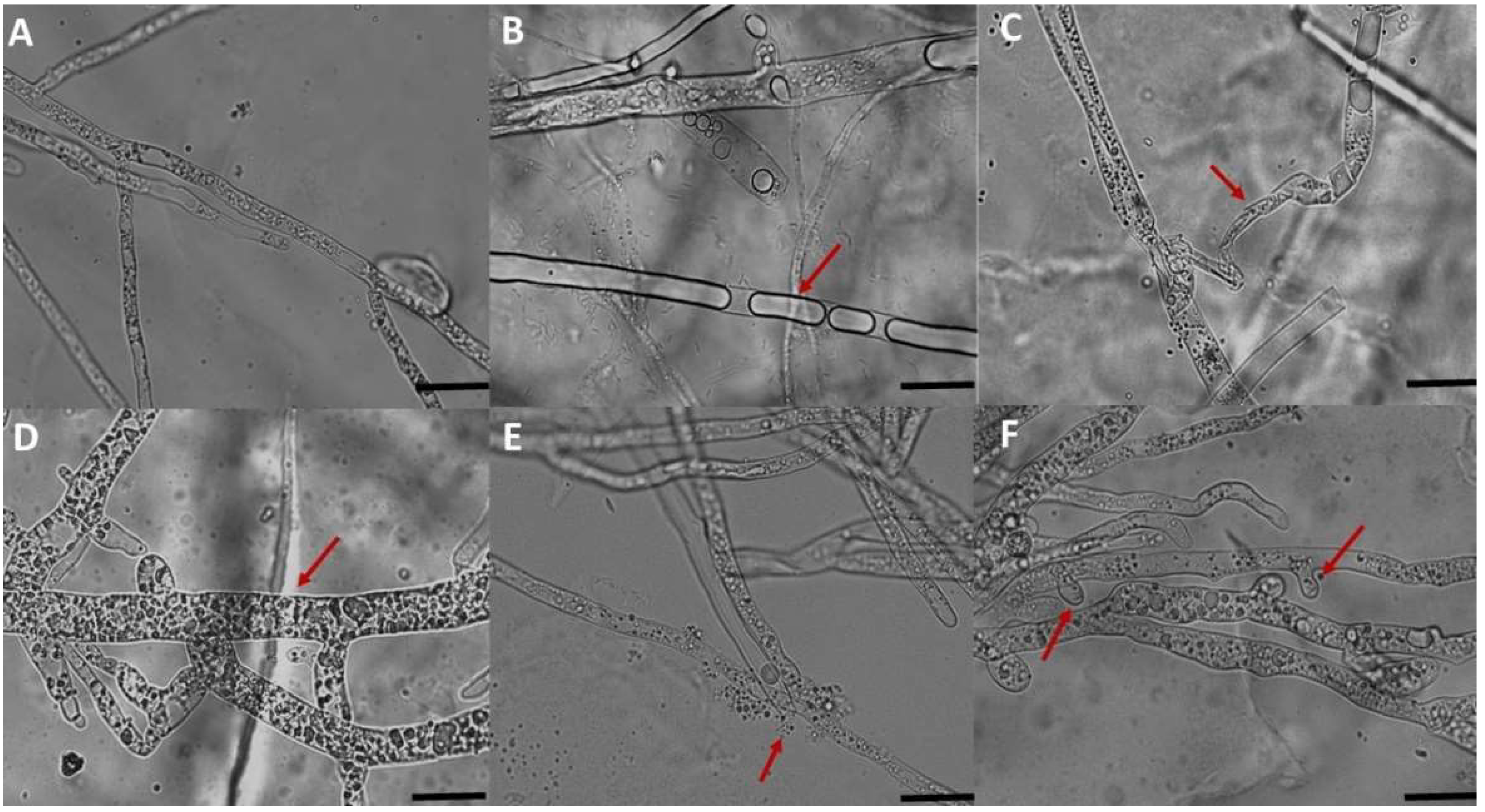
3.2. Microscopic Observation

Microscopic observations of P. schmitthenneri mycelium co-cultured with antagonistic bacteria revealed significantly altered morphology and cytological abnormalities when compared with the untreated control (Figure 2A). Generally, the modifications corresponded to vacuolation, deformation, and hyphal swelling or budding of the mycelium structure (Figure 2B–D,F) and were infrequently linked with mycelium destruction and release of cytoplasmic contents (Figure 2E).

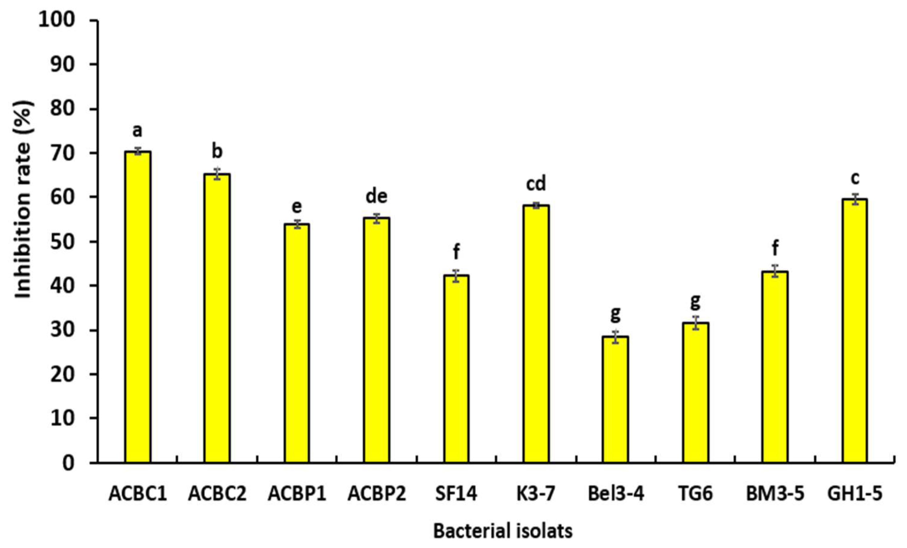
3.3. Effect of Bacterial Volatile Organic Compounds on Mycelial Growth

Statistical analysis demonstrated that bacterial VOCs influenced the mycelial development of P. schmitthenneri, after 6 days of incubation at 25 ± 1 °C, in comparison with the growth of the control (pathogen only) (Figure 3). The antifungal activity of bacterial VOCs ranged from a maximum of 70.32% (ACBC1) to a minimum of 28.37% (Bel3-4). In addition, results showed that six tested isolates gave an inhibition rate higher than 50% (ACBC1, ACBC2, GH1-5, K3-1, ACBP2, and ACBP1).

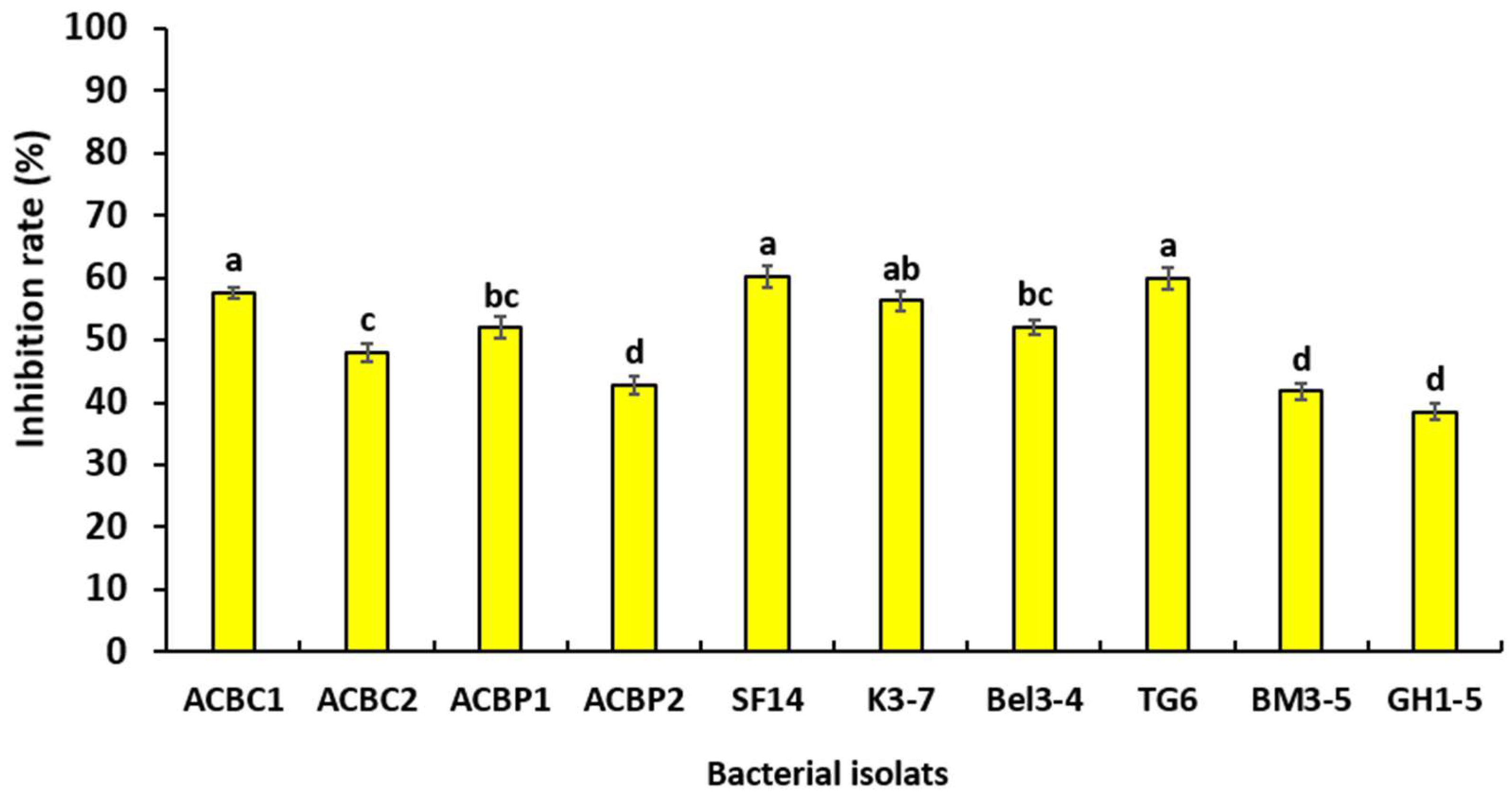
3.4. Effect of Bacterial Filtrates on Mycelial Growth

Statistical analysis showed that there was a significant difference between the inhibition rates of mycelial growth obtained with cell-free bacterial filtrates used against P. schmitthenneri at p ≤ 0.05 (Figure 4). The cell-free filtrates of the bacterial isolates SF14, TG6, and ACBC1 have the highest reduction of mycelial growth with inhibition rates reaching 60.16%, 59.96%, and 57.56%, respectively.

3.5. In Planta Bioassay

The ten bacterial strains were evaluated in the greenhouse based on their in vitro findings to validate their inhibitory effects and abilities to control olive root rot disease. Statistical analysis showed that there was a significant difference between the ten strains at p ≤ 0.05. The strains ACBC1, SF14, and BM3-5 were particularly successful in reducing root rot disease after two months of post-incubation, with disease severity reaching 8.33%, 8.33%, and 25%, respectively (Figure 5A,B). These bacterial isolates showed results that were quite comparable to the negative control (without P. schmitthenneri). However, the remaining bacteria were demonstrated to be less efficient to control P. schmitthenneri infection (Figure 5B).

4. Discussion

The use of microorganisms is a safe and viable alternative to synthetic fungicides for the control of soil-borne diseases. This approach requires searching for possible BCA candidates with strong antagonistic properties. In this sense, the present study evaluated the capacity of ten bacterial isolates, previously characterized and selected for their antagonistic activity against wide pathogens [38,39,40], to control olive root rot disease caused by P. schmitthenneri.
Our findings showed that bacterial strains have a significant in vitro antagonistic capacity on mycelial growth ranging from 60.47 to 85.15%. The highest inhibition rates were observed in treatments with the antagonistic isolates ACBC1 followed by SF14 and then ACBC2. Previous investigations have also shown the effectiveness of antagonistic bacteria in the management of Pythium spp. [52,53,54,55,56]. In accordance with our results, Lahlali et al. [38] reported the antagonistic activity of two strains, namely, Alcaligenes faecalis ACBC1 and Bacillus amyloliquefaciens SF14, in a dual culture test with inhibition rates of 96.3% and 91.9%, respectively, against Monilinia fructigena. Likewise, Bardin et al. [57] found that the Pantoea agglomerans 2-2 strain was successful in reducing Pythium sp. mycelial development in vitro, which was related to its capacity to secrete extracellular protease. In addition, the P. agglomerans ENA1 strain showed a very significant antagonistic activity against Macrophomina phaseolina, with an inhibition rate reaching 89% compared with the control [58]. Stenotrophomonas maltophilia isolate W81, a multidrug-resistant bacteria, has also shown an ability to control P. ultimum through the production of lytic enzymes including protease, pectinase, and chitinase and the secretion of VOCs [59]. The significant antagonistic potential of other bacteria has been extensively studied such as S. maltophilia CR71 against Colletotrichum nymphaea [60]; B. xiamenensis PM14 against P. splendens [61]; B. subtilis DCl1 against P. myriotylum, Phytophthora infestans, and Rhizoctonia solani [62]; and B. subtilis CU12 against Alternaria solani, P. sulcatum, Botrytis cinerea, and F. sambucinum [63]; among others.
In this study, both S. multivorum and other antagonists reduced the mycelial development of P. schmitthenneri but with varying degrees of inhibition related specifically to the mode of action of each isolate. Three tests were conducted in vitro to study the modes of action of selected bacterial isolates. Under a microscope, the results revealed an alteration of mycelial structure in the form of deformation, vacuolation, and swelling of P. schmitthenneri mycelium co-cultured with each isolate. These cytological changes can be due to substances synthesized by the bacteria [64]. In the same line, Cheffi et al. [65] examined the potential of B. velezensis OEE1 to control the strain Fso1 of F. solani, finding substantial cytoplasm vacuolization and mycelial lysis.
The mode of action occurs generally via the production of antifungal substances and VOCs [66,67,68], or parasitism through the secretion of lytic enzymes and lipopeptides [69,70,71]. The production of VOCs has been widely implicated in the bio-control of soil-borne fungi [72,73,74]. For instance, Wang et al. [75] found that B. halotolerans KLBC XJ-5 highly inhibited the hyphal growth of B. cinerea after five days of incubation, with an inhibition rate higher than 73.7%. In that study, the inhibition was correlated with the secretion of lipopeptides. In addition, the isolate B. halotolerans BFOA1/BFOA4 produced a variety of secondary metabolites and successfully controlled B. cinerea, F. oxysporum f. sp. Albedinis, P. infestans, R. bataticola, and A. alternata [76].
In our study, the inhibitory rate of bacterial filtrates ranged from 38.49% to 60.16%, suggesting that the ten isolates used might be sources of diverse secondary metabolites. According to Li et al. [47], the inhibition rates of the bacterial cell-free filtrates are associated with their increasing concentration; the inhibition becomes greater as the filtrate concentrations increase. Extracellular metabolites from B. amyloliquefaciens strain QSB-6 significantly affected Fusarium mycelial development and spore germination [77].
The emission of VOCs has been extensively linked to the biological control of soil-borne pathogens [72,73,74]. The findings of the indirect confrontation in this study showed that the ten strains could generate VOCs and limit the mycelial growth of P. schmitthenneri. According to Sànchez-Fernàndez et al. [78], Nodulisporium sp. GS4d2II1a reduced the hyphal development of P. aphanidermatum and other pathogenic fungi by emitting VOCs. These VOCs are a mixture of small volatile compounds that are present in a gaseous state under ambient temperatures (i.e., 1 atm pressure and 25 °C temperature) due to their low water solubility and high vapor pressure [74,79].
In this work, an in vivo bioassay on the root of olive trees was assessed to validate the antagonistic capacity of the bacterial isolates in field conditions. Tree isolates of A. faecalis ACBC1, B. amyloliquefaciens SF14, and B. halotolerans BM3-5 showed a very significant capacity to decrease the disease severity of root rot. In a greenhouse environment, the strain B. amyloliquefaciens Y1 lowered the incidence of Fusarium wilt disease in tomatoes when compared to the control [80]. Furthermore, this isolate has been effective in the biocontrol of several other pathogens, including Phytophthora capsici, Rhizoctonia solani, and Botrytis cinerea. Taken together, the antagonistic strains A. faecalis ACBC1 and B. amyloliquefaciens SF14 were the most successful in suppressing the pathogen P. schmithenneri in both in vitro and in planta experiments.
Numerous research studies have shown that antagonistic bacteria constitute a major source of lytic enzymes, which are utilized to limit the spread of fungal pathogens and diminish their pathogenicity [81,82]. Our antagonistic bacteria were previously characterized based on their capacity to synthesize lytic enzymes, and all of them could produce at least two lytic enzymes (amylase, cellulose, and protease) (Table S1) [38,39].
The mycelia of P. debaryanum, in this study, were entirely lysed in many regions. In the same context, Salem and Abdel-Rahman et al. [83] reported that B. subtilis MK537378 and Trichoderma reesei MK934489 showed a high capacity for cellulase enzyme production as well as antagonistic activity against P. debaryanum. Aydi Ben Abdallah et al. [84] also revealed that A. faecalis S18 and B. cereus S42 produced protease and chitinase and concluded that the suppression of mycelial growth of F. oxysporm f. sp. lycopersici by these two antagonistic bacteria was due to the synthesis of cell wall destroying enzymes. Moreover, Sen et al. [85] found that the strain SSB17 of A. faecalis might produce the α-amylase. In addition, the antagonistic activity of four Trichoderma strains against P. myriotylum was correlated with the production of protease, cellulase, and xylanase [37].
Furthermore, hydrogen cyanide (HCN) is a volatile compound biosynthesized from glycine using HCN synthase [86]. Our results indicated that only P. agglomerans ACBP1 was able to produce HCN. The latter was reported in various studies to suppress soil-borne pathogens [87,88,89]. Indeed, HCN acts by inhibiting cytochrome oxidase in the electron transport chain and energy supply to cells, leading to the death of phytopathogenic fungi [90].
In this study, the tested antagonistic bacteria exhibit other features. Amongst them, genes implicated in lipopeptide secretion and that are important in antibiosis were also detected in the isolates (Table S1) [38,39]. These antimicrobial compounds can suppress fungal growth, particularly fengycin, which has a potent antifungal activity toward filamentous fungi [91,92]. Bacillus spp. has been shown to produce a wide range of lipopeptides (e.g., surfactins, iturins, and fengycins) [93,94,95,96,97]. According to Zhang et al. [98], genes coding for lipopeptide biosynthesis have been found in most tested Bacillus isolates. Lee et al. [99] suggested that the inhibitory effect of B. amyloliquefaciens DA12 was related to iturin A and volatile heptanones production, making it a good BCA candidate against Fusarium diseases.

5. Conclusions

The effectiveness of antagonistic bacteria in controlling the olive root rot disease caused by P. schmitthenneri was investigated in this research. The three in vitro bioassays revealed a significant effect of the ten bacterial strains on the inhibition of the mycelial growth of the pathogen. Under greenhouse conditions, two bacteria, namely, Alcaligenes faecalis ACBC1 and Bacillus amyloliquefaciens SF14, showed promising results as they were highly effective in controlling the disease severity. The ability of these BCAs to produce lytic enzymes and lipopeptides determined their effectiveness. These results provide new control alternatives for the establishment of biocontrol strategies to manage sustainably the olive root rot disease. Therefore, two bacterial isolates, ACBC1 and SF14, were proposed to be used to control and prevent disease damage. However, additional experiments under natural conditions are needed to confirm their large-scale biocontrol potential before going on to the next stage of formulation of these antagonistic bacterial isolates as commercial bio-fungicide products.

Supplementary Materials

The following supporting information can be downloaded at: www.mdpi.com/article/10.3390/microorganisms10081635/s1, Table S1: Summary of relative hydrolytic enzyme and lipopeptides exhibited by the ten bacteria [38,39].

Author Contributions

Conceptualization, I.L. and R.L.; methodology, I.L. and R.L.; software, I.L. and H.E.H.; validation, I.L., R.L., H.E.H. and E.A.B.; formal analysis, I.L.; investigation, R.L., A.T. and S.A.; data curation, I.L. and R.L.; writing—original draft preparation, I.L.; writing—review and editing, I.L., H.E.H., Z.B., E.A.B. and R.L.; supervision, R.L. and J.A.F.; project administration, R.L. and A.L.; funding acquisition, A.T., R.L., E.A.B. and S.A. All authors have read and agreed to the published version of the manuscript.

Funding

This work was supported by the Phytopathology Unit of the Department of Plant Pathology—Ecole Nationale d’Agriculture de Meknès. This research was financially supported by MESRSI under PRIMA Project Section 2, SIRAM.

Institutional Review Board Statement

Not applicable.

Informed Consent Statement

Not applicable.

Data Availability Statement

The data that support the findings of this study are available from the corresponding author upon reasonable request.

Acknowledgments

The authors wish to acknowledge the olive farmers of the Meknès–Fes region for providing them with plant material for in vivo trials.

Conflicts of Interest

The authors of this work declare no conflict of interest.

References

  1. Fraga, H.; Moriondo, M.; Leolini, L. Mediterranean Olive Orchards under Climate Change: A Review of Future Impacts and Adaptation Strategies. Agronomy 2021, 11, 56. [Google Scholar] [CrossRef]
  2. Chliyeh, M.; Selmaoui, K.; Touhami, A.O.; Abdelkarim, F. Survey of the Fungal Species Associated to Olive-Tree (Olea europaea L.). Int. J. Recent Biotechnol. 2014, 2, 15–32. [Google Scholar]
  3. Ben Amira, M.; Lopez, D.; Triki, A.; Khouaja, A.; Chaar, H.; Fumanal, B.; Gousset-dupont, A.; Bonhomme, L.; Label, P.; Goupil, P.; et al. Beneficial Effect of Trichoderma harzianum Strain Ths97 in Biocontrolling Fusarium solani Causal Agent of Root Rot Disease in Olive Trees. Biol. Control 2017, 110, 70–78. [Google Scholar] [CrossRef]
  4. Mousa, M.S.; Ali, M.K.; Mosa, A.A.; Elewa, I. Root Rot Disease of Olive Transplants and Its Biological Control. Arab Univ. J. Agric. Sci. 2006, 14, 395–409. [Google Scholar] [CrossRef]
  5. Bouzoumita, A.; Metoui, M.; Jemni, M.; Kabaeir, N.; Belhouchette, K.; Ferchichi, A. The Efficacy of Various Bacterial Organisms for Biocontrol of Fusarium Root Rot of Olive in Tunisia. Pol. J. Environ. Stud. 2020, 29, 11–16. [Google Scholar] [CrossRef]
  6. Moustafa, S.M.N.; Elkanzi, N.A.A. Effect of the Newly Synthesized Pyrazole, And Pyrazolo Pyrimidine Derivatives on Pythium aphanidermatum (Edson) Fitzp. Egypt. J. Chem. 2021, 64, 2857–2872. [Google Scholar] [CrossRef]
  7. Gharbi, Y.; Bouazizi, E.; Cheffi, M.; Amar, F. Ben Archives of Phytopathology and Plant Protection Investigation of Soil-Borne Fungi, Causal Agents of Olive Trees Wilt and Dieback in Tunisia. Arch. Phytopathol. Plant Prot. 2020, 53, 828–843. [Google Scholar] [CrossRef]
  8. González, M.; Serrano, M.S.; Sánchez, M.E. First Report of Pythium spiculum Causing Root Rot on Wild-Olive in Spain. Plant Dis. 2016, 100, 1023. [Google Scholar] [CrossRef]
  9. Bizos, G.; Papatheodorou, E.M.; Chatzistathis, T.; Ntalli, N.; Aschonitis, V.G.; Monokrousos, N. The Role of Microbial Inoculants on Plant Protection, Growth Stimulation, and Crop Productivity of the Olive Tree (Olea europea L.). Plants 2020, 9, 743. [Google Scholar] [CrossRef]
  10. González, M.; Pérez-Sierra, A.; Serrano, M.S.; Sánchez, M.E. Two Phytophthora Species Causing Decline of Wild Olive (Olea europaea subsp. europaea Var. sylvestris). Plant Pathol. 2017, 66, 941–948. [Google Scholar] [CrossRef]
  11. Ruano-Rosa, D.; Schena, L.; Agosteo, G.E.; Magnano di San Lio, G.; Cacciola, S.O. Phytophthora oleae Sp. Nov. Causing Fruit Rot of Olive in Southern Italy. Plant Pathol. 2018, 67, 1362–1373. [Google Scholar] [CrossRef]
  12. González, M.; Pérez-Sierra, A.; Sánchez, M.E. Phytophthora oleae, a New Root Pathogen of Wild Olives. Plant Pathol. 2019, 68, 901–907. [Google Scholar] [CrossRef]
  13. Wu, W.; Ogawa, F.; Ochiai, M.; Yamada, K.; Fukui, H. Common Strategies to Control Pythium Disease. Rev. Agric. Sci. 2020, 8, 58–69. [Google Scholar] [CrossRef]
  14. Balla, A.; Silini, A.; Cherif-silini, H.; Bouket, A.C.; Moser, W.K.; Nowakowska, J.A.; Oszako, T.; Benia, F.; Belbahri, L. The Threat of Pests and Pathogens and the Potential for Biological Control in Forest Ecosystems. Forests 2021, 12, 1579. [Google Scholar] [CrossRef]
  15. Bodah, E.T. Root Rot Diseases in Plants: A Review of Common Causal Agents and Management Strategies. Agric. Res. Technol. Open Access J. 2017, 5, 555661. [Google Scholar]
  16. Quyet, N.T.; Cuong, H.V.; Hong, L.T.; Soytong, K. Control Mechanism of Chaetomium Spp. and Its Biological Control of Citrus Root Rot in Pot and Field Experiments in Vietnam. J. Agric. Technol. 2016, 12, 329–336. [Google Scholar]
  17. Chliyeh, M.; Selmaoui, K.; Abdelkarim, F.; El Modafar, C. Geographical Distribution of Phytophtora Palmivora in Different Olive Growing Regions in Morocco. Int. J. Plant Anim. Environ. Sci. 2014, 4, 297–303. [Google Scholar]
  18. Shlevin, E.; Gamliel, A.; Katan, J.; Shtienberg, D. Multi-Study Analysis of the Added Bene Fi Ts of Combining Soil Solarization with Fumigants or Non-Chemical Measures. Crop Prot. 2018, 111, 58–65. [Google Scholar] [CrossRef]
  19. Porter, L.D.; Hamm, P.B.; David, N.L.; Gieck, S.L.; Miller, J.S.; Gundersen, B.; Inglis, D.A. Metalaxyl-M-Resistant Pythium Species in Potato Production Areas of the Pacific Northwest of the USA. Am. J. Potato Res. 2009, 86, 315–326. [Google Scholar] [CrossRef]
  20. Hassanisaadi, M.; Hosein, G.; Bonjar, S.; Hosseinipour, A.; Abdolshahi, R. Biological Control of Pythium aphanidermatum, the Causal Agent of Tomato Root Rot by Two Streptomyces Root Symbionts. Agronomy 2021, 11, 846. [Google Scholar] [CrossRef]
  21. Lookabaugh, E.C.; Kerns, J.P.; Cubeta, M.A.; Shew, B.B.; Pathology, P.; State, N.C. Fitness Attributes of Pythium aphanidermatum with Dual Resistance to Mefenoxam and Fenamidone. Plant Dis. 2018, 102, 1938–1943. [Google Scholar] [CrossRef]
  22. Halo, B.A.; Al-yahyai, R.A.; Al-sadi, A.M. Biological Control of Pythium aphanidermatum-Induced Cucumber and Radish Damping-off by an Endophytic Fungus, Cladosporium omanense Isolate 31R Cucumber and Radish Damping-o Ff by an Endophytic Fungus. Biocontrol Sci. Technol. 2020, 31, 235–251. [Google Scholar] [CrossRef]
  23. Cook, R.J. Making Greater Use of Introduced Microorganisms for Biological Control of Plant Pathogens. Annu. Rev. Phytopathol. 1993, 31, 53–80. [Google Scholar] [CrossRef]
  24. Lahlali, R.; Ezrari, S.; Radouane, N.; Kenfaoui, J.; Esmaeel, Q.; El Hamss, H.; Belabess, Z.; Barka, E.A. Biological Control of Plant Pathogens: A Global Perspective. Microorganisms 2022, 10, 596. [Google Scholar] [CrossRef] [PubMed]
  25. Romanazzi, G.; Sanzani, S.M.; Bi, Y.; Tian, S.; Martínez, P.G.; Alkan, N. Induced resistance to control postharvest decay of fruit and vegetables. Postharvest Biol. Technol. 2016, 122, 82–94. [Google Scholar] [CrossRef]
  26. Sharma, R.R.; Singh, D.; Singh, R. Biological Control of Postharvest Diseases of Fruits and Vegetables by Microbial Antagonists: A Review. Biol. Control 2009, 50, 205–221. [Google Scholar] [CrossRef]
  27. Droby, S.; Wisniewski, M.; Macarisin, D.; Wilson, C. Twenty Years of Postharvest Biocontrol Research: Is It Time for a New Paradigm? Postharvest Biol. Technol. 2009, 52, 137–145. [Google Scholar] [CrossRef]
  28. O’Brien, P.A. Biological Control of Plant Diseases. Australas. Plant Pathol. 2017, 46, 293–304. [Google Scholar] [CrossRef]
  29. Farhaoui, A.; Adadi, A.; Tahiri, A.; El Alami, N.; Khayi, S.; Mentag, R.; Ezrari, S.; Radouane, N.; Mokrini, F.; Belabess, Z.; et al. Biocontrol Potential of Plant Growth-Promoting Rhizobacteria (PGPR) against Sclerotiorum Rolfsii Diseases on Sugar Beet (Beta vulgaris L.). Physiol. Mol. Plant Pathol. 2022, 119, 101829. [Google Scholar] [CrossRef]
  30. Jimtha, J.C.; Mallikarjunaswamy, G.; Najiya, N. Probiotic Rhizospheric Bacillus Sp. from Zingiber officinale Rosc. Displays Antifungal Activity against Soft Rot Pathogen Pythium Sp. Curr. Plant Biol. 2021, 27, 100217. [Google Scholar] [CrossRef]
  31. Ben Khedher, S.; Boukedi, H.; Laarif, A.; Tounsi, S. Biosurfactant Produced by Bacillus Subtilis V26: A Potential Biological Control Approach for Sustainable Agriculture Development. Org. Agric. 2020, 10, 117–124. [Google Scholar] [CrossRef]
  32. Kalantari, S.; Marefat, A.; Naseri, B. Improvement of Bean Yield and Fusarium Root Rot Biocontrol Using Mixtures of Bacillus, Pseudomonas and Rhizobium. Trop. Plant Pathol. 2018, 43, 499–505. [Google Scholar] [CrossRef]
  33. Zhang, J.; Mavrodi, D.V.; Yang, M.; Thomashow, L.S.; Mavrodi, O.V.; Kelton, J.; Weller, D.M. Pseudomonas Synxantha 2-79 Transformed with Pyrrolnitrin Biosynthesis Genes Has Improved Biocontrol Activity against Soilborne Pathogens of Wheat and Canola. Phytopathology 2020, 110, 1010–1017. [Google Scholar] [CrossRef]
  34. Vurukonda, S.S.k.P.; Giovanardi, D.; Stefani, E. Plant Growth Promoting and Biocontrol Activity of Streptomyces Spp. as Endophytes. Int. J. Mol. Sci. 2018, 19, 952. [Google Scholar] [CrossRef]
  35. Newitt, J.T.; Prudence, S.M.M.; Hutchings, M.I.; Worsley, S.F. Biocontrol of Cereal Crop Diseases Using Streptomycetes. Pathogens 2019, 8, 78. [Google Scholar] [CrossRef]
  36. Sood, M.; Kapoor, D.; Kumar, V.; Sheteiwy, M.S.; Ramakrishnan, M.; Landi, M.; Araniti, F.; Sharma, A. Trichoderma: The “Secrets” of a Multitalented. Plants 2020, 9, 762. [Google Scholar] [CrossRef]
  37. Tchameni, S.N.; Cotârle, M.; Ghinea, I.O.; Ampere, M.; Bedine, B. Involvement of Lytic Enzymes and Secondary Metabolites Produced by Trichoderma Spp. in the Biological Control of Pythium myriotylum. Int. Microbiol. 2019, 23, 179–188. [Google Scholar] [CrossRef]
  38. Lahlali, R.; Aksissou, W.; Lyousfi, N.; Ezrari, S.; Blenzar, A.; Tahiri, A.; Ennahli, S.; Hrustić, J.; MacLean, D.; Amiri, S. Biocontrol Activity and Putative Mechanism of Bacillus amyloliquefaciens (SF14 and SP10), Alcaligenes faecalis ACBC1, and Pantoea Agglomerans ACBP1 against Brown Rot Disease of Fruit. Microb. Pathog. 2020, 139, 103914. [Google Scholar] [CrossRef]
  39. Ezrari, S.; Mhidra, O.; Radouane, N.; Tahiri, A.; Polizzi, G.; Lazraq, A.; Lahlali, R. Potential Role of Rhizobacteria Isolated from Citrus Rhizosphere for Biological Control of Citrus Dry Root Rot. Plants 2021, 10, 872. [Google Scholar] [CrossRef]
  40. Bahadou, S.A.; Ouijja, A.; Karfach, A.; Tahiri, A.; Lahlali, R. New Potential Bacterial Antagonists for the Biocontrol of Fire Blight Disease (Erwinia amylovora) in Morocco. Microb. Pathog. 2018, 117, 7–15. [Google Scholar] [CrossRef]
  41. Legrifi, I.; Al Figuigui, J.; Radouane, N.; Ezrari, S.; Belabess, Z.; Tahiri, A.; Amiri, S.; Lahlali, R. First Report of Pythium schmitthenneri on Olive Trees and in Morocco. Australas. Plant Dis. Notes 2022, 17, 3. [Google Scholar] [CrossRef]
  42. Zhu, C.; Lei, M.; Andargie, M.; Zeng, J.; Li, J. Antifungal Activity and Mechanism of Action of Tannic Acid against Penicillium Digitatum. Physiol. Mol. Plant Pathol. 2019, 107, 46–50. [Google Scholar] [CrossRef]
  43. Wahyuni, D.K.; Hafida, S.N.; Ermayanti, T.M.; Eko, B.P.; Wardoyo, H.P.; Utami, E.S.W. Hairy Root Induction on Justicia gendarussa by Various Density of Agrobacterium Rhizogenes Strain LB 510. Biosaintifika J. Biol. Biol. Educ. 2017, 9, 26–32. [Google Scholar] [CrossRef]
  44. Lahlali, R.; Bajii, M.; Jijakli, M.H. Isolation and Evaluation of Bacteria and Fungi as Biological Control Agents. Commun. Agric. Appl. Biol. Sci. 2007, 72, 973–982. [Google Scholar]
  45. Trivedi, P.; Pandey, A.; Palni, L.M.S. In Vitro Evaluation of Antagonistic Properties of Pseudomonas corrugata. Microbiol. Res. 2008, 163, 329–336. [Google Scholar] [CrossRef]
  46. Guevara-avendan, E.; Carrillo, J.D.; Moreno, K.; Méndez-Bravo, A.; Guerrero-Analco, J.A.; Reverchon, F. Antifungal Activity of Avocado Rhizobacteria against Fusarium euwallaceae and Graphium Spp., Associated with Euwallacea Spp. Nr. Fornicatus, and Phytophthora cinnamomi. Antonie Van Leeuwenhoek 2018, 111, 563–572. [Google Scholar] [CrossRef]
  47. Li, Z.; Guo, B.; Wan, K.; Cong, M.; Huang, H.; Ge, Y. Effects of Bacteria-Free Filtrate from Bacillus Megaterium Strain L2 on the Mycelium Growth and Spore Germination of Alternaria Alternata. Biotechnol. Biotechnol. Equip. 2015, 29, 1062–1068. [Google Scholar] [CrossRef]
  48. Moradi, M.; Nejad, F.J.; Hosein, G.; Bonjar, S.; Fani, S.R. Efficacy of Bacillus Subtilis Native Strains for Biocontrol of Phytophthora Crown and Root Rot of Pistachio in Iran. Trop. Plant Pathol. 2018, 43, 306–313. [Google Scholar] [CrossRef]
  49. Santilli, E.; Riolo, M.; La Spada, F.; Pane, A.; Cacciola, S.O. First Report of Root Rot Caused by Phytophthora Bilorbang on Olea europaea in Italy. Plants 2020, 9, 826. [Google Scholar] [CrossRef] [PubMed]
  50. Simamora, A.V.; Stukely, M.J.C.; Barber, P.A.; Hardy, G.E.S.; Burgess, T.I. Age-Related Susceptibility of Eucalyptus Species to Phytophthora Accepted Article Boodjera. Plant Pathol. 2016, 66, 501–512. [Google Scholar] [CrossRef]
  51. Jabiri, S.; Bahra, C.; Maclean, D.; Radouane, N.; Barka, E.A. Phytopythium vexans Associated with Apple and Pear Decline in the Saïss Plain of Morocco. Microorganisms 2021, 9, 1916. [Google Scholar] [CrossRef] [PubMed]
  52. Miao, G.; Han, J.; Wang, C.; Zhang, K.; Miao, G. Growth Inhibition and Induction of Systemic Resistance against Pythium aphanidermatum by Bacillus Simplex Strain HS-2 Growth Inhibition and Induction of Systemic Resistance against. Biocontrol Sci. Technol. 2018, 28, 1114–1127. [Google Scholar] [CrossRef]
  53. Peter, P.; Divakaran, K.; Kaniyala, H.; Nair, A.R. Endophytic Bacillus Spp. from Zingiber zerumbet Rhizome, Rhizosphere and Axenic Cultures: Characterisation and Identification of Isolates with Plant Growth Promoting Activities and Antagonism to Soft-Rot Causative Pythium myriotylum. Arch. Phytopathol. Plant Prot. 2021, 54, 2007–2022. [Google Scholar] [CrossRef]
  54. Al-daghari, D.S.S.; Al-abri, S.A.; Al-mahmooli, I.H. Efficacy of Native Antagonistic Rhizobacteria in the Biological Control of Pythium aphanidermatum -Induced Damping-off of Cucumber in Oman. J. Plant Pathol. 2019, 102, 305–310. [Google Scholar] [CrossRef]
  55. Fatouros, G.; Gkizi, D.; Fragkogeorgi, G.A.; Paplomatas, E.J.; Tjamos, S.E. Biological Control of Pythium, Rhizoctonia and Sclerotinia in Lettuce: The Plant Protective Activity of the Bacterium Paenibacillus Alvei K165 Is Associated with the Induction of Systemic Resistance. Plant Pathol. 2017, 67, 418–425. [Google Scholar] [CrossRef]
  56. Al-hussini, H.S.; Al-rawahi, A.Y.; Al-marhoon, A.A.; Al-abri, S.A.; Al-mahmooli, I.H. Biological Control of Damping-off of Tomato Caused by Pythium aphanidermatum by Using Native Antagonistic Rhizobacteria Isolated from Omani Soil. J. Plant Pathol. 2018, 101, 315–322. [Google Scholar] [CrossRef]
  57. Bardin, S.D.; Huang, H.C.; Liu, L.; Yanke, L.J. Control, by Microbial Seed Treatment, of Damping- off Caused by Pythium Sp. on Canola, Safflower, Dry Pea, and Sugar Beet 1. Can. J. Plant Pathol. 2003, 25, 268–275. [Google Scholar] [CrossRef]
  58. Vasebi, Y.; Alizadeh, A.; Safaie, N. Pantoea Agglomerans ENA1 as a Biocontrol Agent of Macrophomia Phaseolina and Growth Enhancer of Soybean. J. Crop Prot. 2015, 4, 43–57. [Google Scholar]
  59. Dunne, C.; Crowley, J.J.; Mo, Y.; Dowling, D.N.; Bruijn, S.; Gara, F.O. Biological Control of Pythium Ultimum by Stenotrophomonas maltophilia W81 Is Mediated by an Extracellular Proteolytic Activity. Microbiology 1997, 143, 3921–3931. [Google Scholar] [CrossRef]
  60. Alijani, Z.; Amini, J.; Ashengroph, M.; Bahramnejad, B. Volatile Compounds Mediated Effects of Stenotrophomonas maltophilia Strain UN1512 in Plant Growth Promotion and Its Potential for the Biocontrol of Colletotrichum nymphaeae. Physiol. Mol. Plant Pathol. 2020, 112, 101555. [Google Scholar] [CrossRef]
  61. Amna, X.Y.; Farooq, M.A.; Javed, M.T.; Kamran, M.A.; Mukhtar, T.; Ali, J.; Tabassum, T.; ur Rehman, S.; Munis, M.F.H.; Sultan, T.; et al. Plant Physiology and Biochemistry Multi-Stress Tolerant PGPR Bacillus Xiamenensis PM14 Activating Sugarcane (Saccharum officinarum L.) Red Rot Disease Resistance. Plant Physiol. Biochem. 2020, 151, 640–649. [Google Scholar] [CrossRef]
  62. Jayakumar, A.; Nair, I.C.; Radhakrishnan, E.K. Environmental Adaptations of an Extremely Plant Beneficial Bacillus Subtilis Dcl1 Identified Through the Genomic and Metabolomic Analysis. Microb. Ecol. 2020, 81, 687–702. [Google Scholar] [CrossRef] [PubMed]
  63. Wise, C.; Novitsky, L.; Tsopmo, A.; Avis, T.J. Production and Antimicrobial Activity of 3-Hydroxypropionaldehyde from Bacillus Subtilis Strain CU12. J. Chem. Ecol. 2012, 38, 1521–1527. [Google Scholar] [CrossRef] [PubMed]
  64. Sahu, K.P.; Kumar, A.; Patel, A.; Kumar, M.; Gopalakrishnan, S.; Prakash, G.; Gogoi, R. Rice Blast Lesions: An Unexplored Phyllosphere Microhabitat for Novel Antagonistic Bacterial Species Against Magnaporthe oryzae. Ecology 2020, 81, 731–745. [Google Scholar] [CrossRef] [PubMed]
  65. Cheffi, M.; Bouket, A.C.; Alenezi, F.N.; Luptakova, L.; Belka, M.; Vallat, A.; Rateb, M.E.; Tounsi, S.; Triki, M.A. Olea europaea L. Root Endophyte Bacillus velezensis OEE1 Counteracts Oomycete and Fungal Harmful Pathogens and Harbours a Large Repertoire of Secreted and Volatile Metabolites and Beneficial Functional Genes. Microorganisms 2019, 7, 314. [Google Scholar] [CrossRef]
  66. Jimtha, J.C.; Jishma, P.; Arathy, G.B.; Anisha, C.; Radhakrishnan, E.K. Identification of Plant Growth Promoting Rhizosphere Bacillus Sp. WG4 Antagonistic to Pythium myriotylum and Its Enhanced Antifungal Effect in Association with Trichoderma. J. Soil Sci. Plant Nutr. 2016, 16, 578–590. [Google Scholar] [CrossRef]
  67. Gong, A.; Wu, N.; Kong, X.; Zhang, Y.; Hu, M. Inhibitory Effect of Volatiles Emitted from Alcaligenes faecalis N1-4 on Aspergillus flavus and Aflatoxins in Storage. Front. Microbiol. 2019, 10, 1419. [Google Scholar] [CrossRef] [PubMed]
  68. Shan, S.; Wang, W.; Wang, M.; Sun, B. The Symbiotic Bacteria Alcaligenes faecalis of the Entomopathogenic Nematodes Oscheius Spp. Exhibit Potential Biocontrol of Plant- and Entomopathogenic Fungi. Microb. Biotechnol. 2019, 12, 459–471. [Google Scholar] [CrossRef]
  69. Kilany, M.; Ibrahim, E.H.; Al Amry, S.; Roman, S. Microbial Suppressiveness of Pythium Damping-Off Diseases. In Organic Amendments and Soil Suppressiveness in Plant Disease Management; Springer: Cham, Switzerland, 2015; pp. 187–206. [Google Scholar] [CrossRef]
  70. Singh, R.; Sachan, N.S. Review on Biological Control of Soil Borne Fungi in Vegetable Crops. HortFlora Res. Spectr. 2013, 2, 72–76. [Google Scholar]
  71. Abd-el-kareem, F.; Elshahawy, I.E.; Abd-elgawad, M.M.M. Application of Bacillus Pumilus Isolates for Management of Black Rot Disease in Strawberry. Egypt. J. Biol. Pest Control 2021, 31, 25. [Google Scholar] [CrossRef]
  72. Montes-osuna, N.; Cernava, T.; Carmen, G.; Berg, G. Identification of Volatile Organic Compounds Emitted by Two Beneficial Endophytic Pseudomonas Strains from Olive Roots. Plants 2022, 11, 318. [Google Scholar] [CrossRef]
  73. Pascal, M.; Dietel, K.; Grosch, R.; Cernava, T.; Berg, G. Microbiota Associated with Sclerotia of Soilborne Fungal Pathogens—A Novel Source of Biocontrol Agents Producing Bioactive Volatiles. Phytobiomes J. 2019, 3, 125–136. [Google Scholar] [CrossRef]
  74. Tilocca, B.; Cao, A.; Migheli, Q. Scent of a Killer: Microbial Volatilome and Its Role in the Biological Control of Plant Pathogens. Front. Microbiol. 2020, 11, 41. [Google Scholar] [CrossRef] [PubMed]
  75. Wang, F.; Xiao, J.; Zhang, Y.; Li, R.; Liu, L.; Deng, J. Postharvest Biology and Technology Biocontrol Ability and Action Mechanism of Bacillus halotolerans against Botrytis Cinerea Causing Grey Mould in Postharvest Strawberry Fruit. Postharvest Biol. Technol. 2021, 174, 111456. [Google Scholar] [CrossRef]
  76. Slama, H.B.; Cherif-silini, H.; Bouket, A.C.; Qader, M. Screening for Fusarium Antagonistic Bacteria from Contrasting Niches Designated the Endophyte Bacillus halotolerans as Plant Warden against Fusarium. Front. Microbiol. 2019, 9, 3236. [Google Scholar] [CrossRef]
  77. Duan, Y.; Chen, R.; Zhang, R.; Jiang, W.; Chen, X.; Yin, C. Isolation, Identification, and Antibacterial Mechanisms of Bacillus amyloliquefaciens QSB-6 and Its Effect on Plant Roots. Front. Microbiol. 2021, 12, 746799. [Google Scholar] [CrossRef]
  78. Sánchez-fernández, R.E.; Diaz, D.; Duarte, G.; Lappe-oliveras, P.; Sánchez, S.; Macías-rubalcava, M.L.; Macías-rubalcava, M.L. Antifungal Volatile Organic Compounds from the Endophyte Nodulisporium Sp. Strain GS4d2II1a: A Qualitative Change in the Intraspecific and Interspecific Interactions with Pythium aphanidermatum Interaction. Ecology 2015, 71, 347–364. [Google Scholar] [CrossRef] [PubMed]
  79. Morath, S.U.; Hung, R.; Bennett, J.W. Fungal Volatile Organic Compounds: A Review with Emphasis on Their Biotechnological Potential. Fungal Biol. Rev. 2012, 26, 73–83. [Google Scholar] [CrossRef]
  80. Maung, E.C.H.; Choi, T.G.; Nam, H.H.; Kim, K.Y. Role of Bacillus amyloliquefaciens Y1 in the Control of Fusarium Wilt Disease and Growth Promotion of Tomato Role of Bacillus amyloliquefaciens Y1 in the Control of Fusarium Wilt Disease and Growth Promotion of Tomato. Biocontrol Sci. Technol. 2017, 27, 1400–1415. [Google Scholar] [CrossRef]
  81. Khabbaz, S.E.; Zhang, L.; Cáceres, L.A.; Sumarah, M.; Wang, A.; Abbasi, P.A. Characterisation of Antagonistic Bacillus and Pseudomonas Strains for Biocontrol Potential and Suppression of Damping-off and Root Rot Diseases. Ann. Appl. Biol. 2015, 166, 456–471. [Google Scholar] [CrossRef]
  82. Chenniappan, C.; Narayanasamy, M.; Daniel, G.M.; Ramaraj, G.B.; Ponnusamy, P.; Sekar, J.; Ramalingam, P.V. Biocontrol Efficiency of Native Plant Growth Promoting Rhizobacteria against Rhizome Rot Disease of Turmeric. Biol. Control 2019, 129, 55–64. [Google Scholar] [CrossRef]
  83. Salem, A.A.; Abdel-Rahman, H.M. Cellulolytic Activity of Trichoderma Reesei and Bacillus Subtilis against the Plant Pathogen Pythium Debaryanum. Environ. Biodivers. Soil Secur. 2021, 5, 105–119. [Google Scholar] [CrossRef]
  84. Aydi Ben Abdallah, R.; Mokni-Tlili, S.; Nefzi, A.; Jabnoun-Khiareddine, H.; Daami-Remadi, M. Biocontrol of Fusarium Wilt and Growth Promotion of Tomato Plants Using Endophytic Bacteria Isolated from Nicotiana Glauca Organs. Biol. Control 2016, 97, 80–88. [Google Scholar] [CrossRef]
  85. Sen, K.S.; Dora, K.T.; Bandyopadhyay, B.; Das Mohapatra, K.P.; Raut, S. Thermostable Alpha-Amylase Enzyme Production from Hot Spring Isolates Alcaligenes faecalis SSB17—Statistical Optimization. Biocatal. Agric. Biotechnol. 2014, 3, 218–226. [Google Scholar] [CrossRef]
  86. Sehrawat, A.; Sindhu, S.S.; Glick, B.R. Hydrogen Cyanide Production by Soil Bacteria: Biological Control of Pests and Promotion of Plant Growth in Sustainable Agriculture. Pedosphere Int. J. 2022, 32, 15–38. [Google Scholar] [CrossRef]
  87. Sendi, Y.; Pfeiffer, T.; Koch, E.; Mhadhbi, H.; Mrabet, M. Potential of Common Bean (Phaseolus vulgaris L.) Root Microbiome in the Biocontrol of Root Rot Disease and Traits of Performance. J. Plant Dis. Prot. 2020, 127, 453–462. [Google Scholar] [CrossRef]
  88. Jha, B.K.; Gandhi Pragash, M.; Raman, G.; Sakthivel, N. Simultaneous Phosphate Solubilization Potential and Antifungal Activity of New Fluorescent Pseudomonad Strains, Pseudomonas aeruginosa, P. plecoglossicida and P. mosselii. World J. Microbiol. Biotechnol. 2008, 25, 573–581. [Google Scholar] [CrossRef]
  89. Habiba, R.N.; Ali, S.A.; Sultana, V.; Ara, J.; Ehteshamul-Haque, S. Evaluation of Biocontrol Potential of Epiphytic Fluorescent Pseudomonas Associated with Healthy Fruits and Vegetables against Root Rot and Root Knot Pathogens of Mungbean. Pak. J. Bot. 2016, 48, 1299–1303. [Google Scholar]
  90. Rehman, F.U.; Kalsoom, M.; Adnan, M.; Toor, M.D. Plant Growth Promoting Rhizobacteria and Their Mechanisms Involved in Agricultural Crop Production: A Review SunText Review of Biotechnology Plant Growth Promoting Rhizobacteria and Their Mechanisms Involved in Agricultural Crop Production: A Review. SunText Rev. BioTechnol. 2020, 35, 1044–1051. [Google Scholar] [CrossRef]
  91. Defilippi, S.; Groulx, E.; Megalla, M.; Mohamed, R.; Avis, T.J. Fungal Competitors Affect Production of Antimicrobial Lipopeptides in Bacillus Subtilis Strain B9-5. J. Chem. Ecol. 2018, 44, 374–383. [Google Scholar] [CrossRef] [PubMed]
  92. Farace, G.; Fernandez, O.; Jacquens, L.; Coutte, F.; Krier, F.; Jacques, P.; Clément, C.; Barka, E.A.I.T.; Jacquard, C.; Dorey, S. Cyclic Lipopeptides from Bacillus Subtilis Activate Distinct Patterns of Defence Responses in Grapevine. Mol. Plant Pathol. 2014, 16, 177–187. [Google Scholar] [CrossRef]
  93. de Souza Freitas, F.; Coelho de Assis Lage, T.; Ayupe, B.A.L.; de Paula Siqueira, T.; de Barros, M.; Tótola, M.R. Bacillus Subtilis TR47II as a Source of Bioactive Lipopeptides against Gram-Negative Pathogens Causing Nosocomial Infections. 3 Biotech 2020, 10, 474. Available online: https://Link.Springer.Com/Article/10.1007/S13205-020-02459-Z (accessed on 12 May 2022). [CrossRef] [PubMed]
  94. Raaijmakers, J.M.; de Bruijn, I.; Nybroe, O.; Ongena, M. Natural Functions of Lipopeptides from Bacillus and Pseudomonas: More than Surfactants and Antibiotics. FEMS Microbiol. Rev. 2010, 34, 1037–1062. [Google Scholar] [CrossRef]
  95. Ali, N.; Pang, Z.; Wang, F.; Xu, B.; El-Seedi, H.R. Lipopeptide Biosurfactants from Bacillus Spp.: Types, Production, Biological Activities, and Applications in Food. J. Food Qual. 2022, 2022, 3930112. [Google Scholar] [CrossRef]
  96. Cochrane, S.A.; Vederas, J.C. Lipopeptides from Bacillus and Paenibacillus Spp.: A Gold Mine of Antibiotic Candidates. Med. Res. Rev. 2016, 36, 4–31. [Google Scholar] [CrossRef] [PubMed]
  97. Stincone, P.; Veras, F.F.; Pereira, J.Q.; Mayer, F.Q.; Varela, A.P.M.; Brandelli, A. Diversity of Cyclic Antimicrobial Lipopeptides from Bacillus P34 Revealed by Functional Annotation and Comparative Genome Analysis. Microbiol. Res. 2020, 238, 126515. [Google Scholar] [CrossRef] [PubMed]
  98. Zhang, L.; Khabbaz, S.E.; Wang, A.; Li, H.; Abbasi, P.A. Detection and Characterization of Broad-Spectrum Anti-Pathogen Activity of Novel Rhizobacterial Isolates and Suppression of Fusarium Crown and Root Rot Disease of Tomato. J. Appl. Microbiol. 2015, 118, 685–703. [Google Scholar] [CrossRef] [PubMed]
  99. Lee, T.; Park, D.; Kim, K.; Lim, S.M.; Yu, N.H.; Kim, S.; Kim, H.; Jung, K.S.; Jang, J.Y.; Park, J.; et al. Characterization of Bacillus amyloliquefaciens DA12 Showing Potent Antifungal Activity against Mycotoxigenic Fusarium Species. Plant Pathol. 2017, 33, 499–507. [Google Scholar] [CrossRef] [PubMed]
Figure 1. In vitro confrontation revealing antagonistic activity of bacterial strains against P. schmitthenneri on PDA medium after 6 days of incubation at 25 °C. (A) control; (B) ACBC1; (C) ACBC2; (D) ACBP1; (E) ACBP2; (F) SF14; (G) K3-7; (H) Bel3-4; (I) TG6; (J) BM3-5; (K) GH1-5.
Microorganisms 10 01635 g001
Figure 2. Microscopic observation (×40) of the hyphal structure of P. schmitthenneri co-cultured with the antagonistic bacteria after 6 days of incubation. (A) untreated control; (B) vacuolation (SF14); (C) deformation (ACBC2); (D) hyphal swelling (ACBC1); (E) degradation of the mycelium (BM3-5); (F) budding of the mycelium structure (Bel3-4). Changes in the hyphae and mycelia are indicated by arrows. Scale bar = 20 µm.
Microorganisms 10 01635 g002
Figure 3. Effect of volatile organic compounds (VOCs), which were produced by tested bacteria on the inhibition of hyphal growth of P. schmitthenneri after 6 days of incubation at 25 ± 1 °C. Data in the figure are the average of two separate experiments with four replicates. According to the Tukey test, values with the same letter were not significantly different (p ≤ 0.05).
Microorganisms 10 01635 g003
Figure 4. Effect of bacterial cell-free filtrates 10% v/v on the inhibition of mycelial growth of P. schmitthenneri after 6 days of incubation. Data in the figure represent the mean of two independent trials with 4 replicates. Treatments with the same letter were not significantly different according to the Tukey test (p ≤ 0.05).
Microorganisms 10 01635 g004
Figure 5. (A) Symptoms of olive root rot disease on a one-year-old plant treated with bacterial isolates ACBC1, SF14, and BM3-5 and inoculated with P. schmitthenneri after two months within glasshouse conditions. C+: positive control (olive plants inoculated only with the pathogen), and C−: negative control (plants treated only with water in the absence of the pathogen). (B) Observation of disease severity (%) on the root of olive trees treated with bacterial suspension of the ten isolates (2 × 10 8 CFU/mL) and inoculated with P. schmitthenneri, after 2 months of incubation at 25 °C within greenhouse conditions. C+, positive control (pathogen only), and C−, negative control (plants treated only with water in the absence of the pathogen). Histograms represent the mean value of disease severity of two trials over time with six replicates. Error bars represent standard error, and values of plant severity with the same letter were not significantly different according to the Tukey test (p ≤ 0.05).
Microorganisms 10 01635 g005
Table 1. Treatments used in the in vivo bioassays.
TreatmentsCompositionRepetitionsPeriod
ACBC1 SS + pathogen + ACBC162 months
ACBC2 SS + pathogen + ACBC2
ACBP1 SS + pathogen + ACBP1
ACBP2 SS + pathogen + ACBP2
SF14 SS + pathogen + SF14
K3-7 SS + pathogen + K3-7
Bel3-4 SS + pathogen + Bel3-4
TG6 SS + pathogen + TG6
BM3-5 SS + pathogen + BM3-5
GH1-5 SS + pathogen + GH1-5
C+ SS + Pathogen
C− SS alone
SS: sterilized soil.
Table 2. The in vitro inhibition rates (%) of the P. schmitthenneri mycelial growth after 6 days of incubation at 25 °C in darkness.
Bacterial Isolate CodeSpeciesAccession NumbersInhibition Rates (%)
ACBC1Alcaligenes faecalisKY35728585.14 ± 1.13 a
ACBC2Pantoea agglomeransKY35728680.58 ± 1.31 bc
ACBP1P. agglomeransKY35728777.79 ± 0.64 cde
ACBP2P. agglomeransKY35728878.38 ± 0.64 bcd
SF14Bacillus amyloliquefaciensKY35729881.76 ± 1.76 ab
K3-7B. xiamenensisMW84301077.89 ± 1.96 cd
Bel3-4Sphingobacterium multivorumMW85682773.97 ± 0.87 f
TG6B. subtilisMW84762874.45 ± 1.48 ef
BM3-5B. halotoleransMW84795176.49 ± 0.53 def
GH1-5Stenotrophomonas maltophiliaMW84881960.47 ± 1.04 g
Data represent mean ± standard deviation (SD) of two trials over time with three replicates. Inhibition rates with the same letter are not significantly different according to the Tukey test performed on mycelial growth (p ≤ 0.05).
Publisher’s Note: MDPI stays neutral with regard to jurisdictional claims in published maps and institutional affiliations.

