Article

First Report of Two Jaculus Rodents as Potential Reservoir Hosts of Leishmania Parasites in Tunisia

by
Wissem Ghawar
1,2,3,4,*,†,
Melek Chaouch
4,5,6,†,
Afif Ben Salah
1,2,3,4,7,8,‡,
Mohamed Ali Snoussi
1,2,3,4,
Sadok Salem
1,2,3,4,
Ghassen Kharroubi
1,2,3,4,7,
Said Chouchen
3,9,
Amor Bouaoun
3,9,
Dhafer Laouini
2,3,4,
Jihene Bettaieb
1,2,3,4,7,‡ and
Souha Ben Abderrazak
4,5,‡
1
Department of Medical Epidemiology, Institut Pasteur de Tunis, Tunis 1002, Tunisia
2
Laboratory of Transmission, Control and Immunobiology of Infections (LR16IPT02), Institut Pasteur de Tunis, Tunis 1002, Tunisia
3
Clinical Investigation Center (CIC), Institut Pasteur de Tunis, Tunis 1002, Tunisia
4
Campus Universitaire Farhat Hached, University Tunis El Manar, Tunis 1068, Tunisia
5
Laboratory of Medical Parasitology, Biotechnology and Biomolecules (LR16IPT06), Institut Pasteur de Tunis, Tunis 1002, Tunisia
6
Laboratory of Bioinformatics, Biomathematics and Biostatistics (LR16IPT09), Institut Pasteur de Tunis, Tunis 1002, Tunisia
7
Faculty of Medicine of Tunis, University Tunis El Manar, Tunis 1068, Tunisia
8
Department of Family and Community Medicine, College of Medicine and Medical Sciences (CMMS), Arabian Gulf University (AGU), Manama 329, Bahrain
9
Health Regional Directorate of Tataouine, Administrative City, Tataouine Nouvelle, Tataouine 3263, Tunisia
*
Author to whom correspondence should be addressed.
These authors contributed equally to this work.
Microorganisms 2022, 10(8), 1502; https://doi.org/10.3390/microorganisms10081502
Submission received: 15 June 2022 / Revised: 19 July 2022 / Accepted: 21 July 2022 / Published: 25 July 2022

Abstract

:
This study shows, for the first time, natural Leishmania infection among Jaculus spp. in an endemic region of Tataouine, South Tunisia. To better characterize the transmission cycles in this complex focus of mixed transmission, Leishmania detection and species identification were performed by direct examination, internal transcribed spacer-1 (ITS1)-PCR-restriction fragment length polymorphism (RFLP), and sequencing of Jaculus (J.) jaculus (Linnaeus, 1758) and J. hirtipes (Lichtenstein, 1823) rodent species, which are frequently encountered in this area. Leishmania parasites were observed in 19 (41.3%) smears, while DNA parasites were detected in 28 (60.9%) Jaculus spp. spleens; among them, 12 (54.5%) were from 22 J. jaculus individuals and 16 (66.7%) were from 24 J. hirtipes individuals. Leishmania parasites were confirmed as Leishmania (L.) killicki (syn. L. tropica) in two J. hirtipes individuals (4.3%) and L. major (n = 24; 52.2%) in 10 J. jaculus and 14 J. hirtipes individuals. This finding represents the first evidence of natural infection with Leishmania parasites in rodents belonging to the Jaculus genus, providing the rationale to consider them as potential reservoir hosts of Old World Leishmania parasites in Tunisia and North Africa.

1. Introduction

Leishmaniasis is caused by protozoan parasites of the genus Leishmania. Among the three main clinical manifestations of this disease (cutaneous, visceral, and mucocutaneous), the cutaneous form is the most abundant [1]. In Tunisia, human cutaneous leishmaniasis (CL) is widely distributed and is prevalent in central and southern Tunisia. The annual incidence of CL ranges from 1 to 10,000 cases according to climatic factors and the importance of herd immunity [2]. Furthermore, three epidemiological scenarios of cutaneous leishmaniasis have been described: sporadic (caused by Leishmania (L.) infantum Nicolle, 1908), chronic (caused by L. tropica Wright, 1903), and zoonotic (caused by L. major Yakimoff & Shokkor, 1914). The latter is predominant in the central and southern parts of the country [3]. In these areas, zoonotic cutaneous leishmaniasis (ZCL) circulates in an endemoepidemic mode [4] and is recognized as a serious public health problem [2,5].
Leishmaniasis caused by L. major and L. infantum are commonly zoonotic, with some species of rodents belonging to the Muridae family and domestic dogs as reservoir hosts, respectively [6,7]. In contrast, L. killicki (syn. L. tropica), the causative agent of chronic CL in urban endemic microfoci, known to be anthroponotic, has been strongly suspected to be zoonotic, especially with evidence of Ctenodactylus (C.) gundi (Rothmann, 1776) infection [8,9]. Several studies have shown that Leishmania parasites can infect small mammals in Tunisia, including the least weasel [7] and hedgehog [10]. Furthermore, the geographical distribution of CL cases caused by L. killicki in the region of Tataouine suggested the absence of the classically conventional reservoir host, C. gundi, while other small mammals and rodent species frequently encountered in the environment (Regional Directorate of Primary Care, Tataouine) could play a role as reservoir hosts, which remain to be identified. Rodent species belonging to the Jaculus genus were suspicious due to their wide distribution in central and southern Tunisia [11]. Moreover, these rodents were frequently faced by the regional health workers in Tataouine (Regional Directorate of Primary Care, Tataouine). This clinical and epidemiological context justified this study, which aimed to investigate rodent species belonging to the Jaculus genus in an old emerging focus of CL, that is, the Tataouine Governorate in southeastern Tunisia, to explore them as novel reservoir hosts of Leishmania parasites in the region.
This will better characterize transmission cycles and provide stronger evidence for intervention policies.

2. Materials and Methods

2.1. Rodent Trapping and Identification

Rodents were collected between May 2016 and June 2018 using butterfly nets at two study sites: BniMhira and Guermessa, belonging to the Tataouine Governorate, South Tunisia [12].
Some of the captured rodents (n = 41) were identified by amplification of the mitochondrial complete cytochrome b (Cytb) gene, as previously described [12].
To identify all captured rodents consistently (n = 46), we also used polymerase chain reaction (PCR) amplification of the mitochondrial partial Cytb gene after DNA extraction from their spleens using the QIAamp® DNA Mini Kit (QIAgen, Hilden, Germany) according to the manufacturer’s instructions. Modified vertebrate-universal specific primers (cytB1-F and cytB2-R) were used to amplify a 370 bp segment [13]. PCR reactions were conducted in a 50 μL reaction volume, including 0.2 mM dNTPs, 2.5 U of Taq DNA polymerase (GE Healthcare, Chicago, IL, USA), and 60 pmol of each primer (Forward 5′-CCATCCAACATYTCADCATGATGAAA-3′ and Reverse 5′-GCHCCTCAGAATGATATTTGKCCTCA-3′). Reactions were performed in an Applied Biosystems GeneAmp PCR System 2700 (Foster City, CA, USA) using the following cycling conditions: an initial denaturation step at 94 °C for 5 min; 30 cycles of 94 °C for 1 min, 54 °C for 1 min, and 72 °C for 1 min; and a final elongation step at 72 °C for 10 min. The amplicons were electrophoresed on 1% agarose gels and visualized under UV light. PCR products were purified using a QIAquick PCR purification kit (QIAGEN, Hilden, Germany) according to the manufacturer’s instructions. Amplicon sequencing was performed using a conventional Big Dye Terminator Cycle Sequencing Ready Reaction Kit (Perkin Elmer, Applied Biosystems, Foster City, CA, USA) and an ABI373 automated DNA sequencer (Thermo Fisher Scientific, Waltham, MA, USA). The obtained sequences were edited using ChromasPro software (Technelysium Pty Ltd., South Brisbane, Australia). For homology searches, sequences were submitted to the National Center for Biotechnology Information (NCBI) server using the Basic Local Alignment Search Tool (BLAST) (http://blast.ncbi.nlm.nih.gov (accessed on 22 March 2022)). Accession numbers of the sequences are listed in Supplementary Table S1.

2.2. Detection of Infection by Leishmania Parasites

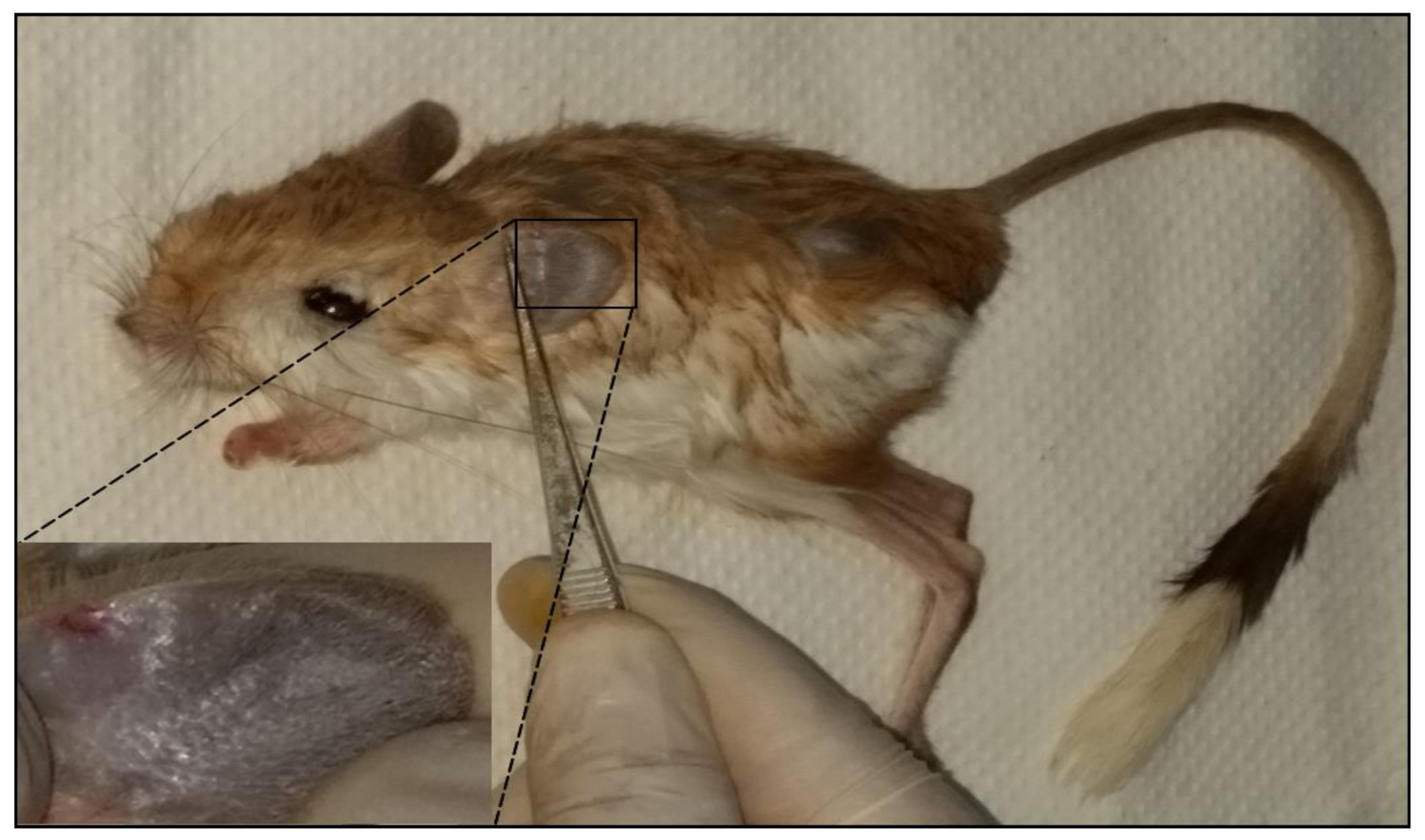
2.2.1. Clinical Examination

Before autopsy, each rodent was examined for cutaneous lesions in different parts of the body and skin. Clinical manifestations, including hyperpigmentation of the higher edge of the ear, depilation, small nodules, infiltration, and/or partial or total organ destruction, were recorded as previously described [7].

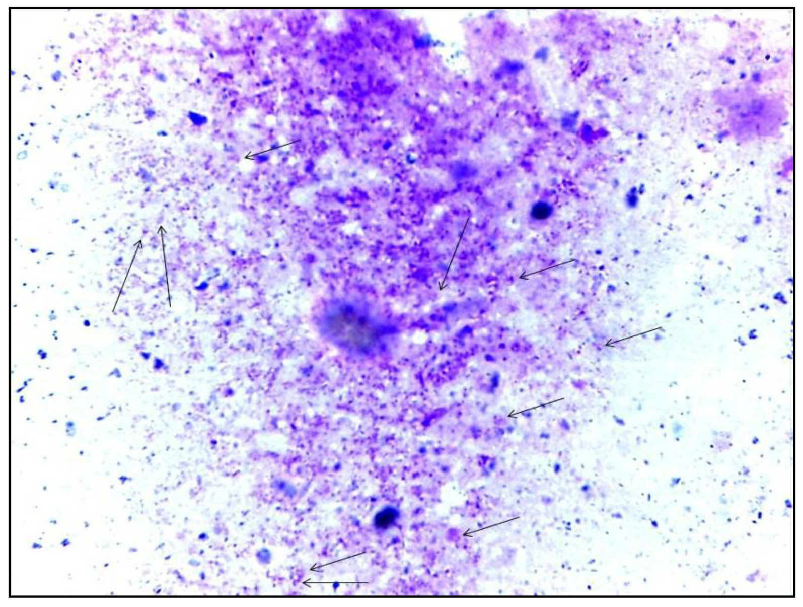
2.2.2. Parasitological Diagnosis

Extracts from two macerated ears of each caught rodent were subjected to microscopic examination (Direct Exam) and parasite culture in Novy–MacNeal–Nicolle (NNN) medium [7].

2.2.3. Molecular Screening

DNA extracted from the spleen of each rodent was used for molecular diagnosis. The presence of Leishmania DNA was tested by PCR targeting ribosomal internal transcribed spacer 1 (ITS1) present in numerous copies of Trypanosoma spp., including Leishmania [14,15]. Amplification was performed using LITSR 5′-CTTG GATCATTTTCCGATG-3′ and L5.8S 5′-TGA TAC CAC TTA TCG CAT T-3′ primers, as previously described [15]. The WHO reference strains L. major MON-25 MHOM/TN2009/S600, L. tropica MON-8 MHOM/TN/2011/MX, and L. infantum MON-1 MHOM/TN/80/IPT1 were used as positive controls.

2.2.4. Leishmania Parasite Typing

Restriction fragment length polymorphism (RFLP) analysis of ITS1-PCR products digested with HaeIII restriction enzyme (Invitrogen, Waltham, MA, USA) was employed to identify the detected Leishmania spp. [15]. In fact, L. major samples present two fragments of 132-bp and 206-bp, L. killicki three fragments of 188-bp, 57-bp, and 26-bp; and L. infantum three fragments of 187-bp, 72-bp, and 55-bp [15].
ITS1-PCR products were directly purified and sequenced in both directions using a conventional Big Dye Terminator Cycle Sequencing Ready Reaction Kit (Perkin Elmer, Applied Biosystems, Foster City, CA, USA) and an ABI373 Automated DNA Sequencer (Thermo Fisher Scientific, Waltham, MA, USA).

2.2.5. Collection of Publicly Available Sequences and Phylogenetic Analyses

For our study, we searched for partial and complete ITS1 sequences belonging to Leishmania parasites in the NCBI database using the following keywords: Leishmania, internal transcribed spacer 1, ITS1, Tunisia, host, and rodents. Three reference sequences for Leishmania spp. (accession numbers: KF152937, MK474651.1, and MK474641.1 for L. major, L. killicki, and L. infantum, respectively) were used to identify the sequences.
Moreover, two datasets were collected: the first included all sequences originating from Tunisia (Supplementary Table S2) and the second regrouped all sequences detected from rodent hosts worldwide (Supplementary Table S3). The retrieved sequences were aligned using the MEGA 6 software (Pennsylvania State University, State College, PA, USA) [16] to study the genetic variability of the ITS1 fragment from the two datasets. The output was edited and regions of unambiguously aligned sequences were retained for the final analysis. Accession numbers of sequences used in the present phylogenetic study are provided in Supplementary Tables S2 and S3.
Maximum likelihood (ML) phylogenetic analysis “SMS: Smart Model Selection in PhyML” (Montpellier University, Montpellier, France) [17] was run to determine the most suitable model of DNA evolution for the two haplotype sets of sequences using hierarchical log-likelihood-ratio tests and the Akaike information criterion.
The best evolutionary model for both datasets was the GTR +G model with estimated gamma distribution shape parameters of 0.915 and 0.497 based on the Tunisian sequences (Supplementary Table S2) and those detected among rodents (Supplementary Table S3), respectively.
Furthermore, identified haplotypes from the two sets of sequences, and their population genetic analysis, including (i) the number of haplotypes (H), (ii) the haplotype diversity (Hd), (iii) the nucleotide diversity (π), (iv) the number of segregating sites (S), (v) the total number of mutations (η), (vi) the average number of nucleotide differences (k), and (vii) the Tajima’s D test for neutrality, were realized and calculated using the DnaSP (Julio Rozas & Universitat de Barcelona, Barcelona, Spain) [18].
These models were used to generate ML haplotype phylogenetic trees with PhyML 3.0 (Montpellier University, Montpellier, France) [19] available in SeaView V4 (Lyon 1 University, Lyon, France) [20], using optimization options and 1000 bootstrap replicates.

2.3. Statistical Analysis

Categorical variables are summarized as frequency counts and percentages. Comparisons between group percentages were performed using the chi-squared test or Fisher’s exact test. Statistical significance was set at p ≤ 0.05. All statistical analyses were performed using STATA software version 11 (StataCorp LLC, TX, USA).

3. Results

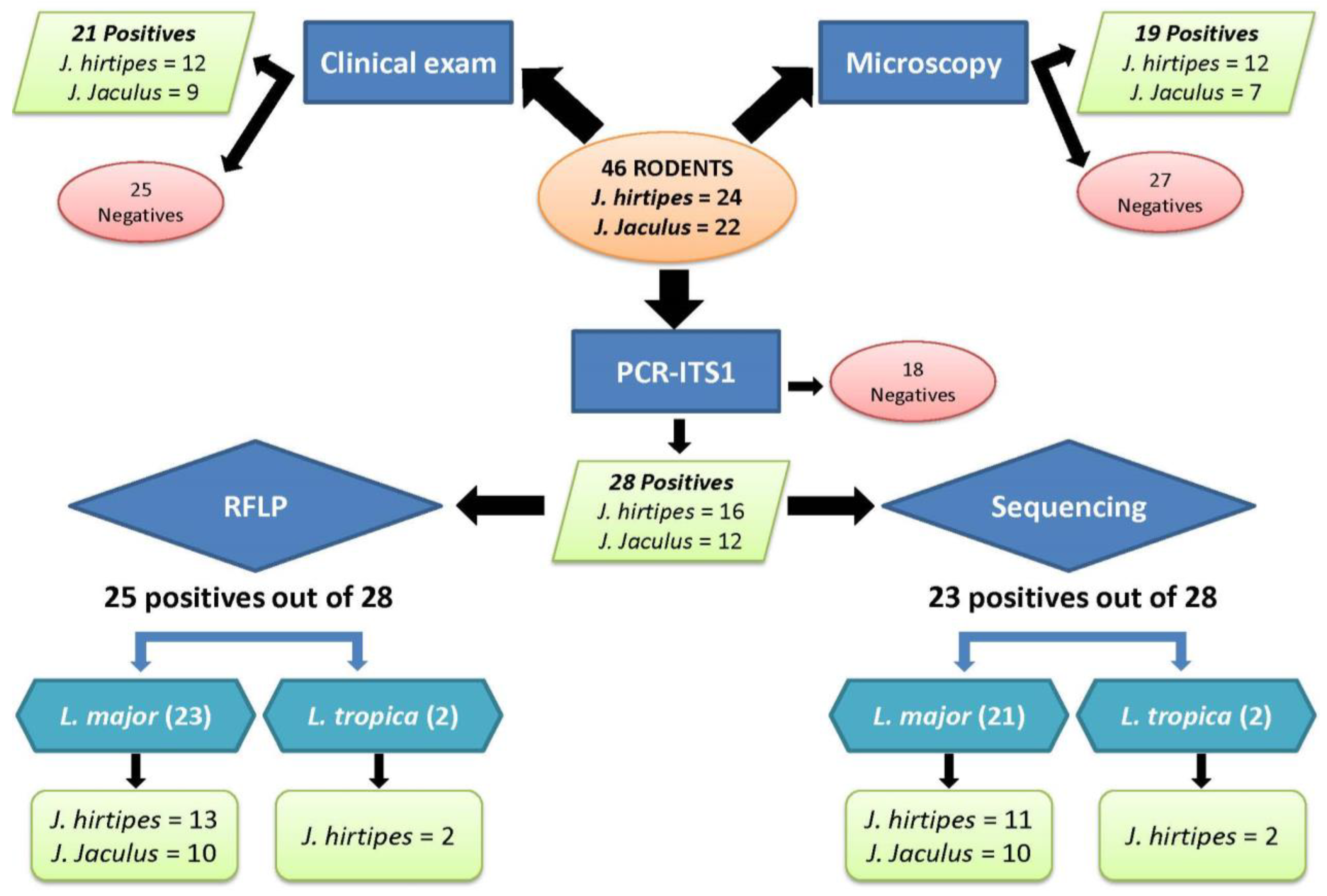
3.1. Rodent Identification

A total of 46 rodents belonging to the Jaculus genus were captured. Phylogenetic analysis of the complete Cytb gene, as previously described [12], identified 21 as belonging to Jaculus (J.) jaculus and 20 to J. hirtipes. A total of 39 short Cytb sequences were successfully amplified, allowing the identification of all rodent populations (Supplementary Table S1). Blasting 360 bp sequences allowed us to identify all the captured rodents as 22 (47.8%) belonging to J. jaculus and 24 (52.2%) to J. hirtipes. No discordance was observed between the two studies. The distribution of these rodents according to capture biotope and gender is illustrated in Table 1.

3.2. Leishmania Infection

The process of Leishmania diagnosis and identification among Jaculus rodents is shown in the flowchart in Figure 1.

3.3. Clinical Manifestations

Clinical examination of captured rodents revealed the presence of skin lesions as small nodules in the back and tail, and, more specifically, in both ears (Figure 2) among 45.7% (21/46) of the rodents tested. Specifically, nine (40.9%) J. jaculus and twelve (50%) J. hirtipes presented at least one clinical sign compatible with CL.

3.4. Leishmania Infection Prevalence

The presence of a confirmed Leishmania infection varied among Jaculus specimens, according to the technique used (Figure 1).

3.4.1. Parasitological Methods

For all studied rodents, the infection prevalence was 41.3% (19/46) by microscopy (Figure 3). Specifically, seven (31.8%) J. jaculus and twelve (50%) J. hirtipes showed the presence of amastigote forms. Unfortunately, we did not succeed in growing Leishmania isolates from these rodents on Novy–MacNeal–Nicolle (NNN) medium because of the high rate of contamination.

3.4.2. Molecular Methods

Leishmania infection determined by ITS1-PCR among all the studied rodents reached 60.9% (28/46) and varied according to Jaculus spp. (Figure 1). Specifically, 12 (54.5%) J. jaculus and 16 (66.7%) J. hirtipes samples were positive.

3.4.3. Combined Methods

Combining these two methods (parasitological and molecular), Leishmania parasites were detected in 37 of the 46 studied Jaculus individuals, reaching a positivity rate of 80.4%. Moreover, there is a non-significant difference between positivity rate of both rodent species (p-value not significant); it was 77.3% (17/22) among J. jaculus and 83.3% (20/24) among J. hirtipes.
A comparison between the overall detected infection and clinical signs revealed a high rate of asymptomatic infection, rising to 59.5% (22/37). According to the rodent spp., asymptomatic infection was 64.7% (11/17) versus 55% (11/20) among J. jaculus and J. hirtipes, respectively (p-value not significant).

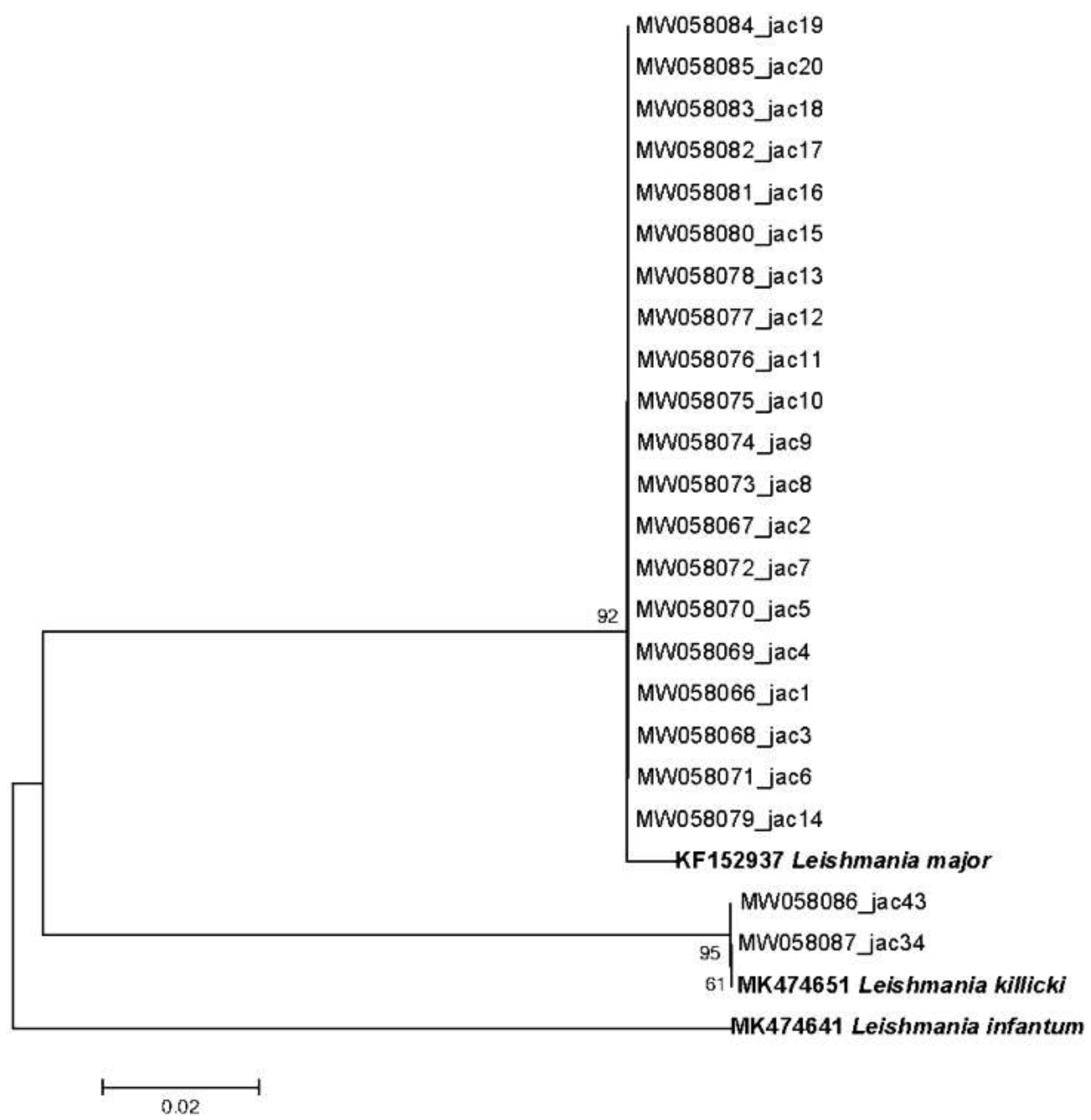
3.5. Leishmania Parasite Typing

Leishmania parasite typing was performed using two methods (Figure 1). First, RFLP analysis allowed the identification of 89% (25/28) of the detected positive ITS1-PCR, such as 23 L. major and 2 L. killicki. In fact, 10 J. jaculus (45.5%) and 13 J. hirtipes (54.2%) individuals were infected by L. major, whereas L. killicki were exclusively found in two J. hirtipes (8.3%) individuals.
Second, 82% (23/28) of the ITS1-PCR-positive samples were successfully sequenced, and their ML phylogenetic tree was constructed using three reference sequences for each Leishmania spp. (accession numbers of Leishmania ITS1 sequences used as references: KF152937, MK474651.1, and MK474641.1 for L. major, L. killicki, and L. infantum), confirming the results of the RFLP analysis (Figure 4).
Only four samples were not double-checked by either method: one of them was not identified by RFLP and found to be L. major by sequencing, and three identified by RFLP as L. major were not successfully sequenced. Finally, two samples were not species-identified either by RFLP or sequencing.
Ultimately, combining the results of both methods, we identified Leishmania spp. in 26 samples: 10 J. jaculus (45.5%) and 14 J. hirtipes (58.3%) were found to be infected by L. major and 2 J. hirtipes (8.3%) were found to be infected by L. killicki.

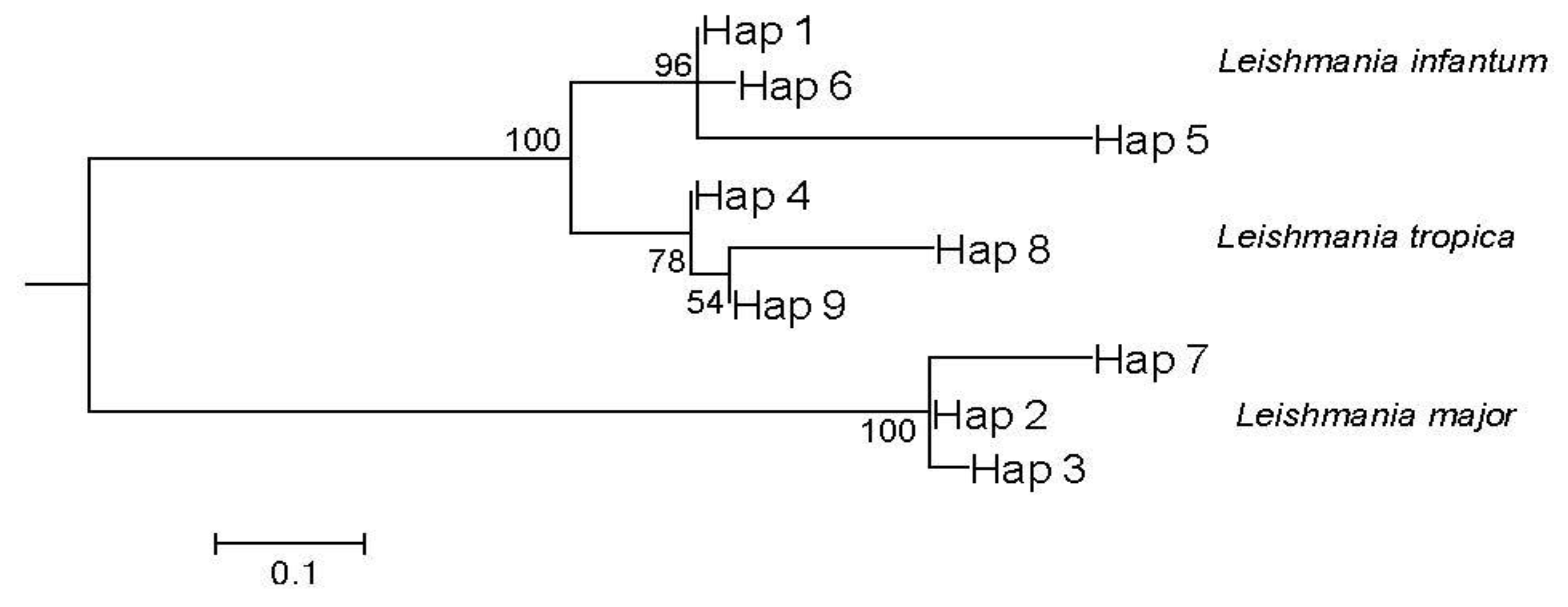
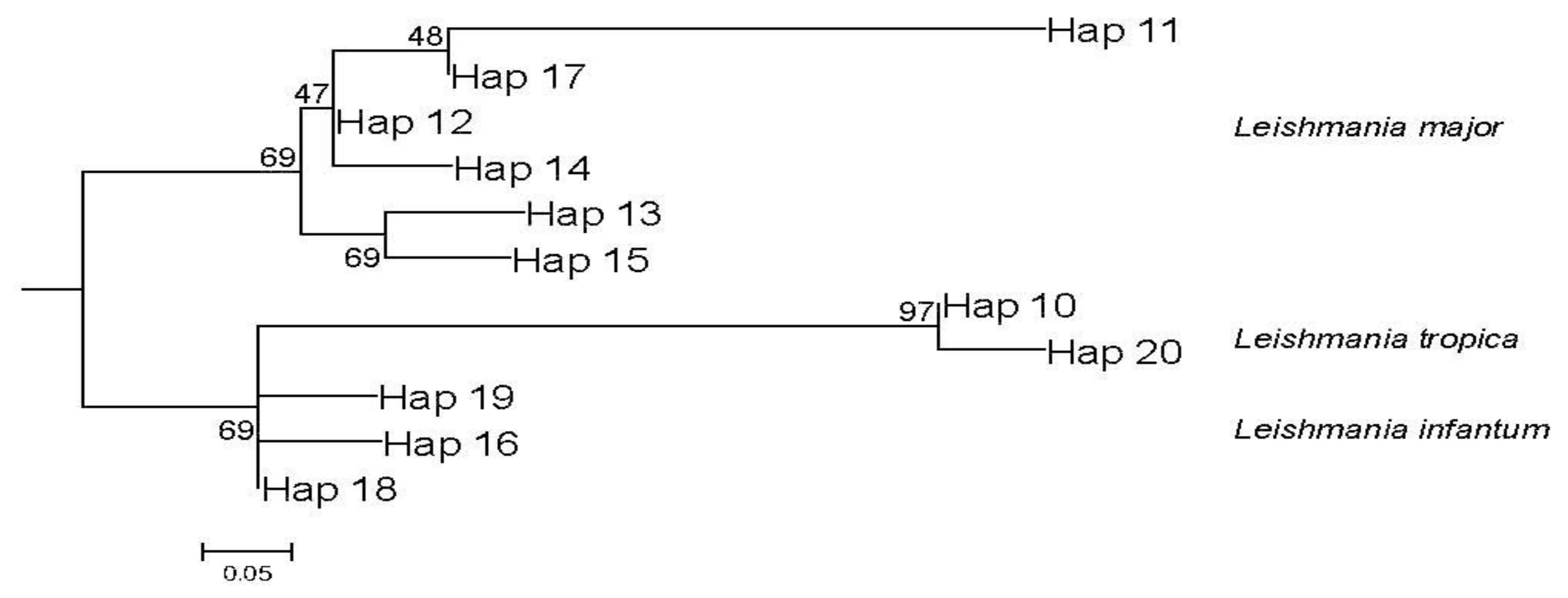
3.6. Leishmania Phylogenetic Analysis

Phylogenetic analysis of all Tunisian Leishmania ITS1 sequences, performed using the ML method, showed three strongly supported clades with high bootstrap values (100) separating Leishmania parasite spp. (Figure 5).
Interestingly, among each of the three clades, we found three different haplotypes: one dominant haplotype encompassing the majority of the sequences regardless of their location, origin, and hosts, and two other haplotypes encompassing only one sequence each. The sequences detected among Jaculus spp. were grouped among the dominant haplotype for either L. major (Hap 2) or L. tropica (Hap 4).
Indices of genetic diversity among the haplotypes determined for the three species of Leishmania parasites are summarized in Table 2. Tajima’s D test, which was used to determine the extent of neutral selection among these Tunisian parasites, generated a positive value (D = 1.2243; p > 0.1).
The same topology was also observed in a phylogenetic study of the worldwide ITS1 Leishmania sequences detected in rodent reservoir hosts. These sequences were sufficient to discriminate Leishmania species (Figure 6).
The number of haplotypes increased compared to the previous phylogenetic analysis. In fact, L. tropica and L. infantum clades were represented by two and three haplotypes, respectively, with a dominant haplotype, while the remaining haplotypes contained a single sequence, whereas the L. major clade comprised six haplotypes with two dominant haplotypes (Hap 12 and Hap 17). Interestingly, each of the latter haplotypes comprised L. major ITS1 sequences from a single geographic location, namely Tunisia and Iran, and each of the four remaining haplotypes contained a single sequence. Similar to the previous analysis, the sequences described in this study were grouped in the dominant haplotype for both parasite spp. (Hap 17 for L. major clade and Hap 10 for L. tropica clade). The indices of genetic diversity among the haplotypes (Figure 6) are summarized in Table 2. Tajima’s D test, used to determine the extent of neutral selection among parasites detected from different rodent reservoir hosts, generated a negative value (D = −0.6; p > 0.05).

4. Discussion

To our knowledge, this is the first study to shed light on the importance of Leishmania infection and its clinical manifestations in two rodent spp. belonging to the Jaculus genus. This novel finding could constitute a breakthrough in understanding the transmission cycles of Old World cutaneous leishmaniasis, with implications for control. Confirmed or suspected reservoir hosts have been identified following the discovery of Leishmania parasites in numerous studies [7,9,21,22,23,24,25,26].
In fact, Jaculus rodents have been found to be infected with many ecto- and endoparasites [27,28,29]. Although Jaculus spp. were found to be susceptible to experimental infection with Leishmania parasites, the infection seems to be chronic and does not impair animal health [30,31], even in Tunisia with L. tropica [32]. However, despite several attempts since 1986, Leishmania parasites have never been isolated and/or detected in these rodents [33,34,35,36,37]. It is worth noting that all of these studies were based on a small sample size of rodents (<10) and were conducted only using parasitological diagnostic methods.
In our study, the Leishmania infection prevalence among Jaculus spp. was 41.3% and 60.9% using parasitological and molecular tools, respectively. These high infection rates fluctuate depending on the rodent spp. Indeed, they increased among J. hirtipes using both methods (50% by direct examination and 66.7% by ITS1-PCR), while they reached only 31.8% by direct examination and 54.4% by ITS1-PCR among tested J. jaculus individuals. Despite these variations, the overall infection prevalence in these rodent spp. (more than 80%) remains the highest compared to previous proportions observed among the potential and/or confirmed rodent reservoir hosts in Tunisia using parasitological, serological, or molecular tools [7,9,21,22,23,38].
Approximately 60% of the overall infections among these rodents were asymptomatic, similar to previous findings in rodent reservoir hosts [9,39]. This might be explained by the fact that Leishmania parasites can disseminate to the spleen during the early stages of infection in asymptomatic animals [40]. This finding, in addition to the clinical manifestation and location of lesions, is in agreement with previous studies performed on reservoir hosts in Tunisia [7,9,22]. This could indicate a long history of co-evolution between these rodent spp. and the detected Leishmania parasites.
RFLP and sequencing approaches, followed by the construction of a phylogenetic tree (Figure 4), allowed for the identification of Leishmania spp. infecting these rodents. Surprisingly, both rodent Jaculus genera were found to be infected with L. major parasites. However, only J. hirtipes was found to be infected with L. killicki. Moreover, two specimens of J. hirtipes infected with L. killicki were captured in Guermessa, which is known to be an old focus of CL owing to L. killicki in Tunisia (according to the Regional Health Directorate and National Control Program of Cutaneous Leishmaniasis). This concomitant infection with two different Leishmania spp. has already been described in other rodents, such as Nesokia indica (L. major and L. infantum) [41], Psammomys spp. (L. major, L. killicki, and L. infantum) [21]; C. gundi (L. major and L. killicki), ref. [9] and Mus musculus Linnaeus, 1758 (L. tropica and L. infantum) [25].
Similar to other rodent reservoir host studies conducted worldwide, our phylogenetic study revealed a low number of available Leishmania ITS1 sequences, regardless of the origin of the host in Tunisia (≤50 sequences (Supplementary Tables S2 and S3)). Furthermore, we confirmed that ITS1 sequences could segregate between Leishmania spp., even for small samples [9,42,43,44,45,46,47,48,49,50].
The Tunisian Leishmania phylogenetic tree (Figure 5) allowing the separation of the parasite spp. failed to show clear grouping according to the sequences, geographical origin, or related hosts. Moreover, this result confirms the heterogeneity of circulating Leishmania parasites in the country, with a dominant haplotype for each parasite sp., even though two more haplotypes were detected containing a single sequence for each Leishmania spp. (Supplementary Table S2).
Furthermore, the genetic diversity among Tunisian sequences was high, as evidenced by the high haplotype diversity (Hd = 0.707) and nucleotide diversity (π = 0.15), which were comparable to the values determined from kinetoplast DNA sequences of other tested Leishmania complexes [51]. This heterogeneity has been reported using other genetic markers among Tunisian Leishmania spp. in previous studies [52,53,54,55].
In addition, the positive value of Tajima’s D test suggests a recent population bottleneck or balancing selection [51,56,57]. The latter seems to be more realistic and can explain the Leishmania parasite evolution in Tunisia, despite the fact that this value was not significant, most likely because of the small number of available Leishmania ITS1 sequences. In addition, genetic variability among Leishmania ITS1 genes at the species level, although not clearly established due to the reduced number of sequences of the determined haplotypes (one sequence for some haplotypes), can be related to the zymodemes of Leishmania parasites circulating in the country. As established for Tunisia, L. major MON-25 and L. killicki MON-8 were the exclusive zymodemes for these dermotropic species, whereas the viscerotropic species were represented by three different zymodemes: MON-24 (the most frequent), MON-1, and MON-80 [2]. In the last decade, many other zymodemes have been detected in Tunisia (L. tropica MON-317) [55] and neighboring countries [2]. This description can explain our results and confirm the presence of other potential zymodemes. These hypotheses should be addressed in future studies.
On the other hand, Leishmania from the phylogenetic tree of rodent hosts (Figure 6) showed a high level of diversity, with 11 haplotypes detected, and a clear clustering according to the geographical origin in L. major clade (Supplementary Table S3). Similarly, the genetic diversity among rodent host sequences was high, as revealed by the high haplotype diversity (Hd = 0.765) and nucleotide diversity (π = 0.03).
In addition, the negative value of the Tajima’s D test suggests, by definition, a population expansion or purifying selection [51]. This could explain the Leishmania parasite evolution among their rodent reservoir hosts, suggesting a purifying geographical link. Enriching databases with more Leishmania ITS1 sequences harbored by rodent reservoir hosts from all over the world could confirm this finding.
This is the first description of Leishmania infection in rodents belonging to the Dipodidae family. In fact, previous studies using ITS1 sequences have described Leishmania infection among rodents belonging to two families: seven species belonging to five genera in the Muridea family and only one species from the Ctenodactylidea family (Figure 6). Confirming that these new rodents are reservoir hosts for cutaneous leishmaniasis might pave the way for more targeted control measures.

5. Conclusions

The present study suggests the role of the two Jaculus spp. as potential reservoir hosts in the transmission cycles of L. major and L. killicki for both the former and only for J. hirtipes in the latter. Despite the small size of this study, the high proportion of infected specimens, the high density of these rodents, and their wide geographic spread in Tunisia [58] and the Old World [59] demand deeper investigations on larger sample sizes with potential striking consequences for CL transmission and control.

Supplementary Materials

The following supporting information can be downloaded at: https://www.mdpi.com/article/10.3390/microorganisms10081502/s1, Table S1: Accession numbers of short and long sequences of Cytochrome b used to identify rodents as belonging to J. jaculus and J. hirtipes.; Table S2: Accessions number of Tunisian Leishmania sequences, their hosts, locations and haplotypes.; Table S3: Accessions number of Leishmania sequences detected from rodent all over the world, their hosts, locations and haplotypes.

Author Contributions

W.G. and A.B.S. conceived the study and acquired the funding; W.G., M.A.S., S.S. and S.C. performed the fieldwork; W.G., M.C., M.A.S., S.S. and A.B. produced the data; W.G. and S.S. performed the laboratory work; W.G., M.C., S.B.A., G.K., D.L. and J.B. analyzed the data; W.G., M.C. and A.B.S. wrote the manuscript with contributions and revisions from all co-authors. All authors have read and agreed to the published version of the manuscript.

Funding

This work was funded in part by the Tunisian Ministry of Higher Education and Scientific Research for the Laboratory of Transmission, Control and Immunobiology of Infections (LR11IPT02), Institut Pasteur de Tunis, and in the frame of the Centre d’Investigation Clinique (PRF-4; CIC2016IPT02). LeiSHield-MATI project “A multi-disciplinary international effort to identify clinical, molecular and social factors impacting cutaneous leishmaniasis” H2020-MSCA-RISE-2017-778298-LeiSHield-MATI partially supported the study. The funding bodies played no role in the design of the study, sample collection, analysis, interpretation, or writing the manuscript.

Institutional Review Board Statement

All animal experiments complied with institutional, national, and international guidelines. The study, rodent collection, and handling protocols were approved by the Biomedical Ethics Committee of the Institut Pasteur de Tunis (document reference: 2016/13E/I/CIC).

Informed Consent Statement

Not applicable.

Data Availability Statement

Cytb and ITS1 sequences were submitted to the GenBank database under accession numbers MT340265-MT340304 and MW058066-MW058088, respectively. The dresses are as follows: www.ncbi.nlm.nih.gov/genbank (accessed on 20 May 2022).

Acknowledgments

We are grateful to the staff of the Regional Directory of Public Health of Tataouine, Tunisia, for their contribution to this work. Our gratitude is also extended to the Regional Direction of Agriculture and Environment and the Forest Directory of Tataouine, which authorized and facilitated the fieldwork. We are also grateful to Samir Taraoui, who contributed to this study by facilitating access to the field.

Conflicts of Interest

The authors declare that they have no competing interest.

References

  1. Klaus, S.N.; Frankenburg, S.; Ingber, A. Epidemiology of cutaneous leishmaniasis. Clin. Dermatol. 1999, 17, 257–260. [Google Scholar] [CrossRef]
  2. Aoun, K.; Bouratbine, A. Cutaneous leishmaniasis in North Africa: A review. Parasite 2014, 21, 14. [Google Scholar] [CrossRef] [PubMed] [Green Version]
  3. Aoun, K.; Amri, F.; Chouihi, E.; Haouas, N.; Bedoui, K.; Benikhlef, R.; Ghrab, J.; Babba, H.; Chahed, M.K.; Harrat, Z.; et al. Epidemiology of Leishmania (L.) infantum, L. major and L. killicki in Tunisia: Results and analysis of the identification of 226 human and canine isolates. Bull. Soc. Pathol. Exot. 2008, 101, 323–328. [Google Scholar] [CrossRef] [PubMed]
  4. Masmoudi, A.; Ayadi, N.; Boudaya, S.; Meziou, T.J.; Mseddi, M.; Marrekchi, S.; Bouassida, S.; Turki, H.; Zahaf, A. Clinical polymorphism of cutaneous leishmaniasis in centre and south of Tunisia. Bull. Soc. Pathol. Exot. 2007, 100, 36–40. [Google Scholar]
  5. Ben Ismail, R.; Gradoni, L.; Gramiccia, M.; Bettini, S.; Ben Rachid, M.S.; Garraoui, A. Epidemic cutaneous leishmaniasis in Tunisia: Biochemical characterization of parasites. Trans. R. Soc. Trop. Med. Hyg. 1986, 80, 669–670. [Google Scholar] [CrossRef]
  6. Benikhlef, R.; Harrat, Z.; Toudjine, M.; Djerbouh, A.; Bendali-Braham, S.; Belkaid, M. Detection of Leishmania infantum MON-24 in the dog. Med. Trop. 2004, 64, 381–383. [Google Scholar]
  7. Ghawar, W.; Snoussi, M.A.; Hamida, N.B.; Boukthir, A.; Yazidi, R.; Chaabane, S.; Chemkhi, J.; Zaatour, A.; Salah, A.B. First report of natural infection of least weasel (Mustela nivalis Linnaeus, 1776) with Leishmania major in Tunisia. Vector Borne Zoonotic Dis. 2011, 11, 1507–1509. [Google Scholar] [CrossRef]
  8. Bousslimi, N.; Ben-Ayed, S.; Ben-Abda, I.; Aoun, K.; Bouratbine, A. Natural infection of North African gundi (Ctenodactylus gundi) by Leishmania tropica in the focus of cutaneous leishmaniasis, Southeast Tunisia. Am. J. Trop. Med. Hyg. 2012, 86, 962–965. [Google Scholar] [CrossRef] [Green Version]
  9. Ghawar, W.; Bettaieb, J.; Salem, S.; Snoussi, M.A.; Jaouadi, K.; Yazidi, R.; Ben-Salah, A. Natural infection of Ctenodactylus gundi by Leishmania major in Tunisia. Acta Trop. 2018, 177, 89–93. [Google Scholar] [CrossRef]
  10. Chemkhi, J.; Souguir, H.; Ali, I.B.; Driss, M.; Guizani, I.; Guerbouj, S. Natural infection of Algerian hedgehog, Atelerix algirus (Lereboullet 1842) with Leishmania parasites in Tunisia. Acta Trop. 2015, 150, 42–51. [Google Scholar] [CrossRef]
  11. Gharaibeh, B.M. Systematics, Distribution, and Zoogeography of Mammals of Tunisia; Texas Tech University: Lubbock, TX, USA, 1997. [Google Scholar]
  12. Ghawar, W.; Chaouch, M.; Ben Abderrazak, S.; Snoussi, M.A.; Salem, S.; Chouchen, S.; Bouaoun, A.; Ben Salah, A.; Bettaieb, J. Evaluation of the Taxonomic Status of Lesser Egyptian Jerboa, Jaculus jaculus: First Description of New Phylogroups in Tunisia. Animals 2022, 12, 758. [Google Scholar] [CrossRef]
  13. Svobodova, M.; Alten, B.; Zidkova, L.; Dvorak, V.; Hlavackova, J.; Myskova, J.; Seblova, V.; Kasap, O.E.; Belen, A.; Votypka, J.; et al. Cutaneous leishmaniasis caused by Leishmania infantum transmitted by Phlebotomus tobbi. Int. J. Parasit. 2009, 39, 251–256. [Google Scholar] [CrossRef]
  14. Njiru, Z.K.; Constantine, C.C.; Guya, S.; Crowther, J.; Kiragu, J.M.; Thompson, R.C.; Davila, A.M. The use of ITS1 rDNA PCR in detecting pathogenic African trypanosomes. Parasitol. Res. 2005, 95, 186–192. [Google Scholar] [CrossRef]
  15. Schonian, G.; Nasereddin, A.; Dinse, N.; Schweynoch, C.; Schallig, H.D.; Presber, W.; Jaffe, C.L. PCR diagnosis and characterization of Leishmania in local and imported clinical samples. Diagn. Microbiol. Infect. Dis. 2003, 47, 349–358. [Google Scholar] [CrossRef]
  16. Tamura, K.; Stecher, G.; Peterson, D.; Filipski, A.; Kumar, S. MEGA6: Molecular Evolutionary Genetics Analysis version 6.0. Mol. Biol. Evol. 2013, 30, 2725–2729. [Google Scholar] [CrossRef] [Green Version]
  17. Lefort, V.; Longueville, J.E.; Gascuel, O. SMS: Smart Model Selection in PhyML. Mol. Biol. Evol. 2017, 34, 2422–2424. [Google Scholar] [CrossRef] [Green Version]
  18. Librado, P.; Rozas, J. DnaSP v5: A software for comprehensive analysis of DNA polymorphism data. Bioinformatics 2009, 25, 1451–1452. [Google Scholar] [CrossRef] [Green Version]
  19. Guindon, S.; Dufayard, J.F.; Lefort, V.; Anisimova, M.; Hordijk, W.; Gascuel, O. New algorithms and methods to estimate maximum-likelihood phylogenies: Assessing the performance of PhyML 3.0. Syst. Biol. 2010, 59, 307–321. [Google Scholar] [CrossRef] [Green Version]
  20. Gouy, M.; Guindon, S.; Gascuel, O. SeaView version 4: A multiplatform graphical user interface for sequence alignment and phylogenetic tree building. Mol. Biol. Evol. 2010, 27, 221–224. [Google Scholar] [CrossRef] [Green Version]
  21. Ben Othman, S.; Ghawar, W.; Chaouch, M.; Ayari, C.; Chemkhi, J.; Cancino-Faure, B.; Tomas-Perez, M.; Alcover, M.M.; Riera, C.; Ben Salah, A.; et al. First detection of Leishmania DNA in Psammomys obesus and Psammomys vexillaris: Their potential involvement in the epidemiology of leishmaniasis in Tunisia. Infect. Genet. Evol. 2018, 59, 7–15. [Google Scholar] [CrossRef]
  22. Ben-Ismail, R.; Ben Rachid, M.S.; Gradoni, L.; Gramiccia, M.; Helal, H.; Bach-Hamba, D. Zoonotic cutaneous leishmaniasis in Tunisia: Study of the disease reservoir in the Douara area. Ann. Soc. Belg. Med. Trop. 1987, 67, 335–343. [Google Scholar] [PubMed]
  23. Ben-Ismail, R.; Khaled, S.; Makni, S.; Ben Rachid, M.S. Anti-leishmanial antibodies during natural infection of Psammomys obesus and Meriones shawi (Rodentia, Gerbillinae) by Leishmania major. Ann. Soc. Belg. Med. Trop. 1989, 69, 35–40. [Google Scholar] [PubMed]
  24. Davami, M.H.; Motazedian, M.H.; Kalantari, M.; Asgari, Q.; Mohammadpour, I.; Sotoodeh-Jahromi, A.; Solhjoo, K.; Pourahmad, M. Molecular Survey on Detection of Leishmania Infection in Rodent Reservoirs in Jahrom District, Southern Iran. J. Arthropod. Borne Dis. 2014, 8, 139–146. [Google Scholar] [PubMed]
  25. Echchakery, M.; Chicharro, C.; Boussaa, S.; Nieto, J.; Carrillo, E.; Sheila, O.; Moreno, J.; Boumezzough, A. Molecular detection of Leishmania infantum and Leishmania tropica in rodent species from endemic cutaneous leishmaniasis areas in Morocco. Parasit. Vectors 2017, 10, 454. [Google Scholar] [CrossRef] [Green Version]
  26. Parvizi, P.; Moradi, G.; Akbari, G.; Farahmand, M.; Ready, P.D.; Piazak, N.; Assmar, M.; Amirkhani, A. PCR detection and sequencing of parasite ITS-rDNA gene from reservoirs host of zoonotic cutaneous leishmaniasis in central Iran. Parasitol. Res. 2008, 103, 1273–1278. [Google Scholar] [CrossRef]
  27. Faleh, A.B.; Annabi, A.; López, S.; Said, K.; Ribas, A. On the helminth parasites of the genus Jaculus (Rodentia: Dipodidae) in Tunisia: A preliminary survey study. Leb. Sci. J. 2012, 13, 1107. [Google Scholar]
  28. Hoogstraal, H. The life cycle and incidence of Hepatozoon balfouri (Laveran, 1905) in Egyptian jerboas (Jaculus spp.) and mites (Haemolaelaps aegyptius Keegan, 1956). J. Protozool. 1961, 8, 231–248. [Google Scholar] [CrossRef]
  29. Morsy, T.A.; el Bahrawy, A.A.; al Dakhil, M.M.; Abdel Mawla, M.M. Babesia infection in rodents trapped in Riyadh Region, Saudi Arabia, with a general discussion. J. Egypt. Soc. Parasitol. 1994, 24, 177–185. [Google Scholar]
  30. Al-Taqi, M.; Mohammed, A.H. Susceptibility of Kuwaiti rodents and experimental mice to isolates of Leishmania spp. Trans. R. Soc. Trop. Med. Hyg. 1981, 75, 133–136. [Google Scholar] [CrossRef]
  31. Archibald, R. A Preliminary Report on some Further Investigations on Kala Azar in the Sudan. BMJ Mil. Health 1914, 23, 479–495. [Google Scholar]
  32. Dedet, P.; Ben Osman, F. Sensibilite experimentale de divers rongeurs de Tunisie vis-a-vis de souches autochtones de Leishmania tropica (Wright, 1903) et de Leishmania donovani (Laveran & Mesnil, 1903). Arch. Inst. Pasteur Tunis 1973, 50, 85–94. [Google Scholar]
  33. Bin, D.S.; Mostafa, O.M.; Abdoon, A.; Al-Quraishy, S.A.; Alqahtani, A.A. Isoenzyme electrophetic characterization of Leishmania major, the causative agent of zoonotic cutaneous Leishmaniasis in North and West Saudi Arabia. J. Egypt. Soc. Parasitol. 2010, 40, 465–478. [Google Scholar]
  34. Hamadto, H.A.; Al, F.A.; Farrag, A.B.; Abdel Maksoud, M.K.; Morsy, T.A. Zoonotic cutaneous leishmaniasis: Reservoir host and insect vector in north Sinai, Egypt. J. Egypt. Soc. Parasitol. 2007, 37, 843–850. [Google Scholar]
  35. Morsy, T.A.; Naser, A.M.; el Gibali, M.R.; Anwar, A.M.; el Said, A.M. Studies on zoonotic cutaneous leishmaniasis among a group of temporary workers in North Sinai Governorate, Egypt. J. Egypt. Soc. Parasitol. 1995, 25, 99–106. [Google Scholar]
  36. Nadim, A.; Faghih, M. The epidemiology of cutaneous leishmaniasis in the Isfahan province of Iran. I. The reservoir. II. The human disease. Trans. R. Soc. Trop. Med. Hyg. 1968, 62, 534–542. [Google Scholar] [CrossRef]
  37. Rioux, J.A.; Dereure, J.; Khiami, A.; Pratlong, F.; Sirdar, K.; Lambert, M. Ecoepidemiology of leishmaniasis in Syria. 1. Leishmania major Yakimoff and Schokhor (Kinetoplastida-Trypanosomatidae) infestation of Psammomys obesus Cretzschmar (Rodentia-Gerbillidae). Ann. Parasitol. Hum. Comp. 1990, 65, 203–207. [Google Scholar] [CrossRef] [Green Version]
  38. Fichet-Calvet, E.; Jomaa, I.; Ben Ismail, R.; Ashford, R.W. Leishmania major infection in the fat sand rat Psammomys obesus in Tunisia: Interaction of host and parasite populations. Ann. Trop. Med. Parasitol. 2003, 97, 593–603. [Google Scholar] [CrossRef]
  39. Ghawar, W.; Toumi, A.; Snoussi, M.A.; Chlif, S.; Zaatour, A.; Boukthir, A.; Hamida, N.B.; Chemkhi, J.; Diouani, M.F.; Ben-Salah, A. Leishmania major infection among Psammomys obesus and Meriones shawi: Reservoirs of zoonotic cutaneous leishmaniasis in Sidi Bouzid (central Tunisia). Vector Borne Zoonotic Dis. 2011, 11, 1561–1568. [Google Scholar] [CrossRef] [Green Version]
  40. Schilling, S.; Glaichenhaus, N. T cells that react to the immunodominant Leishmania major LACK antigen prevent early dissemination of the parasite in susceptible BALB/c mice. Infect. Immun. 2001, 69, 1212–1214. [Google Scholar] [CrossRef] [Green Version]
  41. Pourmohammadi, B.; Mohammadi-Azni, S.; Kalantari, M. Natural infection of Nesokia indica with Leishmania major and Leishmania infantum parasites in Damghan city, Northern Iran. Acta Trop. 2017, 170, 134–139. [Google Scholar] [CrossRef]
  42. Ajaoud, M.; Es-sette, N.; Hamdi, S.; El-Idrissi, A.L.; Riyad, M.; Lemrani, M. Detection and molecular typing of Leishmania tropica from Phlebotomus sergenti and lesions of cutaneous leishmaniasis in an emerging focus of Morocco. Parasit. Vectors 2013, 6, 217. [Google Scholar] [CrossRef] [Green Version]
  43. Chicharro, C.; Llanes-Acevedo, I.P.; Garcia, E.; Nieto, J.; Moreno, J.; Cruz, I. Molecular typing of Leishmania infantum isolates from a leishmaniasis outbreak in Madrid, Spain, 2009 to 2012. Eur. Surveill 2013, 18, 20545. [Google Scholar] [CrossRef] [Green Version]
  44. Ghatee, M.A.; Sharifi, I.; Kuhls, K.; Kanannejad, Z.; Harandi, M.F.; de Almeida, M.E.; Hatam, G.; Mirhendi, H. Heterogeneity of the internal transcribed spacer region in Leishmania tropica isolates from southern Iran. Exp. Parasitol. 2014, 144, 44–51. [Google Scholar] [CrossRef] [PubMed]
  45. Hajjaran, H.; Mohebali, M.; Mamishi, S.; Vasigheh, F.; Oshaghi, M.A.; Naddaf, S.R.; Teimouri, A.; Edrissian, G.H.; Zarei, Z. Molecular identification and polymorphism determination of cutaneous and visceral leishmaniasis agents isolated from human and animal hosts in Iran. Biomed. Res. Int. 2013, 2013, 789326. [Google Scholar] [CrossRef] [Green Version]
  46. Jafari, R.; Najafzadeh, N.; Sedaghat, M.M.; Parvizi, P. Molecular characterization of sandflies and Leishmania detection in main vector of zoonotic cutaneous leishmaniasis in Abarkouh district of Yazd province, Iran. Asian Pac. J. Trop. Med. 2013, 6, 792–797. [Google Scholar] [CrossRef] [Green Version]
  47. Mahdy, M.A.K.; Al-Mekhlafi, H.M.; Al-Mekhlafi, A.M.; Lim, Y.A.L.; Bin Shuaib, N.O.M.; Azazy, A.A.; Mahmud, R. Molecular Characterization of Leishmania Species Isolated from Cutaneous Leishmaniasis in Yemen. PLoS ONE 2010, 5, e12879. [Google Scholar] [CrossRef]
  48. Salloum, T.; Khalifeh, I.; Tokajian, S. Detection, molecular typing and phylogenetic analysis of Leishmania isolated from cases of leishmaniasis among Syrian refugees in Lebanon. Parasite Epidemiol. Control 2016, 1, 159–168. [Google Scholar] [CrossRef] [Green Version]
  49. Spotin, A.; Rouhani, S.; Parvizi, P. The associations of Leishmania major and Leishmania tropica aspects by focusing their morphological and molecular features on clinical appearances in Khuzestan Province, Iran. Biomed. Res. Int 2014, 2014, 913510. [Google Scholar] [CrossRef] [Green Version]
  50. Zahirnia, A.H.; Bordbar, A.; Ebrahimi, S.; Spotin, A.; Mohammadi, S.; Ghafari, S.M.; Ahmadvand, S.; Jabbari, N.; Esmaeili Rastaghi, A.R.; Parvizi, P. Predominance of Leishmania major and rare occurrence of Leishmania tropica with haplotype variability at the center of Iran. Braz. J. Infect. Dis. 2018, 22, 278–287. [Google Scholar] [CrossRef]
  51. Kariyawasam, U.L.; Selvapandiyan, A.; Rai, K.; Wani, T.H.; Ahuja, K.; Beg, M.A.; Premathilake, H.U.; Bhattarai, N.R.; Siriwardena, Y.D.; Zhong, D.B.; et al. Genetic diversity of Leishmania donovani that causes cutaneous leishmaniasis in Sri Lanka: A cross sectional study with regional comparisons. BMC Infect. Dis 2017, 17, 791. [Google Scholar] [CrossRef] [Green Version]
  52. Attia, H.; Sghaier, R.M.; Gelanew, T.; Bali, A.; Schweynoch, C.; Guerfali, F.Z.; Mkannez, G.; Chlif, S.; Belhaj-Hamida, N.; Dellagi, K.; et al. Genetic micro-heterogeneity of Leishmania major in emerging foci of zoonotic cutaneous leishmaniasis in Tunisia. Infect. Genet. Evol. 2016, 43, 179–185. [Google Scholar] [CrossRef] [PubMed]
  53. Harrabi, M.; Bettaieb, J.; Ghawar, W.; Toumi, A.; Zaatour, A.; Yazidi, R.; Chaabane, S.; Chalghaf, B.; Hide, M.; Banuls, A.L.; et al. Spatio-temporal Genetic Structuring of Leishmania major in Tunisia by Microsatellite Analysis. PLoS Negl. Trop. Dis. 2015, 9, e0004017. [Google Scholar] [CrossRef] [PubMed]
  54. Chaouch, M.; Fathallah-Mili, A.; Driss, M.; Lahmadi, R.; Ayari, C.; Guizani, I.; Ben Said, M.; BenAbderrazak, S. Identification of Tunisian Leishmania spp. by PCR amplification of cysteine proteinase B (cpb) genes and phylogenetic analysis. Acta Trop. 2013, 125, 357–365. [Google Scholar] [CrossRef] [PubMed]
  55. Chaara, D.; Ravel, C.; Banuls, A.; Haouas, N.; Lami, P.; Talignani, L.; El Baidouri, F.; Jaouadi, K.; Harrat, Z.; Dedet, J.P.; et al. Evolutionary history of Leishmania killicki (synonymous Leishmania tropica) and taxonomic implications. Parasit. Vectors 2015, 8, 198. [Google Scholar] [CrossRef] [Green Version]
  56. Shafiei, R.; Kalantari, M.; Yousefi, M.; Aspatwar, A.; Arzamani, K.; Bozorgomid, A.; Mirahmadi, H.; Soleimani, A.; Raeghi, S. Bionomics and phylo-molecular analysis of Leishmania species isolated from human lesions using ITS1 genes in northeast of Iran. J. Parasit. Dis. 2021, 45, 754–761. [Google Scholar] [CrossRef]
  57. El Hamouchi, A.; Ajaoud, M.; Arroub, H.; Charrel, R.; Lemrani, M. Genetic diversity of Leishmania tropica in Morocco: Does the dominance of one haplotype signify its fitness in both predominantly anthropophilic Phlebotomus sergenti and human beings? Transbound. Emerg. Dis. 2019, 66, 373–380. [Google Scholar] [CrossRef] [Green Version]
  58. Ben Faleh, A.; Granjon, L.; Tatard, C.; Boratynski, Z.; Cosson, J.F.; Said, K. Phylogeography of two cryptic species of African desert jerboas (Dipodidae: Jaculus). Biol. J. Linn. Soc. 2012, 107, 27–38. [Google Scholar] [CrossRef] [Green Version]
  59. Moutinho, A.F.; Seren, N.; Pauperio, J.; Silva, T.L.; Martinez-Freiria, F.; Sotelo, G.; Faria, R.; Mappes, T.; Alves, P.C.; Brito, J.C.; et al. Evolutionary history of two cryptic species of northern African jerboas. BMC Evol. Biol. 2020, 20, 26. [Google Scholar] [CrossRef]
Figure 1. Flowchart summarizing the process and the results of Leishmania diagnosis and identification among Jaculus species. PCR-ITS1: PCR targeting ribosomal internal transcribed spacer 1; RFLP: Restriction fragment length polymorphism.
Microorganisms 10 01502 g001
Figure 2. Jaculus jaculus presenting leishmaniasis clinical manifestation as a small nodule on the higher edge of the ear.
Microorganisms 10 01502 g002
Figure 3. Leishmania amastigotes in Giemsa-stained smears from the ear of Jaculus species under oil immersion (×1000) (Leica DM50).
Microorganisms 10 01502 g003
Figure 4. Maximum likelihood phylogenetic tree constructed with Leishmania ITS1 sequences detected among Jaculus species and three sequences used as references (bold-faced). The numbers above each sequence are their accession numbers, and those above the branches are bootstrap percentages of 1000 replications.
Microorganisms 10 01502 g004
Figure 5. Maximum likelihood phylogenetic tree for 9 haplotypes of all Tunisian Leishmania parasites based on ITS1-rDNA gene sequences. Sequences fitting for each haplotype were defined in Supplementary Table S2. The numbers above the branches are bootstrap percentages of 1000 replications.
Microorganisms 10 01502 g005
Figure 6. Maximum-likelihood phylogenetic tree for 11 haplotypes of all rodent hosts–Leishmania parasites based on ITS1-rDNA gene sequences. Sequences fitting for each haplotype are defined in Supplementary Table S3. Haplotypes are numbered from 10 to 20 to avoid confusion with the previous figure. The numbers above the branches are bootstrap percentages of 1000 replications.
Microorganisms 10 01502 g006
Table 1. Distribution of the captured rodents by location and gender.
Jaculus (J.) jaculus
n = 22
J. hirtipes
n = 24
Location of capture * n (%)
−  Guermessa8 (36.0)8 (33.3)
−  BniMhira14 (64.0)16 (66.7)
Gender * n (%)
−  Males4 (18.2)6 (25.0)
−  Females18 (81.8)18 (75.0)
*: no significant association.
Table 2. Indices of genetic diversity for the two studied sets of Leishmania sequences.
Sets of SequencesNSHSηkHd (SD)π (SD)
Different hosts
(Tunisia)
739404412.440.707 (0.029)0.1575 (0.004)
Rodents hosts
(all world)
711116182.970.765 (0.031)0.0327 (0.003)
NS: number of sequences, H: number of haplotypes, S: number of segregating sites, η: total number of mutations, k: average number of nucleotide differences, Hd: haplotype diversity, π: nucleotide diversity, SD: standard deviation.
Publisher’s Note: MDPI stays neutral with regard to jurisdictional claims in published maps and institutional affiliations.

