Review

Microbiology and Epidemiology of Escherichia albertii—An Emerging Elusive Foodborne Pathogen

1
Institute for Food Safety and Hygiene, Vetsuisse Faculty, University of Zurich, 8057 Zurich, Switzerland
2
Department of Veterinary and Biosciences, Faculty of Veterinary Medicine, Ghent University, 9820 Merelbeke, Belgium
*
Author to whom correspondence should be addressed.
Microorganisms 2022, 10(5), 875; https://doi.org/10.3390/microorganisms10050875
Submission received: 5 April 2022 / Revised: 19 April 2022 / Accepted: 19 April 2022 / Published: 22 April 2022
(This article belongs to the Section Food Microbiology)

Abstract

:
Escherichia albertii, a close relative of E. coli, is an emerging zoonotic foodborne pathogen associated with watery diarrhea mainly in children and immunocompromised individuals. E. albertii was initially classified as eae-positive Hafnia alvei, however, as more genetic and biochemical information became available it was reassigned to its current novel taxonomy. Its infections are common under conditions of poor hygiene with confirmed transmission via contaminated water and food, mainly poultry-based products. This pathogen has been isolated from various domestic and wild animals, with most isolates being derived from birds, implying that birds among other wild animals might act as its reservoir. Due to the absence of standardized isolation and identification protocols, E. albertii can be misidentified as other Enterobacteriaceae. Exploiting phenotypes such as its inability to ferment rhamnose and xylose and PCR assays targeting E. albertii-specific genes such as the cytolethal distending toxin and the DNA-binding transcriptional activator of cysteine biosynthesis encoding genes can be used to accurately identify this pathogen. Several gaps exist in our knowledge of E. albertii and need to be bridged. A deeper understanding of E. albertii epidemiology and physiology is required to allow the development of effective measures to control its transmission and infections. Overall, current data suggest that E. albertii might play a more significant role in global infectious diarrhea cases than previously assumed and is often overlooked or misidentified. Therefore, simple, and efficient diagnostic tools that cover E. albertii biodiversity are required for effective isolation and identification of this elusive agent of diarrhea.

1. Introduction

The emerging zoonotic foodborne pathogen Escherichia albertii is a Gram-negative rod, nonmotile, non-spore-forming, facultative anaerobe belonging to the Enterobacteriaceae family [1,2]. It is one of five species within the genus Escherichia [3]. Emerging data indicate that E. albertii is a diverse pathogen with two clades divided into eight genetic lineages, that have a geographical distribution bias, described to date [4,5,6]. E. albertii was first identified and reported as Hafnia alvei in diarrhea cases in children from Bangladesh and later reclassified as a new Escherichia species in 2003 [1,7,8]. Since then, E. albertii has been implicated in several outbreaks but is often mistakenly identified as enteropathogenic E. coli (EPEC) or enterohemorrhagic E. coli (EHEC) because of its genetic and phenotypic similarity to these pathogens [9,10,11,12,13,14]. For instance, E. albertii commonly carries the eae gene that encodes intimin, an important virulence factor also harbored by pathogenic subgroups of E. coli [10,13,15,16]. This has probably resulted in underestimation of E. albertii infection numbers. For example, in several gastroenteritis outbreaks the causative pathogen was wrongly diagnosed as EPEC instead of eae-positive E. albertii [9,11,12,14,17,18]. Because of this, several enrichment broths, isolation, and identification protocols have been developed, however, to date, there is no standard method [19,20,21,22,23]. The specificity and sensitivity for most of these protocols though high [24], have been evaluated based on a limited number of strains, thus it is not clear if such protocols cover the biodiversity of E. albertii isolates. In addition, the suggested further identification and characterization approaches such as E. albertii-specific PCR, multilocus sequence type (MLST) analysis, and O-genotyping are too costly, time-consuming, or not available in most routine laboratories, especially in resource-limited areas where such cases might occur more frequently [4,25,26]. Coupled with the fact that E. albertii is underappreciated as an enteric pathogen [27], it reduces the chance of it being included on the differential diagnosis list, further compounding identification. Therefore, E. albertii misclassification will probably continue until a novel, simple and accurate diagnostic tool is created. Moreover, awareness of this enteropathogenic Escherichia species needs to be increased.
E. albertii infections mostly present as watery diarrhea, fever, and abdominal pain, with most cases resolving without complications [5,14]. Genes such as eae, associated with attaching and effacing lesions, cdt cytolethal distending, and Shiga toxins (stx), among others, make part of the virulence arsenal contributing to this pathogens’ clinical manifestations [5,11]. Early outbreak and sporadic case reports indicate that E. albertii is transmitted through contaminated water and food such as salads, chicken, and packed lunches [9,11,12,14,28]. Due to poor hygiene and sanitary conditions that prevail in several countries, such infectious gastroenteritis will remain an important cause of morbidity and mortality [29,30]. Therefore, we need to expand our knowledge on infectious gastroenteritis causative agents to better control and prevent such infections. In this article, we review recent advancements in our understanding of key aspects, e.g., the biochemistry and epidemiology of E. albertii (Figure 1). Emphasis is made on the currently available isolation and identification diagnostic modalities. Knowledge gaps are highlighted to provide a basis for future research on this emerging elusive zoonotic pathogen.

2. Biochemical Properties of Escherichia albertii

Initially, the biochemical properties of E. albertii were clear with key biochemical makers being an inability to produce indole from tryptophan or to ferment lactose, xylose and rhamnose [1,10,20,31,32,33]. These phenotypes are due to the absence of effector proteins such as those encoded by the rhaMDABSRT (rhamnose utilization) and xylBAFGHR (xylose utilization) operons required for these metabolic functions in E. albertii [13]. Based on a limited number of strains, E. albertii was also initially reported as unable to ferment sucrose and melibiose [1,32]. However, as the number of available isolates increased this narrative changed. Some E. albertii strains have been confirmed to be lactose, melibiose, and sucrose fermenters as well as capable of producing indole from tryptophan [26,34,35,36,37]. Interestingly, some of these strains lack the known genes required for the utilization of these sugars, suggestive that they may encode yet-to-be-described pathways for utilization of these carbon sources. For instance, E. albertii strain NIAH_Bird_23 showed a weak lactose fermentation phenotype but lacks the lacA, lacY, and lacI genes required for lactose utilization [13].
With further analysis of more isolates, additional phenotypic variability among E. albertii strains is being observed including utilization of maltose, d-mannitol, d-sorbitol, trehalose, and ortho-nitrophenyl-B-galactoside (ONPG), esculin hydrolysis, β-galactosidase, lysine-decarboxylase and l-prolineaminopeptidase activity (Table 1) [10,33,38]. It is likely that as more isolates are characterized, variability in these biochemical properties will also increase, giving a clearer picture of the true E. albertii distinguishing features. It has been postulated that such phenotypic variability is suggestive that biotypes or biovars may exist within E. albertii. Using some of these phenotypes, three biogroups have been described (Table 2) [36], however, not all E. albertii strains fit in these groups [39].
According to currently available data, isolates of this pathogen analyzed so far, can reduce nitrate, assimilate 3-hydroxybenzoate, utilize glucose, D-mannose, mannitol, and galactose, and are positive for methyl red (Table 1) [1,27,32,38]. This bacterium does not grow on KCN medium or produce H2S and is nonmotile, however, E. albertii carries fully conserved flagellar biosynthesis genes [2,13,20,33]. Most E. albertii strains do not utilize several common and uncommon sugars, such as adonitol, cellobiose, 2-ketogluconate, myoinositol, rhamnose, melibiose xylose, and xylitol, phenotypes potentially exploitable for E. albertii differentiation from other Enterobacteriaceae [26,32,35,37,38]. However, the feasibility of using some of these sugars in routine diagnostic media is restricted due to their cost. E. albertii is also negative for chitinase, oxidase, hydroxyproline deaminase, tripeptidase, and proline deaminase (Table 1) [1,10,31,32,33]. Although not commonly used in most routine laboratories, such phenotypes might be useful in distinguishing E. albertii from other species such as H. alvei and E. coli.

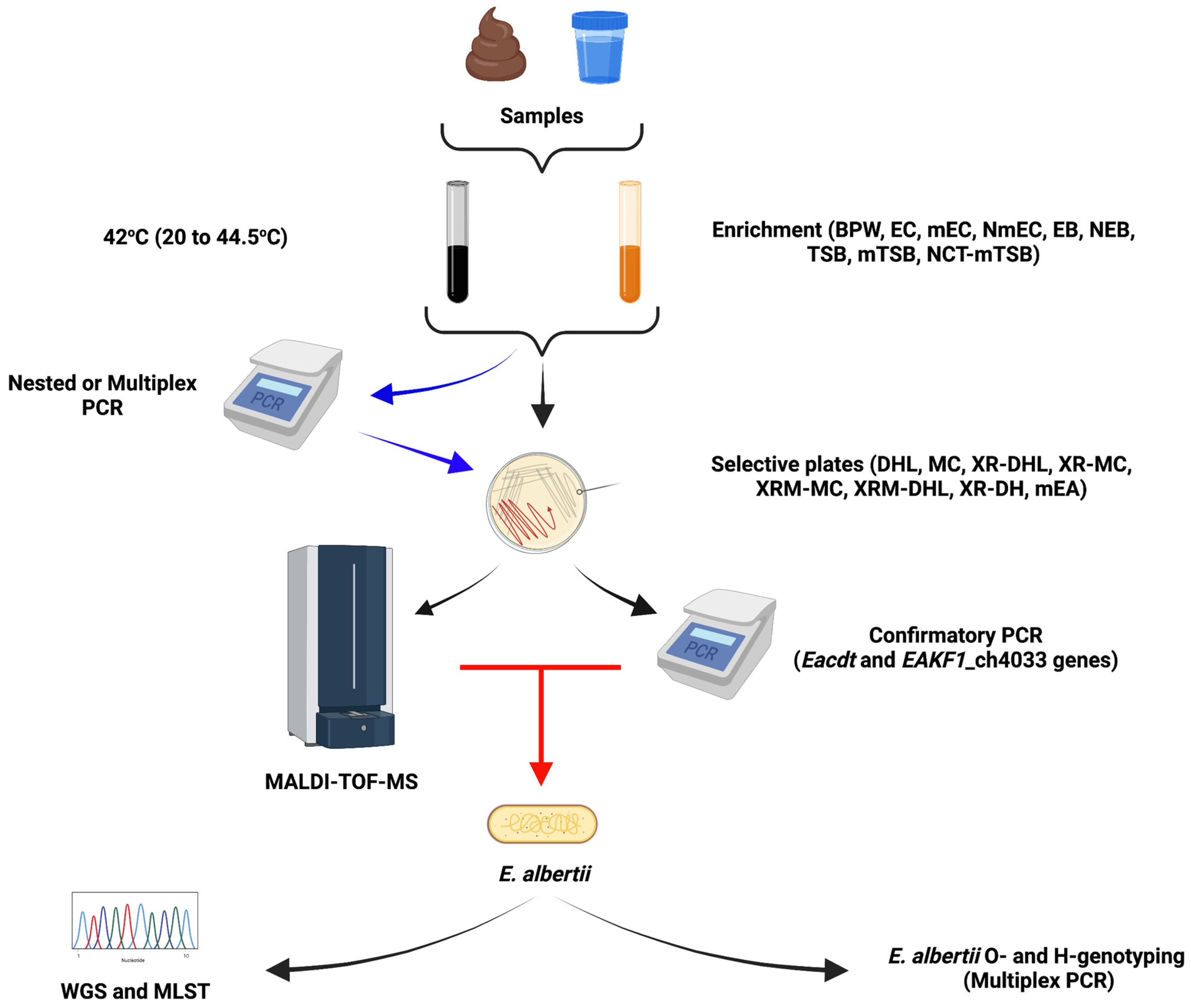
3. Detection and Identification Methods for Escherichia albertii

Discriminating E. albertii from other Enterobacteriaceae is challenging. This zoonotic pathogen is often misclassified as E. coli, H. alvei, Shigella boydi serotype 13, Salmonella enterica, and Yersinia ruckeri due to their similar phenotypic and genetic features [2,7,10,19,26,31,32,41,43]. Moreover, no commercial test is available to differentiate E. albertii from other Enterobacteriaceae. On the systems that currently exist, E. albertii isolates are often misidentified. For instance, on the MicroScan dried overnight panels and Micronaut-E system, they are identified as Y. ruckeri while on the API 20E and Vitek systems some strains are identified as H. alvei [32,36,38].
A multitude of different protocols have been proposed for differentiating E. albertii from other Enterobacteriaceae, however, since these methods have been validated using a minimum number of strains from limited sources, the extent of their accuracy is questionable. Moreover, these methods especially the agar and enrichment broths are not selective for E. albertii, most of them are modifications of methods used for Enterobacteriaceae, hence other species within this family can grow [23]. Moreover, earlier protocols were based on the now invalidated hypothesis that no E. albertii strains can ferment sucrose and lactose which might have also led to the misclassification of some E. albertii strains [2,9,10,12,21,33,34,44,45,46,47].

3.1. Enrichment Broths

Most E. albertii isolation protocols rely on a single enrichment step using various broths including buffered peptone water (BPW), E. coli broth (EC), modified EC (mEC), novobiocin–mEC (NmEC), tryptic soy broth (TSB), modified TSB (mTSB), or novobiocin–cefixime–tellurite supplemented mTSB (NCT-mTSB) at various temperatures ranging from 20 to 44.5 °C [22,23,37,46,48,49,50]. The added novobiocin (20 to 25 mg/L) inhibits most Gram-positive bacteria, while in the NCT-mTSM enrichment broth the tellurite (1 mg/L) and cerufuxim (0.05 mg/L) inhibit most Enterobacteria, while growth at temperatures ≥ 42 °C is postulated to inhibit most background microbiota, Shigella spp. and E. coli [22,23,37]. Enrichment protocols that employ antimicrobial selective pressure though promising, run the risk of selecting only E. albertii strains resistant to these antimicrobial agents especially when they are validated using a few strains from limited sources and or geographical regions. Since most E. albertii were initially identified as E. coli, one can speculate that Enterobacteriaceae enrichment broth (EB) is a good option for the enrichment of samples for E. albertii isolation. However, since some Enterobacteriaceae selective media use lactose as the main carbon source, caution must be exercised as most E. albertii do not ferment lactose and might be outcompeted by lactose fermenting Enterobacteriaceae [10,36].

3.2. Molecular Approaches

Several PCR protocols have been proposed, most of which are based on the lysP, mdh, clpX, yejH, and yejK, EAKF1_ch4033, and the species-specific cdt gene [13,19,22,24,33,51]. The eae gene was also proposed as a gene marker for E. albertii but this was soon discarded as several pathogenic E. coli strains also carry this gene [5,24]. This approach also discriminates against eae-negative E. albertii isolates [6]. In addition, some studies have reported nonspecific amplification in assays targeting lysP and mdh genes indicative of lower sensitivity for such protocols [24,52]. Using 67 E. albertii strains Hinenoya et al. [24], observed varying sensitivities from 98.5 to 100% of different PCR assays developed by Ooka et al. [13], Hyma et al. [19], and Lindsey et al. [51].
Currently, it seems the DNA-binding transcriptional activator of the cysteine biosynthesis gene (EAKF1_ch4033) and the cdtB gene-based PCR assays are receiving the most attention. Three potential E. albertii specific genes (EAKF1_ch3804, EAKF1_ch4075c and EAKF1_ch0408c) have also been proposed [6].
On the National Center for Biotechnology Information database (NCBI; https://www.ncbi.nlm.nih.gov/; accessed on 18 March 2022), there are currently 319 E. albertii whole genome sequences (WGS) available. Using an in-silico PCR analysis approach, we observed that the E. albertii cdt gene primers developed by Hinenoya et al. [24] correctly identify 310 strains (96.1%) as E. albertii and three possibly misclassified strains as non-E. albertii (Table 3). Previous studies have shown 100% specificity and sensitivity of this PCR assay, although a limited number of strains (n = 64) was analyzed [24]. The E. albertii cytolethal distending toxin (Eacdt) gene is homologous to the E. coli cdt-II gene, which is one of five variants of this gene in E. coli (cdt-I to cdt-V) [53], suggesting that E. coli strains encoding this cdt-II gene are misclassified E. albertii [26,41,43].
The E. albertii specific primers proposed by Lindsey et al. [51] targeting EAKF1_ch4033, correctly identified 305 (96.5%) strains and also distinguished the aforementioned three misclassified strains (Table 3). Our analysis shows that these three misclassified strains might belong to the novel species Escherichia ruysiae [3]. Since E. albertii and E. ruysiae have similar biochemistry phenotypes, misidentification of the two species can occur via such tests as well. The three presumably misclassified E. ruysiae strains are isolates 385522, 832738, and 896071, from whole-genome shotgun sequencing projects GenBank: AAVTNX000000000.1, AAWWUJ000000000.1 and AAWWXV000000000.1, respectively. The strains for which the in-silico PCR did not work either lost or had different alleles of these genes or the sequencing quality was not good enough. An E. albertii strain isolated from an asymptomatic cat lacked the Eacdt gene due to a deletion mutation [13]. Luo et al. [6], also reported four isolates that lacked the EAKF1_ch4033 gene. Because of these reasons, some strains which are true E. albertii will be missed by this approach, however, combining the two primers sets in a multiplex PCR would correctly identify all 316 true E. albertii isolates.
Using the in-silico PCR approach described above on the currently available 178 E. coli WGS with inconclusive taxonomy on NCBI, revealed approximately 15% (n = 26) misclassified E. albertii strains positive for the Eacdt and or EAKF1_ch4033 genes (Table 3). These 26 genomes were further confirmed to be E. albertii via MLST analysis. This shows that even with WGS, some strains can still be misclassified, an indication that genome storage databases need to update their taxonomy assigning settings. Interestingly, a previous study showed that 26 of presumed 179 (14.5%) eae-positive EPEC or EHEC isolates were misclassified E. albertii [10]. In another study that analyzed 373 eae-positive E. coli strains using biochemical tests, MLST, and an E. albertii-specific PCR, 17 (4.6%) isolates were reidentified as E. albertii [26].
Sequencing of 16S rRNA and or rpoB genes has been put forward as a discriminatory tool for E. albertii identification [1,2,19,21]. However, studies have demonstrated that, although, rpoB sequencing is promising [21], the differences between 16S rRNA genes of Shigella spp., E. coli, E. fergusonii, and E. albertii are insufficient to effectively distinguish them [54,55]. Thus, 16S sequencing is not the best system for differentiating these pathogens.

3.3. Selective Agar Plates

Post enrichment, cultures are plated on deoxycholate hydrogen sulphide lactose (DHL) or MacConkey (MC) agar and various variations of these media with 1% xylose, 1% rhamnose, and or 1% melibiose supplementation (XR-MC, XR-DHL, XRM-MC, XRM-DHL, and XR-DH) [22,36,56]. E. coli utilize these sugars and grow to produce colored colonies. On the other hand, most E. albertii isolates described to date cannot ferment the above-mentioned sugars and most do not ferment sucrose and lactose, hence white colonies on such plates are presumed to be E. albertii often followed by further validation through E. albertii specific PCR. Some protocols recommend to only plate E. albertii-PCR-positive enriched samples [22]. From our analyses, using the Eacdt and EAKF1_ch4033 gene primers in a multiplex PCR assay would ensure 100% identification. Other genes such as lysP, mdh, and clpX have been used at this step to differentiate E. albertii from other Enterobacteriaceae through multiplex PCR assays, however, they do not detect all E. albertii and can result in false positives [19,20,21,52].
Melibiose, sucrose, and lactose fermenting E. albertii strains have been reported meaning that the selection of white colonies only might result in the exclusion of other E. albertii isolates. In support of this, several studies have reported E. albertii PCR positive samples from which the bacterium could not be isolated [22,37,46,49]. For instance, Hinenoya, et al., [49] reported a recovery rate of 25% (n = 62) from 248 PCR positive raccoon rectal swab samples. Although there might be other explanations for these observations, it is likely that the selection of white colonies (strains that do not ferment lactose, sucrose, or melibiose) might have resulted in the non-inclusion of colored E. albertii colonies from lactose or melibiose fermenting isolates when such approaches were employed. The XRM-MC agar [56] and the recently proposed XR-DH agar [23], lack lactose or both sucrose and lactose, respectively, circumventing the lactose or sucrose fermentation issue, however, other non-E. albertii strains such as Shigella spp. also produce white colonies on these plates introducing another selection challenge.
The chromogenic mEA (E. albertii medium) agar, proposed by Maheux et al. [35], contains cellobiose and peptones as carbon sources also circumventing the lactose and sucrose fermentation challenges. This agar allows isolation of both lactose-positive and -negative E. albertii strains. Bile salts and incubation at 44.5 °C inhibits the growth of most background non-enteric bacteria. β-D-glucuronidase-positive bacteria grow as blue colonies while bacteria that can ferment cellobiose produce acid and appear as pink colonies. Cellobiose non-fermenting bacteria, including E. albertii produce ammonia from peptone metabolism, thus growing as white/colorless colonies. However, at least 18 other species including H. alvei, Acinetobacter baumannii, Bordetella spp., Citrobacter spp., E. fergusonii, E. hermannii, Proteus mirabilis, Salmonella bongori, S. enterica, and some Shigella spp., give false-positive results as they also grow as white colonies on mEA agar [35]. This limits the applicability and selectiveness of this agar especially in samples containing E. albertii together with such species.
Interestingly, a few E. albertii strains have been reported to also utilize xylose (<9%) and rhamnose (<5%) [2,10,31,32,33]. Since inability to utilize these two carbon sources is key in most isolation and identification protocols, caution must be taken when selecting colonies to test especially from PCR-positive cultures with no white colonies.

Matrix-Assisted Laser Desorption Ionization-Time of Flight Mass Spectrometry

Matrix-assisted laser desorption ionization-time of flight mass spectrometry (MALDI-TOF-MS) is an accurate, cost-effective, fast, high-throughput method used for the identification and typing of microorganisms [57,58,59]. Recently, Hatanaka et al. [60] developed a MALDI-TOF-MS-based E. albertii specific identification protocol and mass spectra library which can be used to accurately distinguish E. albertii from E. coli. Using their own refined E. albertii database library and species-specific spectral peaks the authors could correctly identify all tested (n = 58) E. albertii strains [60]. Although, MALDI-TOF-MS is a promising tool for differentiating E. albertii from other Enterobacteriaceae, its use is restricted due to high initial investment cost and limited availability in most laboratories. Moreover, since only the mass spectra for E. coli identification are established, E. albertii-specific database libraries need to be created locally to be able to effectively distinguish this emerging pathogen from other species. For instance, in the above study, when using the manufacturers’ database library only 4 of the 58 tested strains (<7%) were correctly identified [60].
Another recent study reported that with custom-made or commercially available databases, MALDI-TOF-MS was not able to accurately differentiate a significant number of E. coli and Shigella spp. isolates [61]. An earlier study by others, also using a custom reference library, correctly classified 94.4% of the 180 tested strains as E. coli or Shigella spp., but incorrectly classified six (3.3%) isolates, with the results of four (2.2%) strains being non-interpretable [62]. These observations plus the high genetic similarity of E. coli, Shigella spp., and E. albertii indicate that any new E. albertii database must similarly be validated for specificity against Shigella spp. since E. albertii is also often misclassified as Shigella boydii. From our own observation, the novel species E. ruysiae can be misclassified as E. albertii, hence it would be prudent to validate any created database against this species as well. Overall, when coupled with its potential to subtype strains and detect antimicrobial resistances determinates/profiles, MALDI-TOF-MS applied in combination with an E. albertii specific database library can be a superior option over PCR for differentiating E. albertii [58,59,60].

3.4. Characterization by MLST and O-Genotyping

Multilocus sequence analysis of seven housekeeping genes (adk, fumC, gyrB, icd, mdh, purA, and recA), has been proposed as a more accurate discriminatory tool to differentiate between E. albertii and E. coli, however, such a diagnostic tool is not always available in routine diagnostic labs [2,10,13,19,25]. Its use is further hindered by the cost and time-consuming nature of such an approach, making it less favored in most routine labs. Since there is no E. albertii specific serotyping system yet, further strain characterization can be performed by O-genotyping which can assist in understanding E. albertii strain diversity [4]. This system examines the genetic structure of O-antigen gene clusters of this pathogen. To date, forty of such O-genotypes (EAOg1 to EAOg40) have been defined [4,47,63]. These genotypes are detectable via a multiplex PCR-based system targeting the wzx genes and an E. albertii-specific gene [4,5]. This is because the wzx sequences of several EAOgs show high similarity (>95%) to those of other species including E. coli and Shigella serotypes [5,64,65]. However, according to Gomes et al. [5], this system could only genotype around 82% of the analyzed E. albertii genomes, indicating that the system needs to be further improved to cover the remaining 18% strains.
Nakae et al. [66], developed an H-genotyping system to complement the O-genotypes. This system is based on the fliC gene which encodes flagellin. To date, four E. albertii H-genotypes (EAHg1–EAHg4), which are distinct from the 53 known E. coli H-antigens, have been described. Similar to O-genotyping, a multiplex PCR-based H-genotyping system has been developed [66]. However, like most E. albertii identification and classification systems, analysis of more E. albertii strains is required to further validate this typing system.

3.5. Escherichia albertii Whole Genome Sequencing

Because E. albertii is challenging to classify using traditional methods, WGS is the gold standard for identification and further characterization of this pathogen and must always be considered especially in outbreak situations. Besides aiding in outbreak source tracking, it has the added advantage of giving more details on virulence potential, antimicrobial resistance profiles, and stress tolerance capabilities, due to the detection of genes linked to these traits [4,5,6,13,67]. However, WGS application is hampered by its cost and limited availability in some laboratories. As observed with the misclassified E. coli and E. ruysiae genomes described above, WGS is not immune to error. This misclassification is most likely due to the high genomic similarity of E. albertii and other Escherichia species [5,6,13]. On average, the chromosome of E. albertii is analogous in size to that of E. fergusonii but is smaller than that of E. coli [5,13]. Average nucleotide identities (ANIs) ranging between 86 to 90% have been observed between E. albertii and other Escherichia species [5,13]. Some of these differences and their phenotypic consequences such as the inability to utilize rhamnose have been highlighted in this review. Complete 16S rRNA gene sequencing and DNA-DNA hybridization studies have shown a significant difference between E. albertii and its initial misclassification identity H. alvei, with relatedness values of 93.5% and 9 to 17%, respectively [1]. Interestingly, E. albertii strains are highly conserved with ANI values > 98%, with no major phylogenetic relationship differences between animal and human isolates [5,6,13,67]. For instance, Wang et al. [67] recently reported 4 chicken E. albertii strains from the United States of America, which are evolutionarily very close to a human strain isolated from a case of acute diarrhea in Bangladesh.
E. albertii strains can thus far be grouped into two clades (clades 1 and 2) that divide into five distinct phylogroups (G1 to G5) and eight lineages, most of which belong to clade 2 (lineage 2 to 8) [4,5,6]. Due to such high genomic similarity among E. albertii strains, the development of E. albertii-specific primers that detect most, not if all isolates, is possible. However, since the currently available genome-based studies used a limited number of strains from limited sources, studies analyzing more genomes from different sources and geographic locations are required. For more in-depth genome comparison please see the studies by Luo et al. [6] and Ooka et al. [13]. A summary of the E. albertii isolation and identification protocols is given in Figure 2.

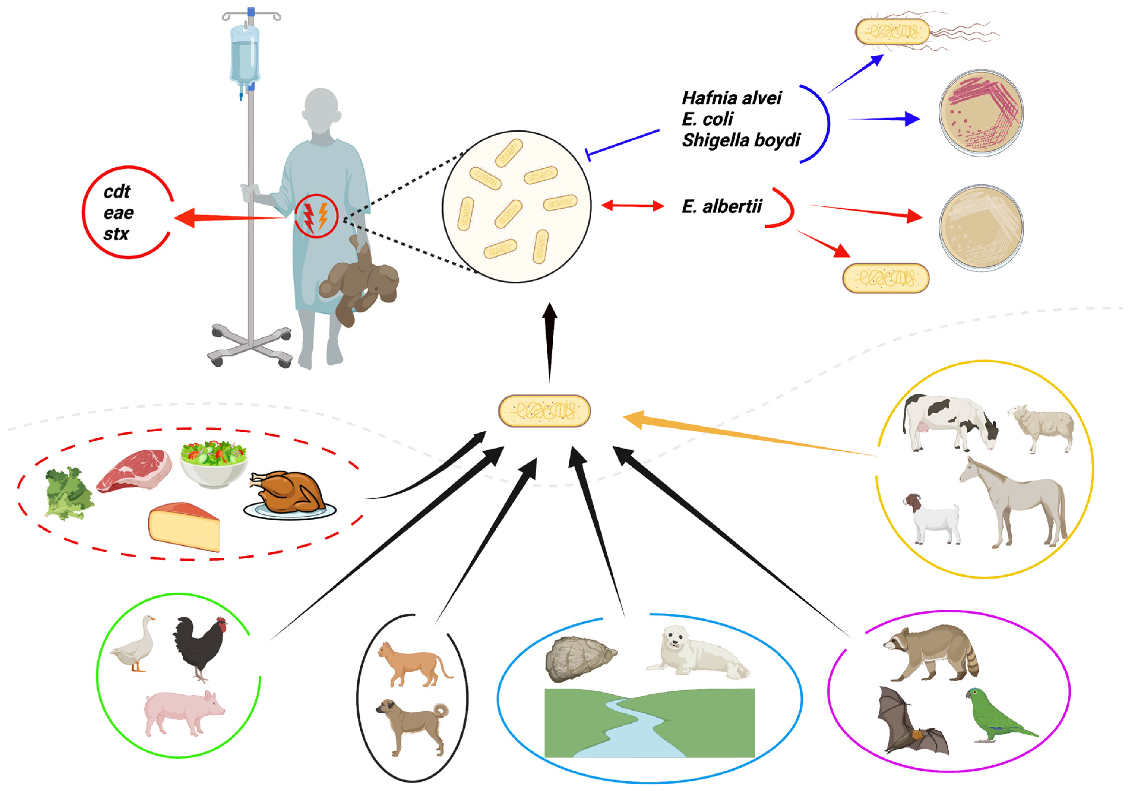
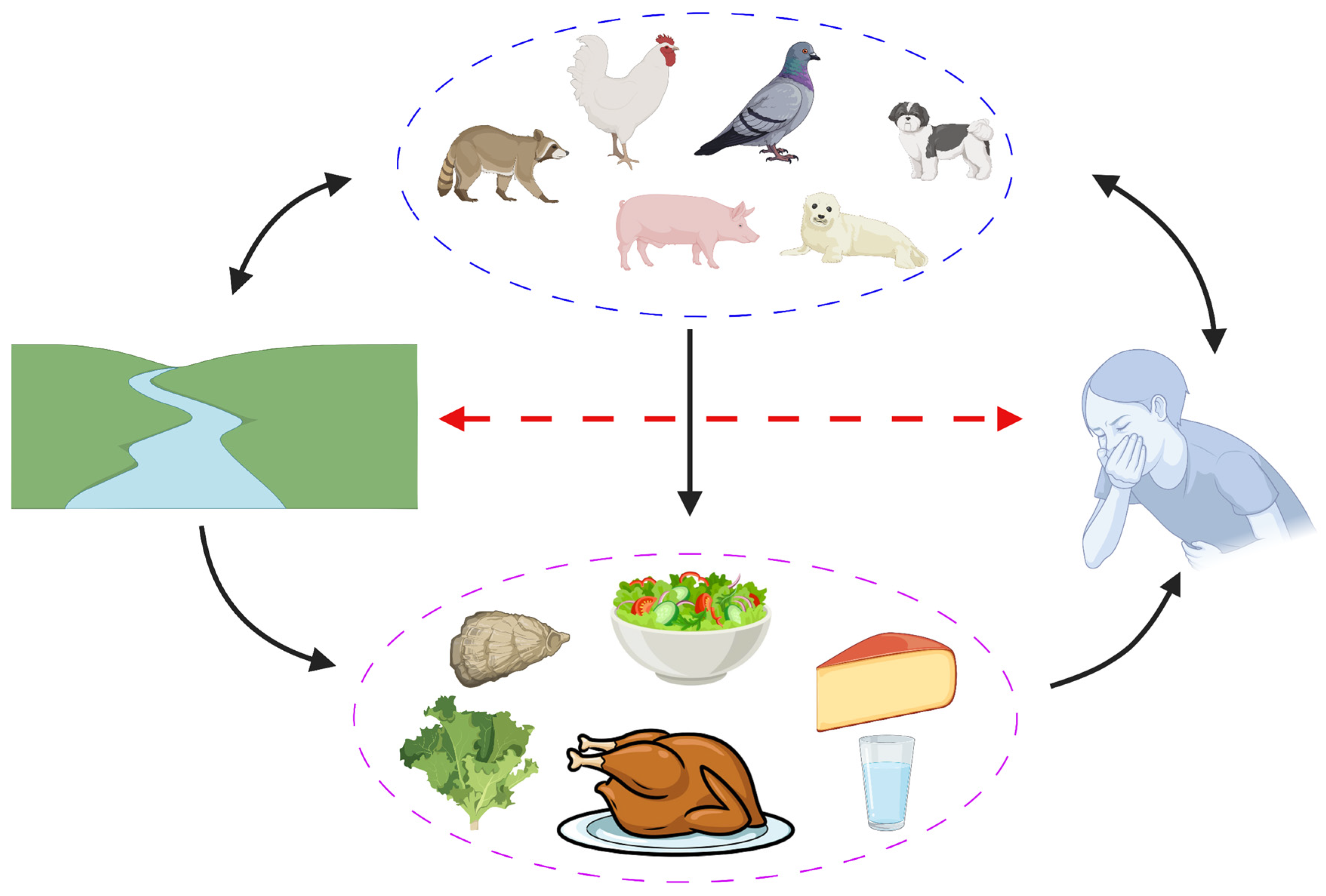
4. Occurrence of Escherichia albertii in Animals

E. albertii has been isolated from various domestic and migratory birds, occasionally causing epidemics worldwide [2,50,67,68,69,70,71]. E. albertii has also been reported in pigs, cats, dogs, bats, raccoons, penguins, and seals [10,41,49,70,72]. Isolation frequency seems highest in birds [44,46,48], hence poultry could act as a reservoir and its meat might pose an increased risk for E. albertii exposure and or infection [50,67]. For instance, surveys in Australia between 1994–2004 and 2010–2011 showed the prevalence of E. albertii of 14.3 to 18% in magpies and 6.7 to 33% in chickens [70,73]. Interestingly, the same analysis did not detect E. albertii in fish, snakes, lizards, crocodiles, and frogs, over a 10-year period [70]. Isolation of this pathogen in raccoons (57.7% prevalence; [49]), bats, seals, and wild birds is suggestive that wild animals more so birds might be reservoirs of this bacteria. It would be advantageous to evaluate E. albertii prevalence in different wild animals and other livestock such as cattle, goats, and sheep from which this pathogen has not yet been detected. However, the isolation of E. albertii from mutton meat supports the occurrence of this pathogen in livestock [48]. The occurrence of E. albertii in apparently healthy animals might suggest the existence of careers that can act as reservoirs of this pathogen in livestock [2,41,71]. E. albertii in companion animals such as cats and dogs as well as farm animals such as chickens and pigs might act as a source or reservoir of this pathogen for people in close contact with these animals. Moreover, animal and human E. albertii isolates are heterogeneous and seem not to be host-specific, therefore, all its strains might have zoonotic potential and represent a significant public health threat [6,50,67].
The clinical picture of active E. albertii infections in animals is rarely described. This might be a consequence of infected animals being subclinical or dying acutely without many observable clinical signs. This possibility coupled with the challenge in differentiating this pathogen from E. coli might result in illness due to E. albertii infection being misdiagnosed, precluding full definition of its clinical presentation both in humans and animals.

5. Occurrence of Escherichia albertii in Food

E. albertii has been isolated from the environment, water, packed lunch, lettuce, salad, pork, chicken, giblets, mutton, duck meat, minced meat, and Damietta cheese indicating that this bacterium can be transmitted through food and water [10,12,17,21,23,25,44,46,48,74,75]. In most outbreaks and sporadic cases, the contaminated vehicle for disease transmission is not identified. Nonetheless, in a few cases, it has been confirmed that salads, water, and chicken are some of the contaminated vehicles [12,14,18,70]. E. albertii was associated with outbreaks in campers who drank contaminated water [14,17]. Furthermore, it has been detected in various water bodies across the world [25,27,39,75]. The occurrence of E. albertii in water is suggestive that seafood is also potentially contaminated with this pathogen. The study by Arai et al. [22] confirmed this as they detected E. albertii in 1.9% of 427 samples of raw Pacific and Japanese rock oysters. The occurrence of this pathogen in other kinds of seafood need to be elucidated. The use of contaminated irrigation water might be responsible for contaminating plants such as lettuce and eventually salads, hence caution must be exercised when using untreated irrigation water, especially in areas where E. albertii contaminated water is expected.
Since the full phenotypic features such as growth potential on different foods and hurdle technique resistance profiles of E. albertii are not yet known, it is challenging to determine the most effective approaches to control this pathogen on food. It is tempting to speculate that hurdle procedures employed for E. coli and other Enterobacteriaceae coupled with good manufacturing practice can minimize E. albertii occurrence in foods. Interestingly, Sharma et al. [76] using five strains reported that E. albertii is less tolerant to heat, acid, and pressure than E. coli O157:H7. Moreover, it is highly likely that E. albertii is more sensitive to osmotic stress compared to E. coli due to the absence of the betIAB operon required for glycine betaine synthesis an important osmo-protective solute [13,77]. However, this needs to be experimentally confirmed. The potential transmission cycle of E. albertii is depicted in Figure 3.

6. Pathogenesis and Virulence Factors of Escherichia albertii

E. albertii is one of the attaching and effacing pathogens, a phenotype confirmed in vivo using the rabbit ileal loop model [8]. Its pathogenesis depends on its ability to adhere to epithelial cells with the formation of attaching-effacing lesions. E. albertii achieves this through the dual activity of the type III secretion system effectors and an outer membrane protein (intimin), encoded by a locus of enterocyte effacement (LEE) and the eae gene, respectively [5,13,78]. Similar effacing lesions are produced by other pathogens such as EHEC and EPEC as well as Citrobacter rodentium [5]. These lesions promote the bacterial invasion process, while the ability of E. albertii to survive intracellularly protects it from intestinal clearance and the immune system, causing prolongation of the diarrheal disease [5]. Invasion of epithelial cells was shown to depend on intimin interactions with its receptor Tir, located on the host cells membrane [79]. This intimin-Tir interaction results in actin polymerization and the formation of pedestal-like structures beneath adherent bacteria [80,81,82,83,84]. It has also been demonstrated that E. albertii can invade HeLa, Caco-2, and T84 cells, and can decrease transepithelial electrical resistance by redistributing tight junction proteins (zona occludenes-1) which leads to higher cell permeability [78,85,86,87].
Interestingly, in E. albertii the highly conserved LEE is integrated almost exclusively on the pheU tRNA locus while in EHEC or EPEC, LEE is found either at the pheU, pheV, or seIC loci suggestive of multiple acquisition events in E. coli [5,10,13,26,27,88,89]. In further contrast to E. coli, some E. albertii strains contain an intact E. coli type III secretion system 2 (ETT2) locus which includes the virulence genes yqeH, ygeF, eprH, epaS, eivG, and etrA, and is integrated at the glyU tRNA locus, however, the importance of this locus for virulence is not yet clear [13,14]. In addition, porcine attaching-effacing associated protein encoded by paa is highly conserved in E. albertii strains, but its occurrence is strain-specific [90].
Almost all reported E. albertii isolates carry a cdtABC locus which encodes the cytolethal distending toxin (Cdt). Of the 5-subtype known to exist in E. coli [53], Cdt-I and Cdt-II have been reported in E. albertii, with Cdt-II being the predominate subtype [6,91]. Only a single strain that encodes both Cdt-I and Cdt-II subtypes has been described to date [91]. Moreover, a new subtype, Cdt-VI was recently reported in E. albertii [6]. The highly conserved EacdtABC operon has so far mostly been detected on the chromosome and not in mobile genetic elements [13,26,43]. However, Gomes et al. [5] recently reported two strains that carry the cdt gene in a prophage region. Cdt protein is composed of three subunits, CdtA and CdtC are critical for translocating the virulence factor CdtB into the host cell. CdtB has DNase I activity and functions through DNA damage, inducing G2/M cell cycle arrest and eventual apoptosis [53,92,93,94]. Although the importance and role played by Cdt in E. albertii pathogenesis is not yet fully understood, it has been demonstrated that Cdt is linked with persistent colonization and invasion by bacteria, which, in turn, affects disease severity [95,96]. Since cdtB is highly conserved amongst E. albertii strains it is now being exploited as a genetic marker for the identification of this pathogen.
Some E. albertii encode the protein synthesis inhibiting cytotoxic Shiga toxin 2 (Stx2a or Stx2f) henceforth referred to as Shiga toxin-producing E. albertii (STEA) [5,10,13,26,33,39,45,49,67,91,97,98,99]. Due to this carriage of the stx gene, some E. albertii isolates have been misclassified as EHEC [10,45]. The Stx2f variant is more common among E. albertii strains [45], with only a few strains encoding the Stx2a subtype being reported to date [5,45]. It seems carriage of the Stx2a toxin increases virulence with bloody diarrhea being one of the consequences of infection [45,100].
Using the CHO and Vero cell cytotoxic assays, studies have shown that some E. albertii strains produce biologically active Cdt and Stx toxins, respectively [10,20,26,49,91,99]. As seen with Shiga toxin-producing E. coli (STEC), it is possible that STEA can cause hemorrhagic colitis, hemolytic-uremic syndrome (HUS), thrombotic microangiopathy, and renal failure [13]. As observed in E. coli, such clinical manifestation is highly likely in strains expressing the stx2a allele [101,102,103]. In E. albertii, the Shiga toxin is mainly harbored on phages, hence clinicians must be aware of the danger of using antibiotics that might induce stx2-harboring bacteriophages, which might worsen the prognosis in STEA infections [104]. In support, some studies have shown that STEA culture supernatants’ toxicity is enhanced by mitomycin C, implying that the stx genes in these isolates, might be harbored on inducible prophages [49].
It is not clear if pathogenicity differences exist between E. albertii strains. However, the presence, distribution, and variants of virulence genes in its strains vary with lineage, suggestive of potential virulence variation [6]. Hinenoya et al. [50], using chicken colonization experiments showed that human isolates have a higher colonization rate than wild bird-derived isolates. Over 44 potential virulence factors have been identified in E. albertii, including those encoded by, ibeA, sepL, ent and fep, iuc-ABCD, iutA, ehxA, entABCES, fepABCDG, hlyABCD, map, espA, espB, nleA, and phoE [6,13,14,67]. These virulence factors contribute towards invasion, virulence factor translocation and regulation, iron acquisition and transport, cytolysis, immune response evasion and dampening, and stress tolerance [33,48,105,106,107,108,109]. This list of virulence factors will continue to grow as more sequence data become available. Therefore, much work is required to elucidate the regulation, structure, and mechanism of action of these virulence factors and their role in the pathogenicity of this bacterium. Table 4 summarizes some of the major virulence factors detected in E. albertii. For a more detailed review of E. albertii pathogenesis please refer to Gomes et al. [5].

7. Clinical Significance of Escherichia albertii

Early evidence suggests that E. albertii may be present in animals or humans as a commensal or pathogen. The clinical significance of E. albertii is not yet fully understood, partially because it is difficult to discriminate it from other Enterobacteriaceae using routine identification protocols. Nevertheless, infectious diarrhea outbreaks and sporadic cases due to E. albertii have been reported in multiple countries including Antarctica, Alaska, Bangladesh, Belgium, Brazil, China, Germany, Guinea-Bissau, Iran, Japan, Mexico, Nigeria, Poland, and the United States of America, signifying a worldwide distribution [2,7,9,11,12,14,48,50,67,72,90,99,110,111,112]. Despite this and several retrospective studies determining that many EPEC and EHEC isolates implicated in disease outbreaks are misidentified E. albertii [9,10,11,12,14,17,18,44], this pathogen is still often overlooked as a causative agent for infectious diarrhea [27].
E. albertii infections mainly present as acute watery diarrhea, fever, and abdominal pain with occasional reports of headache, nausea, dehydration, and abdominal distension [1,7,10,14,112]. In most cases, these infections are self-limiting, with patients often recovering with little to no treatment [14]. This could be suggestive of reduced virulence in E. albertii compared to Shigella spp. and enteroinvasive E. coli. In support of this hypothesis, E. albertii lacks invE or ipaH, which encode important virulence factors in Shigella and enteroinvasive E. coli [13].
Afshin et al. [113] and Zaki et al. [114] reported five and seven cases of E. albertii-associated urinary tract infections, respectively. A single case of E. albertii bacteremia has been reported in an old lady with multiple comorbidities probably because of its ability to translocate from the intestinal lumen to other extraintestinal sites [5,34,115]. This might be a rare clinical presentation, however, due to the potential for misclassification, E. albertii bacteremia might occur more frequently but is misclassified as the more common E. coli. It would be prudent in the future to consider E. albertii in Enterobacteriaceae bacteremia presumptively due to E. coli especially those that have discrepant results for routine etiology molecular identification. In support of this, retrospective analyses have indicated that E. albertii represents a significant portion of strains identified as eae-positive E. coli including Shiga toxin 2f–producing strains [12,17]. Because E. albertii possesses the eae gene, many strains might have been misidentified as EHEC or EPEC.
Incubation periods of E. albertii infections which present as diarrhea are relatively short (average 12–24 h) while mortality and morbidity rates are unknown [14]. To date, several outbreaks have been reported mainly in Japan, with most of these initially misidentified as E. coli outbreaks [10,12,14,17]. In all these outbreaks and other sporadic cases, it appears mortality rates were low or none, however, morbidity rates appeared to be relatively high, i.e., >50% of the exposed population [14]. For instance, in two outbreaks in Japan, one linked to boxed lunches and another associated with eating at the same restaurant, 20 of 31 (64.5%) and 48 of 94 (51.1%) exposed people became ill, respectively [11,12,14]. While one of the largest recorded E. albertii outbreaks, linked to contaminated water had an attack rate of 66.7% (273) among the 409 potentially exposed individuals [14].
Because E. albertii and E. coli share ecological niches and display high genomic similarity such as virulence and stress tolerance effectors [5,6,13,27], one can speculate that these pathogens might pose similar public health threats, therefore measures employed to mitigate EPEC and EHEC might also be required for this emerging zoonotic pathogen.
As with most bacteria, antimicrobial resistance is a major concern. Several strains of E. albertii have been shown to be resistant to a significant number of important antibiotics including tetracycline, macrolides except for azithromycin, lincosamides, ampicillin, penicillin G, oxacillin, gentamicin, ciprofloxacin, trimethoprim, sulfamethoxazole, fusidic acid, rifampicin, meropenenem, imipenem, and norfloxacin [26,38,50,51,67,90,114]. Moreover, multidrug-resistant E. albertii strains [67], that displayed resistance or encode resistance determinates to antibiotics from at least eleven classes have been reported [6,116]. Combined with the virulence potential of this pathogen such strains pose a significant public health threat. Therefore, to effectively prevent and control its infections, much must be done to fully understand all aspects of this emerging zoonotic pathogen including its antimicrobial resistance determinants.

8. Risk Assessment

The infectious dose, transmission routes, incidence rates, prevalence, epidemiology, and predisposing factors of E. albertii have not yet been fully elucidated, making risk assessment difficult. However, early indications suggest that children below 10 years and immunosuppressed individuals with multiple commodities might be at higher risk, but outbreaks have also been reported in seemly healthy people [5,7,8,12,14,34,45,113,114]. Poor hygiene conditions especially during food preparation, consumption of raw or minimally cooked meat particularly poultry, and drinking untreated water seems to increase the likelihood of E. albertii infection [14,17,18,112]. The increased occurrence of clinical disease in younger age groups might be a true association or a reflection of sampling bias. Enteric infections may have more severe negative health outcomes amongst young children [117], such as physical and cognitive development impairments [118], hence in most studies, this population is preferentially targeted. Moreover, in STEC infections, HUS development is more likely in children <5 years old [119]. Though not confirmed we speculate that the risk of HUS development in STEA infection might be higher in children. Dual carriage of stx2 and eae genes reported in some E. albertii strains might increase the risk of HUS development [5,6,45]. While E. albertii strains that lack this eae gene, such as those isolated from water and chicken carcass rinse water might be less pathogenic and pose a lower risk for severe clinical disease [21,25,75].
Some E. albertii strains have been observed to produce biofilms [90,120], a phenotype known to increase resistance against different stresses and the host immune system including the mechanical movements of intestinal peristalsis [121,122]. This might increase the severity and duration of the disease, complicating treatment outcomes. Moreover, biofilm formation can assist in niche colonization and persistence in food processing plants, however, further studies are required to confirm this.

9. Knowledge Gaps and Future Research Needs

E. albertii is an emerging zoonotic foodborne enteropathogenic pathogen, however, there are still many knowledge gaps that need to be bridged to fully understand and be able to effectively control and mitigate the risks this pathogen poses. We need to understand fully its pathogenic potential and clinical relevance. Critical information such as mortality and morbidity rates, infectious dose, and predisposing factors need to be elucidated. Its full epidemiological picture, transmission routes, reservoirs both in water bodies, humans, livestock, and wildlife need to be further elucidated. Risk assessment including characterization of E. albertii strains growth capacity on different foods and hurdle technique resistance profiles are required.
Due to challenges in isolation and identification of this pathogen, more specific enrichment, isolation, and identification tools are required. Protocols that employ PCR-based methods have been developed using a limited number of strains, from limited sources and or regions, hence these primers might be selective for these strains missing E. albertii strains with other alleles of the target genes. Such protocols need to be reviewed using more strains, primers must be validated using more genomes from a diverse strain collection. The three presumed E. ruysiae strains potentially misclassified as E. albertii we identified show that much work still needs to be done to correctly classify this species even at the genome level. It is highly likely that with the discovery of new novel species misclassification will continue to be a problem. Sequence databases need to continually filter and update their genome records to ensure that they are in line with the new identification systems.
The discovery of lactose fermenting E. albertii isolates further complicates isolation and identification, therefore, a selective plate that can effectively identify both strains able and not able to ferment lactose is urgently needed. Most previous protocols selected for non-lactose fermenting E. albertii [22,37,112], it will be interesting to investigate how many E. albertii strains were missed using such an approach. Reports of antimicrobial resistance in E. albertii especially toward extended-spectrum beta-lactam antibiotics are troubling. The extent of such antimicrobial resistance in this species needs further investigation. The clinical and economic importance of E. albertii is currently unknown. Until routine laboratories can identify more strains of this species, its prevalence, disease spectrum, and clinical significance will remain in question. Moreover, it is not clear if the currently studied isolates cover the diversity of E. albertii as mostly only strains from three phylogroups have been analyzed. Therefore, a more diverse strain collection needs to be included in future studies. Care must also be taken that such characterization studies are carried out using diverse strains not multiple isolates of the same strain. For instance, it is not clear whether the reported 64 E. albertii strains isolated from 8 oyster samples [22] or 143 strains from 62 PCR-positive swab samples [49], represent multiple different strains or are simply multiple isolates of the same strain.

10. Conclusions

Although E. albertii strains continue to be misclassified, growing evidence indicates that this bacterium is an important human and animal pathogen. Much remains to be investigated regarding this pathogens’ virulence mechanisms and potentials, nutrient utilization, stress tolerance capacity, and their regulation. Our understanding of its pathobiology, as well as mechanisms of colonization, survival, and dissemination within and between hosts, is still limited and needs more analysis. There is a need for E. albertii surveillance, to establish the full extent of its public health significance and contributions to infectious diarrhea which would provide valuable knowledge for the development of intervention and control strategies. This will only be possible if simple and effective diagnostic tools are made available for its effective isolation and identification.

Author Contributions

Conceptualization R.S. and F.M.; literature review, original draft preparation, and final editing, F.M.; literature review, K.B.; visualization, text review, and editing, R.S., K.H., A.T. and K.B. All authors have read and agreed to the published version of the manuscript.

Funding

This research was funded by the University of Zurich.

Institutional Review Board Statement

Not applicable.

Informed Consent Statement

Not applicable.

Data Availability Statement

Not applicable.

Acknowledgments

The authors are grateful to Michael Biggel of the Institute for Food Safety and Hygiene, Vetsuisse Faculty, University of Zurich, for assistance with bioinformatic analyses.

Conflicts of Interest

The authors declare no conflict of interest.

References

  1. Huys, G.; Cnockaert, M.; Janda, J.M.; Swings, J. Escherichia albertii sp. nov., a diarrhoeagenic species isolated from stool specimens of Bangladeshi children. Int. J. Syst. Evol. Microbiol. 2003, 53 Pt 3, 807–810. [Google Scholar] [CrossRef] [PubMed] [Green Version]
  2. Oaks, J.L.; Besser, T.E.; Walk, S.T.; Gordon, D.M.; Beckmen, K.B.; Burek, K.A.; Haldorson, G.J.; Bradway, D.S.; Ouellette, L.; Rurangirwa, F.R.; et al. Escherichia albertii in wild and domestic birds. Emerg. Infect. Dis. 2010, 16, 638–646. [Google Scholar] [CrossRef] [PubMed]
  3. van der Putten, B.C.L.; Matamoros, S.; Mende, D.R.; Scholl, E.R.; Consortium, C.; Schultsz, C. Escherichia ruysiae sp. nov., a novel Gram-stain-negative bacterium, isolated from a faecal sample of an international traveller. Int. J. Syst. Evol. Microbiol. 2021, 71, 004609. [Google Scholar] [CrossRef] [PubMed]
  4. Ooka, T.; Seto, K.; Ogura, Y.; Nakamura, K.; Iguchi, A.; Gotoh, Y.; Honda, M.; Etoh, Y.; Ikeda, T.; Sugitani, W.; et al. O-antigen biosynthesis gene clusters of Escherichia albertii: Their diversity and similarity to Escherichia coli gene clusters and the development of an O-genotyping method. Microb. Genom. 2019, 5, e000314. [Google Scholar] [CrossRef] [PubMed]
  5. Gomes, T.A.T.; Ooka, T.; Hernandes, R.T.; Yamamoto, D.; Hayashi, T. Escherichia albertii Pathogenesis. EcoSal Plus 2020, 9. [Google Scholar] [CrossRef]
  6. Luo, L.; Wang, H.; Payne, M.J.; Liang, C.; Bai, L.; Zheng, H.; Zhang, Z.; Zhang, L.; Zhang, X.; Yan, G.; et al. Comparative genomics of Chinese and international isolates of Escherichia albertii: Population structure and evolution of virulence and antimicrobial resistance. Microb. Genom. 2021, 7, 000710. [Google Scholar] [CrossRef]
  7. Albert, M.J.; Alam, K.; Islam, M.; Montanaro, J.; Rahaman, A.S.; Haider, K.; Hossain, M.A.; Kibriya, A.K.; Tzipori, S. Hafnia alvei, a probable cause of diarrhea in humans. Infect. Immun. 1991, 59, 1507–1513. [Google Scholar] [CrossRef] [Green Version]
  8. Albert, M.J.; Faruque, S.M.; Ansaruzzaman, M.; Islam, M.M.; Haider, K.; Alam, K.; Kabir, I.; Robins-Browne, R. Sharing of virulence-associated properties at the phenotypic and genetic levels between enteropathogenic Escherichia coli and Hafnia alvei. J. Med. Microbiol. 1992, 37, 310–314. [Google Scholar] [CrossRef]
  9. Konno, T.; Yatsuyanagi, J.; Takahashi, S.; Kumagai, Y.; Wada, E.; Chiba, M.; Saito, S. Isolation and Identification of Escherichia albertii from a Patient in an Outbreak of Gastroenteritis. Jpn. J. Infect. Dis. 2012, 65, 203–207. [Google Scholar] [CrossRef] [Green Version]
  10. Ooka, T.; Seto, K.; Kawano, K.; Kobayashi, H.; Etoh, Y.; Ichihara, S.; Kaneko, A.; Isobe, J.; Yamaguchi, K.; Horikawa, K.; et al. Clinical Significance of Escherichia albertii. Emerg. Infect. Dis. 2012, 18, 488–492. [Google Scholar] [CrossRef] [Green Version]
  11. Ooka, T.; Tokuoka, E.; Furukawa, M.; Nagamura, T.; Ogura, Y.; Arisawa, K.; Harada, S.; Hayashi, T. Human Gastroenteritis Outbreak Associated with Escherichia albertii, Japan. Emerg. Infect. Dis. 2013, 19, 144–146. [Google Scholar] [CrossRef] [PubMed]
  12. Asoshima, N.; Matsuda, M.; Shigemura, K.; Honda, M.; Yoshida, H.; Hiwaki, H.; Ogata, K.; Oda, T. Identification of Escherichia albertii as a Causative Agent of a Food-Borne Outbreak Occurred in 2003. Jpn. J. Infect. Dis. 2014, 67, 139–140. [Google Scholar] [CrossRef] [PubMed] [Green Version]
  13. Ooka, T.; Ogura, Y.; Katsura, K.; Seto, K.; Kobayashi, H.; Kawano, K.; Tokuoka, E.; Furukawa, M.; Harada, S.; Yoshino, S.; et al. Defining the Genome Features of Escherichia albertii, an Emerging Enteropathogen Closely Related to Escherichia coli. Genome Biol. Evol. 2015, 7, 3170–3179. [Google Scholar] [CrossRef] [Green Version]
  14. Masuda, K.; Ooka, T.; Akita, H.; Hiratsuka, T.; Takao, S.; Fukada, M.; Inoue, K.; Honda, M.; Toda, J.; Sugitani, W.; et al. Epidemiological Aspects of Escherichia albertii Outbreaks in Japan and Genetic Characteristics of the Causative Pathogen. Foodborne Pathog. Dis. 2020, 17, 144–150. [Google Scholar] [CrossRef]
  15. Donnenberg, M.S.; Tzipori, S.; McKee, M.L.; O’Brien, A.D.; Alroy, J.; Kaper, J.B. The role of the eae gene of enterohemorrhagic Escherichia coli in intimate attachment in vitro and in a porcine model. J. Clin. Investig. 1993, 92, 1418–1424. [Google Scholar] [CrossRef] [PubMed] [Green Version]
  16. Cabal, A.; García-Castillo, M.; Cantón, R.; Gortázar, C.; Domínguez, L.; Álvarez, J. Prevalence of Escherichia coli Virulence Genes in Patients with Diarrhea and a Subpopulation of Healthy Volunteers in Madrid, Spain. Front. Microbiol. 2016, 7, 641. [Google Scholar] [CrossRef] [Green Version]
  17. Baba, A.; Ebuchi, S.; Uryu, K.; Hiwaki, H.; Ogata, K.; Washimi, E.; Hasegawa, A.; Utiyama, S. An outbreak of water-borne gastroenteritis caused by diarrheagenic Escherichia coli possessing eae gene. Jpn. J. Infect. Dis. 2006, 59, 59–60. [Google Scholar]
  18. Tokuoka, E.; Furukawa, K.; Nagamura, T.; Harada, F.; Ekinaga, K.; Tokunaga, H. Food poisoning outbreak due to atypical EPEC OUT:HNM, May 2011–Kumamoto. Infect. Agents Surveill. Rep. 2012, 33, 8–9. [Google Scholar]
  19. Hyma, K.E.; Lacher, D.W.; Nelson, A.M.; Bumbaugh, A.C.; Janda, J.M.; Strockbine, N.A.; Young, V.B.; Whittam, T.S. Evolutionary Genetics of a New Pathogenic Escherichia Species: Escherichia albertii and Related Shigella boydii Strains. J. Bacteriol. 2005, 187, 619–628. [Google Scholar] [CrossRef] [Green Version]
  20. Murakami, K.; Etoh, Y.; Tanaka, E.; Ichihara, S.; Horikawa, K.; Kawano, K.; Ooka, T.; Kawamura, Y.; Ito, K. Shiga Toxin 2f-Producing Escherichia albertii from a Symptomatic Human. Jpn. J. Infect. Dis. 2014, 67, 204–208. [Google Scholar] [CrossRef] [Green Version]
  21. Lindsey, R.L.; Fedorka-Cray, P.J.; Abley, M.; Turpin, J.B.; Meinersmann, R.J. Evaluating the Occurrence of Escherichia albertii in Chicken Carcass Rinses by PCR, Vitek Analysis, and Sequencing of the rpoB Gene. Appl. Environ. Microbiol. 2015, 81, 1727–1734. [Google Scholar] [CrossRef] [PubMed] [Green Version]
  22. Arai, S.; Yamaya, S.; Ohtsuka, K.; Konishi, N.; Obata, H.; Ooka, T.; Hirose, S.; Kai, A.; Hara-Kudo, Y. Detection of Escherichia albertii in Retail Oysters. J. Food Prot. 2022, 85, 173–179. [Google Scholar] [CrossRef] [PubMed]
  23. Wakabayashi, Y.; Seto, K.; Kanki, M.; Harada, T.; Kawatsu, K. Proposal of a novel selective enrichment broth, NCT-mTSB, for isolation of Escherichia albertii from poultry samples. J. Appl. Microbiol. 2022, 132, 2121–2130. [Google Scholar] [CrossRef] [PubMed]
  24. Hinenoya, A.; Ichimura, H.; Yasuda, N.; Harada, S.; Yamada, K.; Suzuki, M.; Iijima, Y.; Nagita, A.; Albert, M.J.; Hatanaka, N.; et al. Development of a specific cytolethal distending toxin (cdt) gene (Eacdt)-based PCR assay for the detection of Escherichia albertii. Diagn. Microbiol. Infect. Dis. 2019, 95, 119–124. [Google Scholar] [CrossRef]
  25. Maheux, A.F.; Boudreau, D.K.; Bergeron, M.G.; Rodriguez, M.J. Characterization of Escherichia fergusonii and Escherichia albertii isolated from water. J. Appl. Microbiol. 2014, 117, 597–609. [Google Scholar] [CrossRef]
  26. Hinenoya, A.; Ichimura, H.; Awasthi, S.P.; Yasuda, N.; Yatsuyanagi, J.; Yamasaki, S. Phenotypic and molecular characterization of Escherichia albertii: Further surrogates to avoid potential laboratory misidentification. Int. J. Med. Microbiol. 2019, 309, 108–115. [Google Scholar] [CrossRef]
  27. Bhatt, S.; Egan, M.; Critelli, B.; Kouse, A.; Kalman, D.; Upreti, C. The Evasive Enemy: Insights into the Virulence and Epidemiology of the Emerging Attaching and Effacing Pathogen Escherichia albertii. Infect. Immun. 2019, 87, e00254-18. [Google Scholar] [CrossRef] [Green Version]
  28. Takara, T.; Nakama, E.; Kyan, H.; Kakita, T.; Kuba, Y.; Kato, T.; Kudaka, J.; Ameku, C.; Imoto, S.; Nakasone, T.; et al. Escherichia albertii food poisoning due to consumption of Nigana shiraae, seasoned mixed salad of nigana (Ixeris dentata) and tofu, May 2016—Okinawa Prefecture. Infect. Agents Surveill. Rep. 2016, 37, 252–253. [Google Scholar]
  29. Fewtrell, L.; Kaufmann, R.B.; Kay, D.; Enanoria, W.; Haller, L.; Colford, J.M., Jr. Water, sanitation, and hygiene interventions to reduce diarrhoea in less developed countries: A systematic review and meta-analysis. Lancet Infect. Dis. 2005, 5, 42–52. [Google Scholar] [CrossRef]
  30. Karinja, M.; Schlienger, R.; Pillai, G.C.; Esterhuizen, T.; Onyango, E.; Gitau, A.; Ogutu, B. Risk reduction of diarrhea and respiratory infections following a community health education program—A facility-based case-control study in rural parts of Kenya. BMC Public Health 2020, 20, 586. [Google Scholar] [CrossRef]
  31. Janda, J.M.; Abbott, S.L.; Albert, M.J. Prototypal Diarrheagenic Strains of Hafnia alvei Are Actually Members of the Genus Escherichia. J. Clin. Microbiol. 1999, 37, 2399–2401. [Google Scholar] [CrossRef] [PubMed] [Green Version]
  32. Abbott, S.L.; O’Connor, J.; Robin, T.; Zimmer, B.L.; Janda, J.M. Biochemical Properties of a Newly Described Escherichia Species, Escherichia albertii. J. Clin. Microbiol. 2003, 41, 4852–4854. [Google Scholar] [CrossRef] [PubMed] [Green Version]
  33. Nimri, L.F. Escherichia albertii, a newly emerging enteric pathogen with poorly defined properties. Diagn. Microbiol. Infect. Dis. 2013, 77, 91–95. [Google Scholar] [CrossRef]
  34. Inglis, T.J.; Merritt, A.J.; Bzdyl, N.; Lansley, S.; Urosevic, M.N. First bacteraemic human infection with Escherichia albertii. New Microbes New Infect. 2015, 8, 171–173. [Google Scholar] [CrossRef] [Green Version]
  35. Maheux, A.F.; Brodeur, S.; Bérubé, È.; Boudreau, D.K.; Abed, J.Y.; Boissinot, M.; Bissonnette, L.; Bergeron, M.G. Method for isolation of both lactose-fermenting and—non-fermenting Escherichia albertii strains from stool samples. J. Microbiol. Methods 2018, 154, 134–140. [Google Scholar] [CrossRef] [PubMed]
  36. Murakami, K.; Maeda-Mitani, E.; Kimura, H.; Honda, M.; Ikeda, T.; Sugitani, W.; Konno, T.; Kawano, K.; Etoh, Y.; Sera, N.; et al. Non-biogroup 1 or 2 Strains of the Emerging Zoonotic Pathogen Escherichia albertii, Their Proposed Assignment to Biogroup 3, and Their Commonly Detected Characteristics. Front. Microbiol. 2019, 10, 1543. [Google Scholar] [CrossRef] [PubMed] [Green Version]
  37. Arai, S.; Ohtsuka, K.; Konishi, N.; Ohya, K.; Konno, T.; Tokoi, Y.; Nagaoka, H.; Asano, Y.; Maruyama, H.; Uchiyama, H.; et al. Evaluating Methods for Detecting Escherichia albertii in Chicken Meat. J. Food Prot. 2021, 84, 553–562. [Google Scholar] [CrossRef]
  38. Stock, I.; Rahman, M.; Sherwood, K.J.; Wiedemann, B. Natural antimicrobial susceptibility patterns and biochemical identification of Escherichia albertii and Hafnia alvei strains. Diagn. Microbiol. Infect. Dis. 2005, 51, 151–163. [Google Scholar] [CrossRef]
  39. Konno, T.; Takahashi, S.; Suzuki, S.; Kashio, H.; Ito, Y.; Kumagai, Y. Distribution of the O-Genotypes of Escherichia albertii Isolated from Humans and Environmental Water in Akita Prefecture, Japan. Jpn. J. Infect. Dis. 2020. [Google Scholar] [CrossRef]
  40. Ridell, J.; Siitonen, A.; Paulin, L.; Lindroos, O.; Korkeala, H.; Albert, M.J. Characterization of Hafnia alvei by biochemical tests, random amplified polymorphic DNA PCR, and partial sequencing of 16S rRNA gene. J. Clin. Microbiol. 1995, 33, 2372–2376. [Google Scholar] [CrossRef] [Green Version]
  41. Hinenoya, A.; Shima, K.; Asakura, M.; Nishimura, K.; Tsukamoto, T.; Ooka, T.; Hayashi, T.; Ramamurthy, T.; Faruque, S.M.; Yamasaki, S. Molecular characterization of cytolethal dis-tending toxin gene-positive Escherichia coli from healthy cattle and swine in Nara, Japan. BMC Microbiol. 2014, 14, 97. [Google Scholar] [CrossRef] [PubMed] [Green Version]
  42. Nataro, J.P.; Bopp, C.A.; Fields, P.I.; Kaper, J.B.; Strockbine, N.A. Escherichia, shigella, and salmonella. In Manual of Clinical Microbiology, 9th ed.; Murray, P.R., Baron, E.J., Jorgensen, J.H., Landry, M.L., Pfaller, M.A., Eds.; ASM Press: Washington, DC, USA, 2007; pp. 670–687. [Google Scholar]
  43. Hinenoya, A.; Yasuda, N.; Mukaizawa, N.; Sheikh, S.; Niwa, Y.; Awasthi, S.P.; Asakura, M.; Tsukamoto, T.; Nagita, A.; Albert, M.J.; et al. Association of cytolethal distending toxin-II gene-positive Escherichia coli with Escherichia albertii, an emerging enteropathogen. Int. J. Med. Microbiol. 2017, 307, 564–571. [Google Scholar] [CrossRef] [PubMed]
  44. Asoshima, N.; Matsuda, M.; Shigemura, K.; Honda, M.; Yoshida, H.; Oda, T.; Hiwaki, H. Isolation of Escherichia albertii from Raw Chicken Liver in Fukuoka City, Japan. Jpn. J. Infect. Dis. 2015, 68, 248–250. [Google Scholar] [CrossRef] [PubMed] [Green Version]
  45. Brandal, L.T.; Tunsjø, H.S.; Ranheim, T.E.; Løbersli, I.; Lange, H.; Wester, A.L. Shiga Toxin 2a in Escherichia albertii. J. Clin. Microbiol. 2015, 53, 1454–1455. [Google Scholar] [CrossRef] [Green Version]
  46. Maeda, E.; Murakami, K.; Sera, N.; Ito, K.; Fujimoto, S. Detection of Escherichia albertii from chicken meat and giblets. J. Vet. Med. Sci. 2015, 77, 871–873. [Google Scholar] [CrossRef] [Green Version]
  47. Wang, H.; Zheng, H.; Li, Q.; Xu, Y.; Wang, J.; Du, P.; Li, X.; Liu, X.; Zhang, L.; Zou, N.; et al. Defining the Genetic Features of O-Antigen Biosynthesis Gene Cluster and Performance of an O-Antigen Serotyping Scheme for Escherichia albertii. Front. Microbiol. 2017, 8, 1857. [Google Scholar] [CrossRef]
  48. Wang, H.; Li, Q.; Bai, X.; Xu, Y.; Zhao, A.; Sun, H.; Deng, J.; Xiao, B.; Liu, X.; Sun, S.; et al. Prevalence of eae-positive, lactose non-fermenting Escherichia albertii from retail raw meat in China. Epidemiol. Infect. 2016, 144, 45–52. [Google Scholar] [CrossRef] [Green Version]
  49. Hinenoya, A.; Nagano, K.; Awasthi, S.P.; Hatanaka, N.; Yamasaki, S. Prevalence of Escherichia albertii in Raccoons (Procyon lotor), Japan. Emerg. Infect. Dis. 2020, 26, 1304–1307. [Google Scholar] [CrossRef]
  50. Hinenoya, A.; Li, X.; Zeng, X.; Sahin, O.; Moxley, R.A.; Logue, C.M.; Gillespie, B.; Yamasaki, S.; Lin, J. Isolation and characterization of Escherichia albertii in poultry at the pre-harvest level. Zoonoses Public Health 2021, 68, 213–225. [Google Scholar] [CrossRef]
  51. Lindsey, R.L.; Garcia-Toledo, L.; Fasulo, D.; Gladney, L.M.; Strockbine, N. Multiplex polymerase chain reaction for identification of Escherichia coli, Escherichia albertii and Escherichia fergusonii. J. Microbiol. Methods 2017, 140, 1–4. [Google Scholar] [CrossRef] [Green Version]
  52. Maeda, E.; Murakami, K.; Okamoto, F.; Etoh, Y.; Sera, N.; Ito, K.; Fujimoto, S. Nonspecificity of Primers for Escherichia albertii Detection. Jpn. J. Infect. Dis. 2014, 67, 503–505. [Google Scholar] [CrossRef] [PubMed] [Green Version]
  53. Yamasaki, S.; Asakura, M.; Tsukamoto, T.; Faruque, S.M.; Deb, R.; Ramamurthy, T. Cytolethal distending toxin (cdt): Genetic diversity, structure and role in diarrheal disease. Toxin Rev. 2006, 25, 61–88. [Google Scholar] [CrossRef]
  54. Lukjancenko, O.; Wassenaar, T.M.; Ussery, D.W. Comparison of 61 sequenced Escherichia coli genomes. Microb. Ecol. 2010, 60, 708–720. [Google Scholar] [CrossRef] [PubMed] [Green Version]
  55. Maheux, A.F.; Bérubé, È.; Boudreau, D.K.; Cantin, P.; Boissinot, M.; Bissonnette, L.; Rodrigue, L.; Bergeron, M.G. Ability of three DNA-based assays to identify presumptive Escherichia coli colonies isolated from water by the culture-based mFC agar method. Water Res. 2011, 45, 2638–2646. [Google Scholar] [CrossRef]
  56. Hinenoya, A.; Nagano, K.; Okuno, K.; Nagita, A.; Hatanaka, N.; Awasthi, S.P.; Yamasaki, S. Development of XRM-MacConkey agar selective medium for the isolation of Escherichia albertii. Diagn. Microbiol. Infect. Dis. 2020, 97, 115006. [Google Scholar] [CrossRef]
  57. Bizzini, A.; Durussel, C.; Bille, J.; Greub, G.; Prod’Hom, G. Performance of matrix-assisted laser desorption ionization-time of flight mass spectrometry for identification of bacterial strains routinely isolated in a clinical microbiology laboratory. J. Clin. Microbiol. 2010, 48, 1549–1554. [Google Scholar] [CrossRef] [Green Version]
  58. Singhal, N.; Kumar, M.; Kanaujia, P.K.; Virdi, J.S. MALDI-TOF mass spectrometry: An emerging technology for microbial identification and diagnosis. Front. Microbiol. 2015, 6, 791. [Google Scholar] [CrossRef] [Green Version]
  59. Hou, T.-Y.; Chiang-Ni, C.; Teng, S.-H. Current status of MALDI-TOF mass spectrometry in clinical microbiology. J. Food Drug Anal. 2019, 27, 404–414. [Google Scholar] [CrossRef]
  60. Hatanaka, N.; Awasthi, S.P.; Hinenoya, A.; Ueda, O.; Yamasaki, S. Accurate identification of Escherichia albertii by matrix-assisted laser desorption ionization-time of flight mass spectrometry. J. Microbiol. Methods 2020, 173, 105916. [Google Scholar] [CrossRef]
  61. van den Beld, M.J.C.; Rossen, J.W.A.; Evers, N.; Kooistra-Smid, M.A.M.D.; Reubsaet, F.A.G. MALDI-TOF MS Using a Custom-Made Database, Biomarker Assignment, or Mathematical Classifiers Does Not Differentiate Shigella spp. and Escherichia coli. Microorganisms 2022, 10, 435. [Google Scholar] [CrossRef]
  62. Paauw, A.; Jonker, D.; Roeselers, G.; Heng, J.M.; Mars-Groenendijk, R.H.; Trip, H.; Molhoek, E.M.; Jansen, H.-J.; van der Plas, J.; de Jong, A.L.; et al. Rapid and reliable discrimination between Shigella species and Escherichia coli using MALDI-TOF mass spectrometry. Int. J. Med. Microbiol. 2015, 305, 446–452. [Google Scholar] [CrossRef] [PubMed]
  63. Naumenko, O.I.; Zheng, H.; Senchenkova, S.N.; Wang, H.; Li, Q.; Shashkov, A.S.; Wang, J.; Knirel, Y.A.; Xiong, Y. Structures and gene clusters of the O-antigens of Escherichia albertii O3, O4, O6, and O7. Carbohydr. Res. 2017, 449, 17–22. [Google Scholar] [CrossRef] [PubMed]
  64. Zheng, H.; Shashkov, A.S.; Xiong, Y.; Naumenko, O.I.; Wang, H.; Senchenkova, S.N.; Wang, J.; Knirel, Y.A. Structure and gene cluster of the O-antigen of Escherichia albertii O1 resembling the O-antigen of Pseudomonas aeruginosa O5. Carbohydr. Res. 2017, 446–447, 28–31. [Google Scholar] [CrossRef] [PubMed]
  65. Zheng, H.; Naumenko, O.I.; Wang, H.; Xiong, Y.; Wang, J.; Shashkov, A.S.; Li, Q.; Knirel, Y.A. Colitose-containing O-polysaccharide structure and O-antigen gene cluster of Escherichia albertii HK18069 related to those of Escherichia coli O55 and E. coli O128. Carbohydr. Res. 2019, 480, 73–79. [Google Scholar] [CrossRef] [PubMed]
  66. Nakae, K.; Ooka, T.; Murakami, K.; Hara-Kudo, Y.; Imuta, N.; Gotoh, Y.; Ogura, Y.; Hayashi, T.; Okamoto, Y.; Nishi, J. Diversification of Escherichia albertii H-Antigens and Development of H-Genotyping PCR. Front. Microbiol. 2021, 12, 737979. [Google Scholar] [CrossRef] [PubMed]
  67. Wang, H.; Zhang, L.; Cao, L.; Zeng, X.; Gillespie, B.; Lin, J. Isolation and characterization of Escherichia albertii originated from the broiler farms in Mississippi and Alabama. Vet. Microbiol. 2022, 267, 109379. [Google Scholar] [CrossRef]
  68. Foster, G.; Ross, H.M.; Pennycott, T.W.; Hopkins, G.F.; McLaren, I.M. Isolation of Escherichia coli O86:K61 producing cyto-lethal distending toxin from wild birds of the finch family. Lett. Appl. Microbiol. 1998, 26, 395–398. [Google Scholar] [CrossRef]
  69. Pennycott, T.W.; Ross, H.M.; McLaren, I.M.; Park, A.; Hopkins, G.F.; Foster, G. Causes of death of wild birds of the family Fringillidae in Britain. Vet. Rec. 1998, 143, 155–158. [Google Scholar] [CrossRef]
  70. Gordon, D.M. Reservoirs of Infection: The Epidemiological Characteristics of an Emerging Pathogen, Escherichia albertii; Research School of Biology, Australian National University: Canberra, Australia, 2011; Available online: https://pdfs.semanticscholar.org/08fe/1278d6d23e907affbaa25f3b71c413758f3f.pdf (accessed on 4 February 2022).
  71. Oh, J.-Y.; Kang, M.-S.; Hwang, H.-T.; An, B.-K.; Kwon, J.-H.; Kwon, Y.-K. Epidemiological investigation of eaeA-positive Escherichia coli and Escherichia albertii strains isolated from healthy wild birds. J. Microbiol. 2011, 49, 747–752. [Google Scholar] [CrossRef]
  72. Grillová, L.; Sedláček, I.; Páchníková, G.; Staňková, E.; Švec, P.; Holochová, P.; Micenková, L.; Bosák, J.; Slaninova, I.; Šmajs, D. Characterization of four Escherichia albertii isolates collected from animals living in Antarctica and Patagonia. J. Vet. Med. Sci. 2018, 80, 138–146. [Google Scholar] [CrossRef] [Green Version]
  73. Gordon, D.M.; Cowling, A. The distribution and genetic structure of Escherichia coli in Australian vertebrates: Host and geographic effects. Microbiology 2003, 149, 3575–3586. [Google Scholar] [CrossRef] [PubMed] [Green Version]
  74. Lindberg, A.-M.; Ljungh, Å.; Ahrné, S.; Löfdahl, S.; Molin, G. Enterobacteriaceae found in high numbers in fish, minced meat and pasteurised milk or cream and the presence of toxin encoding genes. Int. J. Food Microbiol. 1998, 39, 11–17. [Google Scholar] [CrossRef]
  75. Felfö Ldi, T.; Heéger, Z.; Vargha, M.; Márialigeti, K. Detection of potentially pathogenic bacteria in the drinking water distribution system of a hospital in Hungary. Clin. Microbiol. Infect. 2010, 16, 89–92. [Google Scholar] [CrossRef] [Green Version]
  76. Sharma, M.; Kniel, K.E.; Derevianko, A.; Ling, J.; Bhagwat, A.A. Sensitivity of Escherichia albertii, a potential food-borne pathogen, to food preservation treatments. Appl. Environ. Microbiol. 2007, 73, 4351–4353. [Google Scholar] [CrossRef] [Green Version]
  77. Lamark, T.; Røkenes, T.P.; McDougall, J.; Strøm, A.R. The complex bet promoters of Escherichia coli: Regulation by oxygen (ArcA), choline (BetI), and osmotic stress. J. Bacteriol. 1996, 178, 1655–1662. [Google Scholar] [CrossRef] [Green Version]
  78. Hernandes, R.T.; Silva, R.M.; Carneiro, S.M.; Salvador, F.A.; Fernandes, M.C.D.C.; Padovan, A.C.B.; Yamamoto, D.; Mortara, R.A.; Elias, W.P.; Briones, M.R.D.S.; et al. The localized adherence pattern of an atypical enteropathogenic Escherichia coli is mediated by intimin omicron and unexpectedly promotes HeLa cell invasion. Cell. Microbiol. 2008, 10, 415–425. [Google Scholar] [CrossRef]
  79. Yamamoto, D.; Hernandes, R.T.; Liberatore, A.M.; Abe, C.M.; De Souza, R.B.; Romão, F.T.; Sperandio, V.; Koh, I.; Gomes, T.A.T. Escherichia albertii, a novel human enteropathogen, colonizes rat enterocytes and translocates to extra-intestinal sites. PLoS ONE 2017, 12, e0171385. [Google Scholar] [CrossRef] [Green Version]
  80. DeVinney, R.; Stein, M.; Reinscheid, D.; Abe, A.; Ruschkowski, S.; Finlay, B.B. Enterohemorrhagic Escherichia coli O157:H7 produces Tir, which is translocated to the host cell membrane but is not tyrosine phosphorylated. Infect. Immun. 1999, 67, 2389–2398. [Google Scholar] [CrossRef] [Green Version]
  81. Hartland, E.L.; Batchelor, M.; Delahay, R.M.; Hale, C.; Matthews, S.; Dougan, G.; Knutton, S.; Connerton, I.; Frankel, G. Binding of intimin from enteropathogenic Escherichia coli to Tir and to host cells. Mol. Microbiol. 1999, 32, 151–158. [Google Scholar] [CrossRef]
  82. Liu, H.; Magoun, L.; Luperchio, S.; Schauer, D.B.; Leong, J.M. The Tir-binding region of enterohaemorrhagic Escherichia coli intimin is sufficient to trigger actin condensation after bacterial-induced host cell signalling. Mol. Microbiol. 1999, 34, 67–81. [Google Scholar] [CrossRef] [Green Version]
  83. Goosney, D.L.; DeVinney, R.; Pfuetzner, R.; Frey, E.A.; Strynadka, N.C.; Finlay, B.B. Enteropathogenic E. coli translocated intimin receptor, Tir, interacts directly with alpha-actinin. Curr. Biol. 2000, 10, 735–738. [Google Scholar] [CrossRef] [Green Version]
  84. Shaw, R.K.; Daniell, S.; Frankel, G.; Knutton, S. Enteropathogenic Escherichia coli translocate Tir and form an intimin–Tir intimate attachment to red blood cell membranes. Microbiology 2002, 148, 1355–1365. [Google Scholar] [CrossRef] [PubMed] [Green Version]
  85. Donato, K.A.; Zareie, M.; Jassem, A.N.; Jandu, N.; Alingary, N.; Carusone, S.C.; Johnson-Henry, K.; Sherman, P. Escherichia albertii and Hafnia alvei are candidate enteric pathogens with divergent effects on intercellular tight junctions. Microb. Pathog. 2008, 45, 377–385. [Google Scholar] [CrossRef] [PubMed]
  86. Yamamoto, D.; Hernandes, R.T.; Blanco, M.; Greune, L.; Schmidt, M.A.; Carneiro, S.M.; Dahbi, G.; Blanco, J.E.; Mora, A.; Blanco, J.; et al. Invasiveness as a putative additional virulence mechanism of some atypical Enteropathogenic Escherichia coli strains with different uncommon intimin types. BMC Microbiol. 2009, 9, 146. [Google Scholar] [CrossRef] [PubMed] [Green Version]
  87. Pacheco, V.C.; Yamamoto, D.; Abe, C.M.; Hernandes, R.T.; Mora, A.; Blanco, J.; Gomes, T.A. Invasion of differentiated intestinal Caco-2 cells is a sporadic property among atypical enteropathogenic Escherichia coli strains carrying common intimin subtypes. Pathog. Dis. 2014, 70, 167–175. [Google Scholar] [CrossRef] [Green Version]
  88. Wieler, L.H.; McDaniel, T.K.; Whittam, T.S.; Kaper, J.B. Insertion site of the locus of enterocyte effacement in enteropathogenic and enterohemorrhagic Escherichia coli differs in relation to the clonal phylogeny of the strains. FEMS Microbiol. Lett. 1997, 156, 49–53. [Google Scholar] [CrossRef]
  89. Lacher, D.W.; Steinsland, H.; Blank, T.E.; Donnenberg, M.S.; Whittam, T.S. Molecular Molecular evolution of typical enteropathogenic Escherichia coli: Clonal analysis by multilocus sequence typing and virulence gene allelic profiling. J. Bacteriol. 2007, 189, 342–350. [Google Scholar] [CrossRef] [Green Version]
  90. Lima, M.P.; Yamamoto, D.; Santos, A.C.M.; Ooka, T.; Hernandes, R.T.; Vieira, M.A.M.; Santos, F.F.; Silva, R.M.; Hayashi, T.; Gomes, T. Phenotypic characterization and virulence-related properties of Escherichia albertii strains isolated from children with diarrhea in Brazil. Pathog. Dis. 2019, 77, ftz014. [Google Scholar] [CrossRef]
  91. Hinenoya, A.; Yasuda, N.; Hibino, T.; Shima, A.; Nagita, A.; Tsukamoto, T.; Yamasaki, S. Isolation and Isolation and characterization of an Escherichia albertii strain producing three different toxins from a child with diarrhea. Jpn. J. Infect. Dis. 2017, 70, 252–257. [Google Scholar] [CrossRef] [Green Version]
  92. Jinadasa, R.N.; Bloom, S.E.; Weiss, R.S.; Duhamel, G.E. Cytolethal distending toxin: A conserved bacterial genotoxin that blocks cell cycle progression, leading to apoptosis of a broad range of mammalian cell lineages. Microbiology 2011, 157, 1851–1875. [Google Scholar] [CrossRef] [Green Version]
  93. Fedor, Y.; Vignard, J.; Nicolau-Travers, M.-L.; Boutet-Robinet, E.; Watrin, C.; Salles, B.; Mirey, G. From single-strand breaks to double-strand breaks during S-phase: A new mode of action of the Escherichia coli Cytolethal Distending Toxin. Cell. Microbiol. 2013, 15, 1–15. [Google Scholar] [CrossRef] [PubMed]
  94. Taieb, F.; Petit, C.; Nougayrede, J.-P.; Oswald, E. The enterobacterial genotoxins: Cytolethal distending toxin and colibactin. EcoSal Plus 2016, 7. [Google Scholar] [CrossRef]
  95. Ge, Z.; Feng, Y.; Whary, M.T.; Nambiar, P.R.; Xu, S.; Ng, V.; Taylor, N.S.; Fox, J.G. Cytolethal distending toxin is essential for Helicobacter hepaticus colonization in outbred Swiss Webster mice. Infect. Immun. 2005, 73, 3559–3567. [Google Scholar] [CrossRef] [Green Version]
  96. Scuron, M.D.; Boesze-Battaglia, K.; Dlakic, M.; Shenker, B.J. The Cytolethal Distending Toxin Contributes to Microbial Virulence and Disease Pathogenesis by Acting As a Tri-Perditious Toxin. Front. Cell. Infect. Microbiol. 2016, 6, 168. [Google Scholar] [CrossRef] [PubMed] [Green Version]
  97. Iyoda, S.; Ishijima, N.; Lee, K.; Ishihara, T.; Ohnishi, M. Stx2f-positive Escherichia albertii isolated from a patient with hemolytic uremic syndrome, August 2016. Infect. Agents Surveill. Rep. 2016, 37, 255. [Google Scholar]
  98. De Rauw, K.; Jacobs, S.; Piérard, D. Twenty-seven years of screening for Shiga toxin-producing Escherichia coli in a university hospital. Brussels, Belgium, 1987–2014. PLoS ONE 2018, 13, e0199968. [Google Scholar] [CrossRef] [PubMed]
  99. Ori, E.L.; Takagi, E.H.; Andrade, T.S.; Miguel, B.T.; Cergole-Novella, M.C.; Guth, B.E.C.; Hernandes, R.T.; Dias, R.C.B.; Pinheiro, S.R.S.; Camargo, C.H.; et al. Diarrhoeagenic Escherichia coli and Escherichia albertii in Brazil: Pathotypes and serotypes over a 6-year period of surveillance. Epidemiol. Infect. 2019, 147, e10. [Google Scholar] [CrossRef] [PubMed] [Green Version]
  100. Boerlin, P.; McEwen, S.A.; Boerlin-Petzold, F.; Wilson, J.B.; Johnson, R.P.; Gyles, C.L. Associations between virulence factors of Shiga toxin-producing Escherichia coli and disease in humans. J. Clin. Microbiol. 1999, 37, 497–503. [Google Scholar] [CrossRef] [Green Version]
  101. Tesh, V.L.; Burris, J.A.; Owens, J.W.; Gordon, V.M.; Wadolkowski, E.A.; O’Brien, A.D.; Samuel, J.E. Comparison of the relative toxicities of Shiga-like toxins type I and type II for mice. Infect. Immun. 1993, 61, 3392–3402. [Google Scholar] [CrossRef] [Green Version]
  102. Friedrich, A.W.; Bielaszewska, M.; Zhang, W.-L.; Pulz, M.; Kuczius, T.; Ammon, A.; Karch, H. Escherichia coli harboring Shiga toxin 2 gene variants: Frequency and association with clinical symptoms. J. Infect. Dis. 2002, 185, 74–84. [Google Scholar] [CrossRef] [Green Version]
  103. Fuller, C.A.; Pellino, C.A.; Flagler, M.J.; Strasser, J.E.; Weiss, A.A. Shiga toxin subtypes display dramatic differences in potency. Infect. Immun. 2011, 79, 1329–1337. [Google Scholar] [CrossRef] [PubMed] [Green Version]
  104. Bielaszewska, M.; Idelevich, E.A.; Zhang, W.; Bauwens, A.; Schaumburg, F.; Mellmann, A.; Peters, G.; Karch, H. Effects of antibiotics on Shiga toxin 2 production and bacteriophage induction by epidemic Escherichia coli O104:H4 strain. Antimicrob. Agents Chemother. 2012, 56, 3277–3282. [Google Scholar] [CrossRef] [PubMed] [Green Version]
  105. Kenny, B.; Lai, L.-C.; Finlay, B.B.; Donnenberg, M.S. EspA, a protein secreted by enteropathogenic Escherichia coli, is required to induce signals in epithelial cells. Mol. Microbiol. 1996, 20, 313–323. [Google Scholar] [CrossRef] [PubMed]
  106. O’Connell, C.B.; Creasey, E.A.; Knutton, S.; Elliott, S.; Crowther, L.J.; Luo, W.; Albert, M.J.; Kaper, J.B.; Frankel, G.; Donnenberg, M.S. SepL, a protein required for enteropathogenic Escherichia coli type III translocation, interacts with secretion component SepD. Mol. Microbiol. 2004, 52, 1613–1625. [Google Scholar] [CrossRef]
  107. Yen, H.; Sugimoto, N.; Tobe, T. Enteropathogenic Escherichia coli Uses NleA to Inhibit NLRP3 Inflammasome Activation. PLoS Pathog. 2015, 11, e1005121. [Google Scholar] [CrossRef] [Green Version]
  108. Bhatt, S.; Egan, M.; Jenkins, V.; Muche, S.; El-Fenej, J. The tip of the iceberg: On the roles of regulatory small RNAs in the virulence of enterohemorrhagic and enteropathogenic Escherichia coli. Front. Cell. Infect. Microbiol. 2016, 6, 105. [Google Scholar] [CrossRef]
  109. Egan, M.; Ramirez, J.; Xander, C.; Upreti, C.; Bhatt, S. Lambda red-mediated recombineering in the attaching and effacing pathogen Escherichia albertii. Biol. Proced. Online 2016, 18, 3. [Google Scholar] [CrossRef] [Green Version]
  110. Schroeder, M.R.; Juieng, P.; Batra, D.; Knipe, K.; Rowe, L.A.; Sheth, M.; Smith, P.; Garcia-Toledo, L.; Loparev, V.N.; Lindsey, R.L. High-quality complete and draft genome sequences for three Escherichia spp. and three Shigella spp. generated with Pacific Biosciences and Illumina sequencing and optical mapping. Genome Announc. 2018, 6, e01384-17. [Google Scholar] [CrossRef] [Green Version]
  111. Lindsey, R.L.; Rowe, L.A.; Batra, D.; Smith, P.; Strockbine, N.A. PacBio Genome Sequences of Eight Escherichia albertii Strains Isolated from Humans in the United States. Microbiol. Resour. Announc. 2019, 8, e01663-18. [Google Scholar] [CrossRef] [Green Version]
  112. Sulaiman, M.A.; Aminu, M.; Ella, E.E.; Abdullahi, I.O. Prevalence and risks factors of the novel Escherichia albertii among gastroenteritis patients in Kano State, Nigeria. J. Med. Trop. 2021, 23, 39–45. [Google Scholar] [CrossRef]
  113. Afshin, N.; Bahareh, R.; Azadeh, F. Detection of Escherichia albertii in urinary and gastrointestinal infections in Kermanshah, Iran Research square. Int. J. Enteric Pathog. 2020, 9, 37–42. [Google Scholar] [CrossRef]
  114. Zaki, M.E.S.; Eid, A.E.; El-Kazzaz, S.S.; El-Sabbagh, A.M. Molecular Study of Escherichia albertii in Pediatric Urinary Tract Infections. Open Microbiol. J. 2021, 15, 139–144. [Google Scholar] [CrossRef]
  115. La Ragione, R.M.; McLaren, I.M.; Foster, G.; Cooley, W.A.; Woodward, M.J. Phenotypic and genotypic characterization of avian Escherichia coli O86:K61 isolates possessing a gamma-like intimin. Appl. Environ. Microbiol. 2002, 68, 4932–4942. [Google Scholar] [CrossRef] [PubMed] [Green Version]
  116. Li, Q.; Wang, H.; Xu, Y.; Bai, X.; Wang, J.; Zhang, Z.; Liu, X.; Miao, Y.; Zhang, L.; Li, X.; et al. Multidrug-resistant Escherichia albertii: Co-occurrence of beta-lactamase and MCR-1 encoding genes. Front. Microbiol. 2018, 9, 258. [Google Scholar] [CrossRef] [PubMed] [Green Version]
  117. Kotloff, K.L.; Nataro, J.P.; Blackwelder, W.C.; Nasrin, D.; Farag, T.H.; Panchalingam, S.; Wu, Y.; Sow, S.O.; Sur, D.; Breiman, R.F.; et al. Burden and aetiology of diarrhoeal disease in infants and young children in developing countries (the Global Enteric Multicenter Study, GEMS): A prospective, case-control study. Lancet 2013, 382, 209–222. [Google Scholar] [CrossRef]
  118. Guerrant, R.L.; Oriá, R.B.; Moore, S.R.; Oriá, M.O.; Lima, A.A. Malnutrition as an enteric infectious disease with long-term effects on child development. Nutr. Rev. 2008, 66, 487–505. [Google Scholar] [CrossRef] [Green Version]
  119. Proulx, F.; Seidman, E.G.; Karpman, D. Pathogenesis of Shiga toxin-associated hemolytic uremic syndrome. Pediatr. Res. 2001, 50, 163–171. [Google Scholar] [CrossRef] [Green Version]
  120. Hernandes, R.T.; De la Cruz, M.A.; Yamamoto, D.; Girón, J.A.; Gomes, T.A. Dissection of the role of pili and type 2 and 3 secretion systems in adherence and biofilm formation of an atypical enteropathogenic Escherichia coli strain. Infect. Immun. 2013, 81, 3793–3802. [Google Scholar] [CrossRef] [Green Version]
  121. Costerton, J.W.; Stewart, P.S.; Greenberg, E.P. Bacterial biofilms: A common cause of persistent infections. Science 1999, 284, 1318–1322. [Google Scholar] [CrossRef] [Green Version]
  122. Beloin, C.; Roux, A.; Ghigo, J.M. Escherichia coli biofilms. Curr. Top. Microbiol. Immunol. 2008, 322, 249–289. [Google Scholar] [CrossRef] [Green Version]
Figure 1. Overview of key aspects of E. albertii, an emerging elusive zoonotic foodborne pathogen. E. albertii infection mainly presents as watery diarrhea, with most cases being reported in children under 10 years. Virulence factors encoded by cdtABC, eae, stx2a, and stx2f seem to play a critical role in virulence. Blue arrows: E. albertii can be misclassified as H. alvei, E. coli, and Shigella boydi. Red arrows: based on E. albertii being non-motile and other phenotypes such as its inability to ferment rhamnose and xylose (white colonies) and the Eacdt and/or EAKF1_ch4033 gene presence, it can be differentiated from other similar species. Due to earlier reports that E. albertii cannot ferment lactose some lactose fermenting E. albertii strains might have been misidentified. Black arrows: confirmed sources of E. albertii include various foods with poultry products and water being chief culprits. Companion animals might also pose a risk due to close contact with humans while wild animals and birds are postulated to be potential reservoirs for this pathogen. Yellow arrow: although not yet isolated from livestock (cattle, sheep, goats, and horses), it is highly likely that they might also carry E. albertii. In support of this hypothesis, E. albertii has been isolated from mutton. Since several water sources have been confirmed to be contaminated with this pathogen, they pose a risk and can act as sources of contamination for seafood, aquatic animals, livestock, and vegetables when used for irrigation and drinking water without prior treatment. Figure created with BioRender.com.
Microorganisms 10 00875 g001
Figure 2. Compilation of described E. albertii enrichment and isolation protocols. Most protocols rely on a single enrichment step using various broth including buffered peptone water (BPW), E. coli broth (EC), modified EC (mEC), novobiocin–mEC (NmEC), Enterobacteriaceae enrichment broth (EB), novobiocin supplement EB (NEB), modified tryptic soy broth (mTSB), or novobiocin–cefixime–tellurite supplemented mTSB (NCT-mTSB) at various temperatures ranging from 20 to 44.5 °C. After this first step, most protocols recommend doing an E. albertii specific PCR (blue arrows) and only plating out enrichments with positive PCR results. While some protocols recommend direct plating of all enriched samples onto selective media and selecting for white colonies without doing an E. albertii specific PCR. On all media except XRM-MC, XR-DH, and mEA agar, lactose fermenting E. albertii might be missed through this approach, however, white colonies on these three plates might include other non-E. albertii species such as Shigella spp. Red arrow: Selected white colonies can be confirmed using MALDI-TOF-MS or E. albertii specific PCR targeting the EAKF1_ch4033 and Eacdt genes. However, for MALDI-TOF-MS a local E. albertii specific database library is required. Further strain characterization can be performed by WGS, MLST, H- and O-genotyping. Figure created with BioRender.com.
Microorganisms 10 00875 g002
Figure 3. Potential transmission cycle of E. albertii. Animals act as a source and potential reservoir for E. albertii, either contaminating or being colonized/infected via water. Such water can contaminate plants or food and can be a direct source of E. albertii to humans. Although not confirmed, it is highly likely that ill individuals with diarrhea can contaminate the environment and water (red arrow), potentially contaminating food prepared under poor hygiene conditions. However, human E. albertii carriers have not yet been reported. Figure created with BioRender.com.
Microorganisms 10 00875 g003
Table 1. Biochemical properties of Escherichia albertii.
ConditionPhenotypeConditionPhenotype
Lactose-vAcid from: Glycerol *+v
D-Xylose-v     D-Glucose (+gas)+
Melibiose-v     Adonitol-
Sucrose-v     Amygdalin-
L-Rhamnose-v     D-Arabinosev
D-SorbitolvIndole+v
D-Arabitol-Oxidase-
Cellobiose **-Catalase+
Acetate+vVoges-Proskauer (25 °C)-
D-Mannitol+vVoges-Proskauer (35 °C)-
Trehalose+vLysine decarboxylase+v
MaltosevArginine dihydrolase (4 days)-v
LactulosevOrnithine decarboxylase (4 days)+v
D-Arabinose+vGlutamate decarboxylase+
Sedoheptulose anhydride-β-galactosidase+v
D-Galactose+Pigment-
D-Mannose+Urea-
D-Ribose+MUG-v
D-Fructose+PPA-
Raffinose-vMethyl red +
Salicin-Nitrate reductase+
D-Lyxose-ONPG+v
L-Arabinose+vProtease-
Dulcitol-H2S production on: TSI
GCF
--
Citrate-vPectinase-
D-Fucose-Degradation of: Mucate-
Palatinose-        Elastin-
L-Sorbose+v        Gelatin-
D-Tagatose-v        Hide powder-
alpha-Methyl-D-glucoside-        Polypectate (25 °C)-
Erythritol-        Tyrosine crystals-
i-Inositol-        DNA-
D-Turanose,-        Corn oil (lipase)-
Xylitol-Hydrolysis of: Esculinv
Malonate-        Arbutin-
Growth in KCN broth-L-Prolineaminopeptidasev
3-Hydroxybenzoate assimilation+2-Ketogluconate assimilation-
Myoinositol utilization-Histidine assimilation -
Key: -: negative, +: positive, v: variable strain-dependent positive or negative phenotypes, -v: variable phenotype but most strains are negative, and +v: variable phenotype but most strains are positive. H2S: hydrogen sulphide; TSI: Triple sugar iron agar; GCF: gelatin-cysteine-thiosulfate medium; MUG: Methylumbelliferyl glucuronide cleavage by β-D-glucuronidase; ONPG: o-nitrophenyl-β-D-galactopyranoside; and PPA: phenylpyruvic acid. * Acid production from glycerol is a variable phenotype observed in some strains after prolonged incubation (3–7 days). ** Murakami et al. [36], reported a single strain that could utilize cellobiose. Table 1 is a combination of data derived from [1,2,10,22,26,31,32,33,35,36,37,38,40,41].
Table 2. E. albertii biogroups a.
BiogroupIndole ProductionLysine DecarboxylaseAcid Production from D-Sorbitol
1-++
2+--
3++NA
a Adapted from Murakami et al. [36] and Nataro et al. [42]. E. albertii biogroup 1 and 2 described by Nataro et al. [42] also considered acid production from D-sorbitol, biogroup 1 are D-sorbitol-positive, while biogroup 2 are D-sorbitol-negative. Key: -: negative, +: positive, NA: not applicable.
Table 3. In-silico PCR analysis.
GenePrimers aTotal Tested bE. albertiiMissed cnon-E. albertii d
Eacdtfw: GCTTAACTGGATGATTCTTG
rv: CTATTTCCCATCCAATAGTCT
31931063
EAKF1_ch4033fw: GTAAATAATGCTGGTCAGACGTTA
rv: AGTGTAGAGTATATTGGCAACTTC
319305113
GenePrimers aTotal Tested eE. albertiiMissed cnon-E. alberti f
Eacdtfw: GCTTAACTGGATGATTCTTG
rv: CTATTTCCCATCCAATAGTCT
1782061
EAKF1_ch4033fw: GTAAATAATGCTGGTCAGACGTTA
rv: AGTGTAGAGTATATTGGCAACTTC
1782510
a Adapted from Hinenoya et al. [24] and Lindsey et al. [51]. b,e WGS available on NCBI. c True E. albertii not picked up by the tested primers. d Reclassified as E. ruysiae. f Strain positive on the Eacdt or EAKF1_ch4033 PCR but correctly classified as E. coli.
Table 4. Major virulence factors of E. albertii a.
GeneFunction or Annotation Reference
LEELocus of enterocyte effacement[5,13,78]
eaeIntimin; formation of attaching-effacing lesions[5,13,78]
ETT2E. coli type III secretion system 2[13,14]
paaPorcine attaching-effacing associated protein[90]
cdtABCCytolethal distending toxin[5,6,91]
stxShiga toxin[5,6,13,39,45,91,97,99]
hlyABCDCytotoxicity [6]
iuc-ABCDIron acquisition and transport[6]
ent and fepEnterobactin synthesis/iron acquisition [48,67]
a Functions of some of these virulence factors have not yet been experimentally confirmed.
Publisher’s Note: MDPI stays neutral with regard to jurisdictional claims in published maps and institutional affiliations.

