Article

The Antimicrobial Effect of Various Single-Strain and Multi-Strain Probiotics, Dietary Supplements or Other Beneficial Microbes against Common Clinical Wound Pathogens

1
Faculty of Health Sciences, University of Maribor, Žitna ulica 15, 2000 Maribor, Slovenia
2
Environment and Food, Division of Public Health Microbiology, National Laboratory of Health, Grablovičeva 44, 1000 Ljubljana, Slovenia
3
Institute of Microbiology and Immunology, Faculty of Medicine, University of Ljubljana, Zaloška 4, 1000 Ljubljana, Slovenia
*
Author to whom correspondence should be addressed.
Microorganisms 2022, 10(12), 2518; https://doi.org/10.3390/microorganisms10122518
Submission received: 28 October 2022 / Revised: 9 December 2022 / Accepted: 13 December 2022 / Published: 19 December 2022
(This article belongs to the Special Issue Probiotics and Antimicrobial Effect)

Abstract

:
The skin is the largest organ in the human body and is colonized by a diverse microbiota that works in harmony to protect the skin. However, when skin damage occurs, the skin microbiota is also disrupted, and pathogens can invade the wound and cause infection. Probiotics or other beneficial microbes and their metabolites are one possible alternative treatment for combating skin pathogens via their antimicrobial effectiveness. The objective of our study was to evaluate the antimicrobial effect of seven multi-strain dietary supplements and eleven single-strain microbes that contain probiotics against 15 clinical wound pathogens using the agar spot assay, co-culturing assay, and agar well diffusion assay. We also conducted genera-specific and species-specific molecular methods to detect the DNA in the dietary supplements and single-strain beneficial microbes. We found that the multi-strain dietary supplements exhibited a statistically significant higher antagonistic effect against the challenge wound pathogens than the single-strain microbes and that lactobacilli-containing dietary supplements and single-strain microbes were significantly more efficient than the selected propionibacteria and bacilli. Differences in results between methods were also observed, possibly due to different mechanisms of action. Individual pathogens were susceptible to different dietary supplements or single-strain microbes. Perhaps an individual approach such as a ‘probiogram’ could be a possibility in the future as a method to find the most efficient targeted probiotic strains, cell-free supernatants, or neutralized cell-free supernatants that have the highest antagonistic effect against individual clinical wound pathogens.

1. Introduction

The skin is the largest organ in the human body and is colonized by diverse microbiota. Most of these microbes are harmless or even beneficial and serve as physical barriers, protecting our bodies from potential assaults by foreign organisms or toxic substances. The skin, therefore, prevents disruption of this balance caused by the invasion of pathogens due to skin damage because of illness, surgery, and burns [1,2]. Skin damage can be caused by a variety of different reasons such as trauma (including cuts, abrasions, chemical burns, fire burns, cold, heat, radiation, and surgery), or as a consequence of underlying illnesses such as diabetes [3]. The most common wound pathogens include biofilm-forming bacteria such as Staphylococcus aureus, Pseudomonas aeruginosa, Enterococcus faecalis, Acinetobacter baumannii, Escherichia coli, Klebsiella pneumoniae, Enterobacter spp., Bacteroides spp., Peptostreptococcus spp. [4,5,6,7]. Especially chronic wounds are a prominent health concern as they represent an important cause of morbidity and mortality and can significantly reduce the quality of life of patients due to delayed healing, inflammation process, and excessive scarring. They also result in enormous healthcare expenditures [6,8,9,10]. Wound debridement and the topical application of antibiotics or other antimicrobial substances are the conventional methods usually considered to eradicate wound infection. The main disadvantage of recurrent antibiotics used in the context of delayed wound healing and frequent hospitalizations is exacerbated by the rising risk of therapeutic resistance [3].
Probiotics that are by definition ”live microorganisms that, when administered in adequate amounts, confer a health effect on the host” [11] aid in skin healing by stimulating the production of immune cells. They also exhibit antagonistic effects against pathogens via the competitive exclusion of pathogens [3,9,12]. Interestingly enough, the Organization for Economic Cooperation and Development (OECD) also states that probiotics are a promising alternative therapy to the topical use of antibiotics due to the increasing occurrence and transmission of antibiotic-resistant microorganisms [13]. A recent review found that exogenous and oral application of probiotics has shown a reduction in wound infections, especially when used as an adjuvant to antibiotic therapy, and therefore the potential use of probiotics in this field remains worthy of further studies, perhaps focused more on typical skin inhabitants as next-generation probiotics with high potential [9]. On the other hand, using postbiotics could be a safer adjuvant therapy for wound or skin infections as this would mean a safer version of applying metabolites of beneficial microbes without live cells as postbiotics are by definition a “preparation of inanimate microorganisms and/or their components that confers a health benefit on the host” [14].
Some probiotic strains or their cell-free supernatants, mainly from the lactobacilli group, which was recently divided into several genera [15], have shown strong antimicrobial potential against some common wound pathogens using in vitro studies [9]. The investigated probiotics include Lactiplantibacillus plantarum ATCC 10241 [16,17], Limosilactobacillus fermentum NCIMB 7230 [18], Limosilactobacillus reuteri SD2112 [19], Lacticaseibacillus rhamnosus GG [20], Cutibacterium acnes ATCC 6919 (previously known as Propionibacterium acnes) [21] as well as some multi-strain probiotics [22,23,24,25,26] and the investigated pathogens in these studies mainly include Pseudomonas aeruginosa, Staphylococcus aureus, and Escherichia coli. Animal studies have also shown that topical application of probiotics such as: Lactiplantibacillus plantarum ATCC 10241 [17,27,28,29], ATCC 8014 [30], USM8613 [31], Limosilactobacillus fermentum NCIMB 7230 [32], and Cutibacterium acnes ATCC 6919 [21] were efficient in reducing the pathogen load of skin wounds. The most important type of study to ascertain the efficacy of probiotics is clinical study. In fact, probiotics can only be named as such, if a beneficial effect is supported by at least one well-designed human clinical study [33]. Two human clinical studies have shown that topical application of probiotics reduced pathogen load [5,34] and some recent clinical studies have shown that probiotic consumption indirectly reduced pathogen load via improvement of immune function [9,35,36,37,38,39]. A recent study [40] also addressed the differences between the in vitro and in vivo effects of probiotics on the removal of pathogens using Lactiplantibacillus plantarum ATCC 8014 (PTCC 1058) in simulated wound fluid together with Pseudomonas aeruginosa and Staphylococcus aureus on and animal model. The authors found that the efficacy of probiotics in the presence of different wound pathogens was different and that further investigations are warranted.
To our knowledge, no study has investigated a wide range of single-strain and multiple-strain dietary supplements against a wide range of clinical wound pathogens. Therefore, the aim of our study was to evaluate the antimicrobial effect of eleven single-strain and seven multiple-strain probiotic dietary supplements or other beneficial microbes and their efficiency against fifteen clinical wound pathogens using three methods: agar-spot assay, co-culturing assay, and an agar well diffusion assay, and to statistically compare all results.

2. Materials and Methods

2.1. Microbial Strains and Clinical Isolates

Eleven single-strain and seven multi-strain dietary supplements or other beneficial microbes noted in Table 1 and Table 2 were used.
As noted in Table 1 and Table 2, all multi-strain dietary supplements MS1 to MS7 and single-strain supplements SS1 to SS4 contain different strains of lactobacilli. Eight strains of the modified Lactobacillus genus (MS1 to MS7), seven strains of the Lacticaseibacillus genus (MS1, MS2, MS3, MS4, MS5, MS7, SS01, SS02), three strains of Ligilactobacillus salivarius (MS1, MS2, MS3, MS5, MS7), three strains of Lactiplantibacillus plantarum (MS2, MS6, MS7, SS04), one strain of Levilactobacillus brevis (MS1, MS5) and one strain of Limosilactobacillus reuteri in SS03. The bifidobacteria genus is included in eight samples: MS1, MS2, MS3, MS5, MS6, MS7, SS05, and SS06. All eight samples include strains of the species Bifidobacterium animalis that contains two subspecies: B. animalis subsp. lactis and B. animalis subsp. animalis. Two strains of Bifidobacterium breve W25, PXN 25 and two strains of Bifidobacterium longum W108, PXN 30 in MS5, and MS7, one strain of Bifidobacterium bifidum W23 in MS1, MS2, MS5, and MS7 and one strain of Bifidobacterium infantis in MS7. Three lactococci strains were included in MS1, MS2, MS3, MS5, and MS7. Three propionibacteria strains are included in SS08, SS09, and SS10, [41]. One strain of each of the following bacteria are also included: Enterococcus faecium in MS3, Bacillus subtilis PXN 21 in MS7, Bacillus coagulans MTCC 5260 in SS07, and Streptococcus thermophilus in PXN 66 MS7. Saccharomyces cerevisiae var. boulardii is included in MS6 and SS11. The clinical pathogens were selected from the bacterial strains isolated from the wound samples received at the Institute of Microbiology and immunology at the Faculty of Medicine, University of Ljubljana, Slovenia in 2021. The genera/or species and origin are noted in Table 3.
Clinical strains Staphylococcus aureus, Pseudomonas aeruginosa, Enterococcus faecalis, Escherichia coli, Klebsiella pneumoniae, Enterobacter spp., Acinetobacter spp. and Bacteroides spp. noted in Table 3 were collected from various different skin or wound infections, including skin ulcers, diabetic ulcers, pressure ulcers, inguinal infections, infections at jejunostomy, infections at central venous catheterization, sternal infection, wound dehiscence, surgical wound infection, perianal infection. All pathogens were identified using conventional microbiological methods in the medical diagnostics laboratory Institute of Microbiology and Immunology at the Faculty of Medicine, Ljubljana, Slovenia. All pathogens and probiotics except for those containing lactobacilli were cultured in tryptic soy broth (Fluka, 51228) as overnight cultures, incubated at 35 °C. All lactobacilli containing probiotics or other beneficial microbes were cultured in De Man, Rogosa, and Sharpe broth (Merck, 1.10661) as overnight cultures, incubated at 35 °C.

2.2. Molecular Methods for the Detection of Probiotic Strains

For the detection of bacterial and fungal strains of probiotics and other beneficial microbes used in our assays, genus-specific and species-specific PCR primers were used as shown in Table 4.
Bacterial and fungal genomic DNA was extracted from the suspension of microorganisms using PrepMan Ultra Sample Preparation Reagent (Applied Biosystems) in accordance with the manufacturer’s instructions. Amplification was carried out in a thermal cycler (S Labcycler, Sensoquest, Germany), applying the cycling conditions as presented in Table 5. The reaction mixture (50 μL) contained 2.5 U of Taq DNA Polymerase (Qiagen, Germany), 0.5 μM of each primer, 0.2 mM of each deoxyribonucleotide triphosphate, 1.5 mM of 10× reaction buffer, and different concentrations of MgCl2 2.5 mM MgCl2 for Lacticaseibacillus casei, Limosilactobacillus reuteri, Lactobacillus gasseri, Lactiplantibacillus plantarum, Bacillus subtilis, 2 mM MgCl2 for Lacticaseibacillus rhamnosus, Lactobacillus acidophilus, Bifidobacterium bifidum, Bifidobacterium longum, 1.5 mM MgCl2 for Bifidobacterium animalis, Bacillus coagulans, Lactococcus genus, Saccharomyces cerevisiae, 1 mM MgCl2 for Propionibacterium genus, and Enterococcus faecium and approx. 10 to 100 ng of bacterial or fungal DNA. In the case of single strains, a lower concentration of template was used to avoid inhibition of the reaction.
Aliquots of the amplified products were subjected to electrophoresis (100 V, 45 min) in 1.5% agarose gel in TBE buffer (89 mM Tris base, 89 mM boric acid, 2 mM EDTA, pH 8.0). Gels were stained with 8 μL of Syber Green I and visualized under UV light (312 nm).

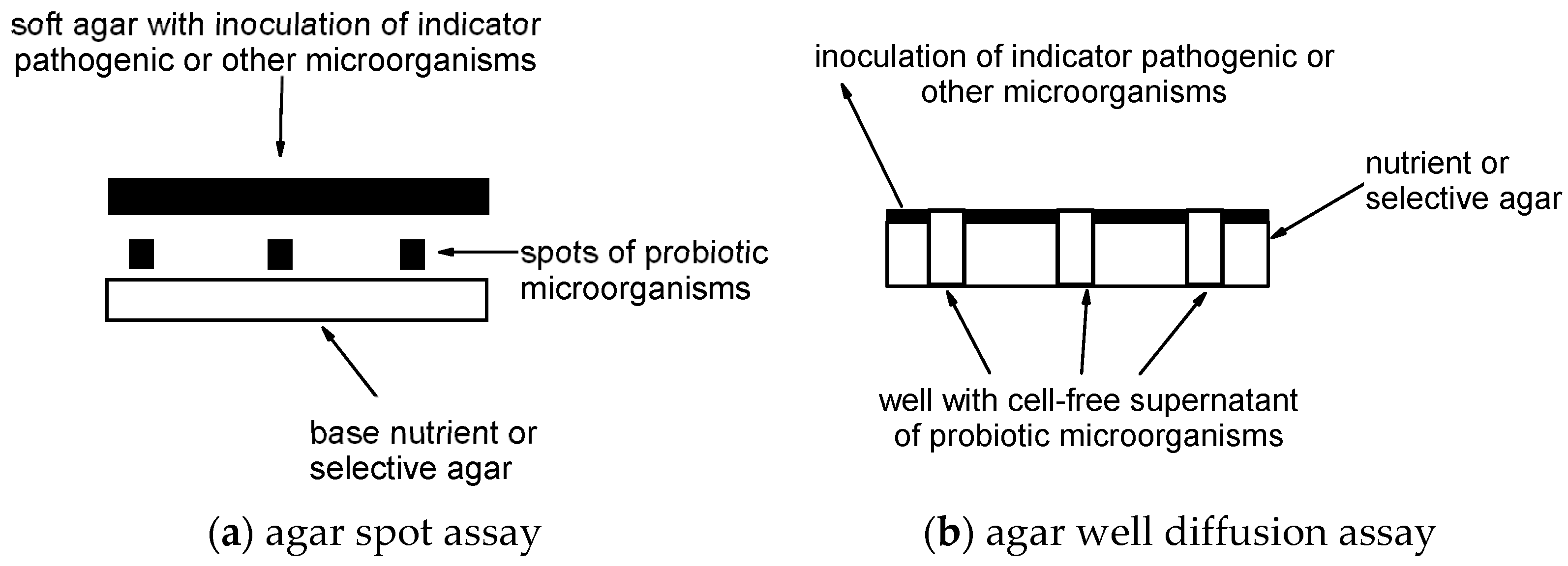
2.3. Agar Spot Assay

The antimicrobial effect of the chosen single-strain and multi-strain dietary supplements, probiotics, or other beneficial microbes against common skin or wound pathogens was determined using the modified agar spot assay [55,56,57]. Briefly, each probiotic overnight culture with a final concentration (108 cfu/mL) was inoculated as spots onto the following media: De Man, Rogosa, and Sharpe agar (Millipore, 1.10660) for all multi-strain probiotics and SS01-SS04, TOS-propionate agar (Sigma-Aldrich, 43314) for SS05 and SS06, Mannitol Egg Yolk Polymyxin agar (Merck, 1.05267) for SS07, Clostridium perfrigens agar (Liofilchen, 610207) for SS08-SS10, and Sabouraud glucose agar (BioMerieux, AEB152202) for SS11.
The plates were dried for 30 min at room temperature. All De Man, Rogosa, and Sharpe agar plates were then incubated anaerobically at 35 °C for 24 h using anaerobic jars together with a Genbag anaerobic pouch. Other agar plates were incubated aerobically for 24 h. All plates were then overlaid with 7 mL of soft agar (15g tryptic soy bujon (Fluka, 51228)/500 mL and 2g agarose (Fluka, 51228)/500 mL) inoculated with overnight cultures of the pathogens (with final concentration 7 log cfu/mL) and incubated at 35 °C for 48 h. Figure 1a shows a scheme of the agar spot assay.
After 48 h of incubation, measurements of inhibition zones around the colonies were measured using a ruler. The diameter of the zone of inhibition measuring from both sides of the clear zone around the colony was measured. The result also included 6–7 mm of the colony. Zones of more than 20 mm, between 10 and 20 mm, and less than 10 mm were considered as strong (3+), intermediate (2+), and low inhibitions (+), respectively. This is a modified scale [57], similar to those proposed by Davis and Stout [58]. If no zone was detected the result was reported as less than 6 mm. This assay was performed in triplicate. The mean of the zones of inhibition as well as the standard deviation SD were calculated.

2.4. Co-Culturing for Microbial Competition Assay

The co-culturing for microbial competition assay of the pathogens and the chosen single-strain and multi-strain dietary supplements, probiotics, or other beneficial microbes was conducted similarly to Tranberg and co-authors [60] as follows: aliquots of 1 mL of an overnight culture of probiotics and 1 mL of the overnight culture of the clinical wound pathogens were inoculated into 500 mL sterile tubes with fresh broth containing 1 mL tryptic soy broth (Fluka, 51228) and 1 mL De Man, Rogosa and Sharpe broth (Merck, 1.10661). As controls, 1 mL overnight cultures of pathogens were grown in 1 mL tryptic soy broth and 1 mL De Man, Rogosa, and Sharpe broth. All samples were incubated overnight at 35 °C for 24 h.
After 24 h of incubation, colonies of surviving pathogens were counted using serial dilutions, ranging from 101 to 108. The following selective media were used for clinical isolates: Baird-Parker agar (Biolife, 4011162) for Staphylococcus aureus isolates, cetrimide agar (22470, Fluka) for Pseudomonas aeruginosa isolates, kanamycin esculin azide agar (Biolife, 4015522) for Enterococcus faecium isolates, violet red bile glucose agar (Fluka, 70189) for Escherichia coli and Enterobacter spp. isolates, HiCrome Klebsiella selective agar (Fluka, 90925) for Klebsiella pneumoniae isolates, MacConkey agar without salt (Sigma Aldrich, 51405) for Acinetobacter spp. isolates and bile esculin agar (Sigma Aldrich, 48300) for Bacteroides spp. isolates. All selective media were then incubated aerobically at 35 °C for 24 or 48 h according to the manufacturer’s recommendation except for bile esculin agar for Bacteroides spp. isolates which were incubated anaerobically at 35 °C for 24 h using anaerobic jars together with a Genbag anaerobic pouch.
The reduction and log step reduction were then calculated as follows:
% R = c f u p a c f u p a + p r o c f u p a × 100
log 10 R = l o g c f u p a + p r o c f u p a
where: %R is the percent of reduction of the pathogen, log10R is the log step reduction, cfupa is the cfu of the pathogen after incubation and cfupa+pro is the cfu of the pathogen after incubation of the pathogen together with the probiotic. A log step reduction of more than six was considered strong inhibition as it corresponds to a 99.9999% reduction in the case of initial concentration of 106 cfu/mL. Between 3 and 6 was considered intermediate inhibition and less than 3 was considered low inhibition. Two separate experiments were conducted, and the average was calculated for each sample.

2.5. Agar Well Diffusion Assay

A slightly modified method of the agar well diffusion assay for the inhibition of pathogens by cell-free supernatants of chosen single-strain and multi-strain dietary supplements, probiotics, or other beneficial microbes by Holder and Boyce [61] was used. Briefly, overnight cultures of pathogens were confluently streaked onto Müller Hinton agar (BioLife, 4017402) plates with sterile cotton swabs and the plates were left to dry for 30 min. Wells (5 mm in diameter) were cut using 1000 µL sterile pipette tips. Cell-free supernatants of overnight cultures of chosen probiotics and other beneficial microbes were prepared by sedimentation of cells with centrifuging (4000× g for 10 min). The cell-free supernatant was filtered through a 0.22 µm pore size syringe filter. Half of each cell-free supernatant was used directly by inoculating 800 µL into the wells. The other half was neutralized using NaOH and adjusted to pH = 7 to achieve a neutralized cell-free supernatant that was inoculated into the remaining wells. Figure 1b shows a scheme of the agar well diffusion assay.
The antibacterial effect was determined by measuring the diameter of the zone of inhibition around the wells. Again, zones of more than 20 mm, between 11 and 20 mm, and less than 10 mm were considered strong (3+), intermediate (2+), and low inhibitions (+), respectively. The mean of the radii measuring from the edges of the colonies to the edges of the clear zones was calculated as well as the standard deviation SD. This assay was also performed in triplicate.
After 48 h of incubation, measurements of inhibition zones around the wells were measured using a ruler. The diameter of the zone of inhibition measuring from both sides of the clear zone around the well was measured. The result also included 6 mm diameters of the wells. Zones of more than 20 mm, between 10 and 20 mm, and less than 10 mm were considered as strong (3+), intermediate (2+), and low inhibitions (+), respectively according to the modified scale by Shokryazdan and co-authors [57]. If no zone was detected, the result was reported as less than 6 mm. This assay was performed in triplicate. The mean of the zones of inhibition as well as the standard deviation SD were calculated.

2.6. Statistics

The mean zones of inhibition were presented as 95% Confidence Intervals (CI) comparing, agar spot assay, co-culturing, and agar well diffusion assay respectively, explored and evaluated with appropriate statistical as needed for various probiotics groupings, such as single-/multi-strain, species. Student t-test was used to compare single-/multi-strains. One-way ANOVA test with post-hoc HSD comparing mean zone was used for multiple probiotic groups and two-factor ANOVA was used to compare agar well diffusion assay interaction with various groups. Assumptions of those tests were also checked. The statistical analysis was performed in the statistical program R (version 4.2.1).

3. Results

3.1. Identification of Species and Genera of Microbial Strains Using Molecular Detection Methods

The results of the polymerase chain reactions (PCR) using genera-specific and species-specific primer pairs for multi-strain probiotic dietary supplements and single-strain probiotics and other beneficial microbes are noted in Table 6 and Table 7, respectively.
The PCR primer pairs LbLMA1-rev and R-16-1 that targets the nucleotide sequence of the spacer between the 16S and 23S rRNA genes in all lactobacilli genera confirmed by a positive band at 220 bp was found for all seven multi-strain probiotics and all single-strain samples that contained lactobacilli (SS1 to SS4). Species-specific PCR using primer pairs noted in Table 4 was run for the following lactobacilli species: Lactobacillus acidophilus, Lactobacillus gasseri, Lacticaseibacillus casei, Lactocaseibacillus rhamnosus, Lactiplantibacillus plantarum and Limosilactobacillus reuteri. Species-specific DNA fragments were found for Lactobacillus acidophilus in MS1, MS2, MS3, MS5, MS6, MS7, Lactobacillus gasseri in MS4, Lacticaseibacillus casei in MS1, MS2, MS3, MS5, MS7, and SS02, Lactocaseibacillus rhamnosus in MS4, MS7, and SS01, Lactiplantibacillus plantarum in MS2, MS6, MS7, and SS04 and Limosilactobacillus reuteri in SS03.
The genus Bifidobacterium using the primer pairs Bif164F and Bif601R for amplifying the 16S ribosomal rRNA fragments confirmed by a positive band at 453 bp was also confirmed for all bifidobacterial containing samples (MS1, MS2, MS3, MS5, MS6, MS7, SS05, SS06). Species-specific PCR using primer pairs noted in Table 4 was run for the following bifidobacterial species: Bifidobacterium animalis, Bifidobacterium bifidum, and Bifidobacterium longum. Species-specific DNA fragments were found for Bifidobacterium animalis in MS1, MS2, MS3, MS5, MS6, MS7, SS5, and SS6, Bifidobacterium bifidum in MS1, MS2, MS5, and MS7 and Bifidobacterium longum in MS5 and MS7. The genera Lactococcus and Propionibacterium were confirmed by primer pairs noted in Table 4 for MS1, MS2, MS3, MS5, and MS7 and SS08, as well as SS09 and SS10, respectively. Species-specific DNA fragments were also found for the bacteria Enterococcus faecium (MS3), Bacillus subtilis (MS7), and Bacillus coagulans (SS07). Saccharomyces cerevisiae species-specific DNA fragments were found (MS6, SS11) thus confirming the presence of Saccharomyces cerevisiae subsp. boulardii.

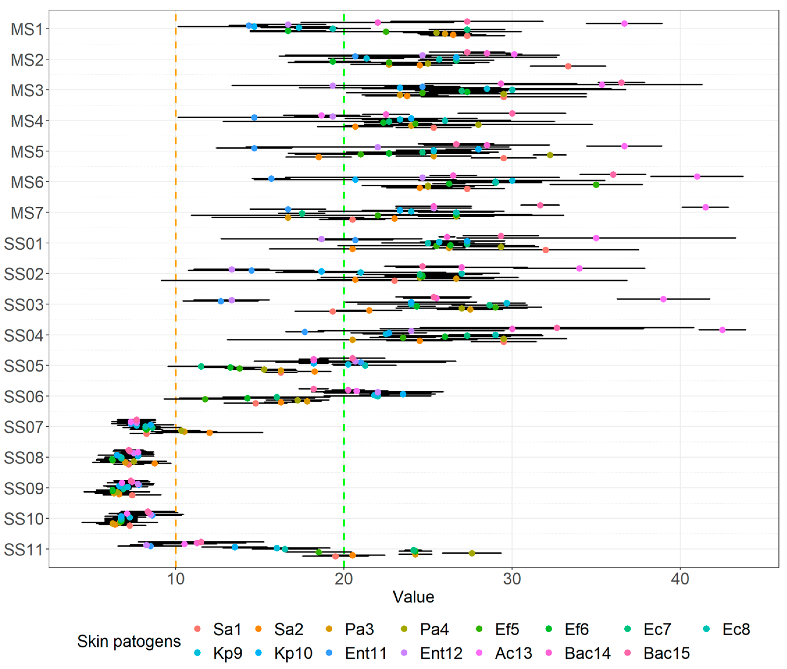
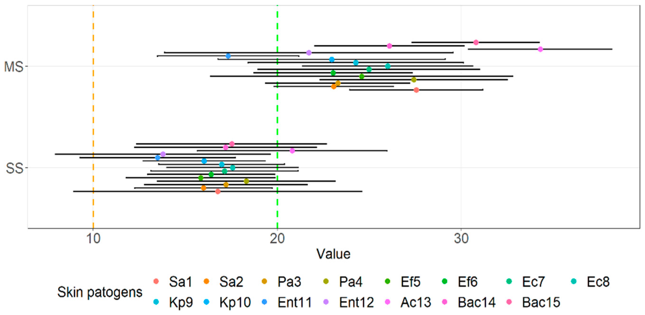
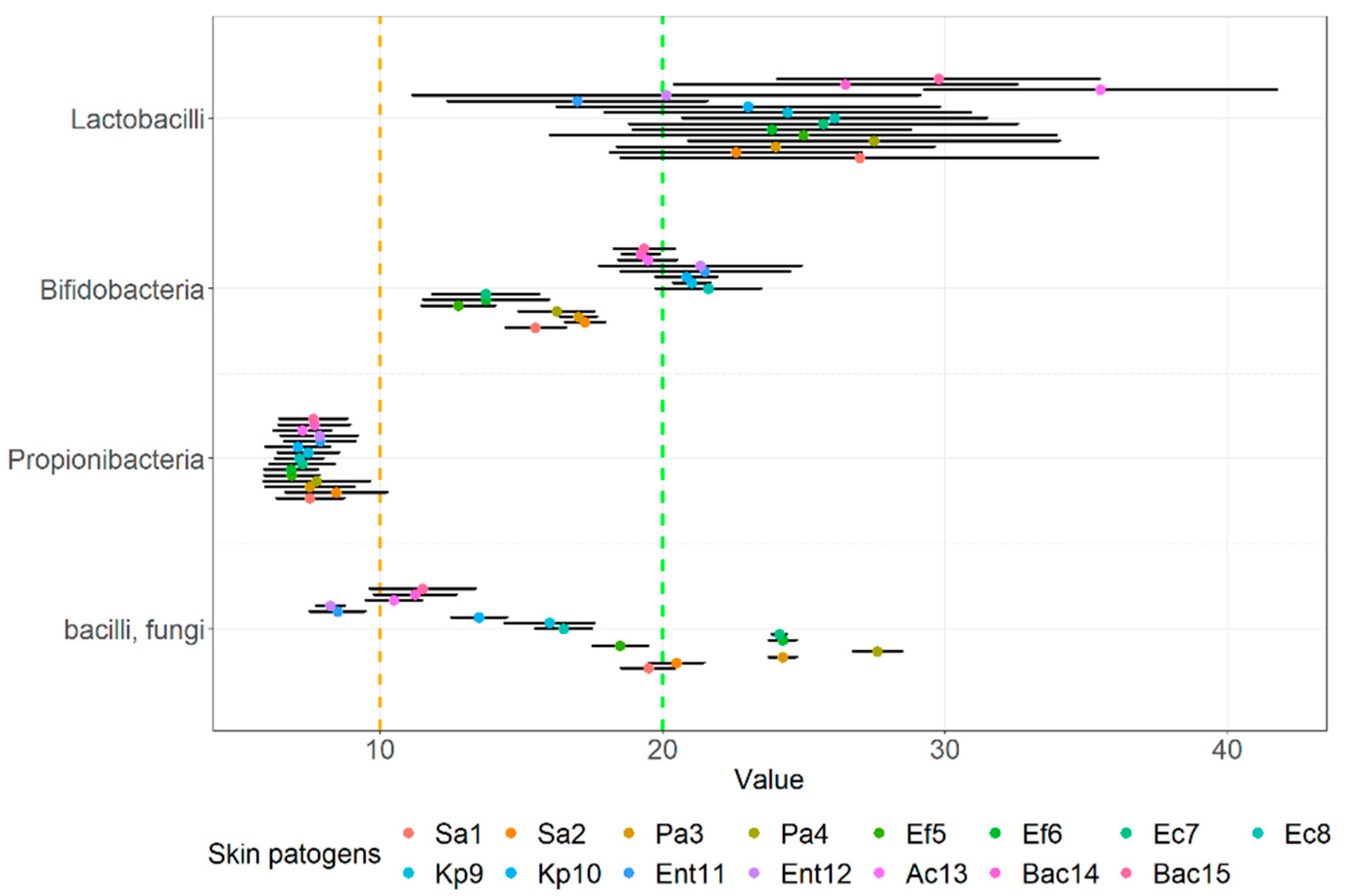
3.2. Agar Spot Assay

The evaluation of the mean zone of inhibition and standard deviation of the agar spot assay for all investigated probiotics and other beneficial microbes against the clinical pathogens, isolated from various skin wounds are noted in Table 8. All results of the zone of inhibition and standard deviation are noted in Supplementary Table S1.
As obvious from Table 8, all multiple-strain probiotics and single-strain probiotics SS01to SS04 (including Lacticaseibacillus paracasei Shirota, Limosilactobacillus reuteri DSM 17938, Lacticaseibacillus rhamnosus GG, and Lactiplantibacillus plantarum DSM 2601) were successful against most clinical wound pathogens as strong inhibition (the zone of inhibition was more than 20 mm) was found in most of the assays. On the other hand, the single strain probiotics Bacillus coagulans MTCC 5260 (SS07), Propionibacterium freudenreichii DSM 20271 (SS08), Propionibacterium propionici DSM 20272 (SS09), and Propionibacterium freudenreichii susp. shermanii (SS10) exhibited only low inhibition (the zone of inhibition was less than 10 mm). Intermediate average inhibition (zone of inhibition was between 10 and 20 mm) was found for both single-strain bifidobacteria: Bifidobacterium lactis HN019 (SS05), Bifidobacterium lactis BB12 (SS06), and the single-strain fungi Saccharomyces boulardii (SS11). The average zone of inhibition of all probiotic strains against individual clinical pathogens was intermediate for most strains and even above 20 mm for one strain of Staphylococcus aureus, Pseudomonas aeruginosa, Escherichia coli, and Acinetobacter and both clinical strains of the Bacteroides genus thus indicating that no specific pathogen stood out or was more resistant to the antimicrobial effect of the chosen probiotics.
The means of the inhibition zone of probiotics against wound pathogens with 95% CI are noted in Figure 2. The Propionibacterium strains and the Bacillus strain (SS07-SS10) seem to have smaller mean zones of inhibition and all multi-strain probiotics seem to have a larger zone of inhibition against all challenge wound pathogens. Checking the mean zone of inhibition against all wound pathogens for the various probiotics we observed statistical differences (F(17.252) = 40.5, p < 0.001).
As obvious in Figure 3, looking just at multi-strain (M = 25.15, SD = 3.95) and single-strain probiotics or beneficial microbes (M = 16.74, SD = 1.74), we showed that the latter has a statistically significant lower mean inhibition zone (t = −7.553, p < 0.001), which is also indicated in Figure 3. Grouping the data along the lines for species we observed the average means of the inhibition zone in descending order as follows: multi-strain probiotics that contained mainly lactobacilli strains and single-strain lactobacilli (M = 25.20, SD = 4.20), single strain bifidobacteria probiotics (M = 18.05, SD = 3.09), probiotic yeast strain and Bacillus strain (M = 16.98, SD = 6.31), and finally the Propionibacterium (M = 7.48, SD = 0.42). These means had a statistically significant difference (F(3.56) = 47.38, p < 0.001).
A post-hoc HSD test comparing pairs showed that the mean zone of inhibition against wound pathogens for all lactobacilli-containing probiotics was higher than others and the mean zone of inhibition of the Propionibacterium strains was lower than the others, which can also be at least partially indicated in Figure 4.

3.3. Co-Culturing for Microbial Competition Assay

The evaluation of the average log step reduction for all investigated probiotics and other beneficial microbes against the clinical pathogens, isolated from various skin wounds using co-culturing is noted in Table 9. The scale of a log step reduction of more than 6 was considered strong inhibition, between 3 and 6 was considered intermediate inhibition and less than 3 was considered low inhibition. All results of the average log step reduction and percentage of reduction are noted in Supplementary Table S2.
As obvious from Table 9 strong reduction of pathogens (log step reduction of more than 6 log steps) was found for three multiple-strain probiotics (MS4, MS5, and MS7), whilst a low reduction of pathogens was found for three single-strain probiotics including Propionibacterium freudenreichii DSM 20271, Propionibacterium propionici DSM 20272, and Saccharomyces boulardii (SS8, SS9, and SS11 respectively). All other probiotics achieved an intermediate reduction of pathogens (log step reduction between 3 and 6 log steps). One clinical pathogen of the Enterobacter genus was less resistant as an average log step reduction above 6 log steps was achieved for all probiotics and one clinical pathogen of Enterococcus faecalis was most resistant as the average log step reduction under 3 log steps was achieved for all probiotics. Comparing the log step reduction of the wound pathogens after co-culturing with probiotics and other beneficial microbes we observed lower inhibition compared to agar spot assays for all probiotics and other beneficial microbes (F(17.252) = 12.08, p < 0.001).
The same was observed when comparing the inhibition effect of multi-strain and single-strain probiotics against wound pathogens (t = −3.962, p < 0.001), where multi-strain probiotics (M = 5.62, SD = 129) achieved a higher log step reduction of all challenge pathogens than single-strain probiotics (M = 3.94, SD = 1.03). When comparing the inhibition effect of probiotic species against all pathogens we found a statistical difference (F(3.56) = 26.79, p < 0.001). When comparing pairs with HSD post-hoc tests, we showed that there was no statistical difference between the inhibition effect of lactobacilli-containing probiotics (M = 5.30, SD = 1.20) and bifidobacteria-containing probiotics (M = 5.51, SD = 1.38) against the wound pathogens. However, there was a difference when comparing both lactobacilli and bifidobacteria containing probiotics to single-strain beneficial microbes that contained propionibacteria, the Bacillus species, and the probiotic yeast. Additionally, there was no difference between the probiotic yeast (M = 2.25, SD = 1.46) and the beneficial microbes that contained propionibacteria (M = 2.78, SD = 0.94).

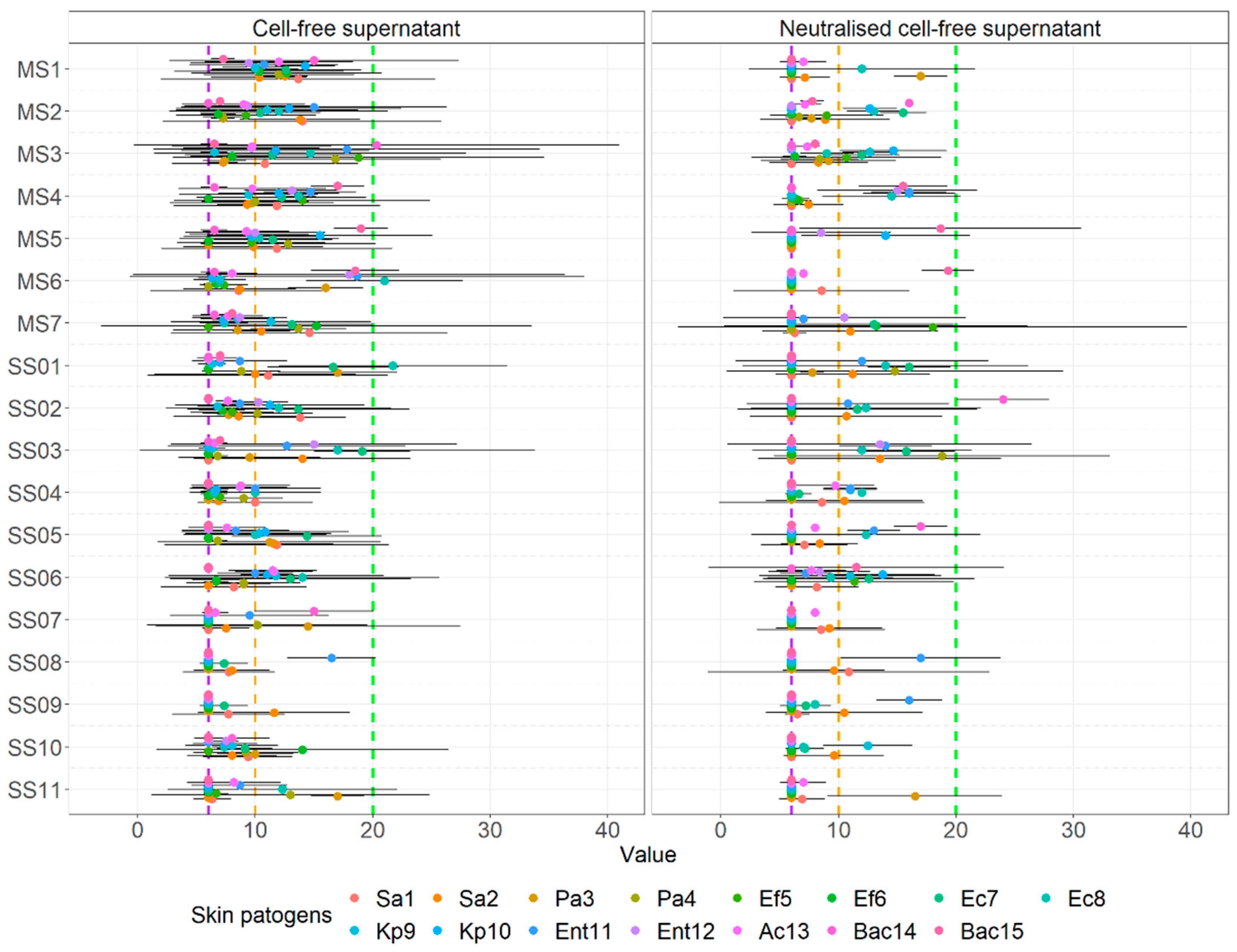
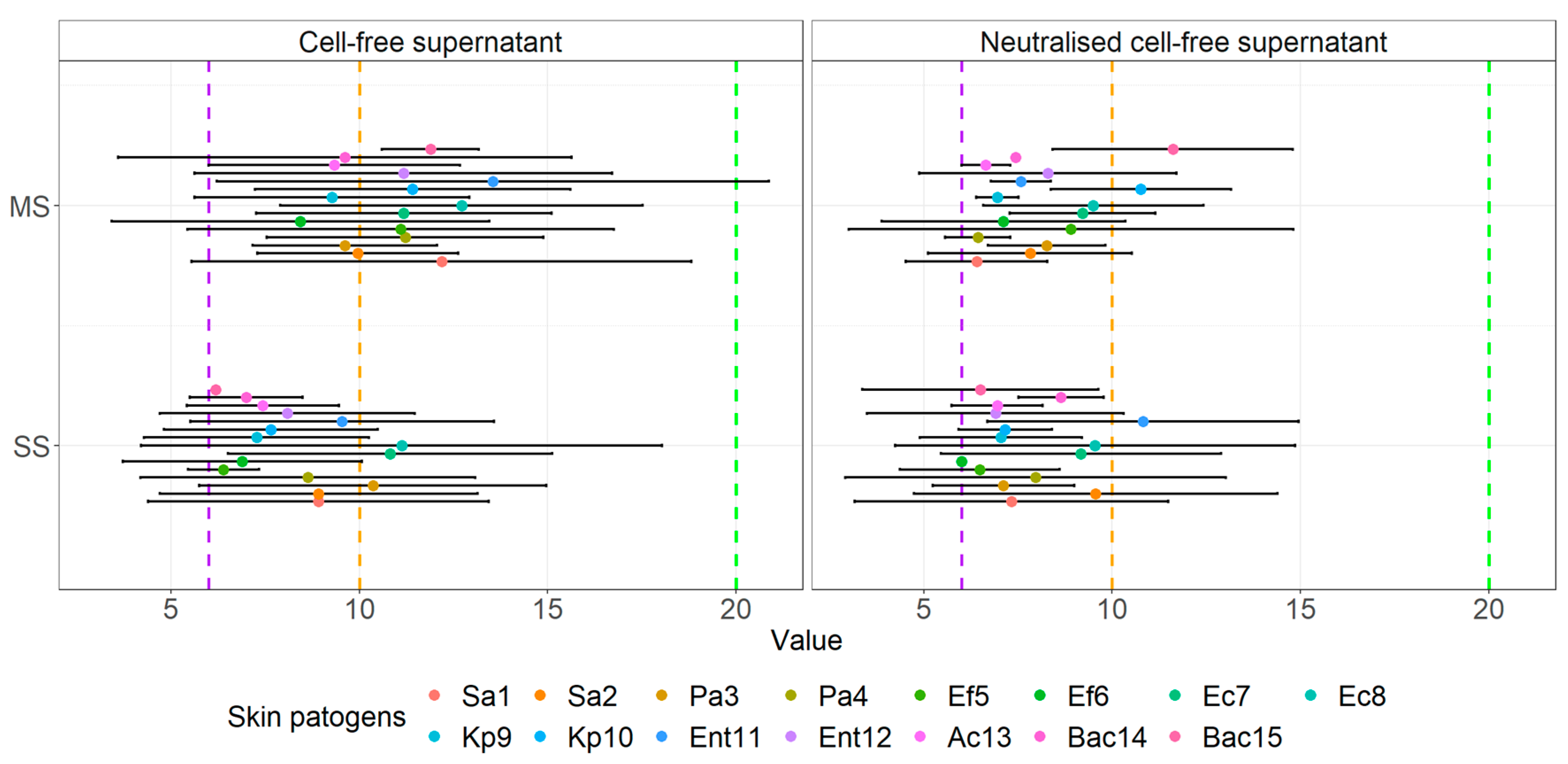
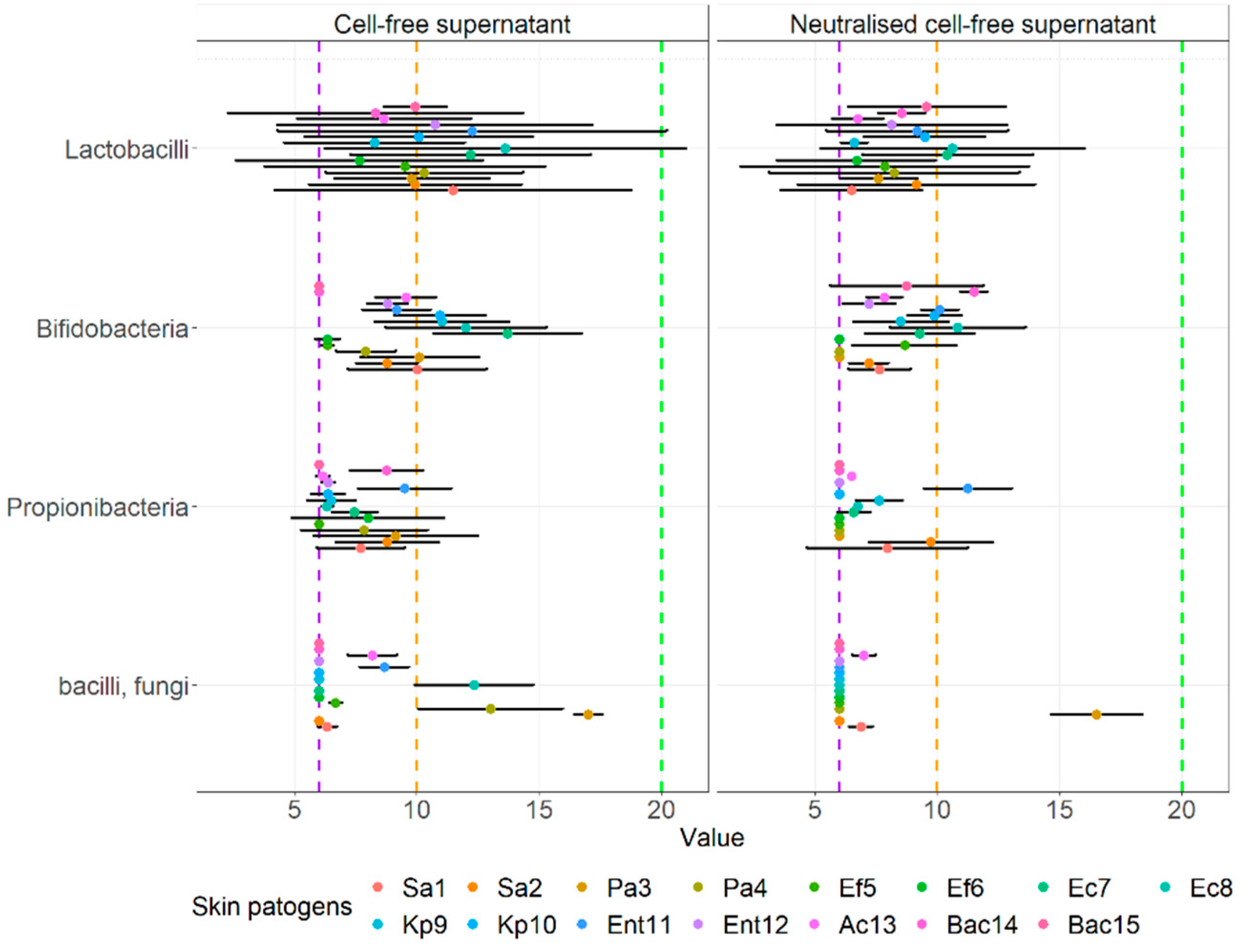
3.4. Agar Well Diffusion Assay

Below are the results of the mean zone of inhibition for all investigated cell-free supernatants (S) (Table 10) and neutralized cell-free supernatants (NS) (Table 10) of probiotics and other beneficial microbes against the clinical pathogens, isolated from various skin wounds. Exact values of inhibition zones and standard deviation are noted in Supplementary Tables S3 and S4.
As obvious from Table 10, the cell-free supernatants of all multiple-strain probiotics except MS7 exhibited an intermediate average inhibition (zone of inhibition was between 10 and 20 mm). All cell-free supernatants of single-strain probiotics and MS7 exhibited only a low inhibition (the zone of inhibition was less than 10 mm). Only two probiotics (MS6 and SS01) exhibited high inhibition of cell-free supernatant, both for the same clinical strain of Escherichia coli. No pathogen stood out in its resistance against the cell-free supernatants. All results show a lower inhibition ability of the cell-free supernatant compared to probiotics.
As obvious from Table 11 (Supplementary Table S4) the neutralized cell-free supernatants of all probiotic strains exhibited only low average inhibition for all investigated clinical pathogens from wounds. Only one neutralized cell-free supernatant of Limosilactobacillus reuteri DSM 17938 exhibited a strong inhibition against one clinical strain from the Bacteroides genus. No pathogen stood out in its resistance against the neutralized cell-free supernatants. All results show a lower ability of the neutralized cell-free supernatant compared to cell-free supernatants.
The visual comparison of the results of the inhibition zones of cell-free supernatants (S) and neutralized cell-free supernatants (NS) of all probiotics and beneficial microbes against clinical pathogens are noted in Figure 5. Figure 6 displays the comparison of the inhibition zones of S and NS of multi- and single- strain probiotics and microbes against clinical wound pathogens and Figure 6 displays the results of the inhibition zones of S and NS for all probiotics and beneficial microbes, divides into main species against the wound pathogens.
Statistically comparing results of the inhibition of cell-free supernatant and neutralized cell-free supernatants agar well diffusion, S and NS respectfully (Figure 6), with the aforementioned groups we observed, that there was no statistically significant interaction between agar well diffusion results and all probiotics of beneficial microbes (F(17.504) = 1.281, p = 0.199), but there was a simple main effect on various probiotics (p < 0.001) and agar well diffusion (p < 0.001) on the mean zone.
Looking at multi- and single- strain probiotics (Figure 7), we can observe a statistically significant interaction between the effect of both S and N supernatants using the agar well diffusion assay against wound pathogens (F(1.56) = 7.475, p = 0.008) as well as simple main effects, more precisely mean zones of supernatants of multi-stain probiotics were higher compared to single-stain supernatants (p < 0.001) and inhibition was higher for S compared to NS (p < 0.001). There was also no interaction between the inhibition of cell-free supernatants of probiotics and other beneficial microbes, divided into main species (F(3.112) = 2.740, p = 0.610) against all wound pathogens. However, both supernatants exhibited significant simple main effects, higher for S than NS (p = 0.007) and also higher for bifidobacteria-containing single strain probiotics (p = 0.030) and lactobacilli-containing probiotics (p = 0.001) than for propionibacteria-containing single strain beneficial microbes (p = 0.005).

4. Discussion

One important attribute of probiotics and probiotic candidates is their antimicrobial effect against pathogens. It is a well-known attribute of the lactobacilli and bifidobacteria genera [62]. The antimicrobial effect against pathogens is mostly attributed to the production of metabolites such as bacteriocins, organic acids, short-chain fatty acids, and hydrogen peroxide. Other important mechanisms of action of probiotics include competitive exclusion, immune modulation, stimulation of host defenses, and the production of signaling molecules that trigger changes in gene expression [55,63,64]. However, appropriate methodology is important in order to determine realistic and repeatable results. Our study used three different in vitro methods for determining the antimicrobial effect: the agar-spot assay, the co-culturing assay, and the agar-well diffusion assay. The first two methods utilised live microbes, whilst the last method utilised cell-free supernatant or postbiotics. The methods presented differences in the results. All dietary supplements achieved a certain level of inhibition of all pathogens, although there were variations between strains and multi-strain supplements, where the latter exhibited higher inhibition of the clinical pathogens than the single strain (p < 0.05), regardless of the method.
Our analysis of the collected data showed that the means of inhibition of probiotics and other beneficial microbes against all wound pathogens were statistically different (F(17.252) = 40.5, p < 0.001), where single strain beneficial microbes containing propionibacteria and Bacillus species (SS07-SS10) exhibited smaller inhibition zones against wound pathogens compared to all other probiotics and all multi-strain probiotics exhibited larger inhibition than single-strain probiotics. Looking at interactions via two-way ANOVA analysis, we observed a statistically significant interaction between multi- and single- strain probiotics or beneficial microbes and agar well diffusion (F(1.56) = 7.475, p = 0.008) as well as higher simple main effects for mean zones of multi-stain probiotics compared to single-stains (p < 0.001) and mean zones in agar well diffusion was lower in neutralized supernatant compared to the supernatant (p < 0.001) against wound pathogens.
The three methods to assess the antimicrobial effect of probiotics and other beneficial microbes or their metabolites against skin pathogens deployed in this study are based on phenotype characteristics that can be used for culturable microorganisms [65]. The most time-consuming is the co-culturing assay which requires the preparation of 10-fold dilutions for the enumeration of the pathogen after incubation with probiotics to determine the reduction effect. There are several modifications to this method, including incubation time, media type, and final detection method [55,66,67,68]. The advantage of this method is that both the probiotics and the pathogens are in a liquid environment enabling more simulation of the natural environment than existing in a colony on a solid surface, where immobilisation restricts growth. Such an environment causes different dynamics, less growth restriction, quorum sensing, and planktonic growth of both the probiotic and pathogen microbes [69,70,71]. Both the agar-well diffusion assay and the agar spot assay are conducted on solid media and require the measurement of the zone of inhibition against challenge pathogens. The agar spot assay investigates the inhibition effect of microbes, grown in a colony, whilst the agar-well diffusion assay investigates the inhibition effect of the cell-free supernatant, either in direct form or neutralized to eliminate the effect of organic acids. Both methods also exhibit several modifications with regard to solid media preparation, incubation conditions, initial concentration, and diffusion of metabolites [21,22,25,66,72,73,74]. Some authors measured either the whole diameter of the zone of inhibition which includes the diameter of the formed probiotic colony or the well with the supernatant [22,66,72,73], whilst other authors measured only the radius of the inhibition zone [25,55,74]. In our study, all cell-free supernatants of lactobacilli-containing multi-strain dietary supplements exhibited some inhibition as the average inhibition was intermediate for all except MS7. However, the cell-free supernatants of single-strain lactobacilli achieved average low inhibition. In the study by Lopes and co-authors [25], all investigated lactobacilli strains exhibited inhibition against examples of possible wound pathogens including Escherichia coli, Pseudomonas aeruginosa, and Staphylococcus aureus. However, when examining the results, it is obvious that in some cases, the radius of the zone of inhibition is only 1 mm, meaning the inhibition was also low for some strains, as found in our study. Similarly, in the study by Tejero-Sarinena and co-authors [74] the radii of the zones of inhibition of the non-adjusted cell-free supernatant of various lactobacilli and bifidobacteria strains were low, between 0.7 mm and up to 2 mm.
Neutralization of the culture supernatants with alkali vastly reduced the antagonistic effects of all our multiple-strain dietary supplements and our single strains thus indicating that the main mechanism of antagonism was the production of organic acids, such as lactic acid, propionic acid, butyric acid, and that bacteriocinogenic potentials were only partially used. On the other hand, the neutralized cell-free supernatant of Lactiplantibacillus plantarum DSM 2601 (SS04), Propionibacterium freudenreichii DSM 20271 (SS08), and Propionibacterium acidipropionici DSM 20272 (SS09) exhibited a somewhat higher average inhibition than the non-adapted cell-free supernatant, thus indicating that bacteriocins, such as perhaps plataricins, pediocins, or other neutral metabolites were produced [75,76,77]. The neutralized cell-free supernatant Bifidobacterium animalis subsp. lactis BB12 had a higher antagonistic effect than the non-neutralized for some pathogens (Enterococcus faecium, Escherichia coli, Klebsiella pneumoniae, and Bacteroides spp.) but not for Pseudomonas aeruginosa, Enterobacter spp. or Acinetobacter spp. Similarly, in the study by Fredua-Agyeman and co-authors [78] the neutralized cell-free supernatants of BB12 and Lactobacillus acidophilus La-5 did not show inhibition against Pseudomonas aeruginosa. Additionally, in the study by Lopes and co-authors [25], the antimicrobial activity of the cell-free supernatant was also attributed to organic acid production as the neutralized supernatant did not exhibit inhibition. The same conclusions were also found in the study by Tejero-Sarinena and co-authors [74].
In order to enable some comparison between all three methods, we created a scale of the co-culturing method based on the disinfection requirement for medical devices according to the Food and Drug Administration [79], where a log step reduction of 6 log steps or more is considered a strong reduction of the pathogen. This was based on the disinfection requirement of disinfectants where the log step reduction of 6 log steps corresponds to a 99.9999% reduction in the case of the initial pathogen concentration of 106 cfu. All chosen probiotics achieved a log step reduction for all challenge pathogens. According to the scale, three of our five chosen multi-strain dietary supplements (MS4, MS5, MS7) exhibited a strong average reduction of pathogens, whilst the other multi-strain dietary supplements achieved intermediate log step reduction of the pathogen. Eight of our chosen single-strain beneficial microbes achieved an average intermediate reduction of pathogens, whilst three achieved a low average reduction of the pathogen (Propionibacterium freudenreichii DSM 20271, Propionibacterium acidipropionici DSM 20272, and Saccharomyces boulardii). Other studies using the co-culturing method found that probiotics or probiotic candidates caused a reduction of pathogens, including Escherichia coli and Staphylococcus aureus co-cultured with Lactobacillus acidophilus La5 and Bifidobacterium longum ATCC 15707 [67], Staphylococcus aureus, and Pseudomonas aeruginosa co-cultured with Limosilactobacillus fermentum [68], Escherichia coli, Salmonella Enteritidis, Salmonella serotype (ser.) Typhimurium, Staphylococcus intermedius, Klebsiella oxytoca, and other pathogens co-cultured with lactobacilli isolated from piglet feces [66].
Molecular methods are much less time-consuming than classical phenotype methods that cannot easily distinguish between various species of the same genera and are also applicable for enumeration [80,81]. Although we did not conduct all species-specific PCR protocols for all species declared in all dietary supplements, we found positive results for all the PCR protocols that we conducted to detect genera or species thus proving that the reliability of the labelling system of probiotic supplements has improved compared to previous years [43,82]. Despite the recent division of lactobacilli into 23 novel genera [15] we found positive bands for all lactobacilli-containing dietary supplements using the primers pairs LbMA1-rev/R-16-1 [42], and it is obvious that these new genera share a common DNA section. Almost no dietary supplements used this new nomenclature. Another interesting finding was the positive band for Lacticaseibacillus paracasei Shirota (SS01, Yakult®), using the primer pairs Prl/CasII for the casei species, published by Walter and co-authors in the year 2000 [44]. As Lacticaseibacillus paracasei Shirota was reclassified from the casei species [83] after the publication of the primers, it is obvious that they are not species-specific and share a common DNA section as they belong to closely related species [84]. Recently the heterogenous genus of propionibacteria was divided into cutaneous (Cutibacterium spp.) and dairy propionic acid-producing bacteria (Propionibacterium and Acidipropionibacterium spp.) [41]; however, using the primer pairs PB1/PB2 [50] all propionibacteria: Propionibacterium freudenreichii subsp. Freudenreichii DSM 20271, Propionibacterium freudenreichii subsp. Shermanii, and Acidipropionibacterium acidipropionici DSM 20271 (SS08-SS10, respectively) were detected. Additionally, the primer pair CS1/SC2 [54] was used to detect Saccharomyces cerevisiae and we found a positive band for MS6 and SS11, which both contain Saccharomyces boulardii according to the manufacturers, confirming it is in fact a variant of Saccharomyces cerevisiae [54]. These findings also indicate that all manufacturers are not up to date with taxonomic changes.
The probiotic Lacticaseibacillus rhamnosus GG, also known as LGG, was the first lactobacilli strain to be patented in 1989 and has proven health benefits as shown by systematic reviews of several clinical studies, focused on antibiotic-associated diarrhoea [85], paediatric diarrhoea [86], gastroenteritis [87] and respiratory tract infections in children [88]. It is a biofilm-forming and immunomodulating probiotic that has shown antimicrobial effect against several pathogens [89] and is often used in in vitro studies as a reference strain for examining the antimicrobial effect of potential new probiotic strains [90,91]. In our study, this strain was SS03 and it also exhibited strong inhibition of most clinical pathogens using the agar spot method. However, using the co-culturing method, our results show that only an intermediate inhibition rate was achieved, thus implying that complex mechanisms of the probiotic are at work in different circumstances and that promising in vitro results using one method does not necessarily correlate with other methods [55] or correlate to statistically significant health benefits in clinical studies [92,93].
Lacticaseibacillus paracasei Shirota and Limosilactobacillus reuteri DSM 17938 are also well-researched probiotic strains (SS01 and SS02). Both strains exhibited the same results as SS03, namely strong average inhibition using the agar spot assay and an intermediate inhibition rate using the co-culturing assay. The same results were also found for the less-researched strain Lactiplantibacillus plantarum DSM 2601 (SS04). The latest clinical studies of Lacticaseibacillus paracasei Shirota (Yakult®), find consumption leads to improvement of depressive symptoms [83], lipid metabolism and intestinal microbiota [94], digestive disorders [95], and immunological function [96]. Lacticaseibacillus paracasei Shirota has also shown antifungal activity [97] and, similarly to our study, antibacterial activity against Escherichia coli and Bacteroides spp. [98]. Limosilactobacillus reuteri DSM 17938 (BioGaia®) has replaced the original strain Limosilactobacillus reuteri ATCC 55730 as it does not contain plasmid-borne antibiotic resistance and both strains exhibit success in the treatment of acute gastroenteritis, especially in children [99]. Although Limosilactobacillus reuteri DSM 17938 exhibits strong antimicrobial potential against major gastric and enteric bacterial pathogens and rotavirus [100], it did not prove effective as eradication therapy for infection with Helicobacter pylori, thus indicating that further studies are needed to establish the role of probiotics as adjuvant therapy, as the authors concluded [101].
Two well-known strains of the same species of bifidobacteria were used as single-strain probiotics: Bifibacterium animalis subs. lactis HN019 and BB-12 (SS05 and SS06, respectively). Both strains exhibited comparable results using the agar spot assay. On the other hand, there were differences in individual results for the inhibition of pathogens for co-culturing and the agar-well diffusion assay, although the average inhibitions were almost the value, proving that many probiotics traits are indeed strain-specific [11] and cannot be generalized to all representatives of the same species. The strain HN019 proved successful against periodontal pathogens in a recent clinical trial [102] and is a well-known probiotic with immune-enhancing properties [103]. In an in vitro study using the co-culturing method Bifidobacterium animalis subs. Lactis, BB-12 successfully inhibited the growth of Clostridoides difficile (previously known as Clostridium difficile) [104]. This strain reduced the risk of respiratory infections in infancy in a clinical study [105].
The challenge propionic acid-producing bacteria used in our study included three strains: Propionibacterium freudenreichii subsp. freudenreichii DSM 20271, Acidipropionibacterium acidipropionici DSM 2072, and Propionibacterium freudenreichii subsp. Shermanii (SS08, 09, and 10, respectively), and achieved only intermediate, low, or even no average inhibition of pathogens, depending on the method. The common feature of these three bacteria is the ability to produce propionic acid. Our results are similar to the study by Dyshlyuk and co-authors [106] where moderate antimicrobial activity using a version of the agar spot method was found for Propionibacterium jensenii B-6085 and Propionibacterium thoenii B-6082, but not for Propionibacterium freudenreichii B-11921 and Propionibacterium acidipropionici B-5723 against pathogens Escherichia coli ATCC 25922, Salmonella enterica ATCC 14028, Staphylococcus aureus ATCC 25923, Pseudomonas aeruginosa B6643, Proteus vulgaris ATCC 63, and Listeria monocytogenes ATCC 7644. Propionibacterium freudenreichii subsp. freudenreichii DSM 20271 is known to produce cobalamin or vitamin B12 [107] and Propionibacterium freudenreichii subsp. shermanii has shown probiotic effect as part of multi-strain dietary supplements in clinical studies against irritable bowel syndrome-related intestinal microbiota stabilization [108], intestinal microbiota changes during anti-Helicobacter pylori treatment [109].
Our challenge spore-forming representative Bacillus coagulans MTCC 5260 (Prolife®) also achieved only intermediate, low, or even no average inhibition of pathogens, depending on the method. Probiotic Bacillus strains used either in spore or vegetative forms have shown antimicrobial, anticancer, antioxidant, and vitamin production properties. However, they can also produce toxins and biogenic amines and transfer antibiotic resistance genes; therefore, their safety is a concern. Studies on the microbiome using probiotic Bacillus strains are limited in humans [110]. The strain MTCC 5260 is also known as Unique IS2 and ATCC PTA-11748 [111] and has documented clinical efficacy against constipation [112]. It also exhibits antimicrobial effectiveness as it was efficient as an adjuvant in the treatment of bacterial vaginosis [113].
Saccharomyces cerevisiae var. boulardii (SS11) is the only representative of probiotic fungi used in our study and it achieved average intermediate or low pathogen reduction, depending on the method used. It is a well-known probiotic that produces various bioactive compounds and is mostly known for its role in treating gastrointestinal diseases [114,115]. Together with Lacticaseibacillus rhamnosus GG, it is even one of the few probiotics recommended by the ESPGHAN (European Society for Paediatric Gastroenterology, Hepatology, and Nutrition) and ESPID (European Society for Paediatric Infectious Diseases) [116,117] for treating acute gastroenteritis in children. Saccharomyces boulardii has also been proposed as an alternative to treating bacterial infections [114], however, our results do not support this claim for our challenge wound pathogens.
In our study, the multi-strain dietary supplement MS7 (Bio-Kult®) was effective in strong average inhibition found against most clinical pathogens using the agar spot method. This dietary supplement was also the most effective mixture against Enterococcus faecalis in another study using the agar spot test [72]. In a clinical study, this multi-strain probiotic was also associated with significant improvement in symptoms in patents with diarrhoea-predominated irritable bowel syndrome [118].
Several commercial dietary supplements including OMNi-BiOTic® Hetox, OMNi-BiOTic® 6, OMNi-BiOTic® Stress repair, OMNi-BiOTic® Flora plus+, and OMNi-BiOTic® Activ (MS1 to MS5) achieved strong average inhibition against all pathogens in our study. MS1, MS4, and MS5 also achieved strong average inhibition using the co-culturing method, whilst the average inhibition of cell-free supernatant was intermediate or even low. The lower effect of cell-free probiotic supernatant indicates that bacterial response is important in cell-cell signaling and/or bacteria-host interaction. The multi-strain dietary supplement MS2 was also used in a clinical study that found that this multi-strain probiotic might be a well-tolerated tool to positively influence the gastrointestinal quality of life as well as mental and somatic health, cognition, and immune response and potentially have effects on psychiatric symptoms [119]. In another clinical study, this multi-strain probiotic positively influenced the gastrointestinal tract of patients with diarrhoea-predominated irritable bowel syndrome [120]. In another study, the multi-strain postbiotic supernatant of the dietary supplement OMNi-BiOTic AAD10 with similar composition exhibited positive antibacterial and antifungal effects in vitro [121].
Our results show that several dietary supplements were efficient in reducing the pathogen loads of the investigated clinical pathogens. The concept that certain bacteria can destroy other, even pathogenic bacteria, especially with respect to the skin, is not new and many historic researchers, such as Metchnikoff, Nissle, Cantini, and others have investigated and proven this concept [9,122,123]. More than a decade ago, Howard and co-authors concluded that probiotics could be beneficial in the prevention and treatment of wound infections [124]. Probiotics also give positive results for wound healing, wound-epithelization, and neovascularization [125]; however, as such treatment represents a shift in the doctrine of wound treatments where using bacteria to fight bacteria is not intuitive [126,127], many more studies are needed to establish a consensus on the efficacy of using probiotics against skin pathogens.

5. Conclusions

The scientific evidence of the health benefits of using probiotics and postbiotics for wounds is becoming more extensive and, therefore, an important possible application of probiotics in the future. In light of our results, it seems that each clinical pathogen was differently susceptible to different probiotic strains, although in general the multispecies probiotics were more efficient than the single-strain probiotics; however, the method deployed also impacted the results. Perhaps a new approach such as a ‘’probiogram’’ or ‘’postbiogram’’ as an analogue to antibiograms could be a possibility in the future in finding the most efficient targeted probiotic strains, cell-free supernatants, or neutralized cell-free supernatants that have the highest antagonistic effect against individual clinical wound pathogens. Additionally, more robust, well-designed clinical trials of probiotics targeting different clinical skin pathogens are needed to establish more knowledge on the exact efficacy and mechanisms of individual probiotics against pathogens to draw evidence-based conclusions for clinical recommendations.

Supplementary Materials

The following supporting information can be downloaded at: https://www.mdpi.com/article/10.3390/microorganisms10122518/s1, (Tables S1–S4). S1: Results of the antagonistic effect of various probiotics and other beneficial microbes against clinical skin pathogens using the agar spot assay. S2: Results of the antagonistic effect of various probiotics and other beneficial microbes against clinical skin pathogens using the co-culturing assay. S3: Results of the antagonistic effect of various cell-free supernatants of probiotics and other beneficial microbes against clinical skin pathogens using the agar well diffusion assay. S4: Results of the antagonistic effect of various neutralised cell-free supernatants of probiotics and other beneficial microbes against clinical skin pathogens using the agar well diffusion assay.

Author Contributions

Conceptualization, S.F. and A.S.; methodology, S.F and M.S.; formal analysis, S.F.; investigation, S.F.; resources, S.F.; data curation, S.F. and P.K.; writing—original draft preparation, S.F.; writing—review and editing, S.F., P.M.V., A.S., and P.K.; visualization, funding acquisition, S.F. All authors have read and agreed to the published version of the manuscript.

Funding

This research was partially funded by Institut Allergosan, Austria.

Data Availability Statement

No additional data is available.

Conflicts of Interest

All authors declare no conflict of interest.

References

  1. McLoughlin, I.J.; Wright, E.M.; Tagg, J.R.; Jain, R.; Hale, J.D.F. Skin microbiome—The next frontier for probiotic intervention. Probiotics Antimicrob. Proteins 2022, 14, 630–647. [Google Scholar] [CrossRef] [PubMed]
  2. Perez Perez, G.I.; Gao, Z.; Jourdain, R.; Ramirez, J.; Gany, F.; Clavaud, C.; Demaude, J.; Breton, L.; Blaser, M.J. Body site is a more determinant factor than human population diversity in the healthy skin microbiome. PLoS ONE 2016, 11, e0151990. [Google Scholar] [CrossRef] [PubMed] [Green Version]
  3. Mihai, M.M.; Preda, M.; Lungu, I.; Gestal, M.C.; Popa, M.I.; Holban, A.M. Nanocoatings for chronic wound repair—Modulation of microbial colonization and biofilm formation. Int. J. Mol. Sci. 2018, 19, 1179. [Google Scholar] [CrossRef] [PubMed] [Green Version]
  4. Scalise, A.; Bianchi, A.; Tartaglione, C.; Bolletta, E.; Pierangeli, M.; Torresetti, M.; Marazzi, M.; Di Benedetto, G. Microenvironment and microbiology of skin wounds: The role of bacterial biofilms and related factors. Semin. Vasc. Surg. 2015, 28, 151–159. [Google Scholar] [CrossRef] [PubMed]
  5. Peral, M.C.; Huaman Martinez, M.A.; Valdez, J.C. Bacteriotherapy with Lactobacillus plantarum in burns. Int. Wound J. 2009, 6, 73–81. [Google Scholar] [CrossRef]
  6. Daeschlein, G. Antimicrobial and antiseptic strategies in wound management. Int. Wound J. 2013, 10, 9–14. [Google Scholar] [CrossRef]
  7. Bowler, P.G.; Duerden, B.I.; Armstrong, D.G. Wound microbiology and associated approaches to wound management. Clin. Microbiol. Rev. 2001, 14, 244–269. [Google Scholar] [CrossRef] [Green Version]
  8. Mahnic, A.; Breznik, V.; Bombek Ihan, M.; Rupnik, M. Comparison between cultivation and sequencing based approaches for microbiota analysis in swabs and biopsies of chronic wounds. Front. Med. 2021, 8, 607255. [Google Scholar] [CrossRef]
  9. Fijan, S.; Frauwallner, A.; Langerholc, T.; Krebs, B.; Ter Haar Née Younes, J.A.; Heschl, A.; Mičetić Turk, D.; Rogelj, I. Efficacy of using probiotics with antagonistic activity against pathogens of wound infections: An integrative review of literature. Biomed Res. Int. 2019, 2019, 7585486. [Google Scholar] [CrossRef] [Green Version]
  10. Martinengo, L.; Olsson, M.; Bajpai, R.; Soljak, M.; Upton, Z.; Schmidtchen, A.; Car, J.; Järbrink, K. Prevalence of chronic wounds in the general population: Systematic review and meta-analysis of observational studies. Ann. Epidemiol. 2019, 29, 8–15. [Google Scholar] [CrossRef]
  11. Hill, C.; Guarner, F.; Reid, G.; Gibson, G.R.; Merenstein, D.J.; Pot, B.; Morelli, L.; Canani, R.B.; Flint, H.J.; Salminen, S.; et al. The international scientific association for probiotics and prebiotics consensus statement on the scope and appropriate use of the term probiotic. Nat. Rev. Gastroenterol. Hepatol. 2014, 11, 506–514. [Google Scholar] [CrossRef] [Green Version]
  12. Reid, G.; Younes, J.A.; Van der Mei, H.C.; Gloor, G.B.; Knight, R.; Busscher, H.J. Microbiota restoration: Natural and supplemented recovery of human microbial communities. Nat. Rev. Microbiol. 2011, 9, 27–38. [Google Scholar] [CrossRef] [PubMed]
  13. Cecchini, M.; Langer, J.; Slawomirski, L. Antimicrobial Resistance in G7 Countries and beyond,” Economic Issues, Policies and Options for Action: Oecd Report; OECD: Paris, France, 2015. [Google Scholar]
  14. Salminen, S.; Collado, M.C.; Endo, A.; Hill, C.; Lebeer, S.; Quigley, E.M.M.; Sanders, M.E.; Shamir, R.; Swann, J.R.; Szajewska, H.; et al. The international scientific association of probiotics and prebiotics (ISAPP) consensus statement on the definition and scope of postbiotics. Nat. Rev. Gastroenterol. Hepatol. 2021, 18, 649–667. [Google Scholar] [CrossRef] [PubMed]
  15. Zheng, J.; Wittouck, S.; Salvetti, E.; Franz, C.; Harris, H.M.B.; Mattarelli, P.; O’Toole, P.W.; Pot, B.; Vandamme, P.; Walter, J.; et al. A taxonomic note on the genus Lactobacillus: Description of 23 novel genera, emended description of the genus Lactobacillus beijerinck 1901, and union of Lactobacillaceae and Leuconostocaceae. Int. J. Syst. Evol. Microbiol. 2020, 70, 2782–2858. [Google Scholar] [CrossRef] [PubMed]
  16. Ramos, A.N.; Sesto Cabral, M.E.; Noseda, D.; Bosch, A.; Yantorno, O.M.; Valdez, J.C. Antipathogenic properties of Lactobacillus plantarum on Pseudomonas aeruginosa: The potential use of its supernatants in the treatment of infected chronic wounds. Wound Repair Regen. 2012, 20, 552–562. [Google Scholar] [CrossRef]
  17. Valdéz, J.C.; Peral, M.C.; Rachid, M.; Santana, M.; Perdigón, G. Interference of Lactobacillus plantarum with Pseudomonas aeruginosa in vitro and in infected burns: The potential use of probiotics in wound treatment. Clin. Microbiol. Infect. 2005, 11, 472–479. [Google Scholar] [CrossRef] [Green Version]
  18. Jones, M.L.; Ganopolsky, J.G.; Labbé, A.; Prakash, S. A novel nitric oxide producing probiotic patch and its antimicrobial efficacy: Preparation and in vitro analysis. Appl. Microbiol. Biotechnol. 2010, 87, 509–516. [Google Scholar] [CrossRef]
  19. Chan, A.P.; Choi, Y.; Brinkac, L.M.; Krishnakumar, R.; Depew, J.; Kim, M.; Hinkle, M.K.; Lesho, E.P.; Fouts, D.E. Multidrug resistant pathogens respond differently to the presence of co-pathogen, commensal, probiotic and host cells. Sci. Rep. 2018, 8, 8656. [Google Scholar] [CrossRef]
  20. Mohammedsaeed, W.; McBain, A.J.; Cruickshank, S.M.; O’Neill, C.A.; Schaffner, D.W. Lactobacillus rhamnosus GG inhibits the toxic effects of Staphylococcus aureus on epidermal keratinocytes. Appl. Environ. Microbiol. 2014, 80, 5773–5781. [Google Scholar] [CrossRef] [Green Version]
  21. Shu, M.; Wang, Y.; Yu, J.; Kuo, S.; Coda, A.; Jiang, Y.; Gallo, R.L.; Huang, C.-M. Fermentation of Propionibacterium acnes, a commensal bacterium in the human skin microbiome, as skin probiotics against methicillin-resistant Staphylococcus aureus. PLoS ONE 2013, 8, e55380. [Google Scholar] [CrossRef]
  22. Al-Malkey, M.K.; Ismeeal, M.C.; Al-Hur, F.J.; Mohammed, S.W.; Nayyel, H.J. Antimicrobial effect of probiotic Lactobacillus spp. on Pseudomonas aeruginosa. J. Contemp. Med. Sci. 2017, 3, 218–223. [Google Scholar] [CrossRef]
  23. Prince, T.; McBain, A.J.; O’Neill, C.A. Lactobacillus reuteri protects epidermal keratinocytes from Staphylococcus aureus-induced cell death by competitive exclusion. Appl. Environ. Microbiol. 2012, 78, 5119–5126. [Google Scholar] [CrossRef] [PubMed] [Green Version]
  24. Thomas, J.G.; Motlagh, H.; Povey, S.B.; Percival, S.L. 2—The role of micro-organisms and biofilms in dysfunctional wound healing. In Advanced Wound Repair Therapies; Farrar, D., Ed.; Woodhead Publishing: Sawston, UK, 2011; pp. 39–76. [Google Scholar]
  25. Lopes, E.G.; Moreira, D.A.; Gullón, P.; Gullón, B.; Cardelle-Cobas, A.; Tavaria, F.K. Topical application of probiotics in skin: Adhesion, antimicrobial and antibiofilm in vitro assays. J. Appl. Microbiol. 2017, 122, 450–461. [Google Scholar] [CrossRef]
  26. Moghadam, S.S.; Khodaii, Z.; Zadeh, S.F.; Ghooshchian, M.; Aghmiyuni, Z.F.; Shabestari, T.M. Synergistic or antagonistic effects of probiotics and antibiotics- alone or in combination- on antimicrobial-resistant Pseudomonas aeruginosa isolated from burn wounds. Arch. Clin. Infect. Dis. 2018, 13, e63121. [Google Scholar] [CrossRef] [Green Version]
  27. Argenta, A.; Satish, L.; Gallo, P.; Liu, F.; Kathju, S. Local application of probiotic bacteria prophylaxes against sepsis and death resulting from burn wound infection. PLoS ONE 2016, 11, e0165294. [Google Scholar] [CrossRef] [PubMed] [Green Version]
  28. Satish, L.; Gallo, P.H.; Johnson, S.; Yates, C.C.; Kathju, S. Local probiotic therapy with Lactobacillus plantarum mitigates scar formation in rabbits after burn injury and infection. Surg. Infect. 2017, 18, 119–127. [Google Scholar] [CrossRef] [PubMed] [Green Version]
  29. Sürmeli, M.; Maçin, S.; Akyön, Y.; Kayikçioğlu, A.U. The protective effect of Lactobacillus plantarum against meticillin-resistant Staphylococcus aureus infections: An experimental animal model. J. Wound Care 2019, 28, s29–s34. [Google Scholar] [CrossRef]
  30. Brachkova, M.I.; Marques, P.; Rocha, J.; Sepodes, B.; Duarte, M.A.; Pinto, J.F. Alginate films containing Lactobacillus plantarum as wound dressing for prevention of burn infection. J. Hosp. Infect. 2011, 79, 375–377. [Google Scholar] [CrossRef]
  31. Ong, J.S.; Taylor, T.D.; Yong, C.C.; Khoo, B.Y.; Sasidharan, S.; Choi, S.B.; Ohno, H.; Liong, M.T. Lactobacillus plantarum USM8613 aids in wound healing and suppresses Staphylococcus aureus infection at wound sites. Probiotics Antimicrob. Proteins 2020, 12, 125–137. [Google Scholar] [CrossRef]
  32. Jones, M.; Ganopolsky, J.G.; Labbé, A.; Gilardino, M.; Wahl, C.; Martoni, C.; Prakash, S. Novel nitric oxide producing probiotic wound healing patch: Preparation and in vivo analysis in a new zealand white rabbit model of ischaemic and infected wounds. Int. Wound J. 2012, 9, 330–343. [Google Scholar] [CrossRef]
  33. Binda, S.; Hill, C.; Johansen, E.; Obis, D.; Pot, B.; Sanders, M.E.; Tremblay, A.; Ouwehand, A.C. Criteria to qualify microorganisms as probiotic in foods and dietary supplements. Front. Microbiol. 2020, 11, 1662. [Google Scholar] [CrossRef]
  34. Peral, M.C.; Rachid, M.M.; Gobbato, N.M.; Martinez, M.A.H.; Valdez, J.C. Interleukin-8 production by polymorphonuclear leukocytes from patients with chronic infected leg ulcers treated with Lactobacillus plantarum. Clin. Microbiol. Infect. 2010, 16, 281–286. [Google Scholar] [CrossRef] [PubMed] [Green Version]
  35. Komatsu, S.; Sakamoto, E.; Norimizu, S.; Shingu, Y.; Asahara, T.; Nomoto, K.; Nagino, M. Efficacy of perioperative synbiotics treatment for the prevention of surgical site infection after laparoscopic colorectal surgery: A randomized controlled trial. Surg. Today 2016, 46, 479–490. [Google Scholar] [CrossRef] [PubMed]
  36. El-Ghazely, M.H.; Mahmoud, W.H.; Atia, M.A.; Eldip, E.M. Effect of probiotic administration in the therapy of pediatric thermal burn. Ann. Burn. Fire Disasters 2016, 29, 268–272. [Google Scholar]
  37. Mayes, T.; Gottschlich, M.M.; James, L.E.; Allgeier, C.; Weitz, J.; Kagan, R.J. Clinical safety and efficacy of probiotic administration following burn injury. J. Burn Care Res. 2015, 36, 92–99. [Google Scholar] [CrossRef] [PubMed]
  38. Kotzampassi, K.; Stavrou, G.; Damoraki, G.; Georgitsi, M.; Basdanis, G.; Tsaousi, G.; Giamarellos-Bourboulis, E.J. A four-probiotics regimen reduces postoperative complications after colorectal surgery: A randomized, double-blind, placebo-controlled study. World J. Surg. 2015, 39, 2776–2783. [Google Scholar] [CrossRef]
  39. Aisu, N.; Tanimura, S.; Yamashita, Y.; Yamashita, K.; Maki, K.; Yoshida, Y.; Sasaki, T.; Takeno, S.; Hoshino, S. Impact of perioperative probiotic treatment for surgical site infections in patients with colorectal cancer. Exp. Ther. Med. 2015, 10, 966–972. [Google Scholar] [CrossRef] [Green Version]
  40. Moraffah, F.; Kiani, M.; Abdollahi, M.; Yoosefi, S.; Vatanara, A.; Samadi, N. In vitro-in vivo correlation for the antibacterial effect of Lactiplantibacillus plantarum as a topical healer for infected burn wound. Probiotics Antimicrob. Proteins 2022, 14, 675–689. [Google Scholar] [CrossRef]
  41. Bücher, C.; Burtscher, J.; Domig, K.J. Propionic acid bacteria in the food industry: An update on essential traits and detection methods. Compr. Rev. Food Sci. Food Saf. 2021, 20, 4299–4323. [Google Scholar] [CrossRef]
  42. Dubernet, S.; Desmasures, N.; Guéguen, M. A PCR-based method for identification of lactobacilli at the genus level. FEMS Microbiol. Lett. 2002, 214, 271–275. [Google Scholar] [CrossRef]
  43. Sul, S.Y.; Kim, H.J.; Kim, T.W.; Kim, H.Y. Rapid identification of Lactobacillus and Bifidobacterium in probiotic products using multiplex PCR. J. Microbiol. Biotechnol. 2007, 17, 490–495. [Google Scholar] [PubMed]
  44. Walter, J.; Tannock, G.W.; Tilsala-Timisjarvi, A.; Rodtong, S.; Loach, D.M.; Munro, K.; Alatossava, T. Detection and identification of gastrointestinal Lactobacillus species by using denaturing gradient gel electrophoresis and species-specific PCR primers. Appl. Environ. Microbiol. 2000, 66, 297–303. [Google Scholar] [CrossRef] [PubMed] [Green Version]
  45. Takahashi, H.; Fujita, T.; Suzuki, Y.; Benno, Y. Monitoring and survival of Lactobacillus gasseri SBT2055 in the human intestinal tract. Microbiol. Immunol. 2006, 50, 867–870. [Google Scholar] [CrossRef] [PubMed]
  46. Gaspar, C.; Palmeira-de-Oliveira, R.; Martinez-de-Oliveira, J.; Neves, J.D.; Pestana, P.G.; Rolo, J.; Donders, G.; Palmeira-de-Oliveira, A. Development and validation of a new one step multiplex-pcr assay for the detection of ten Lactobacillus species. Anaerobe 2019, 59, 192–200. [Google Scholar] [CrossRef]
  47. Bernhard, A.E.; Field, K.G. Identification of nonpoint sources of fecal pollution in coastal waters by using host-specific 16S ribosomal DNA genetic markers from fecal anaerobes. Appl. Environ. Microbiol. 2000, 66, 1587–1594. [Google Scholar] [CrossRef] [Green Version]
  48. Sheu, S.J.; Hwang, W.Z.; Chiang, Y.C.; Lin, W.H.; Chen, H.C.; Tsen, H.Y. Use of Tuf gene-based primers for the PCR detection of probiotic Bifidobacterium species and enumeration of bifidobacteria in fermented milk by cultural and quantitative real-time pcr methods. J. Food Sci. 2010, 75, M521–M527. [Google Scholar] [CrossRef]
  49. Deasy, B.M.; Rea, M.C.; Fitzgerald, G.F.; Cogan, T.M.; Beresford, T.P. A rapid PCR based method to distinguish between Lactococcus and Enterococcus. Syst. Appl. Microbiol. 2000, 23, 510–522. [Google Scholar] [CrossRef]
  50. Rossi, F.; Torriani, S.; Dellaglio, F. Genus- and species-specific PCR-based detection of dairy propionibacteria in environmental samples by using primers targeted to the genes encoding 16S rRNA. Appl. Environ. Microbiol. 1999, 65, 4241–4244. [Google Scholar] [CrossRef] [Green Version]
  51. Majeed, M.; Nagabhushanam, K.; Arumugam, S.; Alli, F. Novel PCR Primers and Methods Thereof for the Identification of Bacillus coagulans. WO 2017/058741, 6 April 2017. [Google Scholar]
  52. Wattiau, P.; Renard, M.E.; Ledent, P.; Debois, V.; Blackman, G.; Agathos, S.N. A PCR test to identify Bacillus subtilis and closely related species and its application to the monitoring of wastewater biotreatment. Appl. Microbiol. Biotechnol. 2001, 56, 816–819. [Google Scholar] [CrossRef]
  53. Cheng, S.; McCleskey, F.K.; Gress, M.J.; Petroziello, J.M.; Liu, R.; Namdari, H.; Beninga, K.; Salmen, A.; DelVecchio, V.G. A PCR assay for identification of Enterococcus faecium. J. Clin. Microbiol. 1997, 35, 1248–1250. [Google Scholar] [CrossRef] [Green Version]
  54. Mitterdorfer, G.; Mayer, H.K.; Kneifel, W.; Viernstein, H. Clustering of Saccharomyces boulardii strains within the species S. cerevisiae using molecular typing techniques. J. Appl. Microbiol. 2002, 93, 521–530. [Google Scholar] [CrossRef] [PubMed]
  55. Fijan, S.; Šulc, D.; Steyer, A. Study of the in vitro antagonistic activity of various single-strain and multi-strain probiotics against Escherichia coli. Int. J. Environ. Res. Public Health 2018, 15, 1539. [Google Scholar] [CrossRef] [PubMed] [Green Version]
  56. Igbafe, J.; Kilonzo-Nthenge, A.; Nahashon, S.N.; Mafiz, A.I.; Nzomo, M. Probiotics and antimicrobial effect of Lactiplantibacillus plantarum, Saccharomyces cerevisiae, and Bifidobacterium longum against common foodborne pathogens in poultry. Agriculture 2020, 10, 368. [Google Scholar] [CrossRef]
  57. Shokryazdan, P.; Sieo, C.C.; Kalavathy, R.; Liang, J.B.; Alitheen, N.B.; Faseleh Jahromi, M.; Ho, Y.W. Probiotic potential of lactobacillus strains with antimicrobial activity against some human pathogenic strains. Biomed Res. Int. 2014, 2014, 927268. [Google Scholar] [CrossRef] [PubMed] [Green Version]
  58. Davis, W.W.; Stout, T.R. Disc plate method of microbiological antibiotic assay. I. Factors influencing variability and error. Appl. Microbiol. 1971, 22, 659–665. [Google Scholar] [CrossRef] [PubMed]
  59. Fijan, S. Antimicrobial effect of probiotics against common pathogens. In Probiotics and Prebiotics in Human Nutrition and Health; Rao, V., Rao, L., Eds.; InTech: Rijeka, Croatia, 2016; ISBN 978-953-51-2476-4. [Google Scholar] [CrossRef] [Green Version]
  60. Tranberg, A.; Klarin, B.; Johansson, J.; Påhlman, L.I. Efficacy of Lactiplantibacillus plantarum 299 and 299v against nosocomial oropharyngeal pathogens in vitro and as an oral prophylactic treatment in a randomized, controlled clinical trial. MicrobiologyOpen 2021, 10, e1151. [Google Scholar] [CrossRef] [PubMed]
  61. Holder, I.A.; Boyce, S.T. Agar well diffusion assay testing of bacterial susceptibility to various antimicrobials in concentrations non-toxic for human cells in culture. Burns 1994, 20, 426–429. [Google Scholar] [CrossRef]
  62. Varela-Trinidad, G.U.; Domínguez-Díaz, C.; Solórzano-Castanedo, K.; Íñiguez-Gutiérrez, L.; Hernández-Flores, T.D.J.; Fafutis-Morris, M. Probiotics: Protecting our health from the gut. Microorganisms 2022, 10, 1428. [Google Scholar] [CrossRef]
  63. Millette, M.; Luquet, F.M.; Lacroix, M. In vitro growth control of selected pathogens by Lactobacillus acidophilus- and Lactobacillus casei-fermented milk. Lett. Appl. Microbiol. 2007, 44, 314–319. [Google Scholar] [CrossRef]
  64. Saxelin, M.; Tynkkynen, S.; Mattila-Sandholm, T.; de Vos, W.M. Probiotic and other functional microbes: From markets to mechanisms. Curr. Opin. Biotechnol. 2005, 16, 204–211. [Google Scholar] [CrossRef]
  65. Wang, H.; Jiang, X. Isolation and characterization of competitive exclusion microorganisms from animal wastes-based composts against Listeria monocytogenes. J. Appl. Microbiol. 2022, 132, 4531–4543. [Google Scholar] [CrossRef] [PubMed]
  66. Dowarah, R.; Verma, A.K.; Agarwal, N.; Singh, P.; Singh, B.R. Selection and characterization of probiotic lactic acid bacteria and its impact on growth, nutrient digestibility, health and antioxidant status in weaned piglets. PLoS ONE 2018, 13, e0192978. [Google Scholar] [CrossRef] [PubMed] [Green Version]
  67. El-Kholy, A.M.; El-Shinawy, S.H.; Meshref, A.M.S.; Korny, A.M. Screening of antagonistic activity of probiotic bacteria against some food-borne pathogens. J. Appl. Environ. Microbiol. 2014, 2, 53–60. [Google Scholar] [CrossRef]
  68. Varma, P.; Nisha, N.; Dinesh, K.R.; Kumar, A.V.; Biswas, R. Anti-infective properties of Lactobacillus fermentum against Staphylococcus aureus and Pseudomonas aeruginosa. J. Mol. Microbiol. Biotechnol. 2011, 20, 137–143. [Google Scholar] [CrossRef]
  69. Jeanson, S.; Floury, J.; Gagnaire, V.; Lortal, S.; Thierry, A. Bacterial colonies in solid media and foods: A review on their growth and interactions with the micro-environment. Front. Microbiol. 2015, 6, 1284. [Google Scholar] [CrossRef] [Green Version]
  70. Fijan, S. Influence of the growth of Pseudomonas aeruginosa in milk fermented by multispecies probiotics and kefir microbiota. J. Probiotics Health 2015, 4, 1–6. [Google Scholar] [CrossRef]
  71. Skandamis, P.N.; Jeanson, S. Colonial vs. Planktonic type of growth: Mathematical modeling of microbial dynamics on surfaces and in liquid, semi-liquid and solid foods. Front. Microbiol. 2015, 6, 1178. [Google Scholar] [CrossRef] [Green Version]
  72. Chapman, C.M.; Gibson, G.R.; Todd, S.; Rowland, I. Comparative in vitro inhibition of urinary tract pathogens by single- and multi-strain probiotics. Eur. J. Nutr. 2013, 52, 1669–1677. [Google Scholar] [CrossRef]
  73. Monteiro, C.; Do Carmo, M.; Melo, B.; Alves, M.; Dos Santos, C.; Monteiro, S.; Bomfim, M.; Fernandes, E.; Monteiro-Neto, V. In vitro antimicrobial activity and probiotic potential of Bifidobacterium and Lactobacillus against species of Clostridium. Nutrients 2019, 11, 448. [Google Scholar] [CrossRef] [Green Version]
  74. Tejero-Sariñena, S.; Barlow, J.; Costabile, A.; Gibson, G.R.; Rowland, I. In vitro evaluation of the antimicrobial activity of a range of probiotics against pathogens: Evidence for the effects of organic acids. Anaerobe 2012, 18, 530–538. [Google Scholar] [CrossRef]
  75. Favaro, L.; Barretto Penna, A.L.; Todorov, S.D. Bacteriocinogenic lab from cheeses—Application in biopreservation? Trends Food Sci. Technol. 2015, 41, 37–48. [Google Scholar] [CrossRef]
  76. Piwowarek, K.; Lipińska, E.; Hać-Szymańczuk, E.; Kieliszek, M.; Ścibisz, I. Propionibacterium spp.—Source of propionic acid, vitamin b12, and other metabolites important for the industry. Appl. Microbiol. Biotechnol. 2018, 102, 515–538. [Google Scholar] [CrossRef] [PubMed] [Green Version]
  77. El Issaoui, K.; Khay, E.O.; Abrini, J.; Zinebi, S.; Amajoud, N.; Senhaji, N.S.; Abriouel, H. Molecular identification and antibiotic resistance of bacteriocinogenic lactic acid bacteria isolated from table olives. Arch. Microbiol. 2021, 203, 597–607. [Google Scholar] [CrossRef] [PubMed]
  78. Fredua-Agyeman, M.; Gaisford, S. Assessing inhibitory activity of probiotic culture supernatants against Pseudomonas aeruginosa: A comparative methodology between agar diffusion, broth culture and microcalorimetry. World J. Microbiol. Biotechnol. 2019, 35, 49. [Google Scholar] [CrossRef] [PubMed] [Green Version]
  79. FDA. Content and Format of Premarket Notification [510(k)] Submissions for Liquid Chemical Sterilants/ High Level Disinfectants; U.S. Department of Health And Human Services, Food and Drug Administration: Washington, DC, USA, 2000.
  80. Matijašić, B.B.; Obermajer, T.; Rogelj, I. Quantification of Lactobacillus gasseri, Enterococcus faecium and Bifidobacterium infantis in a probiotic otc drug by real-time pcr. Food Control 2010, 21, 419–425. [Google Scholar] [CrossRef]
  81. Goldstein, E.J.C.; Tyrrell, K.L.; Citron, D.M. Lactobacillus species: Taxonomic complexity and controversial susceptibilities. Clin. Infect. Dis. 2015, 60, S98–S107. [Google Scholar] [CrossRef] [Green Version]
  82. Patro, J.N.; Ramachandran, P.; Lewis, J.L.; Mammel, M.K.; Barnaba, T.; Pfeiler, E.A.; Elkins, C.A. Development and utility of the fda ‘gutprobe’ DNA microarray for identification, genotyping and metagenomic analysis of commercially available probiotics. J. Appl. Microbiol. 2015, 118, 1478–1488. [Google Scholar] [CrossRef]
  83. Otaka, M.; Kikuchi-Hayakawa, H.; Ogura, J.; Ishikawa, H.; Yomogida, Y.; Ota, M.; Hidese, S.; Ishida, I.; Aida, M.; Matsuda, K.; et al. Effect of Lacticaseibacillus paracasei strain Shirota on improvement in depressive symptoms, and its association with abundance of actinobacteria in gut microbiota. Microorganisms 2021, 9, 1026. [Google Scholar] [CrossRef]
  84. Kim, E.; Yang, S.M.; Cho, E.J.; Kim, H.Y. Novel real-time PCR assay for Lactobacillus casei group species using comparative genomics. Food Microbiol. 2020, 90, 103485. [Google Scholar] [CrossRef]
  85. Szajewska, H.; Kołodziej, M. Systematic review with meta-analysis: Lactobacillus rhamnosus GG in the prevention of antibiotic-associated diarrhoea in children and adults. Aliment. Pharmacol. Ther. 2015, 42, 1149–1157. [Google Scholar] [CrossRef]
  86. Li, Y.-T.; Xu, H.; Ye, J.-Z.; Wu, W.-R.; Shi, D.; Fang, D.-Q.; Liu, Y.; Li, L.-J. Efficacy of Lactobacillus rhamnosus GG in treatment of acute pediatric diarrhea: A systematic review with meta-analysis. World J. Gastroenterol. 2019, 25, 4999–5016. [Google Scholar] [CrossRef] [PubMed]
  87. Szajewska, H.; Skórka, A.; Ruszczyński, M.; Gieruszczak-Białek, D. Meta-analysis: Lactobacillus GG for treating acute gastroenteritis in children—Updated analysis of randomised controlled trials. Aliment. Pharmacol. Ther. 2013, 38, 467–476. [Google Scholar] [CrossRef] [PubMed]
  88. Laursen, R.P.; Hojsak, I. Probiotics for respiratory tract infections in children attending day care centers—A systematic review. Eur. J. Pediatr. 2018, 177, 979–994. [Google Scholar] [CrossRef] [PubMed]
  89. Capurso, L. Thirty years of Lactobacillus rhamnosus GG: A review. J. Clin. Gastroenterol. 2019, 53 (Suppl. 1), S1–S41. [Google Scholar] [CrossRef] [PubMed]
  90. Jang, H.J.; Lee, N.-K.; Paik, H.-D. Probiotic characterization of Lactobacillus brevis Ku15153 showing antimicrobial and antioxidant effect isolated from kimchi. Food Sci. Biotechnol. 2019, 28, 1521–1528. [Google Scholar] [CrossRef]
  91. Wang, Y.; Jia, D.; Wang, J.-H.; Li, H.-H.; Liu, J.-L.; Liu, A.-H.; Wang, J.-M.; Guan, G.-Q.; Luo, J.-X.; Yin, H.; et al. Assessment of probiotic adhesion and inhibitory effect on Escherichia coli and Salmonella adhesion. Arch. Microbiol. 2021, 203, 6267–6274. [Google Scholar] [CrossRef]
  92. Papadimitriou, K.; Zoumpopoulou, G.; Foligné, B.; Alexandraki, V.; Kazou, M.; Pot, B.; Tsakalidou, E. Discovering probiotic microorganisms: In vitro, in vivo, genetic and omics approaches. Front. Microbiol. 2015, 6, 58. [Google Scholar] [CrossRef] [Green Version]
  93. Vinderola, G.; Gueimonde, M.; Gomez-Gallego, C.; Delfederico, L.; Salminen, S. Correlation between in vitro and in vivo assays in selection of probiotics from traditional species of bacteria. Trends Food Sci. Technol. 2017, 68, 83–90. [Google Scholar] [CrossRef]
  94. Li, X.; Liu, Y.; Guo, X.; Ma, Y.; Zhang, H.; Liang, H. Effect of Lactobacillus casei on lipid metabolism and intestinal microflora in patients with alcoholic liver injury. Eur. J. Clin. Nutr. 2021, 75, 1227–1236. [Google Scholar] [CrossRef]
  95. Mai, T.T.; Thi Thu, P.; Thi Hang, H.; Trang, T.T.T.; Yui, S.; Shigehisa, A.; Tien, V.T.; Dung, T.V.; Nga, P.B.; Hung, N.T.; et al. Efficacy of probiotics on digestive disorders and acute respiratory infections: A controlled clinical trial in young vietnamese children. Eur. J. Clin. Nutr. 2021, 75, 513–520. [Google Scholar] [CrossRef]
  96. Macnaughtan, J.; Figorilli, F.; García-López, E.; Lu, H.; Jones, H.; Sawhney, R.; Suzuki, K.; Fairclough, S.; Marsden, J.; Moratella, A.; et al. A double-blind, randomized placebo-controlled trial of probiotic Lactobacillus casei Shirota in stable cirrhotic patients. Nutrients 2020, 12, 1651. [Google Scholar] [CrossRef] [PubMed]
  97. Paniágua, A.L.; Correia, A.F.; Pereira, L.C.; de Alencar, B.M.; Silva, F.B.A.; Almeida, R.M.; de Medeiros Nóbrega, Y.K. Inhibitory effects of Lactobacillus casei Shirota against both Candida auris and Candida spp. Isolates that cause vulvovaginal candidiasis and are resistant to antifungals. BMC Complement. Med. Ther. 2021, 21, 237. [Google Scholar] [CrossRef] [PubMed]
  98. Li, X.; Liang, H. Effects of Lactobacillus casei on iron metabolism and intestinal microflora in rats exposed to alcohol and iron. Turk. J. Gastroenterol. 2022, 33, 470–476. [Google Scholar] [CrossRef] [PubMed]
  99. Szajewska, H.; Urbańska, M.; Chmielewska, A.; Weizman, Z.; Shamir, R. Meta-analysis: Lactobacillus reuteri strain DSM 17938 (and the original strain ATCC 55730) for treating acute gastroenteritis in children. Benef. Microbes 2014, 5, 285–293. [Google Scholar] [CrossRef] [PubMed]
  100. Liévin-Le Moal, V.; Servin, A.L. Anti-infective activities of lactobacillus strains in the human intestinal microbiota: From probiotics to gastrointestinal anti-infectious biotherapeutic agents. Clin. Microbiol. Rev. 2014, 27, 167–199. [Google Scholar] [CrossRef] [Green Version]
  101. Moreno Márquez, C.; Fernández Álvarez, P.; Valdés Delgado, T.; Castro Laria, L.; Argüelles Arias, F.; Caunedo Álvarez, A.; Gómez Rodríguez, B.J. Randomized, double-blind, placebo-controlled clinical trial on the usefulness of probiotic Lactobacillus reuteri in bismuth-containing quadruple eradication therapy for infection with Helicobacter pylori. Rev. Española Enferm. Dig. 2021. [Google Scholar] [CrossRef]
  102. Invernici, M.M.; Furlaneto, F.A.C.; Salvador, S.L.; Ouwehand, A.C.; Salminen, S.; Mantziari, A.; Vinderola, G.; Ervolino, E.; Santana, S.I.; Silva, P.H.F.; et al. Bifidobacterium animalis subsp lactis HN019 presents antimicrobial potential against periodontopathogens and modulates the immunological response of oral mucosa in periodontitis patients. PLoS ONE 2020, 15, e0238425. [Google Scholar] [CrossRef]
  103. Arunachalam, K.; Gill, H.; Chandra, R. Enhancement of natural immune function by dietary consumption of Bifidobacterium lactis (HN019). Eur. J. Clin. Nutr. 2000, 54, 263–267. [Google Scholar] [CrossRef] [Green Version]
  104. Fredua-Agyeman, M.; Stapleton, P.; Basit, A.W.; Beezer, A.E.; Gaisford, S. In vitro inhibition of Clostridium difficile by commercial probiotics: A microcalorimetric study. Int. J. Pharm. 2017, 517, 96–103. [Google Scholar] [CrossRef]
  105. Taipale, T.; Pienihäkkinen, K.; Isolauri, E.; Larsen, C.; Brockmann, E.; Alanen, P.; Jokela, J.; Söderling, E. Bifidobacterium animalis subsp. lactis BB-12 in reducing the risk of infections in infancy. Br. J. Nutr. 2011, 105, 409–416. [Google Scholar] [CrossRef]
  106. Dyshlyuk, L.S.; Milentyeva, I.S.; Asyakina, L.K.; Ostroumov, L.A.; Osintsev, A.M.; Pozdnyakova, A.V. Using Bifidobacterium and Propionibacterium strains in probiotic consortia to normalize the gastrointestinal tract. Braz. J. Biol. 2022, 84, e256945. [Google Scholar] [CrossRef] [PubMed]
  107. Koskinen, P.; Deptula, P.; Smolander, O.P.; Tamene, F.; Kammonen, J.; Savijoki, K.; Paulin, L.; Piironen, V.; Auvinen, P.; Varmanen, P. Complete genome sequence of Propionibacterium freudenreichii DSM 20271(T). Stand. Genom. Sci. 2015, 10, 83. [Google Scholar] [CrossRef] [PubMed] [Green Version]
  108. Lyra, A.; Krogius-Kurikka, L.; Nikkilä, J.; Malinen, E.; Kajander, K.; Kurikka, K.; Korpela, R.; Palva, A. Effect of a multispecies probiotic supplement on quantity of irritable bowel syndrome-related intestinal microbial phylotypes. BMC Gastroenterol. 2010, 10, 110. [Google Scholar] [CrossRef] [Green Version]
  109. Myllyluoma, E.; Ahlroos, T.; Veijola, L.; Rautelin, H.; Tynkkynen, S.; Korpela, R. Effects of anti-Helicobacter pylori treatment and probiotic supplementation on intestinal microbiota. Int. J. Antimicrob. Agents 2007, 29, 66–72. [Google Scholar] [CrossRef] [PubMed]
  110. Lee, N.-K.; Kim, W.-S.; Paik, H.-D. Bacillus strains as human probiotics: Characterization, safety, microbiome, and probiotic carrier. Food Sci. Biotechnol. 2019, 28, 1297–1305. [Google Scholar] [CrossRef] [PubMed]
  111. Ahire, J.J.; Neelamraju, J.; Madempudi, R.S. Behavior of Bacillus coagulans unique IS2 spores during passage through the simulator of human intestinal microbial ecosystem (shime) model. LWT 2020, 124, 109196. [Google Scholar] [CrossRef]
  112. Madempudi, R.S.; Neelamraju, J.; Ahire, J.J.; Gupta, S.K.; Shukla, V.K. Bacillus coagulans unique IS2 in constipation: A double-blind, placebo-controlled study. Probiotics Antimicrob. Proteins 2020, 12, 335–342. [Google Scholar] [CrossRef] [PubMed]
  113. Ratna Sudha, M.; Yelikar, K.A.; Deshpande, S. Clinical study of Bacillus coagulans unique IS-2 (ATCC PTA-11748) in the treatment of patients with bacterial vaginosis. Indian J. Microbiol. 2012, 52, 396–399. [Google Scholar] [CrossRef] [Green Version]
  114. Abid, R.; Waseem, H.; Ali, J.; Ghazanfar, S.; Muhammad Ali, G.; Elasbali, A.M.; Alharethi, S.H. Probiotic yeast Saccharomyces: Back to nature to improve human health. J. Fungi 2022, 8, 444. [Google Scholar] [CrossRef]
  115. Dinleyici, E.C.; Kara, A.; Ozen, M.; Vandenplas, Y. Saccharomyces boulardii CNCM I-745 in different clinical conditions. Expert Opin. Biol. Ther. 2014, 14, 1593–1609. [Google Scholar] [CrossRef]
  116. Szajewska, H.; Guarino, A.; Hojsak, I.; Indrio, F.; Kolacek, S.; Shamir, R.; Vandenplas, Y.; Weizman, Z. Use of probiotics for management of acute gastroenteritis: A position paper by the espghan working group for probiotics and prebiotics. J. Pediatr. Gastroenterol. Nutr. 2014, 58, 531–539. [Google Scholar] [CrossRef] [PubMed] [Green Version]
  117. Guarino, A.; Ashkenazi, S.; Gendrel, D.; Lo Vecchio, A.; Shamir, R.; Szajewska, H. European society for pediatric gastroenterology, hepatology, and nutrition/european society for pediatric infectious diseases evidence-based guidelines for the management of acute gastroenteritis in children in Europe: Update 2014. J. Pediatr. Gastroenterol. Nutr. 2014, 59, 132–152. [Google Scholar] [CrossRef] [PubMed]
  118. Ishaque, S.M.; Khosruzzaman, S.M.; Ahmed, D.S.; Sah, M.P. A randomized placebo-controlled clinical trial of a multi-strain probiotic formulation (Bio-Kult®) in the management of diarrhea-predominant irritable bowel syndrome. BMC Gastroenterol. 2018, 18, 71. [Google Scholar] [CrossRef] [PubMed] [Green Version]
  119. Reininghaus, E.Z.; Wetzlmair, L.C.; Fellendorf, F.T.; Platzer, M.; Queissner, R.; Birner, A.; Pilz, R.; Hamm, C.; Maget, A.; Rieger, A.; et al. Probiotic treatment in individuals with euthymic bipolar disorder: A pilot-study on clinical changes and compliance. Neuropsychobiology 2020, 79, 71–79. [Google Scholar] [CrossRef] [PubMed]
  120. Moser, A.M.; Spindelboeck, W.; Halwachs, B.; Strohmaier, H.; Kump, P.; Gorkiewicz, G.; Högenauer, C. Effects of an oral synbiotic on the gastrointestinal immune system and microbiota in patients with diarrhea-predominant irritable bowel syndrome. Eur. J. Nutr. 2019, 58, 2767–2778. [Google Scholar] [CrossRef] [Green Version]
  121. Kienesberger, B.; Obermüller, B.; Singer, G.; Arneitz, C.; Gasparella, P.; Klymiuk, I.; Horvath, A.; Stadlbauer, V.; Magnes, C.; Zügner, E.; et al. Insights into the composition of a co-culture of 10 probiotic strains (Omni Biotic® AAD10) and effects of its postbiotic culture supernatant. Nutrients 2022, 14, 1194. [Google Scholar] [CrossRef]
  122. Sprunt, K.; Leidy, G. The use of bacterial interference to prevent infection. Can. J. Microbiol. 1988, 34, 332–338. [Google Scholar] [CrossRef] [Green Version]
  123. Florey, H.W. Use of micro-organisms for therapeutic purposes. Br. Med. J. 1945, 2, 635–642. [Google Scholar] [CrossRef] [Green Version]
  124. Howard, J.C.; Reid, G.; Gan, B.S. Probiotics in surgical wound infections: Current status. Clin. Investig. Med. 2004, 27, 274–281. [Google Scholar]
  125. Bekiaridou, A.; Karlafti, E.; Oikonomou, I.M.; Ioannidis, A.; Papavramidis, T.S. Probiotics and their effect on surgical wound healing: A systematic review and new insights into the role of nanotechnology. Nutrients 2021, 13, 4265. [Google Scholar] [CrossRef]
  126. Reid, G.; Jass, J.; Sebulsky, M.T.; McCormick, J.K. Potential uses of probiotics in clinical practice. Clin. Microbiol. Rev. 2003, 16, 658–672. [Google Scholar] [CrossRef] [PubMed]
  127. Siddharthan, R.; Chapek, M.; Warren, M.; Martindale, R. Probiotics in prevention of surgical site infections. Surg. Infect. (Larchmt) 2018, 19, 781–784. [Google Scholar] [CrossRef] [PubMed]
Figure 1. Scheme of the agar spot assay and the agar well diffusion assay. Adapted from Fijan, 2016 [59].
Microorganisms 10 02518 g001
Figure 2. Means of inhibition zone together with 95% CI of various probiotics and other beneficial microbes against clinical skin pathogens using the agar spot assay.
Microorganisms 10 02518 g002
Figure 3. Means of inhibition zone together with 95% CI of multi-strain and single-strain probiotics against clinical skin pathogens using the agar spot assay.
Microorganisms 10 02518 g003
Figure 4. Means of inhibition zones together with 95% CI of probiotics and other beneficial microbes, divided into groups according to main species, against clinical skin pathogens using the agar spot assay.
Microorganisms 10 02518 g004
Figure 5. Means of inhibition zone together with 95% CI of various cell-free supernatants of probiotics and other beneficial microbes against clinical skin pathogens using the agar well diffusion assay.
Microorganisms 10 02518 g005
Figure 6. Means of the inhibition zone together with 95% CI of cell-free supernatants of multi-strain and single-strain probiotics against clinical skin pathogens using the agar well diffusion assay.
Microorganisms 10 02518 g006
Figure 7. Means of inhibition zone together with 95% CI of cell-free supernatants of probiotics and other beneficial microbes, divided into groups according to main species, against clinical skin pathogens using the agar well diffusion assay.
Microorganisms 10 02518 g007
Table 1. Multi-strain dietary supplements containing various probiotic strains.
LabelSupplementLactobacilli 1BifidobacteriaOther Bacteria or Fungi
MS1OMNi-BiOTiC®
HETOX,
Institut Allergosan, Austria
Lacticaseibacillus casei W56
Lactobacillus acidophilus W37
Levilactobacillus brevis W63
Lactobacillus salivarius W24
Bifidobacterium lactis W52
Bifidobacterium bifidum W23
Lactococcus lactis W58
Lactococcus lactis W19
MS 2OMNi-BiOTiC® STRESS Repair,
Institut Allergosan, Austria
Lacticaseibacillus casei W56
Lactobacillus acidophilus W22
Lacticaseibacillus paracasei W20
Lactiplantibacillus plantarum W62
Ligilactobacillus salivarius W24
Bifidobacterium lactis W52
Bifidobacterium lactis W51
Bifidobacterium bifidum W23
Lactococcus lactis W19
MS 3OMNi-BiOTiC® 6,
Institut Allergosan, Austria
Lactobacillus acidophilus W55
Ligilactobacillus salivarius W57
Lacticaseibacillus casei W56
Bifidobacterium animalis W53Enterococcus faecium W54
Lactococcus lactis W58
MS 4OMNi-BiOTiC® FLORA plus+,
Institut Allergosan, Austria
Lactobacillus crispatus LBV88
Lacticaseibacillus rhamnosus LBV96
Lactobacillus gasseri LBV150N
Lactobacillus jensenii LBV116
//
MS 5OMNi-BiOTiC®
Activ,
Institut Allergosan, Austria
Lacticaseibacillus casei W56
Lactobacillus acidophilus W37,
Levilactobacillus brevis W63,
Ligilactobacillus salivarius W24
Bifidobacterium lactis W52,
Bifidobacterium longum W108,
Bifidobacterium breve W25,
Bifidobacterium lactis W51,
Bifidobacterium bifidum W23
Lactococcus lactis W58,
Lactococcus lactis W19,
MS 6NutriVital Ultra SB, NutriVital Ply Ltd., AustraliaLactobacillus acidophilus La14
Lactiplantibacillus plantarum Lp-115
Bifidobacterium animalis subsp. lactis BI-04Saccharomyces cerevisiae var. boulardii
MS 7(Bio-Kult®),
Protexin Lopsen Head, UK,
Lacticaseibacillus casei PXN 37,
Lactiplantibacillus plantarum PXN 47,
Lacticaseibacillus rhamnosus PXN 54,
Lactobacillus acidophilus PXN 35,
Lactobacillus delbrueckii subsp.
bulgaricus PXN 39,
Lactobacillus helveticus PXN 45,
Ligilactobacillus salivarius PXN 57
Bifidobacterium bifidum PXN 23,
Bifidobacterium breve PXN 25,
Bifidobacterium longum PXN 30,
Bifidobacterium infantis PXN 27
Bacillus subtilis PXN 21,
Lactococcus lactis subsp. lactis PXN 63,
Streptococcus thermophilus PXN 66
1 The Lactobacillus genus has been recently divided into novel genera [15], therefore the novel genera have been used.
Table 2. Single-strain dietary supplements and other beneficial microbes.
LabelSupplement Strains
SS01Waya® LGG® forte, Medis GmbH, AustriaLacticaseibacillus rhamnosus LGG
SS02Yakult®, Yakult Honsha Co, Ltd., Yakult Europe, ItalyLacticaseibacillus paracasei Shirota
SS03BioGaia®, TwoPac AB, SwedenLimosilactobacillus reuteri DSM 17938
SS04German Collection of Microorganisms and Cell Cultures GmbHLactiplantibacillus plantarum subsp. plantarum DSM 2601
SS05Probactiol® senior, Metagenics Italia S.r.l., ItaliaBifidobacterium animalis subsp. lactis HN019
SS06Baby Linbi®, Lek Pharmaceutical company d.d., SloveniaBifidobacterium animalis subsp. lactis BB-12
SS07ProLife® sporogenes, Zeta Farmaceutici, S.p.a., ItalyBacillus coagulans MTCC 5260
SS08German Collection of Microorganisms and Cell Cultures GmbHPropionibacterium freudenreichii subsp. freudenreichii DSM 20271
SS09German Collection of Microorganisms and Cell Cultures GmbHAcidipropionibacterium acidipropionici DSM 20272
SS10Optim PropioniBacter, Laboratoire Optim, Bionoto sprl, BelgiumPropionibacterium freudenreichii subsp. shermanii
SS11SB Probiotic, Blooms, Phytologic Pty Ltd., AustraliaSaccharomyces cerevisiae var. boulardii
Table 3. Clinical pathogenic isolates and their origin.
LabelPathogen Origin
1Staphylococcus aureusPatient with leg ulcer infection
2Patient with diabetic ulcer infection
3Pseudomonas aeruginosaPatient with inguinal infection after cardio intervention
4Patient with gastrostomy site infection
5Enterococcus faecalisPatient with infection at central venous catheterization
6Patient with sternal wound infection
7Escherichia coliPatient with surgical wound infection and dehiscence
8Patient with surgical wound infection
9Klebsiella pneumoniaePatient with sternal wound infection
10Patient with surgical wound infection
11Enterobacter spp.Patient with leg ulcer infection
12Patient with inguinal infection after cardio intervention
13Acinetobacter spp.Patient with bedsore (pressure ulcer) infection
14Bacteroides spp.Patient with perianal infection
15Patient with bedsore (pressure ulcer) infection
Table 4. Primer pairs of selected microbial genera or species.
MicroorganismPrimer Pairs (5′–3′)Product SizeReference
Lactobacilli spp.
Lactobacilli spp.LbLMA1-revCTC AAA ACT AAA CAA AGT TTC220 bpDubernet et al., 2002 [42]
R-16-1CTT GTA CAC ACC GCC CGT C
Lacticaseibacillus rhamnosusRham 1GTC GAA CGA GTT CTG ATT ATT G158 bpSul et al., 2007 [43]
RhamRGAA CCA TGC GGT TCT TGG AT
Lactobacillus acidophilusLacidoFCAC TTC GGT GAT GAC GTT GG575 bp
LacidoRCGA TGC AGT TCC TCG GTT AAG C
Lacticaseibacillus caseiPrICAG ACT GAA AGT CTG ACG G200 bpWalter et al., 2000 [44]
CasIIGCG ATG CGA ATT TCT TTT TC
Limosilactobacillus reuteriLfprGCC GCC TAA GGT GGG ACA GAT350 bp
ReuAAC ACT CAA GGA TTG TCT GA
Lactobacillus gasseriLgas-3AGC GAC CGA GAG AGA GAG A360 bpTakahashi et al., 2006 [45]
Lgas-2TGC TAT CGC TTC AAG TGC TT
Lactiplantibacillus plantarumLplanFCGA GAC AGC AAT TCC TGC ACT CG176 bpGaspar et al., 2019 [46]
LplanRCCT CAG AAA CAG TCC GGT TGA C
Bifidobacteria spp.
Bifidobacterium spp.Bif164FGGG TGG TAA TGC CGG ATG453 bpBernhard et al., 2000 [47]
Bif601RTAA GCC ATG GAC TTT CAC ACC
Bifidobacterium bifidumBifFATT TGA GCC ACT GTC TGG TG431 bpSul et al., 2007 [43]
BifRCAT CCG GGA ACG TCG GGA AA
Bifidobacterium longumBiflongFTTC CAG TTG ATC GCA TGG TC831 bp
BiflongRGGG AAG CCG TAT CTC TAC GA
Bifidobacterium animalisBani-tFTCA CGA CAA GTG GGT TGC CA178 bpSheu et al., 2010 [48]
Bani-tRGTT GAT CGG CAG CTT GCC G
Other bacteria and fungi
Lactococcus spp.L1AAC TCT GTT GTT AGA G570 bpDeasy et al., 2000 [49]
L2ATC TCT AGG AAT AGC AC
Propionibacterium spp.PB1AGT GGC GAA GGC GGT TCT CTG GA865 bpRossi et al., 1999 [50]
PB2TGG GGT CGA GTT GCA GAC CCC AAT
Bacillus coagulansBC1-FACA GGG CTT TCA GAT ACC CG990 bpMajeed et al., 2017 2017 [51]
BC1-RCGG GGA TCC GTC CAT CAA AA
Bacillus subtilisBsub5FAAG TCG AGC GGA CAG ATG G595 bpWattiau et al., 2001 [52]
Bsub5RCCA GTT TCC AAT GAC CCT CCC C
Enterococcus faeciumEM1FTTG AGG CAG ACC AGA TTG ACG658 bpCheng et al., 1997 [53]
EM1RTAT GAC AGC GAC TCC GAT TCC
Saccharomyces cerevisiaeSC1AAC GGT GAG AGA TTT CTG TGC1170 bpMitterdorfer et al., 2002 [54]
SC2AGC TGG CAG TAT TCC CAC AG
Table 5. Cycling parameters for polymerase chain reaction programs of selected microbes.
PCR ProgramDenaturation 1AnnealingExtensionNo. of CyclesReference/Modified Program
Lactobacilli spp.30 s at 94 °C30 s at 55 °C30 s at 72 °C20Dubernet, et al., 2002 [42]
Lacticaseibacillus casei,
Limosilalactobacillus reuteri
30Walter et al., 2000 [44]
Lacticaseibacillus rhamnosus, Lactobacillus acidophilus,
Bifidobacterium longum
30 s at 94 °C30 s at 63 °C30 s at 72 °C30Sul, et al., 2007 [43]
Lactobacillus gasseri30 s at 94 °C120 s at 65 °C120 s at 72 °C35Takahashi et al., 2006 [45]
Lactiplantibacillus plantarum15 s at 94 °C30 s at 60 °C60 s at 72 °C40Gaspar et al., 2019 [46]
Bifidobacterium spp.30 s at 94 °C60 s at 53 °C120 s at 72 °C35Bernhard et al., 2000 [47]
Bifidobacterium bifidum30 s at 94 °C45 s at 57 °C30 s at 72 °C35Modified in this study
Bifidobacterium animalis35 s at 94 °C35 s at 60 °C40 s at 72 °C35Sheu et al., 2010 [48]
Propionibacterium spp.30 s at 94 °C15 s at 70 °C60 s at 72 °C40Rossi et al., 1999 [50]
Bacillus coagulans30 s at 94 °C30 s at 60 °C60 s at 72 °C30Majeed et al., 2017 [51]
Bacillus subtilis30 s at 94 °C120 s at 65 °C120 s at 72 °C30Wattiau et al., 2001 [52]
Enterococcus faecium60 s at 94 °C60 s at 54 °C60 s at 72 °C40Fijan et al., 2018 [55]
Lactococcus spp.60 s at 94 °C60 s at 50 °C60 s at 72 °C30Modified in this study
Saccharomyces cerevisiae60 s at 94 °C60 s at 50 °C60 s at 72 °C30Mitterdorfer et al., 2000 [54]
1 Initial denaturation and final extension are 15 min at 95 °C and 7 min at 72 °C respectively for all amplifications.
Table 6. Presence of conducted genera-specific and species-specific PCR products of multi-strain probiotics.
SampleConfirmed LactobacilliConfirmed BifidobacteriaConfirmed Other Bacteria or Fungi
Genus-Specific PCRSpecies-Specific PCRGenus-Specific PCRSpecies-Specific PCRGenus-Specific PCRSpecies-Specific PCR
MS1Lactobacilli 1 spp.L. acidophilus, L. caseiBifidobacteriumB. animalis, B. bifidumLactococcus/
MS2Lactobacilli spp.L. acidophilus, L. casei, L. plantarumBifidobacteriumB. animalis, B. bifidumLactococcus/
MS3Lactobacilli spp.L. acidophilus, L. caseiBifidobacteriumB. animalisLactococcusE. faecium
MS4Lactobacilli spp.L. gasseri, L. rhamnosus////
MS5Lactobacilli spp.L. acidophilus, L. caseiBifidobacteriumB. animalis, B. bifidum, B. longumLactococcus/
MS6Lactobacilli spp.L. acidophilus, L. plantarumBifidobacteriumB. animalis/Saccharomyces cerevisiae
MS7Lactobacilli spp.L. acidophilus, L. casei, L. plantarum, L. rhamnosusBifidobacteriumB. animalis, B. bifidum, B. longumLactococcusB. subtilis
1 The Lactobacillus genus has been recently divided into novel genera [15], therefore the novel genera have been used.
Table 7. Presence of conducted genera-specific and species-specific PCR products of single strain microbes.
Confirmed Genus-Specific PCRConfirmed Species-Specific PCR
SS01Lactobacilli spp.1Lacticaseibacillus rhamnosus
SS02Lactobacilli spp.Lacticaseibacillus casei
SS03Lactobacilli spp.Limosilactobacillus reuteri
SS04Lactobacilli spp.Lactiplantibacillus plantarum
SS05Bifidobacterium genusBifidobacterium animalis
SS06Bifidobacterium spp.Bifidobacterium animalis
SS07(Not conducted)Bacillus coagulans
SS08Propionibacterium spp.(Not conducted)
SS09Propionibacterium spp.(Not conducted)
SS10Propionibacterium spp.(Not conducted)
SS11(Not conducted)Saccharomyces boulardii
1 The Lactobacillus genus has been recently divided into novel genera [15], therefore the novel genera have been used.
Table 8. Evaluation of the antagonistic effect of various probiotics and other beneficial microbes against clinical skin pathogens using the agar spot assay.
Evaluation of Zone of Inhibition Using the Agar Spot Assay *
Sa1Sa2Pa3Pa4Ef5Ef6Ec7Ec8Kp9Kp10Ent11Ent12Ac13Bac14Bac15
MS13+3+3+3+3+2+3+2+2+2+2+2+3+3+3+
MS23+3+3+3+3+2+3+3+3+3+3+3+3+3+3+
MS33+3+3+3+3+3+3+3+3+3+3+2+3+3+3+
MS43+3+3+3+3+3+3+3+3+3+2+2+2+3+3+
MS53+2+3+3+3+3+3+3+3+3+2+3+3+3+3+
MS63+3+3+3+3+3+3+3+3+3+2+3+3+3+3+
MS73+3+2+3+3+3+2+3+3+3+2+3+3+3+3+
SS013+3+3+3+3+3+3+3+3+3+3+2+3+3+3+
SS023+3+3+3+3+3+3+3+3+2+2+2+3+3+3+
SS032+3+3+3+3+3+3+3+3+3+2+2+3+3+3+
SS043+3+3+3+3+3+3+3+3+3+2+3+3+3+3+
SS052+2+2+2+2+2+2+3+3+2+3+3+2+2+3+
SS062+2+2+2+2+2+2+3+3+3+3+3+2+3+2+
SS07+2+2+2++++++++++++
SS08+++++++++++++++
SS09+++++++++++++++
SS10+++++++++++++++
SS112+2+3+3+2+3+3+2+2+2+++2+2+2+
* More than 20 mm was considered strong inhibition (3+), between 11 and 20 mm was considered intermediate inhibition (2+), and less than 10 mm was considered low inhibition (+). The diameter of the colony is included. If no zone of inhibition was detected the result was reported as <6 mm.
Table 9. Evaluation of the antagonistic effect of various probiotics and other beneficial microbes against clinical skin pathogens using the co-culturing assay.
Evaluation of Log Step Reduction Using the Co-Culturing Assay *
Sa1Sa2Pa3Pa4Ef5Ef6Ec7Ec8Kp9Kp10Ent11Ent12Ac13Bac14Bac15
MS12+2+3+3+2+2+3+2++2+3+3+3+3+3+
MS22+2+3+3+2+2+2+2++2+3+3+3+3+2+
MS32+2+2+3+2+2+3+3++3+3+3+3+3+2+
MS42+2+3+3+2+2+3+3+3+3+2+3+3+3+3+
MS52+2+3+3++2+3+3+3+2+3+3+3+2+3+
MS62+2+3+2++2+2+++2+2+3+3+2++
MS72+2+3+3+2+2+3+3+3+3+3+3+3+2+2+
SS012++3+3+++2+2+2++2+3++2++
SS022+2+3+3+2++2+2+2++3+3+2+2++
SS032+2+3+3+2+2+2+3++2+3+3+2+++
SS043+3+3+2+2+2+3+3+3+2+3+3+2+3+2+
SS052+2+3+2+2+2+3+3++3+3+3+2+3+2+
SS063+3+3+3++2+2+3+2+3+3+3+2+++
SS072++3+3+2++2+2+2+2+2+2+2+++
SS08++++++++++++2+2++
SS09+++++++++++++++
SS102++2+++++2+2+2+2+2+2++3+
SS112+2+++++2+++++2++++
* a log step reduction of more than 6 was considered strong inhibition (3+), between 3 and 6 was considered intermediate inhibition (2+) and less than 3 was considered low inhibition (+).
Table 10. Evaluation of the antagonistic effect of various cell-free supernatants of probiotics and other beneficial microbes against clinical skin pathogens using the agar well diffusion assay.
Evaluation of Zone of Inhibition Using the Agar Well Diffusion Assay *
Sa1Sa2Pa3Pa4Ef5Ef6Ec7Ec8Kp9Kp10Ent11Ent12Ac13Bac14Bac15
MS12+2+2+2+2+2+2+2+2+2+2++2+2++
MS22+2+++++2+2+2+2+2++++
MS32+++2+2++2+2++2+2+++2++
MS42+++2+2+2+2++2+2+2+++2+
MS52++2++2+2++2++2+++2+
MS6++2++++3+++2+2+++2+
MS72+2+ 2+2+2++2+++++++
SS012+2+2++2+3++++++
SS022+++2+++2+2++2++2++
SS032+++2+2++2+2+++
SS042+++++2+++2+++
SS052+2+2++2+2+2+2++ +
SS06+ +++2+2+2+2+2+2+2+
SS07+2+2+++2+
SS08+++2+
SS09+2++
SS10++2++2+++++++
SS11+2+2++2++++
* Cell-free supernatant after filtration; more than 20 mm was considered strong inhibition (3+), between 11 and 20 mm was considered intermediate inhibition (2+), and less than 10 mm was considered low inhibition (+). The diameter of the colony is included. If no zone of inhibition was detected the result was reported as <6 mm (–).
Table 11. Evaluation of the antagonistic effect of various neutralized cell-free supernatants of probiotics and other beneficial microbes against clinical skin pathogens using the agar well diffusion assay.
Evaluation of Zone of Inhibition Using the Agar Well Diffusion Assay *
Sa1Sa2Pa3Pa4Ef5Ef6Ec7Ec8Kp9Kp10Ent11Ent12Ac13Bac14Bac15
MS1+2+2+
MS2++++2+2+2++2++
MS3+++2++2++2+2+++
MS4+++2+2+2+2+2+
MS52++2+
MS6++2+
MS7+2+2+2+2++2+
SS012++2+2+2+2+
SS022+2+2+2+3+
SS032+2+2+2+2+2+
SS04+2++2+2+2++
SS05++2+2++2+
SS06+2+2++2+2++++2+
SS07+++
SS082++2+
SS09+2+++2+
SS10+++2+
SS11+2++
* Neutralized cell-free supernatant after filtration with pH = 7 by addition of NaOH. More than 20 mm was considered strong inhibition (3+), between 11 and 20 mm was considered intermediate inhibition (2+), and less than 10 mm was considered low inhibition (+). The diameter of the colony is included. If no zone of inhibition was detected, the result was reported as <6 mm.
Disclaimer/Publisher’s Note: The statements, opinions and data contained in all publications are solely those of the individual author(s) and contributor(s) and not of MDPI and/or the editor(s). MDPI and/or the editor(s) disclaim responsibility for any injury to people or property resulting from any ideas, methods, instructions or products referred to in the content.

