Article

Recent Trend of Antimicrobial Susceptibility among Mycoplasma pneumoniae Isolated from Japanese Children

1
Department of Clinical Infectious Diseases, Kawasaki Medical School, Kurashiki 701-0192, Japan
2
Department of Pediatrics, Kawasaki Medical School, Kurashiki 701-0192, Japan
3
Department of Medical Welfare for Children, Kawasaki University of Medical Welfare, Kurashiki 701-0193, Japan
*
Author to whom correspondence should be addressed.
Microorganisms 2022, 10(12), 2428; https://doi.org/10.3390/microorganisms10122428
Submission received: 28 November 2022 / Revised: 3 December 2022 / Accepted: 6 December 2022 / Published: 8 December 2022
(This article belongs to the Special Issue Recent Advances of Respiratory Infections)

Abstract

:
Macrolide-resistant Mycoplasma pneumoniae (MRMP) infections have become increasingly prevalent, especially in East Asia. Whereas MRMP strains have point mutations that are implicated in conferring resistance, monitoring the antibiotic susceptibility of M. pneumoniae and identifying mutations in the resistant strains is crucial for effective disease management. Therefore, we investigated antimicrobial susceptibilities among M. pneumoniae isolates obtained from Japanese children since 2011. To establish the current susceptibility trend, we analyzed the minimum inhibitory concentrations (MICs) of M. pneumoniae in recent years (2017–2020) in comparison with past data. Our observation of 122 M. pneumoniae strains suggested that 76 were macrolide-susceptible M. pneumoniae (MSMP) and 46 were macrolide-resistant. The MIC ranges (µg/mL) of clarithromycin (CAM), azithromycin (AZM), tosufloxacin (TFLX), and minocycline (MINO) to all M. pneumoniae isolates were 0.001–>128, 0.00012–>128, 0.25–0.5, and 0.125–4 µg/mL, respectively. None of the strains was resistant to TFLX or MINO. The MIC distributions of CAM and AZM to MSMP and MINO to all M. pneumoniae isolates were significantly lower, but that of TFLX was significantly higher than that reported in all previous data concordant with the amount of recent antimicrobial use. Therefore, continuation of appropriate antimicrobial use for M. pneumoniae infection is important.

1. Introduction

Mycoplasma pneumoniae is a major pathogen that causes lower respiratory infections, mainly in children and youth [1], with antibiotics representing the main treatment option. Recently, macrolide-resistant M. pneumoniae [MRMP] strains have emerged in resistance to macrolides, a class of antibiotics that is commonly used to treat M. pneumoniae infections, especially in East Asian countries [2,3,4]. In these situations, alternative antibiotics, such as quinolone or tetracycline agents, should be considered. Approximately 0.5–2% of all M. pneumoniae pneumonia cases are the fulminant type, with a reported mortality rate or 3–5% in the 1980s [5]. The frequency of fulminant M. pneumoniae pneumonia due to MRMP is unclear. However, resistance causes difficulty in the management of M. pneumoniae infections. Therefore, the mortality of M. pneumoniae pneumonia due to MRMP is hypothesized to be not less than that of all M. pneumoniae pneumonia.
Therefore, monitoring the antibiotic susceptibility of M. pneumoniae is crucial. Multiple studies on MRMP in recent years [2,3,6,7,8,9,10,11,12,13,14] have identified point mutations in the V domain of the 23S rRNA sequence related to macrolides, such as the A2063G and A2064G transitions, using real-time PCR [15]. However, other mutations related to macrolide resistance and the occurrence of M. pneumoniae isolates that are resistant to alternative antibiotics cannot be ruled out. Therefore, the minimum inhibitory concentration (MIC) of M. pneumoniae in macrolides and other antibiotics must be determined.
In Japan, two epidemics of M. pneumoniae infections occurred in 2011–2012 and 2015–2016. However, no report on the MIC of M. pneumoniae was published after these pandemics.
Therefore, we aimed to investigate the antimicrobial susceptibility of M. pneumoniae isolates from Japanese children from 2017 to 2020, comparing recent data with those of past epidemics.

2. Materials and Methods

2.1. Sample Collection

All samples were collected from pediatric patients with acute respiratory tract infections at 85 institutions located in eight areas throughout Japan (20 institutions in Kyushu, 25 in Chugoku, 3 in Shikoku, 11 in Kinki, 7 in Chubu, 3 in Kanto, 2 in Tohoku, and 3 in Hokkaido) from 2011 to 2020. The study protocol was approved by the Ethics Committee of Kawasaki Medical School, Kurashiki, Japan, on 8 September 2021 (no. 3119-04), and we obtained parental consents for this study.

2.2. M. pneumoniae Isolation

M. pneumoniae isolates were obtained by specimen cultivation. Pleuropneumonia-like organism broth (PPLO) (Oxoid, Hampshire, UK) supplemented with 0.5% glucose (FUJIFILM Wako Pure Chemical Corporation, Osaka, Japan), 20% mycoplasma supplement G (Oxoid), and 0.0025% phenol red (Sigma-Aldrich, St. Louis, MO, USA) was used for isolation and MIC determination for selection of only M. pneumoniae as described in [16].

2.3. Antimicrobial Susceptibility Testing

The MICs of antimicrobial agents for the isolated strains were determined using microdilution methods [17]. Medium containing 105–106 CFU/mL M. pneumoniae was added to 96-well microplates and incubated at 37 °C for 6–8 days. The MIC was defined as the lowest concentration of antimicrobial agent at which the metabolism of the organism was inhibited, which was evidenced by a lack of color change in the medium three days after the drug-free control first exhibited color change. The reference strain, FH, was used as a drug-susceptible control. Clarithromycin (CAM), azithromycin (AZM), tosufloxacin (TFLX), and minocycline (MINO) were the antimicrobial agents used for MIC determination. Each antibiotic concentration was set from 0.000013 to 128 μg/mL as described in [16].

2.4. Statistical Methods

GraphPad Prism 5 (GraphPad Software Inc., San Diego, CA, USA) was used for statistical analysis. Differences between the two groups were analyzed using the chi-squared test, Student’s t-test, Fisher’s exact text, or Mann–Whitney U test, and the 95% confidence interval was determined. Results are expressed as mean ± standard deviation (SD). p values of <0.05 were considered significant.

3. Results

3.1. In Vitro Antimicrobial Activity

Table 1 lists the last four years of data representing the in vitro antimicrobial activity of the selected agents for the treatment of M. pneumoniae infections from 2017 to 2020.
The MIC ranges of two macrolide agents, CAM and AZM, for all M. pneumoniae were notably large, and the MIC50 and MIC90 values for macrolide-susceptible M. pneumoniae (MSMP) were notably low. However, these values were high for MRMP. The MIC ranges of the quinolone and tetracycline agents TFLX and MINO were relatively small. Furthermore, the MIC50 and MIC90 values of these two agents regarding MRMP and MSMP were near identical.

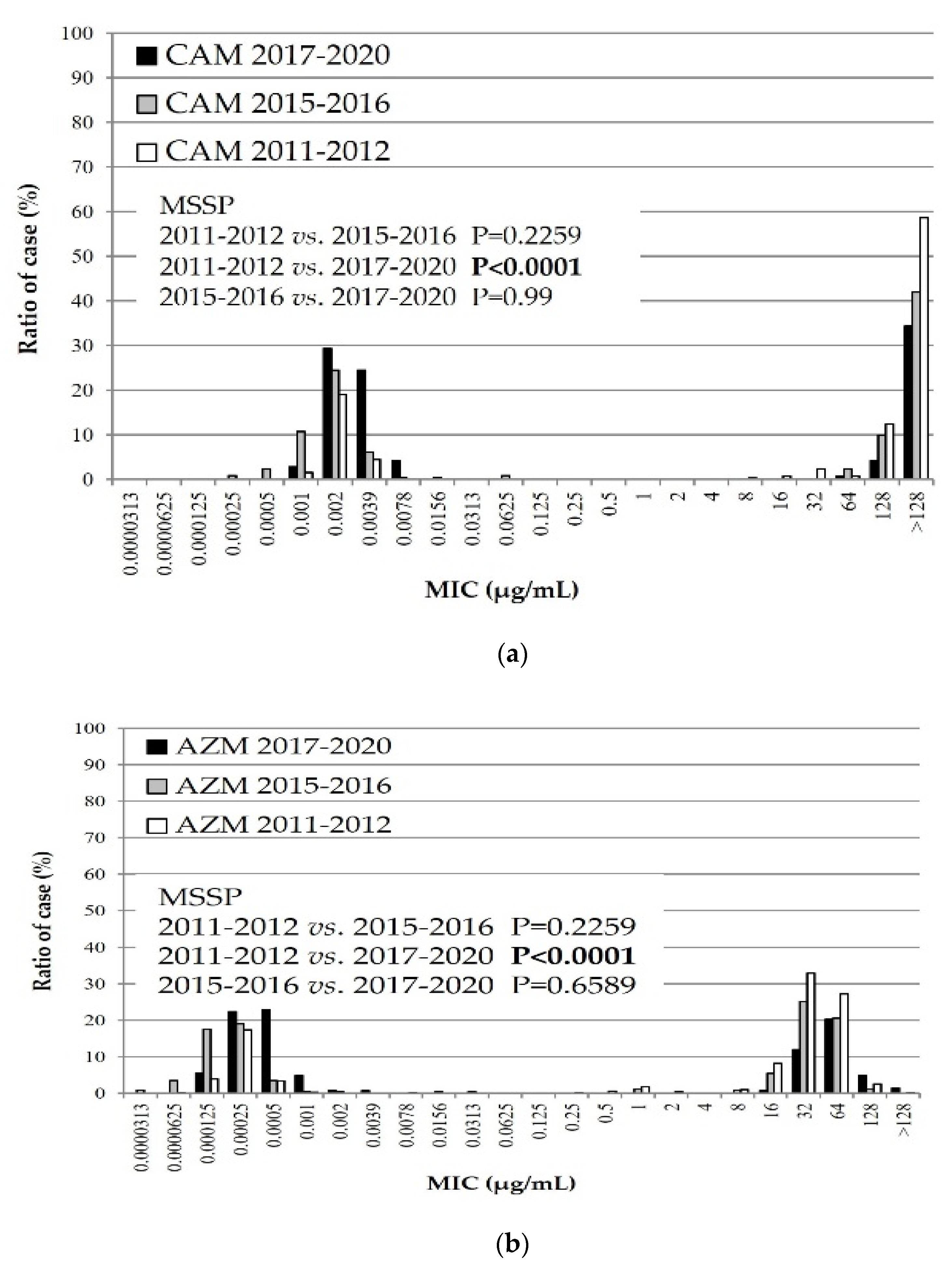
3.2. MIC Distribution of Macrolide Agents against M. pneumoniae Isolates during Three Time Periods

Figure 1 shows the cumulative distribution of the MICs of two macrolide agents, CAM and AZM, during three periods: the first recent epidemic of 2011 and 2012, the second recent epidemic of 2015 and 2016, and the most recent epidemic from 2017 to 2020.
As depicted in Figure 1, all isolates of M. pneumoniae tested against both macrolide agents were grouped into two masses. The masses on the left and right sides predominantly represent MSMP and MRMP groups, respectively. In the MSMP group, the MICs of CAM and AZM were significantly lower during the last few years than during the first recent epidemic.

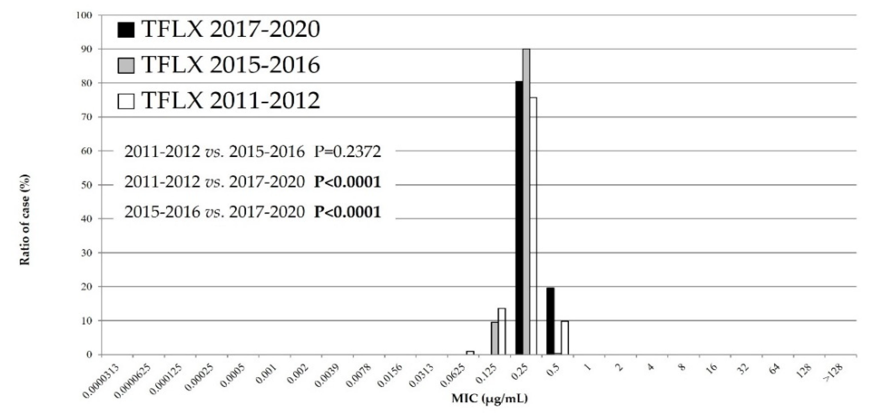
3.3. The MIC Distribution of TFLX against M. pneumoniae Isolates during Three Time Periods

The cumulative MIC distribution of TFLX is indicated in Figure 2. The TFLX MICs of the isolates were significantly higher in recent years than during the first and second epidemics (p < 0.0001). However, all TFLX MICs were grouped under a single mass, as shown in Figure 2. Accordingly, no resistant isolates against TFLX were detected during the investigated periods.

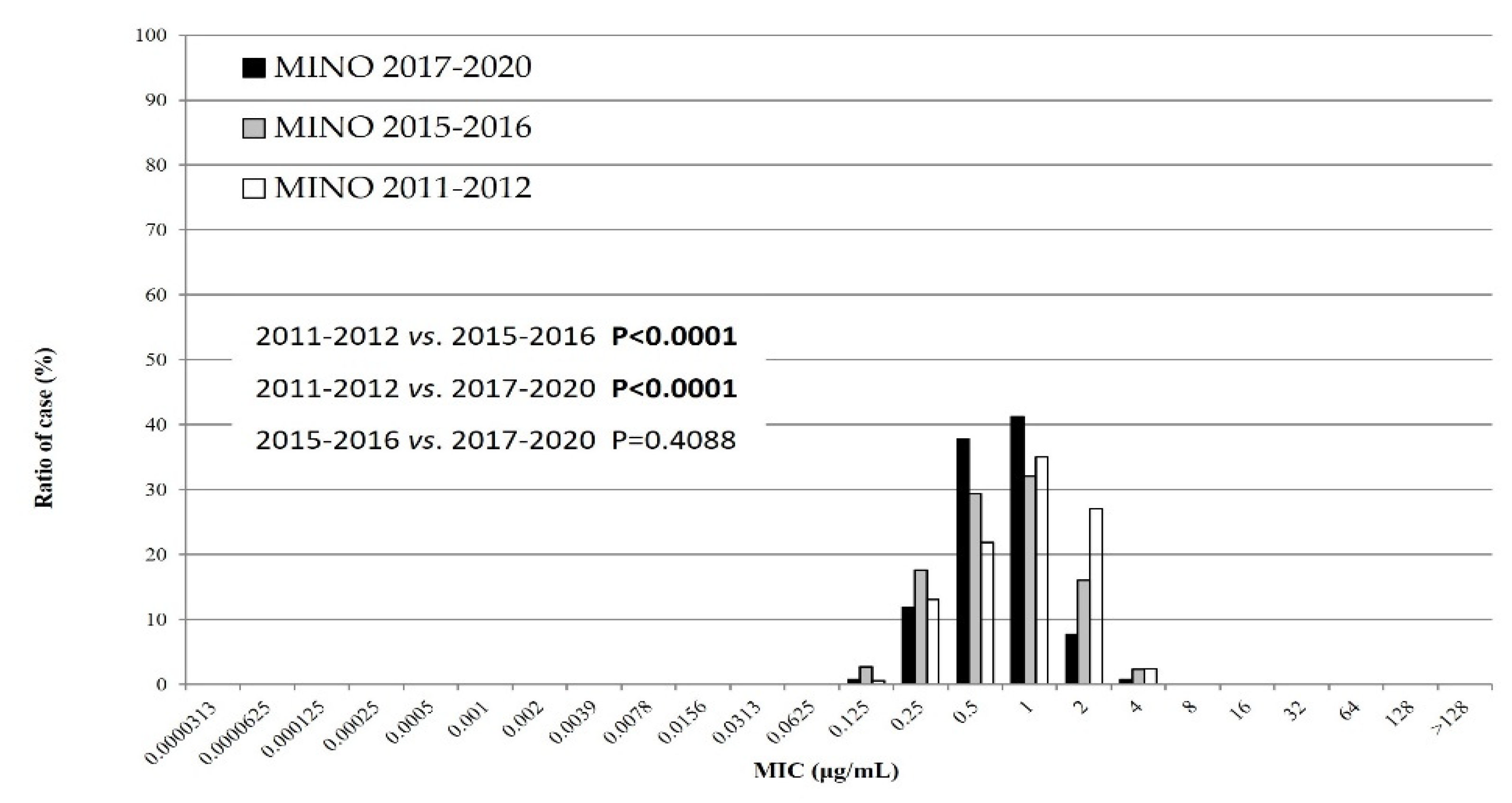
3.4. MIC Distribution of MINO against M. pneumoniae Isolates during Three Time Periods

Figure 3 represents the cumulative MIC distribution of MINO; the MICs of MINO during the second and third epidemics were significantly lower than those observed during the first recent epidemic (p < 0.0001). All MICs of MINO against isolates belonged to a single mass, suggesting that no MINO-resistant isolate was detected in recent years.

4. Discussion

In the MIC distribution of antimicrobial agents, the macrolide MICs of MSSP were significantly lower during the last few years than during the first recent epidemic. The TFLX MICs were significantly higher during the third epidemic than in the first and second recent epidemics. Finally, the MICs of MINO were significantly lower during the third epidemic than during the first and second recent epidemics. These results have two explanations. First, the Japanese guidelines for M. pneumoniae infections were published in 2014 [18]. Specifically, these guidelines state that macrolides are recommended as the first-line drug of choice for treatment of M. pneumoniae infections. The macrolide efficacy has a relatively high accuracy in the presence or absence of defervescence within 48–72 h of initiating macrolide treatment. Second, the use of TFLX or tetracyclines may be considered when required for patients with pneumonia who do not respond to macrolides. Therefore, we assume that clinicians prescribed antimicrobial agents appropriately, that is, they did not continue to prescribe macrolides for insensitive infections, and antimicrobial agents other than macrolides were used more often. Okubo Y et al. (2018) reported that the use of macrolide agents for pediatric M. pneumoniae infections has recently decreased, whereas that of quinolone agents has increased [19], suggesting that the amount of antibiotics used by clinicians is influenced the MIC change. Future considerations include rapid diagnosis kits to detect M. pneumoniae antigens and other factors such as point mutations that confer macrolide resistance [20,21]. Clinicians have been able to immediately identify whether patients with M. pneumoniae infections harbor macrolide-resistant strains. Therefore, the advent of these diagnostic kits has seemingly facilitated appropriate antimicrobial usage. However, tetracyclines such as MINO are contraindicated in children younger than eight years of age per the Japanese guidelines [16], and the average age of patients with Mycoplasma infections is approximately six years. Thus, many suspected MRMP cases have been prescribed TFLX instead of MINO. Furthermore, pediatricians in Japan frequently prescribe TFLX rather than MINO, which is a common practice.
The MIC distribution of TFLX is higher than that in the past; however, it has been approved for treatment of children with M. pneumoniae infection in Japan since 2010 and has played an important role against MRMP infections. Several patients with infections have been cured promptly by TFLX, and its growing use has suppressed the occurrence of MRMP. Notably, the rate of MRMP among Japanese children has decreased in recent years [12]. Ouchi et al. (2017) reported the clinical effectiveness and efficient eradication rates (including MRMP) of TFLX [22]. Thus, MRMP was effectively inhibited by TFLX, consecutively lowering the rate of MRMP. Among M. pneumoniae isolates, no resistant strains were identified against TFLX. Therefore, TFLX must be continually prescribed to effectively combat M. pneumoniae infections.
This study is subject to some limitations. First, we did not analyze the backgrounds of children affected by M. pneumoniae. However, we collected many samples throughout Japan, which may minimize the magnitude of differences among the samples. Second, we only examined the MICs of antimicrobial agents but did not analyze other factors such as genetics and molecular epidemiology. Therefore, in future studies, our analysis should be broadened to include such factors.
In conclusion, we investigated the MICs of antibiotics against M. pneumoniae isolated from Japanese children and MIC distributions of macrolide agents. We found that MINO MICs are lower, whereas those of TFLX are higher than those in the past, in accordance with the increased usage of these drugs. We did not identify quinolone- or tetracycline-resistant M. pneumoniae; however, constant surveillance is required in the future.

Author Contributions

Conceptualization, D.Y., T.N. and K.O.; formal analysis, D.Y.; writing—review and editing, T.N.; Writing—original draft, T.O.; supervision, K.O. All authors have read and agreed to the published version of the manuscript.

Funding

This work was supported by Grants-in-Aid for Scientific Research (KAKENHI” (20K08171)).

Data Availability Statement

Not applicable.

Acknowledgments

We thank Reiji Kimura and Moeka Fujii for their technical assistance and all e clinicians who participated by collecting samples in the Atypical Pathogen Study Group. Individuals from the various facilities who participated in the Atypical Pathogen Study Group and the study include Hideki Asaki (Asaki Pediatric Clinic), Kazutoyo Asada (National Mie Hospital), Tomohiro Ichimaru (Saga Prefectural Hospital, Koseikan), Toshio Inada (Inada Clinic), Takuya Inoue (Chayamati Pediatric Clinic), Masakazu Umemoto (Umemoto Pediatric Clinic), Kanetsu Okura (Okura Clinic), Kenji Okada (Fukuoka National Hospital), Takashige Okada (Okada Pediatric Clinic), Teruo Okafuji (Okafuji Pediatric Clinic), Yasuko Okamoto (Okamoto Clinic), Shinichiro Oki (Higashisaga National Hospital), Keiko Oda (Kawasaki Medical School Kawasaki Hospital), Jin Ochiai (Ochiai Pediatric Clinic), Seiko Obuchi (Obuchi Clinic), Yoji Kanehara (Kanehara Pediatric Clinic), You Kanematsu (Kanematsu Pediatric Clinic), Shoji Kouno (Shomonoseki City Central Hospital), Makoto Kuramitsu (Aoba Pediatric Clinic), Katsuji Kuwakado (Kurashiki Central Hospital), Satoshi Kuwano (Kuwano Kids Clinic), Tatsuo Koga (Koga Pediatric Clinic), Hayashi Komura (Komura Pediatric Clinic), Hiroshi Sakata (Asahikawa-Kosei General Hospital), Takahisa Sakuma (Sakuma Pediatric Clinic), Kazuhide Shiotsuki (Shiotsuki Internal Medicine Pediatric Clinic), Yasushi Shimada (Shimada Clinic), Makio Sugita (Kurashiki Riverside Hospital), Toru Sugimura (Sugimura Pediatric Clinic), Shumei Takeda (Takeda Pediatric Clinic), Isao Tanaka (Mizushima Central Hospital), Hiroyuki Tanaka (Tanaka Family Clinic), Naohumi Tomita (Tomita Clinic), Kensuke Nagai (Nagai Pediatric Internal Medicine Clinic), Yoshikuni Nagao (Mabi Memorial Hospital), Hidekazu Nakashima (Kojima Central Hospital), Tadashi Nagata (Nagata Pediatric Clinic), Kimiko Nakamura (Enoura Clinic), Kazuyo Nomura (Kama Red Cross Hospital), Kanoko Hashino (Hashino Pediatric Clinic), Yuko Hirata (Hirata Internal Medicine Pediatric Clinic), Kazumi Hiraba (Mokubo Pediatric Clinic), Takuji Fujisawa (Fujisawa Pediatric Clinic), Akiko Maki (Hashima Pediatric Clinic), Toshinobu Matsuura (Yoshino Pediatric Clinic), Nobuyoshi Mimaki (Kurashiki Medical Center), Tatsuhiko Moriguchi (Sakai Hospital Kinki University Faculty of Medicine), Shigeru Mori (Momotaro Clinic), Yoichiro Yamaguchi (Yamaguchi Pediatric Clinic), Syuji Yamada (Yamada Pediatric Clinic), Teruyo Fujimi (Fujimi Clinic), Norio Tominaga (Isahaya Health Insurance General Hospital), Syunji Hasegawa (Yamaguchi University Graduate School of Medicine), Kiyoko Nishimura (Nishimura Pediatric Clinic), Mihoko Mizuno (Daido Clinic), Jiro Iwamoto (Iizuka Hospital), Toshiyuki Iizuka (Hakuai Hospital), Shigeru Yamamoto (Daido Municipal Pediatric Clinic), Tomomichi Kurasaki (Kurosaki Pediatric Clinic), Tadashi Matsubayashi (Seirei Hamamatsu General Hospital), Musiaki Ryousuke (Shigei Medical Research Hospital), Oura Toshihiro (Sendai City Hospital), Hasegawa Sumio (Hasegawa Pediatric Clinic), Matsubara Keita (Hiroshima City Funairi Citizens Hospital), Kawasaki Kouzou (Kobe City Medical Center General Hospital), Ikeda Masanori (National Hospital Organization Fukuyama Medical Center), Nagata Ikuo (child care Nagata Child Clinic), Inoue Sachiko (Inoue Internal Medicine and Pediatric Clinic), Tokuda Kiriko (Yawatahama City General Hospital), Ozaki Takanori (Ozaki Pediatric Clinic), Ichikawa Masataka (Ichikawa Pediatric Clinic), Hayakawa Hiroshi (Hayakawa Pediatric Clinic), Nariai Syoukichi (Shimane Prefectural Central Hospital), Tsumura Kumi (Tsumura family Clinic Kumi Pediatrics) Miura Yuuichi (Miura Pediatric Clinic), Ninomiya Takahito National (Hospital Organization Fukuoka Hospital), Okasora Teruo (Okasora Pediatric Clinic), Yamane Tatsuya (Kazenomachi Pediatric Clinic), Matsubara Kazuyo (Dokkyo Medical University Saitama Medical Center), and Mori Toshihiko (NTT East Sapporo Hospital).

Conflicts of Interest

O.T. and T.N. received a research grant from FUJIFILM Toyama Chemical Co., Ltd. The funders had no role in the design of the study; in the collection, analyses, or interpretation of data; in the writing of the manuscript; or in the decision to publish the results.

References

  1. Bradley, J.S.; Byington, C.L.; Shah, S.S.; Alverson, B.; Carter, E.R.; Harrison, C.; Kaplan, S.L.; Mace, S.E.; McCracken, G.H., Jr.; Moore, M.R.; et al. The management of community-acquired pneumonia in infants and children older than 3 months of age: Clinical practice guidelines by the Pediatric Infectious Diseases Society and the Infectious Diseases Society of America. Clin. Infect. Dis. 2011, 53, 25–76. [Google Scholar] [CrossRef] [PubMed] [Green Version]
  2. Chang, C.-H.; Tsai, C.-K.; Tsai, T.-A.; Wang, S.-C.; Lee, Y.-C.; Tsai, C.-M.; Liu, T.-Y.; Kuo, K.-C.; Chen, C.-C.; Yu, H.-R. Epidemiology and clinical manifestations of children with macrolide-resistant Mycoplasma pneumoniae pneumonia in Southern Taiwan. Pediatr. Neonatol. 2021, 62, 536–542. [Google Scholar] [CrossRef] [PubMed]
  3. Kawakami, N.; Namkoong, H.; Saito, F.; Ishizaki, M.; Yamazaki, M.; Mitamura, K. Epidemiology of macrolide-resistant Mycoplasma pneumoniae by age distribution in Japan. J. Infect. Chemother. 2020, 27, 45–48. [Google Scholar] [CrossRef] [PubMed]
  4. Akashi, Y.; Hayashi, D.; Suzuki, H.; Shiigai, M.; Kanemoto, K.; Notake, S.; Ishiodori, T.; Ishikawa, H.; Imai, H. Clinical features, and seasonal variations in the prevalence of macrolide-resistant Mycoplasma pneumoniae. J. Gen. Fam. Med. 2018, 19, 191–197. [Google Scholar] [CrossRef] [PubMed] [Green Version]
  5. Chan, E.D.; Welsh, C.H. Fulminant Mycoplasma pneumoniae pneumonia. West. J. Med. 1995, 162, 133–142. [Google Scholar]
  6. Lanatai, M.M.; Wang, H.; Everhart Moore-Clingenpeel, M.; Ramilo, O.; Leber, A. Macrolide-Resistant Mycoplasma pneumoniae Infections in Children, Ohio, USA. Emerg. Infect. Dis. 2021, 27, 1588–1597. [Google Scholar] [CrossRef]
  7. Xiao, L.; Ratliff, A.E.; Crabb, D.M.; Mixon, E.; Qin, X.; Selvarangan, R.; Tang, Y.-W.; Zheng, X.; Bard, J.D.; Hong, T.; et al. Molecular Characterization of Mycoplasma pneumoniae Isolates in the United States from 2012 to 2018. J. Clin. Microbiol. 2020, 58, e00710-20. [Google Scholar] [CrossRef]
  8. Hung, H.-M.; Chuang, C.-H.; Chen, Y.-Y.; Liao, W.-C.; Li, S.-W.; Chang, I.Y.-F.; Chen, C.-H.; Li, T.-H.; Huang, Y.-Y.; Huang, Y.-C.; et al. Clonal spread of macrolide-resistant Mycoplasma pneumoniae sequence type-3 and type-17 with recombination on non-P1 adhesin among children in Taiwan. Clin. Microbiol. Infect. 2020, 27, 1169.e1–1169.e6. [Google Scholar] [CrossRef]
  9. Zhou, Y.; Wang, J.; Chen, W.; Shen, N.; Tao, Y.; Zhao, R.; Luo, L.; Li, B.; Cao, Q. Impact of viral coinfection and macrolide-resistant mycoplasma infection in children with refractory Mycoplasma pneumoniae pneumonia. BMC Infect. Dis. 2020, 20, 1–10. [Google Scholar] [CrossRef]
  10. Lee, J.K.; Choi, Y.Y.; Sohn, Y.J.; Kim, K.-M.; Kim, Y.K.; Han, M.S.; Park, J.Y.; Cho, E.Y.; Choi, J.H.; Choi, E.H. Persistent high macrolide resistance rate and increase of macrolide-resistant ST14 strains among Mycoplasma pneumoniae in South Korea, 2019–2020. J. Microbiol. Immunol. Infect. 2021, 55, 910–916. [Google Scholar] [CrossRef]
  11. Morozumi, M.; Tajima, T.; Sakuma, M.; Shouji, M.; Meguro, H.; Saito, K.; Iwata, S.; Ubukata, K. Sequence Type Changes Associated with Decreasing Macrolide-Resistant Mycoplasma pneumoniae, Japan. Emerg. Infect. Dis. 2020, 26, 2210–2213. [Google Scholar] [CrossRef]
  12. Nakamura, Y.; Oishi, T.; Kaneko, K.; Kenri, T.; Tanaka, T.; Wakabayashi, S.; Kono, M.; Ono, S.; Kato, A.; Kondo, E.; et al. Recent acute reduction in macrolide-resistant Mycoplasma pneumoniae infections among Japanese children. J. Infect. Chemother. 2020, 27, 271–276. [Google Scholar] [CrossRef]
  13. Chen, J.; Zhang, J.; Lu, Z.; Chen, Y.; Huang, S.; Li, H.; Lin, S.; Yu, J.; Zeng, X.; Ji, C.; et al. Mycoplasma pneumoniae among Chinese Outpatient Children with Mild Respiratory Tract Infections during the Coronavirus Disease 2019 Pandemic. Microbiol. Spectr. 2022, 10, e0155021. [Google Scholar] [CrossRef] [PubMed]
  14. Rivaya, B.; Lluch, E.J.; Rivas, G.F.; Molinos, S.; Campos, R.; Hernández, M.M.; Matas, L. Macrolide resistance and molecular typing of Mycoplasma pneumoniae infections during a 4-year period in Spain. J. Antimicrob. Chemother. 2020, 75, 2752–2759. [Google Scholar] [CrossRef] [PubMed]
  15. Morozumi, M.; Takahashi, T.; Ubukata, K. Macrolide-resistant Mycoplasma pneumoniae: Characteristics of isolates and clinical aspects of community-acquired pneumonia. J. Infect. Chemother. 2010, 16, 78–86. [Google Scholar] [CrossRef]
  16. Miyashita, N.; Kawai, Y.; Yamaguchi, T.; Ouchi, K.; Oka, M.; Atypical Pathogen Study Group. Clinical potential of diagnostic methods for the rapid diagnosis of Mycoplasma pneumoniae pneumonia in adults. Eur. J. Clin. Microbiol. Infect. Dis. 2010, 30, 439–446. [Google Scholar] [CrossRef]
  17. Waites, K.B.; Crabb, D.M.; Bing, X.; Duffy, L.B. In Vitro Susceptibilities to and Bactericidal Activities of Garenoxacin (BMS-284756) and Other Antimicrobial Agents against Human Mycoplasmas and Ureaplasmas. Antimicrob. Agents Chemother. 2003, 47, 161–165. [Google Scholar] [CrossRef] [Green Version]
  18. Committee of the Japanese Society of Mycoplasmology. Guiding Principles * for Treating Mycoplasma Pneumoniae Pneumonia; Japanese Society of Mycoplasmology: Tokyo, Japan, 2014; Available online: http://square.umin.ac.jp/jsm/Eng%20shisin.pdf (accessed on 13 November 2022).
  19. Okubo, Y.; Michihata, N.; Morisaki, N.; Uda, K.; Miyairi, I.; Ogawa, Y.; Matsui, H.; Fushimi, K.; Yasunaga, H. Recent trends in practice patterns and impact of corticosteroid use on pediatric Mycoplasma pneumoniae -related respiratory infections. Respir. Investig. 2017, 56, 158–165. [Google Scholar] [CrossRef] [PubMed]
  20. Morinaga, Y.; Suzuki, H.; Notake, S.; Mizusaka, T.; Uemura, K.; Otomo, S.; Oi, Y.; Ushiki, A.; Kawabata, N.; Kameyama, K.; et al. Evaluation of GENECUBE Mycoplasma for the detection of macrolide-resistant Mycoplasma pneumoniae. J. Med. Microbiol. 2020, 69, 1346–1350. [Google Scholar] [CrossRef]
  21. Kakiuchi, T.; Miyata, I.; Kimura, R.; Shimomura, G.; Shimomura, K.; Yamaguchi, S.; Yokoyama, T.; Ouchi, K.; Matsuo, M. Clinical Evaluation of a Novel Point-of-Care Assay To Detect Mycoplasma pneumoniae and Associated Macrolide-Resistant Mutations. J. Clin. Microbiol. 2021, 59, e0324520. [Google Scholar] [CrossRef]
  22. Ouchi, K.; Takayama, S.; Fujioka, Y.; Sunakawa, K.; Iwata, S. A phase III, randomized, open-label study on 15% tosufloxacin granules in pediatric Mycoplasma pneumoniae pneumonia. Jpn. J. Chemother. 2017, 65, 585–596. [Google Scholar]
Figure 1. Minimum inhibitory concentration (MIC) distribution of macrolide agents, (a) CAM and (b) AZM, against Mycoplasma pneumoniae isolates during three periods: 2011–2012, 2015–2016, and 2017–2020. CAM: clarithromycin, AZM: azithromycin.
Microorganisms 10 02428 g001
Figure 2. Minimum inhibitory concentration (MIC) distribution of tosufloxacin against Mycoplasma pneumoniae isolates during three periods: 2011–2012, 2015–2016, and 2017–2020. TFLX: tosufloxacin.
Microorganisms 10 02428 g002
Figure 3. MIC distribution of minocycline against M. pneumoniae isolates during the three periods: 2011–2012, 2015–2016, and 2017–2020. MINO: minocycline.
Microorganisms 10 02428 g003
Table 1. In vitro antimicrobial activity against clinical isolates of Mycoplasma pneumoniae strains from 2017 to 2020. Antimicrobial susceptibility testing was performed as reported in [16].
Organism
(Number of Strains)
(n = 122)
Antimicrobial AgentsMIC (μg/mL)
MIC RangeMIC50MIC90
Mycoplasma pneumoniae
(122)
CAM0.001>1280.0039>128
AZM0.00012 >1280.000564
TFLX0.250.50.250.5
MINO0.12540.51
Macrolide-susceptible
M. pneumoniae (76)
CAM0.00780.0010.0020.0039
AZM0.00012 0.00390.00050.001
TFLX0.250.50.250.5
MINO0.25411
Macrolide-resistant
M. pneumoniae (46)
CAM16>128>128>128
AZM64 >12864128
TFLX0.250.50.250.25
MINO0.12520.52
TFLX: tosufloxacin, MINO: minocycline, CAM: clarithromycin, AZM: azithromycin; MIC: minimum inhibitory concentration.
Publisher’s Note: MDPI stays neutral with regard to jurisdictional claims in published maps and institutional affiliations.

