Article

Improvements to the Rapid Detection of the Marine Pathogenic Bacterium, Vibrio harveyi, Using Loop-Mediated Isothermal Amplification (LAMP) in Combination with SYBR Green

by
Ahmad Mukhlis Abdul Rahman
1,2,
Julian Ransangan
3 and
Vijay Kumar Subbiah
1,*
1
Biotechnology Research Institute, Universiti Malaysia Sabah, Jln UMS, Kota Kinabalu 88400, Sabah, Malaysia
2
Faculty of Chemical Engineering & Technology, Uniciti Alam Campus, Universiti Malaysia Perlis, Sg. Chuchuh, Padang Besar 02100, Perlis, Malaysia
3
Borneo Marine Research Institute, Universiti Malaysia Sabah, Jln UMS, Kota Kinabalu 88400, Sabah, Malaysia
*
Author to whom correspondence should be addressed.
Microorganisms 2022, 10(12), 2346; https://doi.org/10.3390/microorganisms10122346
Submission received: 11 November 2022 / Revised: 23 November 2022 / Accepted: 24 November 2022 / Published: 27 November 2022
(This article belongs to the Special Issue Host–Bacteria Interactions in Aquaculture Systems)

Abstract

:
The common methods that are presently used to identify Vibrio harveyi include microscopic examination and biochemical, immunological and PCR-based assays. These methods require technical expertise, and can be time-consuming. A rapid method is required for the high-throughput screening of large number of samples. As such, we have developed a rapid, simple yet sensitive and specific detection method based on the use of the loop-mediated isothermal amplification (LAMP) of DNA. A set of six primers, i.e., two outer, two inner and two loop primers, was designed based on the in silico analysis of a large pool of 39 strains of the toxR gene sequence of V. harveyi. The addition of the loop primers decreased the reaction time of the LAMP by more than half. Furthermore, with the application of SYBR Green, the result can be obtained as quickly as in 10 to 15 min without the need of gel electrophoresis. The specificity of the method primers was then determined by performing LAMP with Vibrio and non-Vibrio samples. LAMP has a greater sensitivity than PCR reaction. The sensitivity of PCR was at 0.6 pg concentration of V. harveyi recombinant plasmid DNA standard, while LAMP was able to detect lower amounts even at 0.6 fg. The development of the LAMP assay will provide a valuable tool for the high-throughput rapid detection of V. harveyi contamination both in laboratories and in the field.

1. Introduction

Vibrio harveyi is a gram-negative, rod-shaped, facultative anaerobic microorganism capable of being able to switch between fermentative and respiratory metabolism [1,2]. It occurs naturally in marine habitats, but some strains have developed into significant pathogens of wild and cultured marine fish and invertebrates, especially in warm water. The pathogen is also a zoonosis, as it can infect humans through skin punctures or wounds and may cause severe inflammatory responses [3]. However, V. harveyi has found notoriety as a marine pathogen, as it causes severe economic losses for the aquaculture and mariculture industries [4,5]. Diseased fish may exhibit a range of symptoms such as a chronic skin ulcers, gastro-enteritis and deep dermal lesions [6,7,8,9], and it is the main cause of luminous vibriosis in fish, mollusks and crustaceans worldwide [10,11].
Prior to the advent of PCR, the diagnosis of V. harveyi was based on the culture in bacterial agar and identification through microscopic examination or biochemical assay [2]. These methods are still important today, particularly when examining the immunological and serological characteristics of the host–pathogen response [12,13]. Nevertheless, the PCR-based approach has become widespread as the definitive technique in the detection of V. harveyi. This usually entails the PCR amplification of the 16S rRNA region, the DNA sequencing of the amplicon, and is followed by BLAST analysis for species identification [14,15,16]. Some researchers have developed multiple sets of specific primers for detecting several genes at once in a multiplex PCR approach to increase the specificity of the detection V. harveyi and other Vibrio species concurrently [17]. Others have used real-time PCR, which offers a more rapid and sensitive approach for the detection of the pathogen compared with conventional PCR [18,19,20]. Here, the detection of amplicons is based on the presence of fluorescent signals (either through the use of fluorescent labeled probes or the intercalating dye SYBR Green) [21].
PCR-based amplification methods (conventional, multiplex or real-time) are usually expensive and time-consuming with the need for specific laboratory equipment, and are not suitable to be used onsite at hatcheries or fish farms that require a high-throughput turnaround time. With this in mind, many researchers have turned to the use of isothermal polymerases, as it does not require the use of the cycling temperatures employed in PCR [22]. Three such methods that are commonly used with isothermal polymerases are loop-mediated isothermal amplification (LAMP) [23], recombinase polymerase amplification (RPA) [24] and rolling circle amplification (RCA) [25]. Of these, LAMP is the more commonly used technique, even though the design of its primers is rather complicated and it requires prior template-sequence information [22]. LAMP was first developed in 2000 for the molecular detection of viruses [26], and it is the method of choice that we have used here. On the other hand, RPA requires substantial protocol optimizations, as the background noise can interfere with the results, and RCA only works with a circular nucleic acid template [22].
The most significant advantage of LAMP is its ability to amplify target DNA under isothermal condition, usually between 60 °C and 65 °C, with high specificity, efficiency and speed. This LAMP method has been tested in the diagnoses of viruses [27,28], bacteria [29], protozoa [30] and fungi [31], and it became popular after 2003 following the emergences of the West Nile and SARS viruses [32,33] and more recently due to SARS-CoV-2 [34,35]. It has also been applied for the rapid detection and identification of aquaculture pathogens [36]. In terms of product visualization, the tube containing products of LAMP can be visualized in the presence of fluorescent intercalating dyes such as SYBR Green. For positive amplification, the color of the dye will change from orange to green when the presence of LAMP products are detected [32,37].
Several papers have previously reported the development of LAMP for V. harveyi. Noteworthy among these are Cao and colleagues, who in 2010 were the first to develop LAMP for the detection of V. harveyi utilizing the toxR gene using two inner and outer primers [38]. This was followed by Caipang et al., who designed the primers for LAMP based on the dnaJ gene instead of the toxR [39]. Both methods had limitations, as the primer sequences were not conserved for most V. harveyi strains. Due to this, others have used the VhhP2 gene instead as the target for LAMP, as the gene is said to be widely distributed in V. harveyi strains from different geographical locations and sources [40,41]. It should be noted that Yu and colleagues [40] targeted the VhhP2 gene due to the fact that they were developing a triplex LAMP simultaneously along with the amplification of the toxR gene from V. anguillarum and the collagenase gene of V. alginolyticus. However, all of the studies above reported a longer detection time (usually more than 90 min) than the methods that we described here, and may not be practical in cases where immediate point-of-care is required [42]. In 2002, Nagamine et al. developed a method that accelerates the LAMP reaction by using additional primers, termed as loop primers [43]. Loop primers have now been incorporated in many LAMP assay for the detection of pathogens [44], but are not available for the detection of V. harveyi.
With this in mind, we attempted to reexamine and improve the LAMP assay by incorporating loop primers that will hybridize the stem-loops structure of DNA and prime strand displacement DNA synthesis, which could help to expedite the reaction for rapid detection. In addition, we designed new sets of inner and outer primers that were based on conserved regions of a comprehensive set of lists of all available V. harveyi strains-sequence information of the toxR gene in GenBank (www.ncbi.nlm.nih.gov (accessed on 1 February 2020)). The changes made here significantly improved the detection of V. harveyi, and may be freely incorporated into existing protocols.

2. Materials and Methods

2.1. Sampling and Isolation of Genomic DNA

The Vibrio harveyi strain VHJR7 was obtained from the Borneo Marine Research Institute, Universiti Malaysia Sabah (UMS), Malaysia. The strain was previously isolated from an infected seabass, Lates calcarifer (Bloch), cultured in open net cages in Sabah, Malaysia, during a vibriosis outbreak in February 2008 [45]. The bacterial strain had been previously identified using 16S rRNA and the hemolysin gene through DNA sequencing for species confirmation. Genomic DNA was isolated from the V. harveyi strain VHJR7 using the I-Genomic DNA Extraction Kit (Intron Biotechnology, Gyeonggi-do, Republic of Korea).

2.2. Primer Design for PCR and LAMP Assays

A set of six newly developed primers for the LAMP assay (Table 1), consisting of two inner primers (ToxR2-FIP and ToxR2-BIP), two outer primers (ToxR2-F3 and ToxR2-B3) and two loop primers (ToxR2-LF and ToxR-LB) were designed according to the published sequences of the toxR gene of Vibrio harveyi (GenBank ID: DQ640259.1). The primers were designed using the default parameters of Primer Explorer Version 4 at http://primerexplorer.jp/elamp4.0.0/index.html (accessed on 1 February 2020) (Figure 1).
We ensured that the primers were conserved to target all 39 V. harveyi strains found in Genbank (Appendix A Table A1). We also performed in silico verification to ensure that the primers were specific to V. harveyi and not to any other species under the same genera. The outer primers (ToxR2-F3 and ToxR2-B3) were used to obtain the PCR amplicon, which was sequenced for validation and subsequently used to produce the template plasmid DNA.

2.3. Preparation of DNA Template

The target site of the V. harveyi toxR gene (245 bp) was amplified by PCR with approximately 80 ng of V. harveyi genomic DNA using the ToxR2-F3 and ToxR2-B3 primers. The PCR reaction was performed as described previously, with some modifications [46]. The PCR was conducted in a 25 µL total reaction consisting of 1x PCR buffer, 0.2 mM of dNTP mix (Promega, USA), 10 pmol each ToxR2-F3 and ToxR2-B3 primers, 1.5 mM MgCl2 and 1.25 U of Taq DNA polymerase (Promega, USA). The PCR reaction protocol involved an initial denaturation time of 3 min at 95 °C, followed by 40 cycles of amplification at 95 °C for 30 s, 56 °C for 30 s and 72 °C for 30 s, with a final extension step at 72 °C for 5 min. The PCR product was then purified using a PCR purification Kit (Qiagen, Germantown, MD, USA).

2.4. Recombinant Plasmid Construction for PCR and LAMP Analyses

The PCR amplicon (which was the target sequence for PCR and LAMP) was ligated into the plasmid pGEM-T easy vector (Promega, Madison, WI, USA) according to the manufacturer’s protocol. Next, the recombinant plasmid was transformed into 100 µL of E. coli strain JM109, followed by blue–white colony selection. Positive colonies indicated by a white color were collected and cultured for 16 h in an LB broth medium containing 100 µg ml−1 of ampicillin. Then, the plasmid extraction was performed using the PureYield TM Plasmid Miniprep System (Promega, Madison, WI, USA) according to the manufacturer’s protocol. The plasmid was then electrophoresed on a 1.5% agarose gel and purified for subsequent downstream processes.

2.5. Optimization of LAMP Reaction Condition with UV Analysis

The LAMP reactions were performed in a 25 µL reaction mixture containing 32 pmol each of the inner primers ToxR2-FIP and ToxR2-BIP, 8 pmol each of the outer primers ToxR2-F3 and ToxR2-B3, 32 pmol of the loop primers ToxR2-LB and ToxR2-LF, 1.4 mM deoxyribonucleotide triphosphate (dNTPs), 0.8 M betaine (Sigma-Aldrich), 20 mM Tris-HCl, 10 mM KCl, 10 mM (NH4)2SO4, 8 mM MgSO4, 0.1% Tween-20, 8 U Bst DNA Polymerase (NEB) and 5 µL of DNA sample. The LAMP reaction was performed on a heating block at 65 °C. The reaction was performed at 30, 40, 50 and 60 min for the reaction mixture without loop primers, and at 5, 10, 15 and 20 min for the reaction mixture with loop primers. The final products of the LAMP reaction were analyzed by 1.5% agarose gel electrophoresis.

2.6. Visualization of Amplification Products for LAMP Reaction Using SYBR Green

The LAMP reaction was carried out at 65 °C for 15 min. The resulting amplicons were electrophoresed using a 1.5% agarose gel, stained with ethidium bromide and visualized under UV light. For visualization using SYBR Green, approximately 2 µL of 1:10 diluted SYBR Green I nucleic acid gel stain was added directly into all reaction tubes containing the remaining LAMP-amplification products, and any changes of color was visually observed.

2.7. Sensitivity Test for the Detection of V. harveyi Using PCR and UV Analyses

A set of a 10-fold serial dilution of V. harveyi recombinant plasmid DNA samples was prepared from 60 ng to 0.6 fg. The PCR reactions were performed for all V. harveyi recombinant plasmid DNA samples using the ToxR2-F3 and ToxR2-B3 primers, yielding a PCR amplicon of approximately 245 bp. The amplification products were detected by 1.5% agarose gel electrophoresis followed by ethidium bromide staining and visualization by a UV transilluminator.

2.8. Sensitivity Test for the Detection of V. harveyi Using LAMP-UV Analysis and LAMP-SYBR Green

The same set of 10-fold serial dilutions of the V. harveyi recombinant plasmid DNA samples (60 ng to 0.6 fg) was used for the sensitivity test of the LAMP. The amplification products were then visualized in two ways; UV analysis and SYBR Green dye. The LAMP reaction was performed for all V. harveyi recombinant plasmid DNA samples at 65 °C for 15 min. The resulting amplicon of the LAMP reactions which were stained with ethidium bromide was analyzed on 1.5% agarose gel electrophoresis with the aid of a gel-documentation system. For visualization using SYBR Green dye, approximately 2 µL of 1:10 diluted SYBR Green I nucleic acid gel stain was added directly to all reaction tubes containing LAMP product amplicons, and any changes of color were visually observed.

2.9. Specificity Test of LAMP with UV Analysis for the Detection of Vibrio harveyi

In order to ensure that the developed procedure was specific to V. harveyi, the reference strains of the Vibrio and non-Vibrio bacterial samples were included in a specificity evaluation of the LAMP assays. These strains were Vibrio parahaemolyticus ATCC 17802, Vibrio alginolyticus ATCC 17749, Vibrio aguillarum ATCC 19264 and Aeromonas salmonicida subsp. salmonicida ATCC strain 33658. DNA templates were isolated from the bacterial cultures as described above for V. harveyi, according to the manufacturer’s procedures. The LAMP assay was carried out at 65 °C for 15 min.

2.10. Evaluation of LAMP-SYBR Green Assay with Grouper Infected with Vibrio harveyi

For the field evaluation, cell suspensions of V. harveyi in PBS were prepared prior to inoculation into healthy tiger groupers (Epinephelus fuscoguttatus). Initially, glycerol stock of V. harveyi was cultured in tryptic soy agar (TSA) supplemented with 2% NaCl at 30 °C overnight. Next, 1–2 mL of the cultured bacteria cells was transferred into a 2 mL microcentrifuge tube. Then, the tube was centrifuged for 1 min at 13,000 rpm, and the supernatant was discarded. To wash the cell, the pellet was re-suspended using PBS, centrifuged for 1 min at 13,000 rpm and the supernatant was discarded. This step was repeated three times. Finally, the pellet was re-suspended with approximately 1 mL of PBS. Three groupers were injected intraperitoneally with 104 c.f.u/g of the V. harveyi PBS cell suspension. After the mortalities among the test group, we performed rapid detection using LAMP-SYBR green. A control group was included for comparison purposes. Fish from both the treated and control groups were dissected, and the internal tissues were used for the subsequent preparation of the DNA template using the rapid-boiling method [46]. Briefly, 200 µL of 0.025 N sodium hydroxide (NaOH) and 0.0125% sodium dedocyl sulphate (SDS) was added to the tissues and allowed to boil for 5 min. Approximately 200 µL of the supernatant solution was used directly for the subsequent LAMP-SYBR Green assay.

3. Results

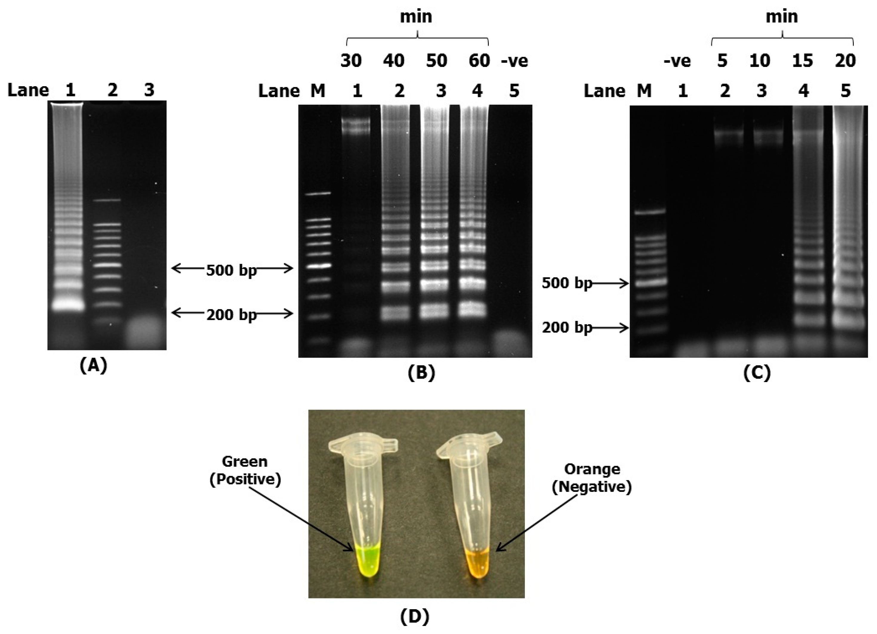
3.1. LAMP Reaction Condition

The LAMP assay was performed to enable the quick identification of V. harveyi. For this purpose, we initially performed LAMP with just the four inner and outer primers, without the addition of the loop primers. However, we found that the best result was obtained with the addition of the loop primers. The loop primers used here consisted of a forward loop primer (ToxR2-LF) and a backward loop primer (ToxR2-LB). A ladder-like band was obtained after the LAMP amplicons were electrophoresed, stained with ethidium bromide and visualized under UV light. The ladder-like band was indicative of a successful LAMP reaction (Figure 2A).
All LAMP reactions were performed on a heating block at 65 °C. For comparison purposes, the assay was conducted at 30, 40, 50 and 60 min without loop primers, and 5, 10, 15 and 20 min for the reaction mixture with loop primers. The different interval periods were due to the time taken to obtain the ladder-like bands. The LAMP reaction without loop primers was able to detect V. harveyi (producing ladder-like bands) after about 40 to 60 min (Figure 2B). However, the LAMP reactions with loop primers only required about 10 to 15 min to detect V. harveyi (Figure 2C). Loop primers were used in all subsequent procedures throughout this study due to the fact that the reaction time was reduced by more than half compared with the LAMP reaction without loop primers. The primers hybridized to the loop region of the stem-loop DNA structure before initiating strand-displacement DNA synthesis. DNA synthesized from the loop primers was displaced by the extension from the 3′ end of the target DNA, thus generating structures which were not present in the conventional LAMP [26]. This enabled the drastic amplification and reduced the reaction time by more than half of that of the original LAMP reaction.

3.2. Visual Detection of Amplification Products of LAMP by SYBR Green

LAMP amplicons can be detected visually with naked eye either through turbidity or color changes by intercalating dye or fluorescent-labeled primers [47,48,49]. The addition of the SYBR Green I nucleic acid gel stain resulted in a color change from orange to green for V. harveyi toxR gene-positive reactions (Figure 2D), while negative reactions remained orange in color. We chose to use SYBR Green for easier visualization of the color changes in the field by less technically trained personnel. The use of the LAMP reaction also eliminated the need of gel electrophoresis with a gel-documentation system or a turbidimeter in the field.

3.3. Sensitivity Test of PCR-UV Analysis, LAMP-UV Analysis and LAMP-SYBR Green for the Detection of V. harveyi

The sensitivity detection of the PCR assay was performed using a set of V. harveyi recombinant plasmid samples with different concentrations from 60 ng to 0.6 fg. The PCR reaction was performed using ToxR2-F3 and ToxR2-B3 primers. Both primers acted as the outer forward and reverse primers that bound at specific flanking regions, yielding a PCR amplicon of approximately 245 bp. The sensitivity of PCR was at 0.6 pg (Figure 3A, top).
The sensitivity level for the LAMP assay was also successfully tested using the same set of V. harveyi recombinant plasmid samples with the DNA plasmid concentration of 60 ng to 0.6 fg. The LAMP reaction was performed at 65 °C for 15 min using the optimized protocol, as described in the methodology section. The sensitivity test of the LAMP assay shows that the method is able to detect even as low as 0.6 fg of V. harveyi recombinant plasmid samples (Figure 3A, bottom).
The sensitivity tests of LAMP and SYBR Green were successfully conducted using the same V. harveyi recombinant plasmid samples from 60 ng to 0.6 fg, and the assay was also able to detect the V. harveyi recombinant plasmid samples at 0.6 fg (Figure 3B).

3.4. Specificity Test of LAMP and UV Analysis

The specificity test was successfully performed with the genomic DNA of the reference species as negative controls i.e., Vibrio parahaemolyticus, V. alginolyticus, V. aguillarum and Aeromonas salmonicida sub sp. salmonicida. We observed positive test results of ladder-like bands with only the V. harveyi toxR gene (Figure 4). All of the other species showed negative test results. The results for the specificity test show that the designed LAMP assay is specific to the V. harveyi toxR gene only, due to the fact that there were not any other positive results observed.
When designing the LAMP primers, we took care to avoid using conserved regions which were mutual with other Vibrio or closely related species. A BLAST search result of the target sequence shows that it is specific and unique to the reported strains of V. harveyi. The design of the primers was meticulously carried out in order to capture the various V. harveyi reported strains. The large capture pool (39 strains of V. harveyi as reported in GenBank) enabled the reduction of false negatives (Appendix A Table A1).

3.5. In Vivo Evaluation of Infected Grouper Using LAMP-SYBR Green

We infected groupers with a cell suspension of the V. harveyi strain VHJR7 and evaluated whether the newly designed LAMP primers could detect the presence of the pathogen in the fish. We used the rapid-boiling method used to avoid a lengthy DNA-isolation procedure. This took an additional 5 min on top of the 15 min required for LAMP amplification. The results indicate that the rapid-boiling method coupled with the newly designed primers was able to rapidly detect the presence of V. harveyi. Consequently, we could not detect the presence of V. harveyi in the control uninfected fish samples (Figure 5).

4. Discussion

A rapid, high-throughput yet simple and cost-effective detection method is needed to detect for the presence of V. harveyi. With this in mind, we optimized a procedure for the rapid detection of the pathogen by using the LAMP assay with the addition of two loop primers and a re-examination of the toxR gene sequences available at GenBank (www.ncbi.nlm.nih.gov (accessed on 1 February 2020)).
For successful LAMP amplification, the target region and design of the primers should be given considerable attention. A search conducted in GenBank resulted in more hits for the toxR genes compared with the other two genes, dnaJ and VhhP2, which were also reported to be used in LAMP assay for V. harveyi [38,39,40]. Hence, in order to capture as much sequence information as possible and translate them into a usable output, we decided to stick to the toxR gene initially used by Cao et al. [38]. However, we redesigned the primers to target conserved regions, taking into consideration all available sequence data in GenBank. With the limited complete genomes available in GenBank [50], selecting conserved regions for designing the LAMP primers was difficult. We took cognizant that V. harveyi strains are heterogeneous, as pointed out in several studies [51,52], which could impact the utility of the LAMP primers. Any mutations at the primer-binding regions of the various global strains would provide false negatives in the LAMP or PCR assays. Thus, we carefully screened all 39 available toxR V. harveyi sequences at GenBank.
To start off, we downloaded published sequences of the Vibrio harveyi toxR gene from GenBank, along with the initial reference sequence DQ640259.1. A blast search was performed to capture the conserved regions of the sequences and avoid nucleotide variants in the V. harveyi strains (Appendix A Table A1). After a manual search, we managed to identify a conserved target region with a length of approximately 246 bp. A set of six primers for LAMP was then designed according to the published sequence of the toxR gene of V. harveyi (GenBank ID: DQ640259.1). The set of six primers included the forward inner primer (ToxR2-FIP), backward inner primer (ToxR2-BIP), forward outer primer (ToxR2-F3), backward outer primer (ToxR2-B3), forward loop primer (ToxR2-LF) and backward loop primer (ToxR2-LB) (Figure 1). Primer Explorer (website link given in the methodology), which is a primer-designing software for LAMP, was used for this purpose. Two of the outer primers (ToxR2-F3 and ToxR2-B3) were initially used to perform PCR for the purpose of generating copies of the V. harveyi DNA amplicon to be used as templates for LAMP, in line with the approach used by others [46,53].
From our observations, the design of the LAMP primers for the toxR gene should be given special attention to prevent no or false amplification. In this regard, four key factors are important and need to be considered. These are the melting temperature (Tm), GC content, stability at the end of each primer and the formation of secondary structures. The optimal melting temperature (Tm) for each flanking region in this research was designed to range between 64 and 66 °C for F1c and B1c and between 59 and 61 °C for F2, B2, F3 and B3 to ensure appropriate primer-template complementarity for the generation of the ‘dumbbell’-like structure, which is unique to LAMP. In addition, the initiation of template amplification is highly sensitive to the ends of the primer sequences, and should contain a certain degree of stability. We ensured that the 3′ ends of the ToxR2-F2 or ToxR2-B2, ToxR2-F3 or ToxR2-B3 and the 5′ ends of ToxR2-F1c or ToxR2-B1c were designed so that the free energy was –4 kcal/mol or less for effective amplifications. The 5′ end of the ToxR2-F1c after amplification corresponded to the 3′ end of the ToxR2-F1. As such, stability is important as the reaction proceeds toward a negative change in free energy for effective LAMP reactions. Accordingly, due to the fact that the annealing reaction between the primer and the target DNA is an equilibrium reaction, it should proceed with a smaller change in free energy [54].
On another note, the GC contents of the inner and outer primer of our newly designed primers were between 50–61%, which was within the optimal range for effective amplification, i.e., 40% to 65% [54]. In addition, it is also important to note that the inner LAMP primers were designed so that they would not form secondary structures. The 3′ ends of the primers should be designed without complementary sequences to prevent the formation of primer dimers, which could interfere with the amplification.
A forward loop primer (ToxR2-LF) and backward loop primer (ToxR2-LB) are not essential requirements for LAMP. However, we included the loop primers as they managed to speed up the reaction, producing a rapid amplification of the LAMP products. With regards to time, the addition of loop primers resulted in the detection of LAMP products within 10–15 min, which was faster compared with the previously reported work on V. harveyi by Cao et al. [38] of approximately 60 min without the addition of loop primers. Furthermore, loop primers increase the specificity of the reaction because they require the recognition of eight distinct target sequences to form the LAMP structure needed for positive amplification [43]. Thus, they confer a higher amplification specificity compared with previous LAMP reactions without loop primers, which used only six distinct targets for the detection of V. harveyi [38,39,41]. In other pathogens, the addition of loop primers also resulted in a reduction of the detection time to 20 min for an infectious spleen and kidney necrosis virus (ISKNV) [55], 30 min for a hematopoietic necrosis virus (IHHNV) [46] and 30 min for a citrus bacterial cancer (CBC) [56].
From the sensitivity test, the detection limit of PCR was lower than the LAMP reaction. The sensitivity of PCR was only at 0.6 pg, while LAMP was able to detect lower amounts of the DNA sample, even at 0.6 fg (Figure 3A). We also observed a marked improvement in terms of sensitivity when using LAMP compared with end-point PCR assay [38].
With regards to the specificity test, the LAMP assay correctly managed only to detect the V. harveyi toxR gene and none of the controls, i.e., three Vibrio species (V. aguillarium, V. alginolyticus and V. parahaemolyticus) and one non-Vibrio (Aeromonas salmonicides subsp. salmonicida) strain (Figure 4). However, we do take note of the limited number of controls used in the specificity test, as we only used what was available to us at hand. A more-thorough screening will be needed for further confirmation.
The ability of SYBR Green to visualize the positive detection of V. harveyi using the naked eye through a color change, without the need for gel electrophoresis and a gel-documentation system, allows for it to be applied for a non-digital assay. Others have used different visualization dyes which could work as well, though with varying degrees of success, such as Calcein [57], PicoGreen [58], hydroxynaphthol blue dye [59], malachite green [60] or GelRed [61].
We proceeded to perform a test to detect the presence of V. harveyi in groupers inoculated with the pathogen. The boiling method seems to work well in generating rapid DNA templates for the LAMP assay. The color difference between orange to green was evident in the control and infected fish. However, we need to point out that while the data seems encouraging, we would need to test different tissue samples and with varying inoculation doses. There is a need to test the efficacy of the protocol and its limits in field trials, which were not conducted here.
Others working on LAMP-SYBR Green have pointed out that it may be difficult to differentiate between intermediate colors when low bacterial copies are present [22,23]. Thus, the need to move away from visual inspection to digitization could improve the reliability of the technique. One way to do this would be to use a turbidity meter, as this would be the cheapest and most cost-effective approach to obtain numerical measurements for cut-off purposes [62]. The use of a biosensor device coupled with LAMP would be another approach that could be employed [63,64]. For example, the integration of microfluidic chips with a real-time LAMP assay (dual-sample on-chip LAMP) can provide the simultaneous detection of multiple pathogens [65]. A more unconventional approach than that was the use of a nuclear-magnetic-resonance (NMR)-based biosensor approach utilizing iron nanoparticles coated with target-specific biomarkers for the rapid detection of a closely related Vibrio species, i.e., V. parahaemolyticus [66]. While these approaches are rapid and can minimize human errors (due to the manual visualization of color changes), one would need to consider the high cost to be borne by fish farmers.
Another factor to consider is that while we may be able to detect low copies of V. harveyi with LAMP or qPCR, this may not necessarily indicate the presence of the disease. The pathogen should be present in a sufficient amount in the environment targeting a susceptible host to trigger an infection. The body has innate and/or adaptive immune responses to deal with nascent contact. This has led to other methods to detect V. harveyi infection based on the response signals of host species. One such approach is to use small, non-coding RNA molecules such as microRNA (miRNA) [67,68] and Piwi-interacting RNA (piRNA) [69,70] obtained from the extracellular vesicles of skin mucus. The up or downregulations of the expressed piRNA and miRNA genes can be used as biomarkers to predict the onset of the diseases. Alternatively, certain protein biomarkers such as Ferritin, toll-like receptor 5S protein and calcium-transporting ATPase, Histone H2B and Eukaryotic translation-initiation factor 5A have been implicated in the disease’s formation [71]. These novel, non-invasive approaches have been shown to be able to differentiate between diseased and healthy fish (Cynoglossus semilaevis). Incorporating the miRNA and piRNA biomarkers with LAMP and integrating it with a rapid-visualization approach may have practical value for large-scale disease screening. The downside of this is that the expression profile of the miRNA, piRNA and protein biomarkers must be consistently expressed across all marine species to be effectively used in the wide host range of the V. harveyi infection.
With regards to future developments, we proceeded to incorporate LAMP in conjunction with lateral-flow dipstick (LFD) for V. harveyi and to perform more comprehensive field trials, which will be presented separately. We feel that researchers could benefit from the LAMP-SYBR Green procedure used here as it is straightforward and without the need for much optimization.

5. Conclusions

The development of the LAMP-SYBR Green assay will provide a valuable tool for the rapid detection of the V. harveyi infection, both in laboratories and especially in the field. While we acknowledge the need to conduct field trials, we thought that it would be best to put out the LAMP primers and protocols for researchers to start testing them, as it would be impossible to obtain all strains of V. harveyi for screening. The assay can be adapted easily for the rapid surveillance and detection of V. harveyi in fish and other aquatic animals in developing countries with low operation expenditures and without the need for costly equipment or skilled personnel.

Author Contributions

Conceptualization, V.K.S. and J.R.; methodology, A.M.A.R. and J.R.; formal analysis, A.M.A.R. and V.K.S.; investigation, A.M.A.R., J.R. and V.K.S.; resources, J.R. and V.K.S.; writing—original draft preparation, A.M.A.R.; writing—review and editing, V.K.S. and J.R.; supervision, V.K.S. and J.R.; project administration, V.K.S.; funding acquisition, J.R. and V.K.S. All authors have read and agreed to the published version of the manuscript.

Funding

This research was funded by the Science Fund grant, Ministry of Science, Technology and Innovation of Malaysia (project code: SCF0066).

Informed Consent Statement

This article does not contain any studies with human participants. Ethical approval for the in vivo test on grouper was according to the clearance from the Borneo Marine Research Institute, Universiti Malaysia Sabah.

Data Availability Statement

Not applicable.

Acknowledgments

The authors are grateful to the Borneo Marine Research Institute for providing the bacterial strains.

Conflicts of Interest

The authors declare that there is no conflict of interest.

Appendix A

Table A1. BLAST search for the specific target sequence of Vibrio harveyi based on the toxR gene. The BLAST search produced a hit of 39 strains of the V. harveyi toxR gene (Parameters: nucleotide collection (nr/nt) and highly similar sequence).
NoSequence DescriptionIdentitiesMax identicalAccession Number
1V. harveyi strain ATCC 35084245/245100%JF930599
2V. harveyi strain VIB 645245/245100%DQ640259
3V. harveyi strain CAIM 363245/245100%DQ640256
4V. harveyi strain CAIM 79245/245100%DQ640255
5V. harveyi strain CAIM 520245/245100%DQ517446
6V. harveyi strain 7207244/24599%JF930600
7V. harveyi strain LPD 1-3-12244/24599%FR719019
8V. harveyi strain VIB 572244/24599%DQ640258
9V. harveyi strain CAIM 1792244/24599%DQ640257
10V. harveyi strain M028243/24599%JF930597
11V. harveyi strain ATCC 43516243/24599%JF930595
12V. harveyi strain ATCC 33868243/24599%JF930596
13V. harveyi strain LMG 4044243/24599%JF930594
14V. harveyi strain LPD 1-3-31243/24599%FR719020
15V. harveyi strain LPD 1-3-11243/24599%FR719018
16V. harveyi strain LPD 1-3-10243/24599%FR719017
17V. harveyi strain LPD 1-1-79243/24599%FR719015
18V. harveyi strain LPD 1-1-39243/24599%FR719014
19V. harveyi strain SA F3s 55243/24599%FR719011
20V. harveyi strain SA F3s 38243/24599%FR719010
21V. harveyi strain DPD 4-1-7243/24599%FR719009
22V. harveyi strain LPD 1-3-35243/24599%FM202687
23V. harveyi strain LPD 1-3-27243/24599%FM202686
24V. harveyi strain LPD 1-1-10243/24599%FM202684
25V. harveyi strain LPD 1-3-1243/24599%FM202685
26V. harveyi strain DL F5 57243/24599%FM202681
27V. harveyi strain H2243/24599%FM202680
28V. harveyi strain SA F3s 40243/24599%FM202679
29V. harveyi strain culture collection 243/24599%FM202678
30V. harveyi strain VIB 658243/24599%DQ640261
31V. harveyi strain VIB 653243/24599%DQ640260
32V. harveyi strain CAIM 512243/25999%DQ503438
33V. harveyi strain CAIM NBRC 15634243/25999%DQ403146
34V. harveyi ToxR (toxR)243/24599%AY247418
35V. harveyi strain C259242/24599%JF930598
36V. harveyi strain LPD 1-1-7242/24599%FR719012
37V. harveyi strain SA F 4s 17241/24598%FM202683
38V. harveyi strain SA F 1s 10241/24598%FM202682
39V. harveyi strain H050704-1241/24598%EF645830

References

  1. Montánchez, I.; Kaberdin, V.R. Vibrio harveyi: A brief survey of general characteristics and recent epidemiological traits associated with climate change. Mar. Environ. Res. 2020, 154, 104850. [Google Scholar] [CrossRef] [PubMed]
  2. Colwell, R.R.; Chun, J. The genus Vibrio and related genera. In Practical Handbook of Microbiology, 2nd ed.; Goldman, E., Green, L.H., Eds.; CRC Press: London, UK, 2008; pp. 287–294. [Google Scholar] [CrossRef]
  3. Yu, G.; Yu, H.; Yang, Q.; Wang, J.; Fan, H.; Liu, G.; Wang, L.; Bello, B.K.; Zhao, P.; Zhang, H.; et al. Vibri o harveyi infections induce production of proinflammatory cytokines in murine peritoneal macrophages via activation of p38 MAPK and NF-κB pathways, but reversed by PI3K/AKT pathways. Dev. Comp. Immunol. 2022, 127, 104292. [Google Scholar] [CrossRef] [PubMed]
  4. Mohd Yazid, S.H.; Mohd Daud, H.; Azmai, M.N.A.; Mohamad, N.; Mohd Nor, N. Estimating the Economic Loss Due to Vibriosis in Net-Cage Cultured Asian Seabass (Lates calcarifer): Evidence From the East Coast of Peninsular Malaysia. Front. Vet. Sci. 2021, 8, 644009. [Google Scholar] [CrossRef] [PubMed]
  5. Cano-Gomez, A.; Bourne, D.G.; Hall, M.R.; Owens, L.; Høj, L. Molecular identification, typing and tracking of Vibrio harveyi in aquaculture systems: Current methods and future prospects. Aquaculture 2009, 287, 1–10. [Google Scholar] [CrossRef]
  6. Zhang, X.H.; He, X.; Austin, B. Vibrio harveyi: A serious pathogen of fish and invertebrates in mariculture. Mar. Life Sci. Technol. 2020, 2, 231–245. [Google Scholar] [CrossRef] [Green Version]
  7. Tendencia, E.A. Vibrio harveyi isolated from cage-cultured seabass Lates calcarifer Bloch in the Philippines. Aquac. Res. 2002, 33, 455–458. [Google Scholar] [CrossRef]
  8. Ruwandeepika, H.A.D.; Jayaweera, T.S.P.; Bhowmick, P.P.; Karunasagar, I.; Bossier, P.; Defoirdt, T. Pathogenesis, virulence factors and virulence regulation of vibrios belonging to the Harveyi clade. Rev. Aquac. 2012, 4, 59–74. [Google Scholar] [CrossRef]
  9. Ransangan, J.; Lal, T.M.; Al-Harbi, A.H. Characterization and experimental infection of Vibrio harveyi isolated from diseased Asian seabass (Lates calcarifer). Malays. J. Microbiol. 2012, 8, 104–115. [Google Scholar] [CrossRef]
  10. Diggles, B.K.; Moss, G.A.; Carson, J.; Anderson, C.D. Luminous vibriosis in rock lobster Jasus verreauxi (Decapoda: Palinuridae) phyllosoma larvae associated with infection by Vibrio harveyi. Dis. Aquat. Org. 2000, 43, 127–137. [Google Scholar] [CrossRef]
  11. Austin, B.; Zhang, X.H. Vibrio harveyi: A significant pathogen of marine vertebrates and invertebrates. Lett. Appl. Microbiol. 2006, 43, 119–124. [Google Scholar] [CrossRef]
  12. Abdelsalam, M.; Elgendy, M.Y.; Elfadadny, M.R.; Ali, S.S.; Sherif, A.H.; Abolghait, S.K. A review of molecular diagnoses of bacterial fish diseases. Aquacult. Int. 2022. [Google Scholar] [CrossRef]
  13. Montánchez, I.; Ogayar, E.; Plágaro, A.H.; Esteve-Codina, A.; Gomez-Farrido, J.; Orruno, M.; Arana, I.; Kaberdin, V.R. Analysis of Vibrio harveyi adaptation in sea water microcosms at elevated temperature provides insights into the putative mechanisms of its persistence and spread in the time of global warming. Sci. Rep. 2019, 9, 289. [Google Scholar] [CrossRef] [PubMed] [Green Version]
  14. Yuan, Y.; Zhang, Y.; Qi, G.; Ren, H.; Gao, G.; Jin, X.; Fang, H. Isolation, identification, and resistance gene detection of Vibrio harveyi from Scophthalmus maximus. Aquacult. Int. 2021, 29, 2357–2368. [Google Scholar] [CrossRef]
  15. Wei, Z.; Xin, L.; Zhang, W.; Bai, C.; Wang, C.; Li, C. Isolation and characterization of Vibrio harveyi as a major pathogen associated with mass mortalities of ark clam, Scapharca broughtonii, in summer. Aquaculture 2019, 511, 734248. [Google Scholar] [CrossRef]
  16. Xie, J.; Bu, L.; Jin, S.; Wang, X.; Zhao, Q.; Zhou, S.; Xu, Y. Outbreak of vibriosis caused by Vibrio harveyi and Vibrio alginolyticus in farmed seahorse Hippocampus kuda in China. Aquaculture 2020, 523, 735168. [Google Scholar] [CrossRef]
  17. Kim, K.I.; Won, K.M.; Lee, E.S.; Cho, M.; Jung, S.H.; Kim, M.S. Detection of Vibrio and ten Vibrio species in cage-cultured fish by multiplex polymerase chain reaction using house-keeping genes. Aquaculture 2019, 506, 417–423. [Google Scholar] [CrossRef]
  18. Hernandez, G.; Olmos, J. Molecular identification of pathogenic and nonpathogenic strains of Vibrio harveyi using PCR and RAPD. Appl. Microbiol. Biotechnol. 2004, 63, 722–727. [Google Scholar] [CrossRef]
  19. Costa, C.; Ferreira, G.D.; Simões, M.; Silva, J.L.; Campos, M.J. Real-Time PCR Protocol for Detection and Quantification of Three Pathogenic Members of the Vibrionaceae Family. Microorganisms 2022, 10, 2060. [Google Scholar] [CrossRef]
  20. Xiao, Y.; Huang, Z.; Yu, K.; Wang, M.; Gao, H.; Bai, X.; Jiang, M.; Wang, D. Distribution and Molecular Characteristics of Vibrio Species Isolated from Aquatic Environments in China, 2020. Microorganisms 2022, 10, 2007. [Google Scholar] [CrossRef]
  21. Mougin, J.; Roquigny, R.; Travers, M.A.; Grard, T.; Bonnin-Jusserand, M.; Le Bris, C. Development of a mreB-targeted real-time PCR method for the quantitative detection of Vibrio harveyi in seawater and biofilm from aquaculture systems. Aquaculture 2020, 525, 735337. [Google Scholar] [CrossRef]
  22. Loo, K.Y.; Law, J.W.F.; Tan, L.T.H.; Pusparajah, P.; Letchumanan, V.; Lee, L.H. Diagnostic techniques for rapid detection of Vibrio species. Aquaculture 2022, 561, 738628. [Google Scholar] [CrossRef]
  23. Garg, N.; Ahmad, F.J.; Kar, S. Recent advances in loop-mediated isothermal amplification (LAMP) for rapid and efficient detection of pathogens. Curr. Res. Microbial Sci. 2022, 3, 100120. [Google Scholar] [CrossRef] [PubMed]
  24. Pang, J.; Wang, Q.; Fei, Y.; Zhu, P.; Qiao, L.; Huang, H.; Dang, C.; Gao, W. A real-time recombinase polymerase amplification assay for the rapid detection of Vibrio harveyi. Mol. Cell. Probes 2019, 44, 8–13. [Google Scholar] [CrossRef] [PubMed]
  25. Yuan, N.; Yang, H.; Zhang, Y.; Xu, H.; Lu, X.; Xu, H.; Zhang, W. Development of real-time fluorescence saltatory rolling circle amplification for rapid detection of Vibrio parahaemolyticus in seafood. Int. J. Food Sci. Technol. 2022, 57, 610–618. [Google Scholar] [CrossRef]
  26. Notomi, T.; Okayama, K.; Masubuchi, H.; Yonekawa, T.; Watanabe, K.; Amino, N.; Hase, T. Loop-mediated Isothermal Amplification of DNA. Nucleic Acid Res. 2000, 28, e63. [Google Scholar] [CrossRef] [Green Version]
  27. Liu, Z.; Teng, Y.; Xie, X.; Li, H.; Lv, J.; Gao, L.; Tian, F.; Jiang, Y. Development and evaluation of a one-step loop isothermal amplification for detection of spring viraemia of carp virus. J. Appl. Microbiol. 2008, 105, 1220–1226. [Google Scholar] [CrossRef]
  28. Wei, Z.; Wang, X.; Feng, H.; Ji, F.; Bai, D.; Dong, X.; Huang, W. Isothermal nucleic acid amplification technology for rapid detection of virus. Crit. Rev. Biotechnol. 2022, 1–18. [Google Scholar] [CrossRef]
  29. Yamazaki, W.; Ishibashi, M.; Kawahara, R.; Inoue, K. Development of a loop mediated isothermal amplification assay for sensitive and rapid detection of Vibrio parahaemolyticus. BMC Microbiol. 2008, 8, 163. [Google Scholar] [CrossRef] [Green Version]
  30. Thekisoe, O.M.M.; Kuboki, N.; Nambota, A.; Fujiksaki, K.; Sugimoto, C.; Igarashi, I.; Yasuda, J.; Inoue, N. Species-specific loop-mediated isothermal amplification (LAMP) for diagnosis of trypanosomosis. Acta Trop. 2007, 102, 182–189. [Google Scholar] [CrossRef]
  31. Tomlinson, J.; Barker, I.; Boonham, N. Faster, simpler, more-specific methods for improved molecular detection of Phytophthora ramorum in the field. Appl. Environ. Microbiol. 2007, 73, 4040–4047. [Google Scholar] [CrossRef] [Green Version]
  32. Parida, M.; Sannarangaiah, S.; Dash, P.K.; Rao, P.V.L.; Morita, K. Loop mediated isothermal amplification (LAMP): A new generation of innovative gene amplification technique; perspectives in clinical diagnosis of infectious diseases. Rev. Med. Virol. 2008, 18, 407–421. [Google Scholar] [CrossRef]
  33. Kim, J.H.; Kang, M.; Park, E.; Chung, D.R.; Kim, J.; Hwang, S.S. A simple and multiplex loop-mediated isothermal amplification (LAMP) assay for rapid detection of SARS-CoV. BioChip J. 2019, 13, 341–351. [Google Scholar] [CrossRef] [Green Version]
  34. Eftekhari, A.; Alipour, M.; Chodari, L.; Dizaj, S.M.; Ardalan, M.; Samiei, M.; Sharifi, S.; Vahed, S.Z.; Huseynova, I.; Khalilov, R.; et al. A Comprehensive Review of Detection Methods for SARS-CoV-2. Microorganisms 2021, 9, 232. [Google Scholar] [CrossRef] [PubMed]
  35. Fellner, M.; Bonaventura, R.; Basiletti, J.; Avaro, M.; Benedetti, E.; Campos, A.; Dattero, M.; Russo, M.; Vladmirsky, S.; Molina, V.; et al. Evaluation of RT-qPCR and Loop-Mediated Isothermal Amplification (LAMP) Assays for the Detection of SARS-CoV-2 in Argentina. Genes 2021, 12, 659. [Google Scholar] [CrossRef]
  36. Biswas, G.; Sakai, M. Loop-mediated isothermal amplification (LAMP) assays for detection and identification of aquaculture pathogens: Current state and perspectives. Appl. Microbiol. Biotechnol. 2014, 98, 2881–2895. [Google Scholar] [CrossRef]
  37. Chen, D.; Liang, Z.; Ren, S.; Alali, W.; Chen, L. Rapid and Visualized Detection of Virulence-Related Genes of Vibrio cholerae in Water and Aquatic Products by Loop-Mediated Isothermal Amplification. J. Food Prot. 2022, 85, 44–53. [Google Scholar] [CrossRef] [PubMed]
  38. Cao, Y.T.; Wu, Z.H.; Jian, J.C.; Lu, Y.S. Evaluation of a loop-mediated isothermal amplification method for the rapid detection of Vibrio harveyi in cultured marine shellfish. Lett. Appl. Microbiol. 2010, 51, 24–29. [Google Scholar] [CrossRef]
  39. Caipang, C.M.A.; Pakingking Jr, R.V.; Apines-Amar, M.J.S. Screening of vibriosis in Asian seabass, Lates calcarifer using loop-mediated isothermal amplification (LAMP) assay. Hum. Vet. Med. 2012, 4, 52–57. [Google Scholar]
  40. Yu, L.P.; Hu, Y.H.; Zhang, Z.H.; Sun, B.G. Development of a triplex loop-mediated isothermal amplification method for rapid on-site detection of three Vibrio species associated with fish diseases. Aquaculture 2013, 414–415, 267–273. [Google Scholar] [CrossRef]
  41. Thongkao, K.; Longyant, S.; Silprasit, K.; Sithigorngul, P.; Chaivisuthangkura, P. Rapid and sensitive detection of Vibrio harveyi by loop-mediated isothermal amplification combined with lateral flow dipstick targeted to vhhP2 gene. Aquac. Res. 2015, 46, 1122–1131. [Google Scholar] [CrossRef]
  42. Park, J.W. Principles and Applications of Loop-Mediated Isothermal Amplification to Point-of-Care Tests. Biosensors 2022, 12, 857. [Google Scholar] [CrossRef] [PubMed]
  43. Nagamine, K.; Hase, T.; Notomi, T. Accelerated reaction by loop-mediated isothermal amplification using loop primers. Mol. Cell. Probes 2002, 16, 223–229. [Google Scholar] [CrossRef] [PubMed]
  44. Soroka, M.; Wasowicz, B.; Rymaszewska, A. Loop-Mediated Isothermal Amplification (LAMP): The Better Sibling of PCR? Cells 2021, 10, 1931. [Google Scholar] [CrossRef]
  45. Ransangan, J.; Mustafa, S. Identification of Vibrio harveyi isolated from diseased Asian seabass Lates calcarifer by use of 16S ribosomal DNA sequencing. J. Aquat. Anim. Health 2009, 21, 150–155. [Google Scholar] [CrossRef]
  46. Arunrut, N.; Prombun, P.; Saksmerprome, V.; Flegel, T.W.; Kiatpathomchai, W. Rapid and sensitive detection of infectious hypodermal and hematopoietic necrosis virus by loop-mediated isothermal amplification combined with a lateral flow dipstick. J. Virol. Methods 2011, 171, 21–25. [Google Scholar] [CrossRef] [PubMed]
  47. Mori, Y.; Nagamine, K.; Tomita, N.; Notomi, T. Detection of loop-mediated isothermal amplification reaction by turbidity derived from magnesium pyrophosphate formation. Biochem. Biophys. Res. Commun. 2001, 289, 15–154. [Google Scholar] [CrossRef] [PubMed]
  48. Soliman, H.; El-Matbouli, M. An inexpensive and rapid diagnostic method of Koi Herpesvirus (KHV) infection by loop-mediated isothermal amplification. Virol. J. 2005, 2, 83. [Google Scholar] [CrossRef] [Green Version]
  49. Fukuta, S.; Nagai, H.; Suzuki, R.; Matsumoto, Y.; Kato, S.; Saka, N.; Horikawa, H.; Kato, S.; Miyake, N. Detection of Fomitiporia torreyae and Fulviformes umbrinellus by multiplex loop-mediated isothermal amplification (mLAMP) for diagnosis of Japanese pear dwarf. Ann. Appl. Biol. 2017, 170, 170–178. [Google Scholar] [CrossRef]
  50. Yao, Y.; Zhou, Q.; Feng, Y.; Leptihn, S.; Yu, Y.; Hua, X. Complete genome sequence of Vibrio harveyi strain ATCC 33866. Microbiol. Resour. Announc. 2022, 11, 7. [Google Scholar] [CrossRef]
  51. Yang, A.; Li, W.; Tao, Z.; Ye, H.; Xu, Z.; Li, Y.; Gao, Y.; Yan, X. Vibrio harveyi isolated from marine aquaculture species in eastern China and virulence to the large yellow croaker (Larimichthys crocea). J. Appl. Microbiol. 2021, 131, 1710–1721. [Google Scholar] [CrossRef] [PubMed]
  52. Pavlinec, Ž.; Zupičić, I.G.; Oraić, D.; Lojkić, I.; Fouz, B.; Zrnčić, S. Biochemical and molecular characterization of three serologically different Vibrio harveyi strains isolated from farmed Dicentrarchus labrax from the Adriatic Sea. Sci. Rep. 2022, 12, 7309. [Google Scholar] [CrossRef] [PubMed]
  53. Nimitphak, T.; Meemetta, W.; Arunrut, N.; Senapin, S.; Kiatpathomchai, W. Rapid and sensitive detection of Penaeus monodon nucleopolyhedrovirus (PemoNPV) by loop-mediated isothermal amplification combined with a lateral-flow dipstick. Mol. Cell. Probes 2010, 24, 1–5. [Google Scholar] [CrossRef]
  54. Eiken Chemical Co., Ltd. A Guide to LAMP Primer Designing (Primer Explorer V4). 2009. Available online: https://primerexplorer.jp/e/v4_manual/index.html (accessed on 1 February 2020).
  55. Ding, W.C.; Chen, J.; Shi, Y.H.; Lu, X.J.; Li, M.Y. Rapid and sensitive detection of infectious spleen and kidney necrosis virus by loop-mediated isothermal amplification combined with a lateral flow dipstick. Arch. Virol. 2010, 155, 385–389. [Google Scholar] [CrossRef] [PubMed]
  56. Rigano, A.L.; Marano, M.R.; Castagnaro, P.A.; Amaral, A.M.D.; Vojnov, A.A. Rapid and sensitive detection of Citrus Bacterial Cancer by loop mediated isothermal amplification combined with simple visual evaluation methods. BMC Microbiol. 2010, 10, 176–184. [Google Scholar] [CrossRef] [Green Version]
  57. Zeng, Y.; Wu, C.; He, Y. Loop-Mediated Isothermal Amplification–Based Microfluidic Platforms for the Detection of Viral Infections. Curr. Infect. Dis. Rep. 2022, 1–11. [Google Scholar] [CrossRef]
  58. Ren, X.F.; Li, P.C. Development of reverse transcription loop-mediated isothermal amplification for rapid detection of porcine epidemic diarrhea virus. Virus Genes 2011, 42, 229–235. [Google Scholar] [CrossRef] [PubMed]
  59. Cardoso, T.C.; Ferrari, H.F.; Bregano, L.C.; Silva-Frade, C.; Rosa, A.C.; Andrade, A.L. Visual detection of turkey coronavirus RNA in tissues and feces by reverse-transcription loop-mediated isothermal amplification (RT-LAMP) with hydroxynaphthol blue dye. Mol. Cell. Probes 2010, 24, 415–417. [Google Scholar] [CrossRef] [PubMed]
  60. Nzelu, C.O.; Gomez, E.A.; Cáceres, A.G.; Sakurai, T.; Martini-Robles, L.; Uezato, H.; Mimori, T.; Katakura, K.; Hashiguchi, Y.; Kato, H. Development of a loop-mediated isothermal amplification method for rapid mass-screening of sand flies for Leishmania infection. Acta Trop. 2014, 132, 1–6. [Google Scholar] [CrossRef]
  61. Wassermann, M.; Mackenstedt, U.; Romig, T. A loop-mediated isothermal amplification (LAMP) method for the identification of species within the Echinococcus granulosus complex. Vet. Pathol. 2014, 200, 97–103. [Google Scholar] [CrossRef] [PubMed]
  62. Yuan, X.Y.; Wang, Y.L.; Meng, K.; Zhang, Y.X.; Xu, H.Y.; Ai, W. LAMP real-time turbidity detection for fowl adenovirus. BMC Vet. Res. 2019, 15, 256. [Google Scholar] [CrossRef]
  63. Toubanaki, D.K.; Margaroni, M.; Prapas, A.; Karagouni, E. Development of a nanoparticle-based lateral flow strip biosensor for visual detection of whole Nervous Necrosis Virus particles. Sci. Rep. 2020, 10, 529. [Google Scholar] [CrossRef] [PubMed] [Green Version]
  64. Jia, N.; Xiong, Y.; Wang, Y.; Lu, S.; Zhang, R.; Kang, Y.; Du, Y. A novel surface-enhanced Raman scattering method for detecting fish pathogenic bacteria with Fe3O4@PEI nanocomposite and concentrated Au@Ag. J. Raman Spectrosc. 2022, 53, 211–221. [Google Scholar] [CrossRef]
  65. Zhou, Q.; Lu, J.; Su, X.; Jin, J.; Li, S.; Zhou, Y.; Wang, L.; Shao, X.; Wang, Y.; Yan, M.; et al. Simultaneous detection of multiple bacterial and viral aquatic pathogens using a fluorogenic loop-mediated isothermal amplification-based dual-sample microfluidic chip. J. Fish Dis. 2021, 44, 401–413. [Google Scholar] [CrossRef] [PubMed]
  66. Hash, S.; Martinez-Viedma, M.P.; Fung, F.; Han, J.E.; Yang, P.; Wong, C.; Doraisamy, L.; Menon, S.; Lightner, D. Nuclear magnetic resonance biosensor for rapid detection of Vibrio parahaemolyticus. Biomed. J. 2019, 42, 187–192. [Google Scholar] [CrossRef] [PubMed]
  67. Zhao, N.; Zhang, B.; Xu, Z.; Jia, L.; Li, M.; He, X.; Bao, B. Detecting Cynoglossus semilaevis infected with Vibrio harveyi using micro RNAs from mucous exosomes. Mol. Immunol. 2020, 128, 268–276. [Google Scholar] [CrossRef]
  68. Zhao, N.; Jia, L.; Li, G.; He, X.; Zhu, C.; Zhang, B. Comparative mucous miRomics in Cynoglossus semilaevis related to Vibrio harveyi caused infection. Mar. Biotechnol. 2021, 23, 766–776. [Google Scholar] [CrossRef] [PubMed]
  69. Zhao, N.; Zhang, B.; Jia, L.; He, X.; Bao, B. Extracellular vesicles piwi-interacting RNAs from skin mucus for identification of infected Cynoglossus semilaevis with Vibrio harveyi. Fish Shellfish Immunol. 2021, 111, 170–178. [Google Scholar] [CrossRef]
  70. Zhao, N.; Deng, Q.; Zhu, C.; Zhang, B. Mucus piRNAs profiles of Vibrio harveyi-infected Cynoglossus semilaevis: A hint for fish disease monitoring. J. Fish Dis. 2022, 45, 165–175. [Google Scholar] [CrossRef]
  71. Zhao, N.; Jia, L.; He, X.; Zhang, B. Proteomics of mucosal exosomes of Cynoglossus semilaevis altered when infected by Vibrio harveyi. Dev. Comp. Immunol. 2021, 119, 104045. [Google Scholar] [CrossRef] [PubMed]
Figure 1. The flanking regions of the inner (green), outer (yellow) and loop (blue) primers based on the toxR gene of V. harveyi for LAMP reaction.
Microorganisms 10 02346 g001
Figure 2. Gel electrophoresis of the LAMP assay and visualization with SYBR Green (A) An optimized LAMP protocol was developed for subsequent downstream tests, lane 1: V. harveyi (genomic DNA), lane 2: 100 bp DNA ladder, lane 3: negative control (distilled water). (B) Reaction mixture without loop primers, lane M: 100 bp DNA ladder, lane 5: negative control (distilled water); the reaction time of LAMP is indicated on the top of each lane. (C) Reaction mixture with loop primers, lane M: 100 bp DNA ladder, lane 1: negative control (distilled water); the reaction time of LAMP is indicated on the top of each lane. All products were analyzed by electrophoresis on a 1.5% agarose gel and stained with ethidium bromide. (D) Visual appearance of LAMP coupled with SYBR Green. Positive sample (green) and negative sample (orange).
Microorganisms 10 02346 g002
Figure 3. Results of sensitivity tests using PCR, LAMP-UV and LAMP-SYBR Green using different concentrations of recombinant plasmid of V. harveyi from 60 ng to 0.6 fg. End-point PCR (A, top), LAMP-UV analysis (A, bottom) and LAMP-SYBR Green (B). The concentrations of samples are indicated on the top of each lane and tube. Lane M: 100 bp DNA ladder, Lane 10: negative control (distilled water).
Microorganisms 10 02346 g003
Figure 4. Specificity test for LAMP-SYBR green indicating the presence of amplicons for only V. harveyi. (A) LAMP amplicons visualized with gel electrophoresis in a 1.5% agarose gel and stained with ethidium bromide. (B) The corresponding LAMP assay visualized with SYBR green. Lane 1: 100 bp DNA ladder, Lanes 2–3: negative controls (distilled water), Lane 4: Aeromonas salmonicides subsp. salmonicida ATCC strain 33658, Lane 5: V. aguillarium ATCC 19264, Lane 6: V. alginolyticus ATCC 17749, Lane 7: V. parahaemolyticus ATCC 17802, Lane 8: V. harveyi strain VHJR 7.
Microorganisms 10 02346 g004
Figure 5. Evaluation of the improved LAMP-SYBR Green protocol on Vibrio harveyi infected grouper. (A) Sample of infected fish. (B) Sample of control non-infected fish. Tissue samples were obtained after 48 hrs of treatment, and the DNA template was prepared using the rapid-boiling method. The corresponding LAMP assay showed as being positive (green) for all three replicates, while the controls were negative (orange).
Microorganisms 10 02346 g005
Table 1. The sequences and lengths of the inner, outer and loop primers based on the toxR gene of V. harveyi. The primers were designed using the default parameters of Primer Explorer Version 4 at http://primerexplorer.jp/elamp4.0.0/index.html (accessed on 1 February 2020).
NameSequence (5′-3′)Length (bp)
ToxR2-FIPATGGTTGAGCTGTCGGTGCTTGTTTCAGAGCCCACTGCTGAGA43
ToxR2-BIPTCATCGTGTTAGTTGCCCTGCTTTTTTGTGATTCTGCAGGGTTGG45
ToxR2-F3GAAGCTCCAGTCGTTGACTT20
ToxR2-B3CATCACTGGCACGTTGTGA19
ToxR2-LFTCGACGGCTGTTTCTGCTTTT21
ToxR2-LBTCCTGTTGGCGTGTTAATGCT21
Publisher’s Note: MDPI stays neutral with regard to jurisdictional claims in published maps and institutional affiliations.

