Article

Distribution and Characterization of Antimicrobial Resistant Pathogens in a Pig Farm, Slaughterhouse, Meat Processing Plant, and in Retail Stores

1
Research Institute of Pharmaceutical Science, College of Pharmacy, Gyeongsang National University, Jinju 52828, Korea
2
Garam Ltd., Eumseong-gun 27617, Chungcheongbuk-do, Korea
3
Companion Animal Health, Inje University, Gimhae 50834, Korea
4
Department of Food Science and Biotechnology, Ewha Womans University, Seoul 03760, Korea
5
Bacterial Disease Division, Animal and Plant Quarantine Agency, Gimcheon 39660, Korea
*
Authors to whom correspondence should be addressed.
Microorganisms 2022, 10(11), 2252; https://doi.org/10.3390/microorganisms10112252
Submission received: 3 August 2022 / Revised: 3 November 2022 / Accepted: 9 November 2022 / Published: 14 November 2022

Abstract

:
The emergence of antibiotic resistance in foodborne pathogens isolated from meat pro-ducts and their producing environment has been an increasing and leading threat to public health. The aim of the study was to identify pathogens and their antimicrobial resistance isolated from pig production to pork meat distribution phases. Through this study, food spoilage and foodborne or clinical pathogenic bacteria were isolated and identified from pork (belly and neck) meat product and its related environmental samples that include pig swabs, diets, feces, liquid manure, workers’ gloves, dust fan swabs, carcass swabs, floor swabs, and drain water in the affiliated farm, slaughterhouse, meat processing plant, and in retail stores. All carcasses at the slaughterhouse and meat products at the meat processing plant were tracked from pigs at a targeted farm. Nine different selective media agars were used to effectively isolate various pathogenic bacteria. A total of 283 presumptive pathogenic bacteria isolated from 126 samples were selected and identified using MALDI-ToF MS. Twenty-three important foodborne pathogens were identified, and some of them, Shiga-toxin-producing E. coli (STEC), Listeria monocytogenes, Staphylococcus aureus, and Yersinia enterocolitica, were further confirmed using PCR. The PFGE patterns of 12 STEC isolates were grouped by sample source or site. All the foodborne pathogens used in the study were not resistant to amoxicillin/clavulanate, ciprofloxacin, and gentamicin, whereas some of the STEC, L. monocytogenes, and S. aureus isolates were resistant to various antibiotics, including ampicillin, erythromycin, tetracycline, and vancomycin. The most common antimicrobial resistance pattern in the pathogenic STEC isolates was AMP-KAN-STR-SXT-TET. Consequently, this study provides valuable information for the distribution of antimicrobial-resistant pathogens along the pork meat production chain and can assist farmers and stakeholders to develop a systematic strategy for reducing the current emergence and spread of antimicrobial resistance in the different phases of pig production and distribution.

1. Introduction

The increased demand for animal protein according to the steady growth of the world population has resulted in the expansion of the pig population and large-sized pig farms. The increased use of antimicrobials at the farms, particularly in the phase of weaning, may have accompanied increased antimicrobial-resistant bacteria [1,2,3]. As a result, the increased antimicrobial resistance (AMR) can be a burden for human and animal health, since AMR can be transferred between humans, animals, and their environment through horizontal gene transfer by transformation or conjugation [4,5]. In addition, the prevalence of the pathogens with multidrug resistance (MDR) is raising serious concerns for human and animal health and welfare through the limitation of antimicrobials available to treat diseases. These issues can be also important for food safety and security, as well as global trade [6]. The production of pork meat is globally expanding due to economical and nutritional values. Therefore, the inhibition of increased antimicrobial-resistant bacteria in pork meat and its byproducts is one of the main concerns for farmers, food policymakers, stakeholders, and consumers, and is highlighted as there is an increased demand for the consumption of the safe meat products. The development of AMR is a typical evolutionary process in microbes, and the increasing use of antibiotics has led to the emergence of AMR, which limits the treatment of various bacterial infections [7]. Currently, extensive efforts have been conducted in South Korea to minimize AMR. These include the Korean National Antimicrobial Resistance Safety Control Program headed by the Korean national action plan on AMR, ruled by the Korean Ministry of Health and Welfare from 2016 to 2020 and the Korean Ministry of Food and Drug Safety from 2003 to 2013 [8].
The use of antimicrobials in the livestock sector for over 60 years has resulted in the dissemination and coselection of antimicrobial-resistant bacteria in food-producing animals [9,10]. Consequently, the transmission of antimicrobial-resistant traits or bacteria between humans and livestock, and the associated environment, has been frequently present [11]. Currently, the therapeutic use of antimicrobials is allowed to treat diseased animals, whereas the use of antimicrobials for the growth promotion and prophylactic purposes is strictly prohibited in most developed countries [12]. However, antimicrobials are still used for this purpose in many developing countries due to the lack of legislative systems and veterinary infrastructure [2,13]. Recently, foodborne pathogens with high MDR in the phases from pork production to distribution have emerged [14,15,16]. Lunha and colleagues showed that 23% of the E. coli isolates from healthy pigs’ feces in the medium-scale commercial swine farms in northeastern Thailand were shown to be MDR, with the most common pattern being CHL-TET-SXT. Another study presented that various foodborne pathogens, including Salmonella spp., Clostridium perfringens, and Staphylococcus aureus, were isolated in edible pig offal samples from 11 pig slaughterhouses in S. Korea [17]. Recently, antimicrobial-resistant bacteria, including methicillin-resistant S. aureus (MRSA), Salmonella spp., and extended-spectrum β-lactamase-producing (ESBL) Enterobacteriaceae, were isolated from fresh and processed retail pork meat products [18].
In the United States and S. Korea, Salmonella as a potential microbiological hazard in carcasses and pork meat products along with other pathogens are primarily monitored to confirm the existence of the pathogenic isolates and assess the hygiene status of pork meats. According to the CDC, the most frequent pathogens associated with outbreaks from 1998 to 2015 through the consumption of pork products were Salmonella, S. aureus, C. perfringens, Bacillus cereus, Y. enterocolitica, and Shigella [19]. Additionally, those pathogens with MDR have been threatening both human and animal health [20]. Therefore, making an effort to reduce AMR should be performed globally and systematically. With continued efforts, monitoring the prevalence of pathogens in pork products and their producing environment is also necessary to make a preventive plan for reducing AMR bacteria. Until now, most studies have fragmentarily monitored the emergence of the foodborne pathogens from each of the farm, slaughterhouse, meat processing plant, or retail store. Accordingly, the purpose of the current study was to examine whether various pathogens with AMR in the pork meats exist and transmit from their producing phase (at the farm, slaughterhouse, and meat processing plant levels) to their distribution phase (at retail meat and grocery stores).

2. Materials and Methods

2.1. Sampling and Treatment

Pork and samples from the production environment were collected from a midsized swine farm (holding 5000 pigs), and the subsequent slaughterhouse, meat processing plant, and retail stores located in Gyeongki-do, South Korea. Pig samples transferred from different locations, such as from the farm to the slaughterhouse, then to the meat processing plant, and finally the processed meat transported to the retail stores, were strictly tracked using the Korean animal product tracking system (animal products traceability, https://mtrace.go.kr/, accessed on 21 May 2021). A total of 126 samples (n = 126) were collected from at least three pigs, carcasses, meats, and their related environment (Supplementary data Table S1). Three finishing pig skins’ swab, diet, feces, liquid manure, floor swab, workers’ gloves, and dust fan swab samples were collected from three pig barns. Each of the three carcass swabs, floor swabs, gloves, carcass halves knife swabs, washing water, and drain water samples were collected from the slaughterhouse. In addition, three processed meat swabs, floor swabs, water, table swabs, gloves, and drain water samples were collected from the meat processing plant, and three pork belly and neck meat products were obtained from three meat retail stores. All samples were sequentially collected from the farm to markets for 4 consecutive days in May 2021 to track the transmission of pathogens to meat samples from their production environments. In detail, the carcass, processed meat, and market meat samples collected from each phase were obtained from the pigs raised from the swine farm. Carcasses, processed meat, meat product, and other swab samples were collected using Whirl-Pak® Speci-Sponge® Environmental Surface Sampling Bag (Nasco, Fort Atkinson, WI, USA) according to the manufacturers’ directions. The areas swabbed at the back of the carcasses and the meats at the meat processing plant were approximately 400 cm2. Water and drain water samples were collected using 2 L sterilized sampling bottles (Mediland, Seoul, Korea) and 50 mL conical centrifuge tubes (SPL Lifesciences Co., Ltd., Pocheon, Korea), respectively. A new sterilized sponge, sampling bag, and glove were used for collecting each swab sample. All samples collected for the current study were placed in the icebox and transported to the laboratory within 4 h. An amount of 100 g of meat products from the retail stores, 25 g of feces, and other environmental swab samples were rinsed using 225 mL buffered peptone water (Oxoid, Hampshire, United Kingdom), and rinsates and water and drain water samples were placed in 3MTM filtered sample bags (3M, St. Paul, MN, USA) to remove the insoluble residues. The filtered samples were then centrifugated at maximum speed (7193× g), the pellets were dispersed with 3 mL of phosphate buffered saline (Biosesang, Gyeonggi-do, Korea), and the concentrated samples were stored at −4 °C until use.

2.2. Pathogen’s Isolation and Identification

One hundred microliters of watery samples or rinsates stored at refrigerator were directly spread on various selective (Tryptose Sulfite Cycloserine (TSC), Oxford, MacConkey, Xylose Lysine Deoxycholate (XLD), Tellurite Cefixime-Sorbitol MacConkey (TC-SMAC), 5-Bromo-4-Chloro-3-Indolyl-β-D-Glucuronide (BCIG), Mannitol Egg Yolk Polymyxin (MYP), Baird–Parker, and modified Charcoal Cefoperazone Deoxycholate (mCCDA)) and Brain Heart Infusion (BHI) growth agar plates and incubated aerobically or anaerobically at 30 °C to 42 °C for 24 h to 48 h to effectively isolate major foodborne pathogens, including C. perfringens, Listeria monocytogenes, nontyphoidal Salmonella spp., Yersinia enterocolitica, pathogenic E. coli, Staphylococcus aureus, and Campylobacter jejuni/coli. If needed, the samples were serially diluted. About one to ten single colonies from each of the selective agar plate were picked up, the single colony was inoculated in BHI broth (Becton Dickinson, Sparks, MD, USA), and the cultures mixed with 40% of glycerol were stored at −80 °C until use. The freshly grown pathogens were identified using the Matrix-Assisted Laser Desorption/Ionization-Time of Flight Mass Spectrometry (MALDI-TOF MS; Bruker Daltonics, Bremen, Germany) and the MALDI-TOF MS Biotyper 3.1 software. A total of 283 of 1, 400 colonies were identified as presumptive pathogens or spoilage bacteria, and 23 important foodborne pathogens (1 L. monocytogenes, 9 Y. enterocolitica, 12 Shiga-toxin-producing E. coli (STEC), 1 S. aureus) were then confirmed using PCR with the primer sets (Table 1).

2.3. Antimicrobial Susceptibility Assay

Antimicrobial susceptibility tests (ASTs) on the major foodborne pathogens using a disc diffusion assay were evaluated according to the Clinical and Laboratory Standards Institute guidelines [26]. Eleven antibiotics, including ampicillin (AMP), amoxicillin/clavulanate (AMC), chloramphenicol (CHL), ciprofloxacin (CIP), erythromycin (ERY), gentamicin (GEN), kanamycin (KAN), streptomycin (STR), tetracycline (TET), rifampicin (RIF), and trimethoprim-sulfamethoxazole (SXT), were used for Gram-negative bacteria. Vancomycin (VAN) was additionally used for Gram-positive bacteria. The purely isolated pathogens’ colonies were cultured in Mueller Hinton (MH) media (Becton Dickinson). The cultures were measured 0.15 at OD600 using the SpectraMax® microplate spectrophotometer (Molecular Devices, San Jose, CA, USA). MH agar plates and antibiotic diffusion assay discs (Becton Dickinson) were used. Antibiotic resistant, intermediate, and susceptible assessment were determined according to the manufacturers’ instructions. S. aureus (ATCC 25923) and E. coli (ATCC 29212) were used as control strains.

2.4. Pulsed-Field Gel Electrophoresis (PFGE) Analysis

PFGE was performed for genetic characterization of the 12 STEC isolates using the standard operating procedure for U.S. Centers for Disease Control and Prevention PulseNet PFGE of E. coli (http://www.cdc.gov/pulsenet/PDF/listeria-pfge-protocol-508c.pdf, accessed on 22 September 2022), slightly modified. In brief, bacterial isolates were grown in LB broth at 37 °C for 16 h. Two-hundred microliters of cultures were adjusted to an OD610 of 0.43 for making plugs. The adjusted cultures were gently mixed with 200 μL of 1.0% agarose (SeaKem Gold Agarose, Cambrex, ME, USA) and 10 μL of proteinase K stock solution (20 mg/mL). Bacterial cells in the plugs were lysed, washed, and stored at 4 °C until use. The plugs were cut to 2 mm. The cut plug slices containing bacterial DNA were placed into and digested in 1.5 mL microcentrifuge tubes containing XbaI (New England Biolabs, Ipswich, MA, USA) restriction buffer (50 U per plug slice) for 4 h at 37 °C. The restricted PFGE plugs were finally washed, loaded into 1% SeaKem Gold agarose gels, and electrophoresed using CHEF Mapper (Bio-Rad Laboratories, Hercules, CA, USA) at 6 V/cm for 19 h. The initial and final switch times were 2.16 s and 54.17 s, respectively. Images were obtained by Molecular Analyst software version 1.1 (Bio-Rad Laboratories). The PFGE patterns for DNA finger printing were analyzed by Bionumerics software v.8.1 (Applied Maths, Inc., Austin, TX, USA). The similarity of PFGE banding patterns for STEC isolates was determined using unweighted pair group method with arithmetic mean (UPGMA).

3. Results

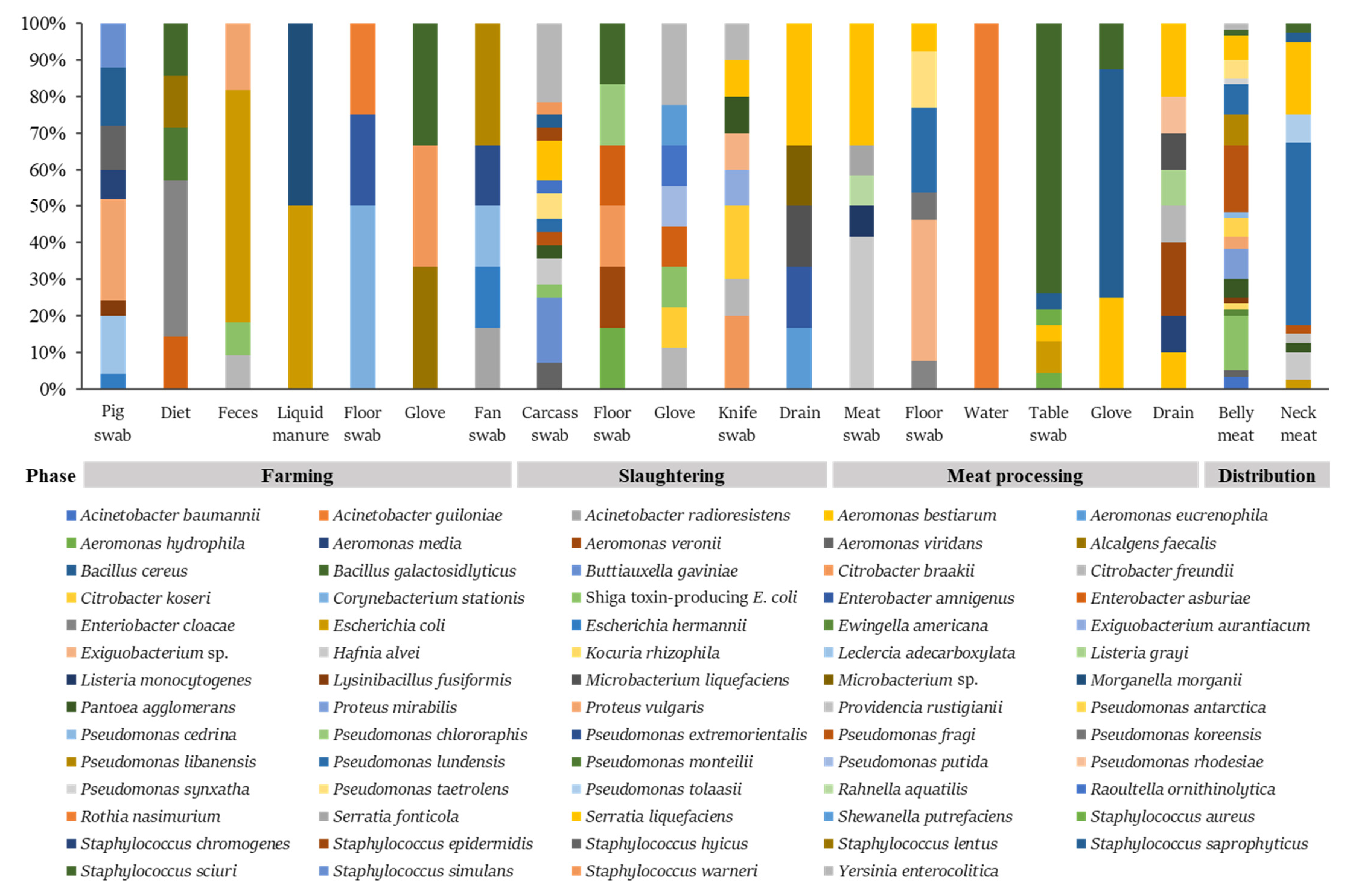
3.1. Distribution of the Pathogens in the Pork Meats and Their Producing Environmental Samples

Pathogenic bacteria isolated from the pork production chain and corresponding environment samples were identified and confirmed using MALDI-TOF MS and PCR, respectively. From 126 samples, 1400 colonies were collected, and 283 of them were identified as belonging to the bacteria focused on in this study (Figure 1). Four major foodborne pathogens, STEC, S. aureus, L. monocytogenes, and Y. enterocolitica, were found in the study. Particularly, nine STEC were isolated from belly meat at a high rate (Figure 1). Supplementary data Table S1 shows that 12 STEC were distributed in feces at the pig farm, on carcasses and gloves at the slaughterhouse, and in belly meats at the retail stores. Other E. coli isolates were found in feces and liquid manure at the farm, the working table at the meat processing plant, and the neck meat at the retail stores. L. monocytogenes and S. aureus were recovered from the pork meat and the working table at the meat processing plant, respectively (Supplementary data Table S1). Ten Y. enterocolitica were mostly found in the slaughterhouse (carcasses, workers’ gloves, and cutting knife). Besides the major foodborne pathogens, the clinically important pathogens associated with the WHO global priority pathogens list of antibiotic-resistant bacteria included Acinetobacter baumannii, Enterobacter cloacae, Morganella morganii, Proteus mirabilis, Providencia rustigianii, and Serratia liquefaciens, found in various sampling sites from swine production to the distribution phase (Supplementary data Table S1). Of the bacteria identified, isolates of Pseudomonas lundensis, S. liquefaciens, and Staphylococcus sciuri isolates were most abundant in the meat and environments. S. saprophyticus and S. sciuri isolates were found in all the phases of production, slaughtering, meat processing, and distribution, whereas A. baumannii, Ewingella americana, Kocuria rhizophila, P. mirabilis, Providencia rustigianii, Pseudomonas antarctica, Pseudomonas synxatha, and Pseudomonas tolaasii isolates were found only in the pork belly or neck meat products at the distribution phase. There were no pathogenic bacteria in the water used in the slaughterhouse and meat processing plant.

3.2. Antimicrobial Resistance in the Important Foodborne Pathogens

Foodborne pathogens such as Y. enterocolitica, STEC, S. aureus, and L. monocytogenes were tested for antimicrobial susceptibility. Twelve and eleven different antibiotic disks used in the study were used for Gram-positive and Gram-negative bacteria, respectively (Table 2). VAN was not used for Gram-negative bacteria due to inefficiency. Data showed that most of the Y. enterocolitica isolates were susceptible to the antimicrobials used in this study (Table 2). Only one pathogen, Y. enterocolitica (GNU-YE6), was resistant to ampicillin and intermediate to ERY and RIF. GNU-YE5 and GNU-YE7 isolates were intermediate to AMC, AMP, ERY, and RIF resistance. There were no multidrug-resistant Y. enterocolitica isolates. On the other hand, most of the STEC isolates were resistant to multiple drugs, including AMP, CHL, ERY, KAN, STR, SXT, and TET. In addition, the most common antimicrobial resistance pattern in the pathogenic E. coli isolates was AMP-KAN-STR-SXT-TET. L. monocytogenes and S. aureus were also shown to be MDR to AMP, ERY, and VAN. All the important foodborne pathogens were susceptible to CIP and GEN.

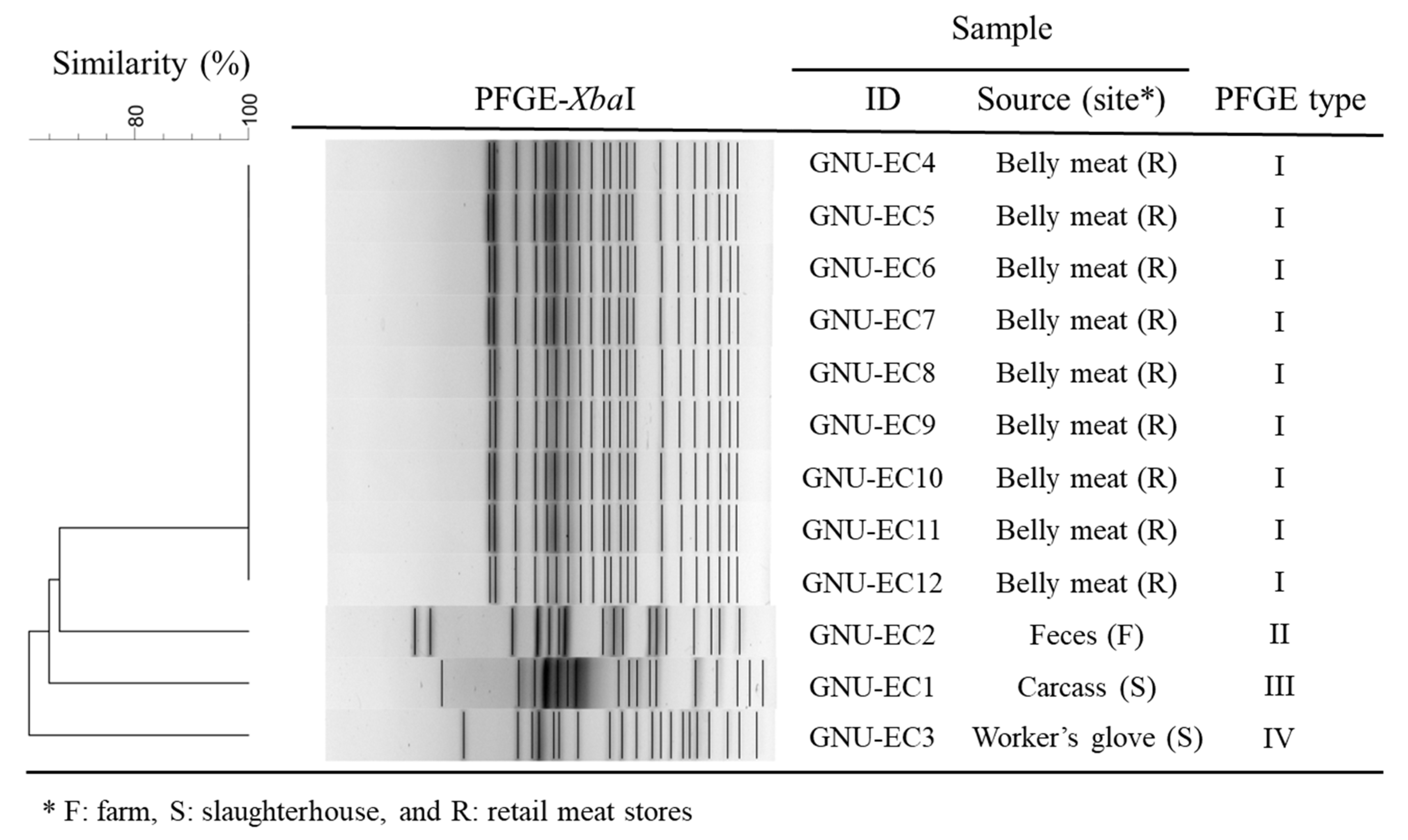
3.3. Genetic Diversity of STEC Isolates

Genomic DNA of 12 STEC isolate strains were digested with XbaI to determine genetic relatedness of the isolates for tracking the pathogens from farm to distribution. The isolates were classified into four pulsotypes based on PFGE banding patterns with more than 99% similarity (Figure 2). The dendrogram of PFGE profiles by XbaI-digestion shows that all STEC isolates grouped in pulsotype I were isolated from belly meats at the retail stores. On the other hand, the other isolates (GNU-EC1, GNU-EC2, and GNU-EC3) showed exceedingly different PFGE band patterns by sample source or site.

4. Discussion

As far as we know, this study is conducted for the first time to track pathogens from pigs and the pork-meat-producing environment to pork meat products via the affiliated slaughterhouse and meat processing plant where the targeted pigs were treated and traced. Thus, this study provides valuable information for the transmission of pathogens between pork meat products and their producing environment. Nine selective agar plates were used to efficiently isolate various bacteria in the samples. Although many colonies with the right form and color to presumably expected pathogens on TSC, XLD, MYP, and mCCDA agar plates were picked up and identified, highly important foodborne pathogens, including Clostridium perfringens, Salmonella spp., Bacillus cereus, and Campylobacter jejuni/coli, were not found in this study. Nevertheless, we found that various foodborne pathogens are present in the pork meats and their producing-related environment. STEC isolates of the pathogens were sequentially found in the farm, slaughterhouse, and retail meat stores, unlike the other pathogens (Supplementary data Table S1). Through subtyping for the STEC colonies, we found that the STEC strains (pulsotype 2, 3, and 4) isolated in the samples from the farm and slaughterhouse environment and carcasses seemed not to be transmitted into the pork meats due to different pulsotypes (Figure 2). STEC strains found in the pork meats were likely to be contaminated from the workers or environments at the retail stores.
The STEC isolates from the pork meat products in retail stores were resistant to various antimicrobials (AMP, KAN, STR, SXT, and TET) than those that at farm and slaughterhouse. A previous report presented that more than 50% of E. coli isolates from fecal and carcass samples were resistant to AMP, CHL, STR, SUL, and TET. In the study, STEC GNU-EC 1 and GNU-EC 2 strains were shown to be resistant to AMP and ERY and AMP and CHL, respectively. In addition, the strains were intermediate to TET (Table 2). The resistance of these antibiotics would be closely associated with the annual antibiotic consumption in the pig industry in S. Korea because antibiotics in the penicillin, tetracycline, and phenicol classes are widely used [3].
With the current results, Y. enterocolitica has been dominantly isolated from food-chain animals and meats, and the bacteria have exhibited to be resistant to various antibiotic classes, including penicillin, cephalosporin, macrolide, aminoglycoside, fluoroquinolone, and tetracycline [27,28,29]. The Korean Ministry of Food and Drug Safety recently reported that Y. enterocolitica was found in 15 out of 289 kimchi products imported from China (www.aninews.in/news/world/asia/bacteria-causing-plague-detected-in-korean-delicacy-kimchi-exported-from-china20210520232537/, accessed on 29 July 2022). It was magnified as an important issue in the food safety sector in S. Korea, since the pathogen, causing gastroenteritis, was reported to cause 35 deaths annually in the US [30]. The pathogens were isolated from the carcasses, the workers’ gloves, and the cutting knife at slaughterhouse and the pork belly meat product (Supplementary data Table S1). In addition, the isolates were susceptible to almost all antibiotics used in the study, except one isolate resistant to AMP (Table 2). P. mirabilis isolates have shown to have different antimicrobial resistance patterns to CHL, ERY, and TET (data not shown). The pathogen has been known to cause urinary tract infection and food poisoning outbreaks [31,32,33]. P. mirabilis is a normal flora in the digestive tracts of humans and animals and is widely found in the natural environment, including soil and water [32]. However, many foodborne poisoning cases associated with the pathogen were recently reported worldwide via nosocomial infection and contaminated food ingestion [34]. Although P. mirabilis isolates were not found in the farm, slaughterhouse, and meat processing plant, the pathogen was found in the pork meat products. It is suggested that the pathogen may be cross-contaminated at the stores. We failed to collect environmental samples in the stores due to exceed repulsion of the owners for collecting the samples. This can be a drawback of the current study. A recent study showed that 5.63% pork samples were contaminated with P. mirabilis producing β-lactamase [35]. However, all P. mirabilis isolated in the study were susceptible to β-lactams. Van was not used for the antibiotic test for Gram-negative pathogens, since VAN has large molecular size and inability to penetrate the outer membrane of the bacteria.
With respect to prevention of the spread of AMR, proper and prudent use of antimicrobials for the pig production is imperative to minimize the emergence and development of bacteria resistant to clinically important antibiotics. In addition to the endeavor to restrict the antibiotic use, monitoring the AMR patterns of clinically important pathogens is also an important step to ensure the safety of meat products and public health. Through the current study, we have tracked clinically important foodborne pathogens from farm to table. Some of the pathogenic or clinically important bacteria, including STEC, Proteus vulgaris, Serratia liquefaciens, and Y. enterocolitica, were found in the pork meat products as well as the farm, slaughterhouse, or meat processing plant. These bacteria contaminated with the pork meats can be transmitted from farm to the meat processing environment. However, other pathogens, such as Acinetobacter baumannii, P. mirabilis, and Providencia rustigianii, were only found in the pork meat products, suggesting that the pathogens would be transmitted via cross contamination at the retail stores. As previously mentioned, the study was conducted for the first time wherein pathogens were tracked from targeted pigs to processed meat products with the pigs’ carcasses, as well as their environment in which the pathogens possibly reside. Consequently, the present study may provide valuable information for presumable sites that clinically important pathogens can be highly transmitted to the pork meat products during the slaughtering and processing phases. Furthermore, the results may help farmers, stakeholders, and government to develop a systematic strategy for reducing the emergence and spread of antimicrobial resistance in pork meat and its producing environment to ensure meat safety and ultimately promote public health.

Supplementary Materials

The following supporting information can be downloaded at: https://www.mdpi.com/article/10.3390/microorganisms10112252/s1, Table S1: Pathogenic or spoilage bacteria isolated from the pig farms, slaughterhouse, meat processing plant, and retail stores.

Author Contributions

Conceptualization, D.B. and S.A.K.; methodology, D.B.; investigation, W.A., S.P., H.J.K. and B.K.; data curation, D.M.M.; writing—original draft preparation, D.B.; writing—review and editing, D.M.M., J.-W.C., G.R.R., G.-H.B., S.A.K., J.S.M. and M.G.K.; supervision, M.G.K.; project administration, D.B.; funding acquisition, D.B. All authors have read and agreed to the published version of the manuscript.

Funding

This research was supported by a grant (MFDS, research project number—20162MFDS011) from the Korean Ministry of Food and Drug Safety in 2020.

Data Availability Statement

Not applicable.

Acknowledgments

This research was also partially supported by Basic Science Research Capacity Enhancement Project through Korea Basic Science Institute (National Research Facilities and Equipment Center) grant funded by Ministry of Education (grant no. 2022R1A6C101B724).

Conflicts of Interest

All authors declare no conflict of interest.

References

  1. Van Boeckel, T.P.; Pires, J.; Silvester, R.; Zhao, C.; Song, J.; Criscuolo, N.G.; Gilbert, M.; Bonhoeffer, S.; Laxminarayan, R. Global trends in antimicrobial resistance in animals in low- and middle-income countries. Science 2019, 365, 1266. [Google Scholar] [CrossRef] [PubMed] [Green Version]
  2. Dewulf, J.; Sternberg-Lewerin, S.; Ryan, M. Tackling antimicrobial resistance in the food and livestock sector. In Challenges to Tackling Antimicrobial Resistance; Anderson, M., Cecchini, M., Mossialos, E., Eds.; Cambridge University Press: Cambridge, UK, 2020; pp. 99–123. [Google Scholar]
  3. APQA. Animal and Plant Quarantine Agency. In Korean Veterinary Antimicrobial Resistance Monitoring Report; APQA: Gimcheon, Korea, 2019. [Google Scholar]
  4. Bae, D.; Kweon, O.; Khan, A.A. Isolation and characterization of antimicrobial-resistant nontyphoidal Salmonella enterica serovars from imported food products. J. Food Protect. 2016, 79, 1348–1354. [Google Scholar] [CrossRef] [PubMed]
  5. Pollock, J.; Muwonge, A.; Hutchings, M.R.; Mainda, G.; Bronsvoort, B.M.; Gally, D.L.; Corbishley, A. Resistance to change: AMR gene dynamics on a commercial pig farm with high antimicrobial usage. Sci. Rep. 2020, 10, 1708. [Google Scholar] [CrossRef] [PubMed] [Green Version]
  6. Avraam, C.; Lambrou, A.S.; Jiang, W.; Siddiqui, S. Antimicrobial resistance and livestock trade for low and middle income countries: Regional analysis of global coordination policies. Front. Sustain. Food Syst. 2021, 5, 650315. [Google Scholar] [CrossRef]
  7. Hughes, D. Selection and evolution of resistance to antimicrobial drugs. IUBMB Life 2014, 66, 521–529. [Google Scholar] [CrossRef] [PubMed]
  8. Ryu, S. The new Korean action plan for containment of antimicrobial resistance. J. Glob. Antimicrob. Resist. 2017, 8, 70–73. [Google Scholar] [CrossRef]
  9. Ceccarelli, D.; Hesp, A.; van der Goot, J.; Joosten, P.; Sarrazin, S.; Wagenaar, J.A.; Dewulf, J.; Mevius, D.J.; Effort Consortium, O. Antimicrobial resistance prevalence in commensal Escherichia coli from broilers, fattening turkeys, fattening pigs and veal calves in European countries and association with antimicrobial usage at country level. J. Med. Microbiol. 2020, 69, 537–547. [Google Scholar] [CrossRef]
  10. Vanderhaeghen, W.; Dewulf, J. Antimicrobial use and resistance in animals and human beings. Lancet Planet Health 2017, 1, E307–E308. [Google Scholar] [CrossRef]
  11. Zwirzitz, B.; Wetzels, S.U.; Dixon, E.D.; Stessl, B.; Zaiser, A.; Rabanser, I.; Thalguter, S.; Pinior, B.; Roch, F.F.; Strachan, C.; et al. The sources and transmission routes of microbial populations throughout a meat processing facility. Npj Biofilms Microbiomes 2020, 6, 26. [Google Scholar] [CrossRef]
  12. Caekebeke, N.; Jonquiere, F.J.; Ringenier, M.; Tobias, T.J.; Postma, M.; van den Hoogen, A.; Houben, M.A.M.; Velkers, F.C.; Sleeckx, N.; Stegeman, J.A.; et al. Comparing farm biosecurity and antimicrobial use in high-antimicrobial-consuming broiler and pig farms in the Belgian-Dutch border region. Front. Vet. Sci. 2020, 7, 558455. [Google Scholar] [CrossRef]
  13. OIE. Annual Report on Antimicrobial Agents Intended for Use in Animals. Available online: https://www.oie.int/fileadmin/Home/eng/Our_scientific_expertise/docs/pdf/AMR/Annual_Report_AMR_3.pdf (accessed on 12 June 2020).
  14. Holmer, I.; Salomonsen, C.M.; Jorsal, S.E.; Astrup, L.B.; Jensen, V.F.; Hog, B.B.; Pedersen, K. Antibiotic resistance in porcine pathogenic bacteria and relation to antibiotic usage. BMC Vet. Res. 2019, 15, 449. [Google Scholar] [CrossRef] [Green Version]
  15. Trongjit, S.; Angkititrakul, S.; Tuttle, R.E.; Poungseree, J.; Padungtod, P.; Chuanchuen, R. Prevalence and antimicrobial resistance in Salmonella enterica isolated from broiler chickens, pigs and meat products in Thailand-Cambodia border provinces. Microbiol. Immunol. 2017, 61, 23–33. [Google Scholar] [CrossRef] [PubMed] [Green Version]
  16. Lunha, K.; Leangapichart, T.; Jiwakanon, J.; Angkititrakul, S.; Sunde, M.; Jarhult, J.D.; Hallenberg, G.S.; Hickman, R.A.; Van Boeckel, T.; Magnusson, U. Antimicrobial resistance in fecal Escherichia coli from humans and pigs at farms at different levels of intensification. Antibiotics 2020, 9, 662. [Google Scholar] [CrossRef] [PubMed]
  17. Im, M.C.; Seo, K.W.; Bae, D.H.; Lee, Y.J. Bacterial quality and prevalence of foodborne pathogens in edible offal from slaughterhouses in Korea. J. Food Protect. 2016, 79, 163–168. [Google Scholar] [CrossRef]
  18. Ballash, G.A.; Albers, A.L.; Mollenkopf, D.F.; Sechrist, E.; Adams, R.J.; Wittum, T.E. Antimicrobial resistant bacteria recovered from retail ground meat products in the US include a Raoultella ornithinolytica co-harboring blaKPC-2 and blaNDM-5. Sci. Rep. 2021, 11, 14041. [Google Scholar] [CrossRef]
  19. Self, J.L.; Luna-Gierke, R.E.; Fothergill, A.; Holt, K.G.; Vieira, A.R. Outbreaks attributed to pork in the United States, 1998–2015. Epidemiol. Infect. 2017, 145, 2980–2990. [Google Scholar] [CrossRef] [PubMed] [Green Version]
  20. Doyle, M.E. Multidrug-resistant pathogens in the food supply. Foodborne Pathog. Dis. 2015, 12, 261–279. [Google Scholar] [CrossRef]
  21. Nogva, H.K.; Rudi, K.; Naterstad, K.; Holck, A.; Lillehaug, D. Application of 5’-nuclease PCR for quantitative detection of Listeria monocytogenes in pure cultures, water, skim milk, and unpasteurized whole milk. Appl. Environ. Microbiol. 2000, 66, 4266–4271. [Google Scholar] [CrossRef] [Green Version]
  22. Wang, J.Z.; Duan, R.; Liang, J.R.; Huang, Y.; Xiao, Y.C.; Qiu, H.Y.; Wang, X.; Jing, H.Q. Real-time TaqMan PCR for Yersinia enterocolitica detection based on the ail and foxA genes. J. Clin. Microbiol. 2014, 52, 4443–4444. [Google Scholar] [CrossRef] [Green Version]
  23. Paton, A.W.; Paton, J.C. Detection and characterization of Shiga toxigenic Escherichia coli by using multiplex-PCR assays for stx1, stx2, eaeA, enterohaemorrhagic E. coli hlyA, rfbO111 and rfbO157. J. Clin. Microbiol. 1998, 36, 598–602. [Google Scholar] [CrossRef]
  24. Chen, J.; Griffiths, M.W. PCR differentiation of Escherichia coli from other gram-negative bacteria using primers derived from the nucleotide sequences flanking the gene encoding the universal stress protein. Lett. Appl. Microbiol. 1998, 27, 369–371. [Google Scholar] [CrossRef] [PubMed]
  25. Chiefari, A.K.; Perry, M.J.; Kelly-Cirino, C.; Egan, C.T. Detection of Staphylococcus aureus enterotoxin production genes from patient samples using an automated extraction platform and multiplex real-time PCR. Mol. Cell Probe 2015, 29, 461–467. [Google Scholar] [CrossRef] [PubMed]
  26. CLSI. Performance Standards for Antimicrobial Susceptibility Testing. In Twentieth Informational Supplement; CLSI: Wayne, PA, USA, 2018. [Google Scholar]
  27. Gkouletsos, T.; Patas, K.; Lambrinidis, G.; Neubauer, H.; Sprague, L.D.; Ioannidis, A.; Chatzipanagiotou, S. Antimicrobial resistance of Yersinia enterocolitica and presence of plasmid pYV virulence genes in human and animal isolates. New Microbes New Infect. 2019, 32, 100604. [Google Scholar] [CrossRef] [PubMed]
  28. Younis, G.; Mady, M.; Awad, A. Yersinia enterocolitica: Prevalence, virulence, and antimicrobial resistance from retail and processed meat in Egypt. Vet. World 2019, 12, 1078–1084. [Google Scholar] [CrossRef]
  29. Baumgartner, A.; Kuffer, M.; Suter, D.; Jemmi, T.; Rohner, P. Antimicrobial resistance of Yersinia enterocolitica strains from human patients, pigs and retail pork in Switzerland. Int. J. Food Microbiol. 2007, 115, 110–114. [Google Scholar] [CrossRef]
  30. CDC. Yersinia enterocolitica (Yersiniosis). Available online: https://www.cdc.gov/yersinia/faq.html (accessed on 6 October 2021).
  31. Wang, Y.; Zhang, S.Y.; Yu, J.Y.; Zhang, H.; Yuan, Z.Q.; Sun, Y.S.; Zhang, L.; Zhu, Y.F.; Song, H.B. An outbreak of Proteus mirabilis food poisoning associated with eating stewed pork balls in brown sauce, Beijing. Food Control 2010, 21, 302–305. [Google Scholar] [CrossRef]
  32. Drzewiecka, D. Significance and roles of Proteus spp. bacteria in natural environments. Microb Ecol. 2016, 72, 741–758. [Google Scholar] [CrossRef] [Green Version]
  33. Hola, V.; Peroutkova, T.; Ruzicka, F. Virulence factors in Proteus bacteria from biofilm communities of catheter-associated urinary tract infections. Fems Immunol. Med. Mic. 2012, 65, 343–349. [Google Scholar] [CrossRef] [Green Version]
  34. Gong, Z.L.; Shi, X.L.; Bai, F.; He, X.L.; Zhang, H.Y.; Li, Y.B.; Wan, Y.; Lin, Y.M.; Qiu, Y.Q.; Chen, Q.C.; et al. Characterization of a novel diarrheagenic strain of Proteus mirabilis associated with food poisoning in China. Front. Microbiol. 2019, 10, 2810. [Google Scholar] [CrossRef] [Green Version]
  35. Chinnam, B.K.; Nelapati, S.; Tumati, S.R.; Bobbadi, S.; Peddada, V.C.; Bodempudi, B. Detection of beta-lactamase-producing Proteus mirabilis strains of animal origin in Andhra Pradesh, India and their genetic diversity. J. Food Protect. 2021, 84, 1374–1379. [Google Scholar] [CrossRef]
Figure 1. The ratio of pathogens isolated from the pork meat and its producing environment. Bacteria isolated from various samples were identified using the MALDI-TOF MS and the MALDI-TOF MS Biotyper 3.1 software. Important foodborne pathogens, including STEC, Listeria monocytogenes, Staphylococcus aureus, and Y. enterocolitica were found in the pork meat products and their producing environment from the farm, slaughterhouse, or meat processing plant. Additionally, clinically important pathogens (e.g., Acinetobacter baumannii, E. coli, Enterobacter spp., Serratia spp., Proteus mirabilis, Providencia rustigianii, and Morganella morganii) highly related to antimicrobial resistance were found in in the pig farms, slaughterhouse, meat processing plant, and retail stores.
Microorganisms 10 02252 g001
Figure 2. Analysis of PFGE banding patterns of STEC strains isolated from feces at swine farm, carcass, and the workers’ gloves at meat processing plant, and belly meats at retail stores. Dendrogram of the PFGE patterns from 12 STEC meat and environmental isolates using XbaI-digestion is shown by 4 pulsotypes according to the similarity of PFGE band profiles of more than 99% within a group using UPGMA clustering method.
Microorganisms 10 02252 g002
Table 1. Selective agars, cultivation conditions, and primer sequences used in the study.
Selective AgarTarget BacteriaCharacteristics/Cultivation ConditionsPrimer SequencesGene/Product SizeReferences
OxfordListeria monocytogenesGray colony with a black halo, 37 °C for 24 hF: TGCAAGTCCTAAGACGCCA
R: CACTGCATCTCCGTGGTATACTAA
hlyA/113 bp[21]
MacConkeyYersinia enterocoliticaPale pink or colorless colony, 30 °C for 36 hF: TTTGGAAGCGGGTTGAATTG
R: GCTCACGGAAAGGTTAAGTCATCT
ail/101 bp[22]
TC-SMACSTECClear or colorless colony, 37 °C for 24 hF: ATAAATCGCCATTCGTTGACTAC
R: AGAACGCCCACTGAGATCATC
F: GGCACTGTCTGAAACTGCTCC
R: TCGCCAGTTATCTGACATTCTG
stx1/180 bp
stx2/255 bp
[23]
BCIGE. coliBluish green colony, 37 °C for 24 hF: CCGATACGCTGCCAATCAGT
R: CTGGTATCAGCGCGAAGTCT
uspA/884bp[24]
Baird-ParkerStaphylococcus aureusGray to less black with opaque zone, 35 °C for 48 hF: AAGTGCCGATCAATTTATGGCTA
R: CCTGAACAGTTACATTTTTCTTATTCGT
entA/90 bp[25]
Table 2. Antimicrobial 1 susceptibility test of major pathogens using disc diffusion assay.
IDSpeciesAMCAMPCHLCIPGENERYKANRIFSTRSXTTETVANSource (Site 2)
GNU-YE 1Y. enterocoliticaNACarcass (S)
GNU-YE 2Y. enterocoliticaNACarcass (S)
GNU-YE 3Y. enterocoliticaNACarcass (S)
GNU-YE 4Y. enterocoliticaNACarcass (S)
GNU-YE 5Y. enterocolitica++++NAGlove (S)
GNU-YE 6Y. enterocolitica+++++NAKnife (S)
GNU-YE 7Y. enterocolitica++++NAGlove (S)
GNU-YE 8Y. enterocolitica+NABelly meat (R)
GNU-YE 9Y. enterocoliticaNACarcass (S)
GNU-EC 1STEC 3+++++NACarcass (S)
GNU-EC 2STEC+++++++NAFeces (F)
GNU-EC 3STEC++++++++++NAGlove (S)
GNU-EC 4STEC+++++++++++++NABelly meat (R)
GNU-EC 5STEC++++++++++++NABelly meat (R)
GNU-EC 6STEC+++++++++++NABelly meat (R)
GNU-EC 7STEC++++++++++++NABelly meat (R)
GNU-EC 8STEC++++++++++++NABelly meat (R)
GNU-EC 9STEC+++++++++++++NABelly meat (R)
GNU-EC 10STEC+++++++++++++NABelly meat (R)
GNU-EC 11STEC+++++++++++++NABelly meat (R)
GNU-EC 12STEC++++++++++++++NABelly meat (R)
GNU-SA 1S. aureus++++++++Table (P)
GNU-LM 1L. monocytogenes+++++++Meat (P)
++, Resistant; +, intermediate; −, susceptible; 1 AMC: amoxicillin and clavulanic acid, AMP: ampicillin, CHL: chloramphenicol, CIP: ciprofloxacin, ERY: erythromycin, GEN: gentamicin, KAN: kanamycin, RIF: rifampicin, STR: streptomycin, SXT: trimethoprim and sulfamethoxazole, TET: tetracycline, VAN: vancomycin; 2 F: farm, S: slaughterhouse, P: meat processing plant, and R: retail meat stores; 3 Shiga-toxin-producing E. coli.
Publisher’s Note: MDPI stays neutral with regard to jurisdictional claims in published maps and institutional affiliations.

