Article

Screening of Endophytic Bacteria of Leucojum aestivum ‘Gravety Giant’ as a Potential Source of Alkaloids and as Antagonist to Some Plant Fungal Pathogens

by
Yuka Munakata
1,2,
Rosella Spina
1,2,
Sophie Slezack-Deschaumes
1,
Julie Genestier
1,
Alain Hehn
1 and
Dominique Laurain-Mattar
1,2,*
1
Université de Lorraine—INRAE, LAE, F-54000 Nancy, France
2
Université de Lorraine—CNRS, L2CM, F-54000 Nancy, France
*
Author to whom correspondence should be addressed.
Microorganisms 2022, 10(10), 2089; https://doi.org/10.3390/microorganisms10102089
Submission received: 22 September 2022 / Revised: 17 October 2022 / Accepted: 18 October 2022 / Published: 21 October 2022

Abstract

:
Leucojum aestivum is a medicinal plant belonging to the Amaryllidaceae family well known as a producer of alkaloids such as galanthamine and lycorine. However, the endophytic microbes that colonize different plant tissues without causing any damage have not been reported in this plant. Here, we explored the different endophytic bacterial communities isolated from different surface disinfected tissues of L. aestivum ‘Gravety giant’ and screened bacterial isolates producing alkaloids and their potential use as biocontrol agent against wheat pathogens. For that purpose, endophytic bacteria were isolated from bulbs, roots and shoots of L. aestivum. After taxonomical characterization, these microorganisms were screened for their ability to produce alkaloids using high-performance thin-layer chromatography (HPTLC) and untargeted liquid chromatography-Mass Spectrometry/Mass Spectrometry (LC-MS/MS) strategies. We isolated 138 bacteria belonging to four phyla and 42 genera, mainly from roots and shoots. The most abundant genera were Rahnella in shoot, Patulibacter in bulb and Bacillus in roots. Among the different bacterial isolates, the methanolic extracts of Luteibacter rhizovicinus (LaBFB3301) and Commamonas denitrificans (LaBFS2103) slightly delayed the growth of F. graminearum colonies in in vitro dual tests against F. graminearum and M. nivale strains with 15.5% and 19.9% inhibition rates, respectively. These isolates are able to produce an indolic alkaloid tryptophol (C10H11NO, [M + H]+ 162.0913). These endophytic bacteria might be investigated to characterize the plant protection effect and the plant growth promotion effect.

1. Introduction

Endophytic microbes are found in all plant species studied so far [1]. Although endophytes involve archaea, protists and viruses, most of them are represented by bacteria and fungi. Even though fungal endophytes have been more intensively studied than bacterial endophytes, such bacterial endophytes are of interest in terms of plant growth promotion effects and the robustness of the plant immune system against fungal/oomycete plant pathogens [2]. These bacteria are also able to produce bioactive specialized metabolites with various biological activities, and endophytes may modulate plant metabolite synthesis. However, the metabolic interactions between plants and endophytes remain largely unknown [3]. A great number of endophytic bacteria have been reported, including Proteobacteria, Actinobacteria, Bacteroidetes and Firmicutes [4,5,6]. The population densities of endophytic bacteria are extremely variable considering plant species and organs [7]. Roots and below-ground tissues tend to yield the highest density of bacteria compared to above-ground tissues, while there are a small number of reports about endophytes in flowers, fruits and seeds [8,9].
The microbes isolated from inner plant tissues are attractive and might constitute original factories for producing new bioactive molecules [10,11,12]. Furthermore, some endophytes have also been reported to mimic the synthesis of plant compounds, such as paclitaxel and vinblastine [1,13,14]. It is known that these microorganisms are also able to produce different specialized metabolites such as non-ribosomal synthesized peptides, polyketides, alkaloids, sesquiterpenes, flavonoids, saponins, lactones and organic acids [15,16], which confer ecological benefits to their host plants. These compounds can possess pharmaceutical functions such as antifungal, antibacterial, antivirus, anticancer as well as anti-inflammatory properties. Bioactive molecules such as actinoallolides A (anti-trypanosomal activity) [17], ecomycins (antifungal activity) [18] and maytansine (cytotoxicity) [19] were discovered and isolated from bacterial endophytes. It was also demonstrated that a Bacillus strain isolated from an Icacinaceae plant was also able to produce camptothecin, a quinoline alkaloid, independently from the host plant [14,20]. Some specialized metabolites isolated from endophytic bacteria have shown agronomical interests such as phenolic compounds, volatile organic compounds that show phytotoxic activity [21], and antifungal and nematicidal properties [22,23]. These examples clearly establish that endophytic bacteria might be used as bioreactors or as genomic resources to create cell factories for producing phytochemicals independently of their host plants or to characterize new leads for further agrochemical and pharmaceutical applications.
Bulbous plants of the Amaryllidaceae family are known for the synthesis of isoquinoleic alkaloids, also known as Amaryllidaceae alkaloids, including lycorine and galantamine [24]. These alkaloids exhibit a wide range of biological activities, such as acetylcholinesterase inhibition for galantamine, which led to their use as drugs. Only a few studies on the molecular identification of endophytes isolated from Amaryllidaceae plants and the identification of compounds extracted from these endophytic microorganisms were reported. Hexacyclopeptides have been identified in the endophytic fungus Fusarium salani from Narcissus tazetta [25]. From Leucojum aestivum’s in vitro bulblets, five Amaryllidaceae alkaloids, four amino acids and fatty acids were identified in the endophytic bacteria belonging to the Bacillus genus [26]. In this study, we analyze endophytic bacteria from bulbs, roots and shoots of L. aestivum ‘Gravety giant’. Because it is reported that Amaryllidaceae alkaloids differently accumulate depending on plant tissues [27], we assumed that the different tissues host different endophytic communities in terms of taxonomy and metabolites. We screened bacterial isolates with Dragendorff’s reagent for the detection of alkaloids on HPTLC plates and performed fine LC-MS/MS analysis of methanolic extracts from bacterial pellets. The isolates were taxonomically identified using 16S RNA sequencing, and their potential activities towards some wheat fusarium wilt agents such as Fusarium graminearum and Microdochium nivale were investigated.

2. Materials and Methods

2.1. Plant Material and Culture Conditions

The soil used was a clay soil (FAO-WRB classification) sampled from the 0–20 cm layer at the Bouzule research farm in northeastern France (48°74′ N, 6°35′ E). The bulbs of Leucojum aestivum ‘Gravety giarance). The bulbs were sont’ were purchased from a French market (Pépinière Lepage ‘Val de Loire’, Les Ponts-de-Cé, Fwn in a container (76 cm × 38.5 cm × 23 cm, 5 kg of soil) placed indoors and watered with 2 L of water once a week at 20 °C with a 16/8 h light/dark cycle until bacterial isolation. After 90 days, 3 plants showing shoots about 10 cm long were harvested.
The plant materials were washed with running tap water and the different tissues were separated using blades into aerial parts (shoots), bulbs and roots. Shoot and root parts were washed again with tap water and then dried on paper towels. For bulbus parts, the outer tunic was peeled by blade and by hand, and the basal plate was removed by blade because of their difficulty for disinfection. The treated bulb was cut in half and individual scales were separated by hand. Each tissue material was weighed just after sampling. The fresh materials were then cut into certain sizes: shoot and bulbus materials were 1 to 1.5 cm in horizontal length and 2 to 3 cm in vertical length; root materials were around 3 cm in length. The trimmed materials were randomly distributed by around 1.1 g in a sterile 50 mL tube and stored at 4 °C. They were used for bacterial isolation within 48 h after being harvested.

2.2. Bacterial Isolation and Cultivation

The plant materials were processed as described by Munakata et al. [28], with a minor modification depending on the plant tissue. Briefly, for the surface sterilization, the shoot materials were treated in 70% ethanol for 1 min and in 2.6% bleach solution for 2.5 min; the bulb materials were treated in 70% ethanol for 1 min and in 2.6% bleach solution for 5 min; the root materials were treated in 70% ethanol for 1 min 3 times and in 2.6% bleach solution for 5 min. At last, all the materials were rinsed in sterile ultrapure water for 1 min, 3 times. All the treatments and the rinses were performed in 20 mL solution with agitation. The final rinse solution was used for testing the efficiency of the surface sterilization by spreading 100 µL on 10% Tryptic soy agar (10% TSA) media (Sigma-Aldrich, St. Quentin Fallavier, France) containing cycloheximide at 100 mg/L (10% TSAC). The treated plant materials were cut into smaller pieces using a sterile blade and then grinded by sterile tubes on sterile Petri dishes. They were then incubated in 10 mL of 0.85% (w/v) sodium chloride for 1 h with agitation at 60 rpm at 28 °C. Then, 100 µL of the incubated suspension was spread on three plates of 10% TSAC, with a dilution series of 1 time and 10 times. The plates were incubated at 28 °C for 20 days. The number of colonies was expressed as CFU/g of fresh plant material. Colonies were purified by repeated sub-culturing, and single colonies were picked for subsequent analysis. The pure isolates were also stored in 15% glycerol solution at −80 °C.

2.3. Molecular Identification

The bacteria were taxonomically identified by PCR amplification of the 16S rDNA regions, performed with a method described in Munakata et al. [28]. For some isolated bacteria, DNA was sequenced using the DNeasy® UltraClean® Microbial Kit (QIAGEN, Hilden, Germany) and used as a template for PCR amplification with the primer sets of 27F (5′–GAGAGTTTGATCCTGGCTCAG–3′) and 1492R (5′–CTACGGCTA CCTTGTTACGA–3′). The PCR 50 µL reaction mix contained 1 µL of PrimeSTAR® GXL DNA Polymerase (Takara, St. Germain-en-Laye, France), 10 µL of 5×PrimeStar GXL buffer, 4 µL of dNTPs (2.5 mM for each), 1 µL of each primer at 10 µM and 1 µL of genomic DNA. PCR amplification was performed using the following program: 10 s 98 °C, followed by 30 cycles of 10 s 98 °C, 15 s 55 °C, 2 min 68 °C, with the final elongation at 68 °C for 2 min. PCR products were commercially sequenced (Eurofins, Nantes, France). DNA sequences were edited and screened against those in the GenBank database using BLAST (http://www.ncbi.nlm.nih.gov/ accessed on 20 September 2022). The sequences have been deposited in GenBank under accession numbers (OL307013 to OL307079, accessed on 31 October 2021), shown in Table S1.

2.4. Crude Extract Preparation of Bacterial Pellet

Crude extracts for alkaloid detection were prepared from bacterial pellets. A preculture of the bacteria was performed in 2 mL nutrient broth (NB) (Sigma) at 28 °C and 180 rpm for 24 h. Then, 100 µL of the precultures was added to 50 mL NB in 250 mL Erlenmeyer flasks. After 72 h of cultivation at 28 °C 180 rpm, the bacterial pellets were collected by centrifugation at 4000× g at room temperature for 40 min. The pellets were suspended in 1.5 mL methanol and treated with ultrasonication for 30 min, followed by maceration for 30 min at room temperature. The supernatant was then obtained by centrifugation at 12,800× g at room temperature for 10 min.

2.5. High-Performance Thin-Layer Chromatography (HPTLC) Analysis and Alkaloid Detection with Dragendorff’s Reagent

Briefly, 20 µL of the extracts and 5 µL of a standard mixture of lycorine and galantamine at 1 mg/mL each were sprayed with LINOMAT 5 (Camag®, Muttenz, Switzerland) onto HPTLC plates 60 F254 (Merck, Darmstadt, Germany), 20 × 10 cm. The plates were developed in the mobile phase consisting of 10 mL of ethyl acetate, 1.1 mL of acetic acid, 1.1 mL of formic acid and 2.5 mL of ultrapure water. After the development, the plates were air-dried. To screen the presence of alkaloids, the plate was sprayed with fresh Dragendorff’s reagent containing a mixture of solution A, solution B, acetic acid and water (1:1:4:20 v/v). Herein, solution A was 0.85 g basic bismuth nitrate in 10 mL acetic acid and 40 mL ultrapure water; solution B was 8 g potassium iodide in 30 mL ultrapure water. The colorized plates were observed successively just after spraying, after 30 min and after one day. The orange coloration of a spot indicated the potential detection of an alkaloid. The standards galanthamine and lycorine were purchased from Medchem Express, tryptophol from VWR and indole-3-acetic acid from Sigma-Aldrich (St. Quentin Fallavier, France).

2.6. Dual-Culture Assay to Test the Antagonistic Activity of Endophytic Bacteria against Two Cereal Pathogens

The antagonistic activities were analyzed using an in vitro dual-inoculation method. F. graminearum strain was kindly provided by Institut Français de la Brasserie et de la Malterie (IFBM), and M. nivale strain (MUCL 18523) was purchased from BCCL/MUCL. They were cultivated on potato dextrose agar (PDA) (Sigma) at 28 °C for F. graminearum and at 20 °C for M. nivale in darkness. Mycelial plugs of these fungi, 8 mm in diameter, were prepared from an active growing zone of the fresh cultures. One plug was inoculated at the center of a plate containing 25 mL of PDA. Then, 50 µL of a bacterial suspension obtained from a liquid culture of a bacterial isolate in 2 mL of NB at 28 °C for 24 h or 72 h was dropped on 4 spots that were 3 cm from the center of the plates. Three replicates were made for each bacterial isolate. The plates were incubated at 28 °C for F. graminearum and at 25 °C for M. nivale until the fungal colonies reached the edge of the plate. The diameter of the fungal colony was measured and averaged among replicates. Plates inoculated with fungi without bacterial cultures were used as controls. Antagonistic activities were expressed as the inhibitory rate (%), calculated as follows: ((dcontrol − dassay)/dcontrol × 100), where d is the diameter of the colony of the fungi.

2.7. Diffusion Assay of Cell-Free Culture Broth and Methanolic Extracts of the Selected Strains against F. graminearum mycelia

The crude extracts of bacterial isolates were prepared as described above. The 24-h-cultured bacterial broth in 2 mL NB was centrifuged at 15,000 rpm for 10 min and filtrated with Acrodisc® Syringe Filter 0.2 µm Supor® Membrane. For the diffusion assay, φ5.3 mm paper discs (Whatman, Merck, Darmstadt, Germany) were autoclaved and dried at 70 °C. Then, 25 µL of each sample was applied to one paper disc and the discs were dried under a laminar flow hood. Four discs per 20 mL PDA plate were placed diagonally 1 cm from the center. Fungal plugs were prepared and placed at the center as described above. The plates were incubated at 28 °C for F. graminearum and 20 °C for M. nivale. The diameters of the fungal colonies were measured every day from day 3. The growth inhibition rate was calculated as in the previous section.

2.8. LC-MS Equipment and Analysis

Untargeted LC-MS analysis was performed with UHPLC-Q-Exactive-Orbitrap (Thermo Fisher Scientific, Waltham, MA, USA). The column was Kinetex XB-C18 (150 × 2.1 mm × 2.6 µm) (Phenomenex, Le Pecq, France). The mobile phase consisted of 0.1% formic acid in water (A) and 0.1% formic acid in methanol (B) with 200 µL/min. Gradient elution was performed with the following program: 0–1 min, 10–10% B; 1–15 min, 10–60% B; 15–30 min, 60–95% B; 30–35 min, 95–95% B. MS data were obtained from an Orbitrap IDX mass spectrometer (Thermo Fisher Scientific, Waltham, MA, USA) using positive ionization mode. The ionization was achieved with the heated electrospray ionization (HESI) source and the scan range was 120 to 1200 m/z. The spectrometer worked with the data-dependent scan. MS2 fragments were obtained with collision-induced dissociation (HCD) at 30% energy. The resolution was 60,000 for MS1 and 30,000 for MS2.
The results were analyzed by Compound Discoverer 3.1 (Thermo Fisher Scientific, Waltham, MA, USA). The peaks were extracted with the threshold at minimum intensity = 50,000 m/z and then aligned and identified for metabolites, comparing with public databases such as mzCloud (https://www.mzcloud.org/ accessed on 20 September 2022). The peaks having a full match in mzCloud were used for further analysis, and categorized as ‘enriched’ in test samples when they satisfied log2 fold change to medium control >1 and p < 0.05.
To identify tryptophol and confirm the absence of lycorine, galanthamine and IAA, the bacterial methanolic extracts were analyzed by the LC-MS/MS system using positive ionization mode. It was realized by ESI high-resolution mass spectrometry (HRMS) with micrOTOFQTM (Bruker Daltonics, Bruker, Bremen, Germany) apparatus. The column was Hypersil gold (100 mm × 3 µm) (Thermo Scientific, Bellefonte, PA, USA) and the flow rate was 200 µL/min using the following program: 5% solvent B for 5 min, a gradient from 5% solvent B at 5 min to 99% of solvent B at 40 min, followed by 99% solvent B for 5 min. Solvent A was composed of 1% formic acid in water with LC/MS grade, and solvent B was composed of pure acetonitrile. The calibration was performed with a peak of sodium formate in each analysis. The spectrometer worked with the auto MS/MS mode. Each detected mass peak was isolated and fragmented with collision energy at 20 eV. The results were analyzed by Bruker Compass® software.

2.9. Data Analysis

R studio with R version 3.6.3. was used for statistical tests and analysis. The Bray–Curtis distance was calculated with the R package ‘vegan’. This package was also used for performing PERMANOVA (permutations = 10,000) with adonis. Principal coordinate analysis (PCoA) and scatter plots were illustrated using the ggplot2 package. The Tukey–Kramer test was performed with the R package ‘multcomp’.

3. Results

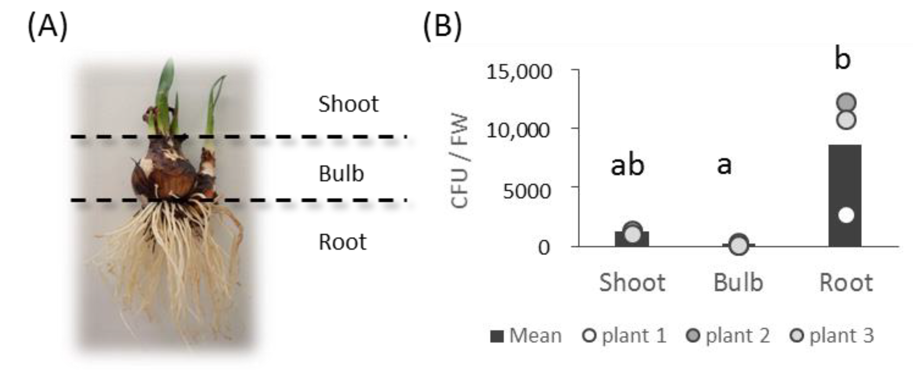
3.1. Bacterial Collection Isolated from L. aestivum ‘Gravety Giant’

Bacteria were isolated from three different organs of L. aestivum ‘Gravety giant’ plants at the vegetative stage, showing leaves about 7 cm long (Figure 1, Figure S1). The average density of culturable bacterial endophytes varied depending on the plant organs and was higher in roots (8569 colony-forming units per 1 g of fresh material weight (CFU/FW) compared to shoots (1210 CFU/FW) and bulbs (169 CFU/FW) (Figure 1).

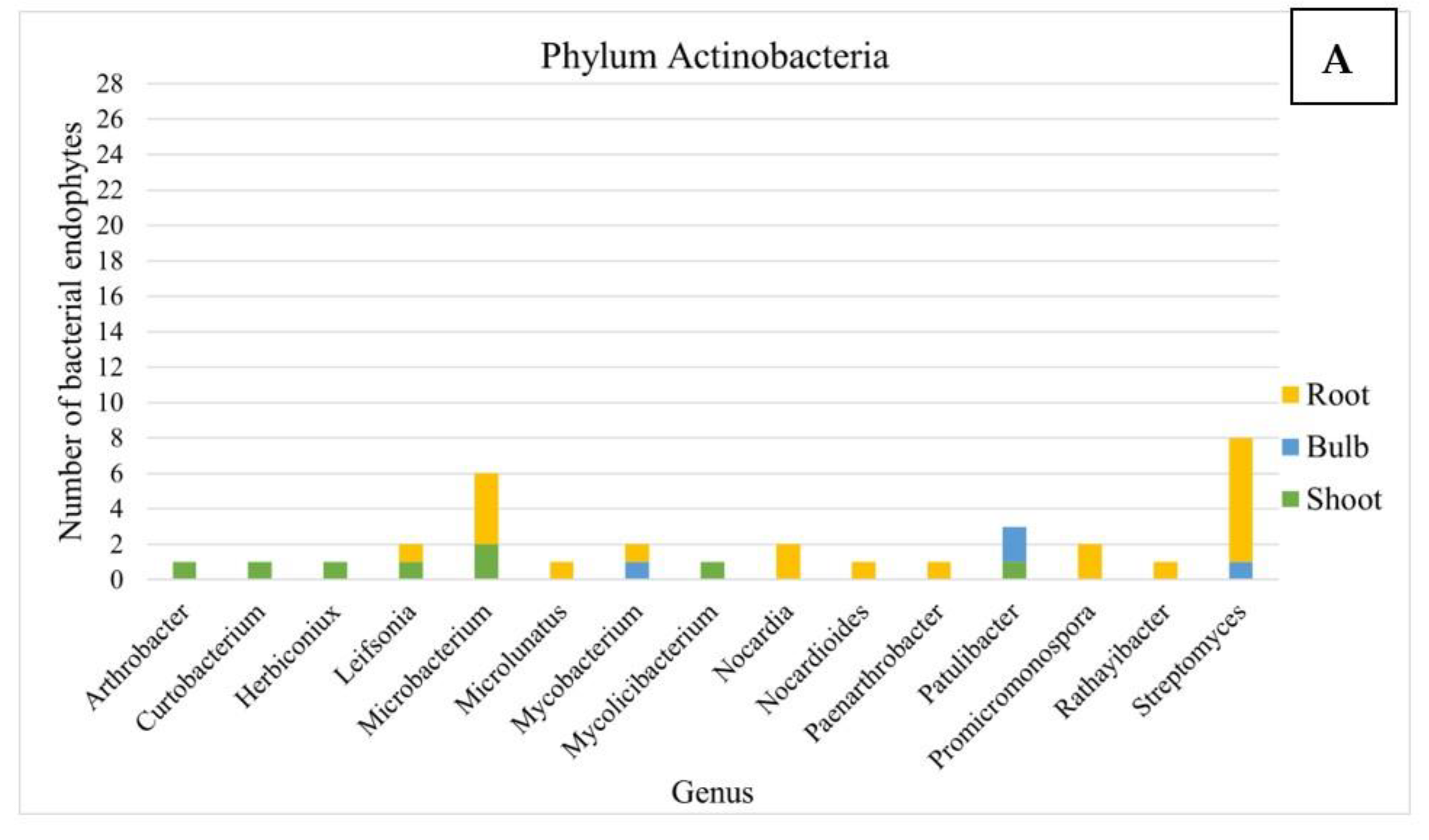
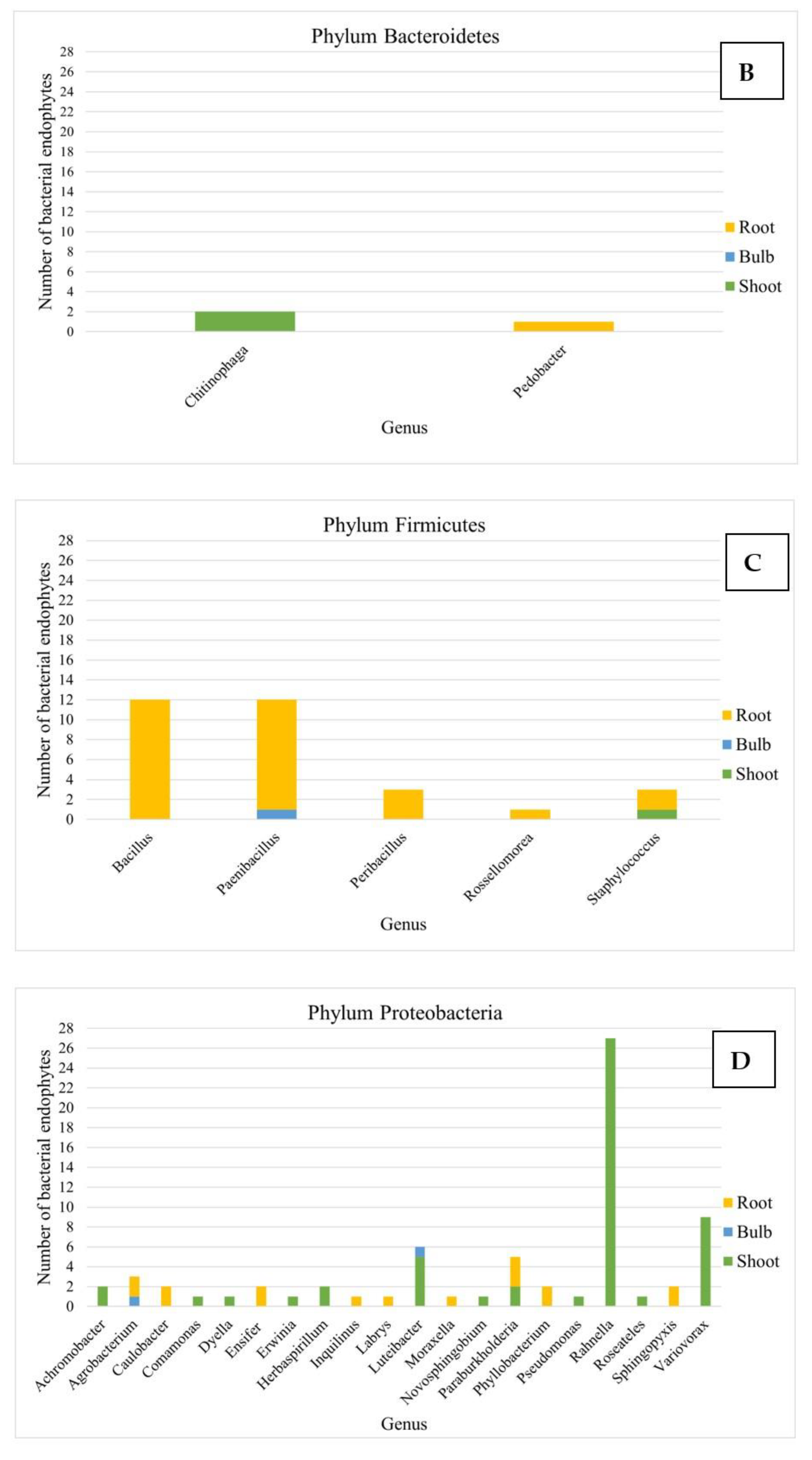
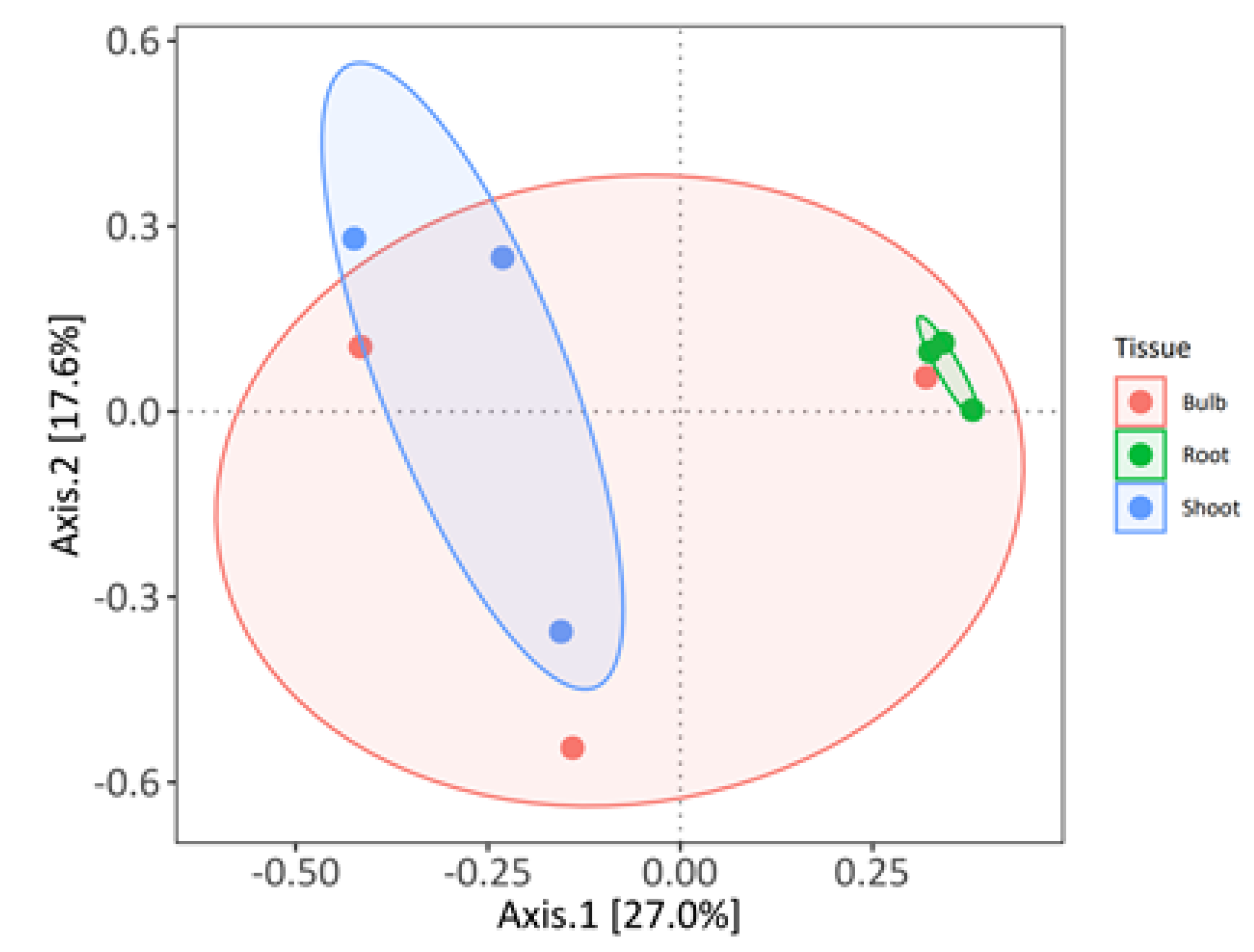
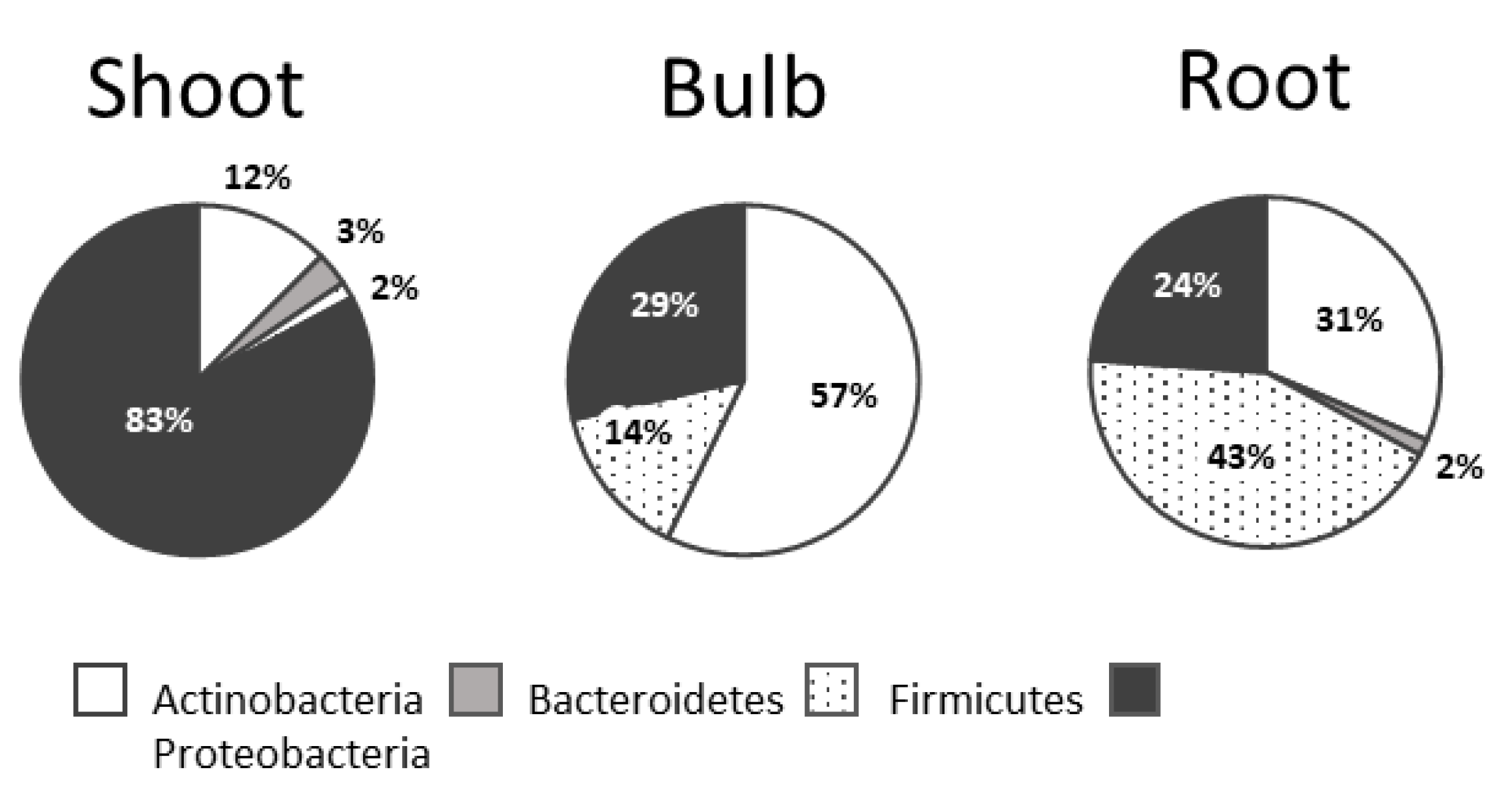
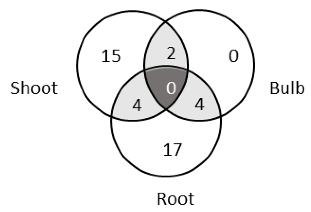
3.2. Analysis of the Composition of Cultivable Endophytic Bacteria from L. aestivum ‘Gravety Giant’

A total of 138 bacteria were successfully isolated and purified (Figure 2, Table S1). This bacterial collection consisted of 67 strains isolated from roots, 64 from the shoot material and only 7 from bulbs.
The strains were taxonomically characterized after partial 16S rDNA sequencing. A total of 42 genera belonging to four phyla were identified. To assess the relative abundance of the compositions of the isolates from the three tissues at the genus level, we conducted a principal coordinate analysis (PCoA) using Bray–Curtis distance (Figure 3).
The first two axes explained 34.7% of the variation. The analysis showed that the bacterial communities from roots and shoots were well separated along the first axis, whereas the bacterial community from bulbs was not discriminated from the two other communities. This observation was validated by permutation ANOVA (permutations = 10,000), suggesting that the culturable bacterial communities from L. aestivum ‘Gravety giant’ are structured by plant organs (p-value = 0.0351).
Proteobacteria were dominant in shoots (83%), whereas Firmicutes were mainly detected in roots (43%) (Figure 4).
At the genus level, 21 genera, 25 genera and 6 genera were found in the shoots, the roots and the bulbs, respectively. However, no genus was common among all three plant tissues (Figure 5).
The isolates from the bulbs were affiliated to six genera, which were shared with either shoot (two genera) or root (four genera) parts, and no genera were unique in bulbs. In shoots, the most abundant genera were Rahnella, Variovorax and Luteibacter, while in roots, Bacillus, Paenibacillus and Steptomyces were the dominant genera (Figure 2).

3.3. Alkaloid Screening Using High-Performance Thin-Layer Chromatography (HPTLC) Analysis

Endophytic bacteria extracts were investigated for their phytochemical characteristics. For this purpose, the methanolic extracts obtained from bacterial pellets were screened for their potential to produce alkaloid using Dragendorff’s reagent after the development of HPTLC plates (Figure S2). Over time after the Dragendorff spraying, some orange signals were observed. Among 69 tested strains, around 14 isolates showed some clear coloring reaction for Dragendorff’s reagent (Table 1), but none of these molecules had the same Rf value as the standard compounds of lycorine and galantamine, which are known Amaryllidaceae alkaloids produced by L. aestivum.

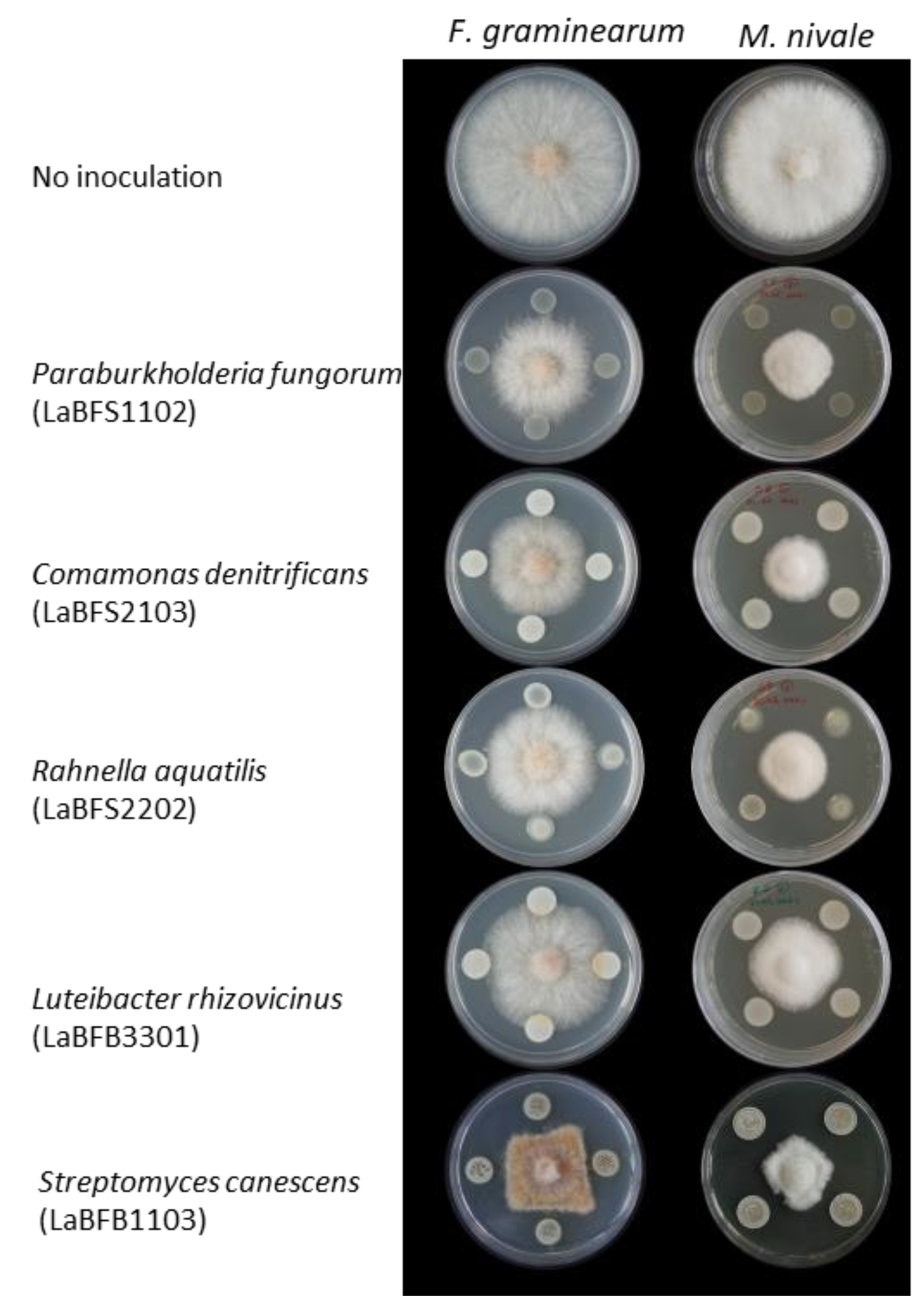
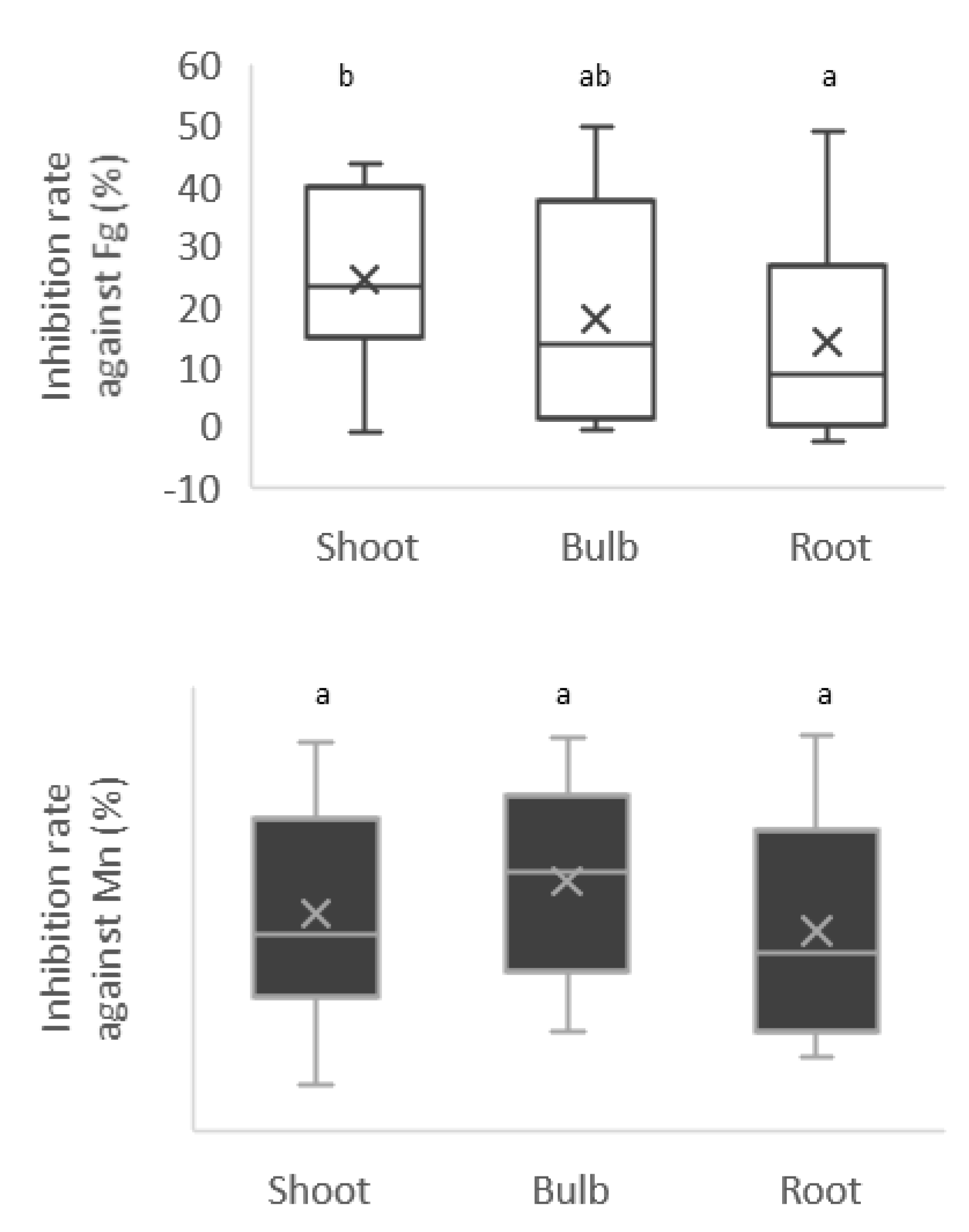
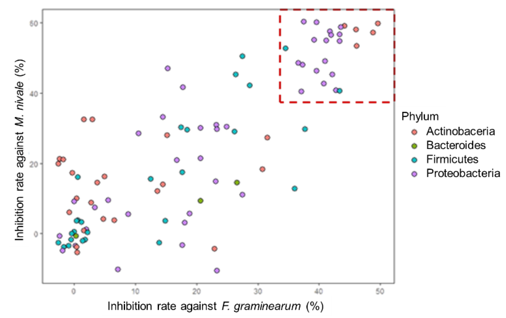
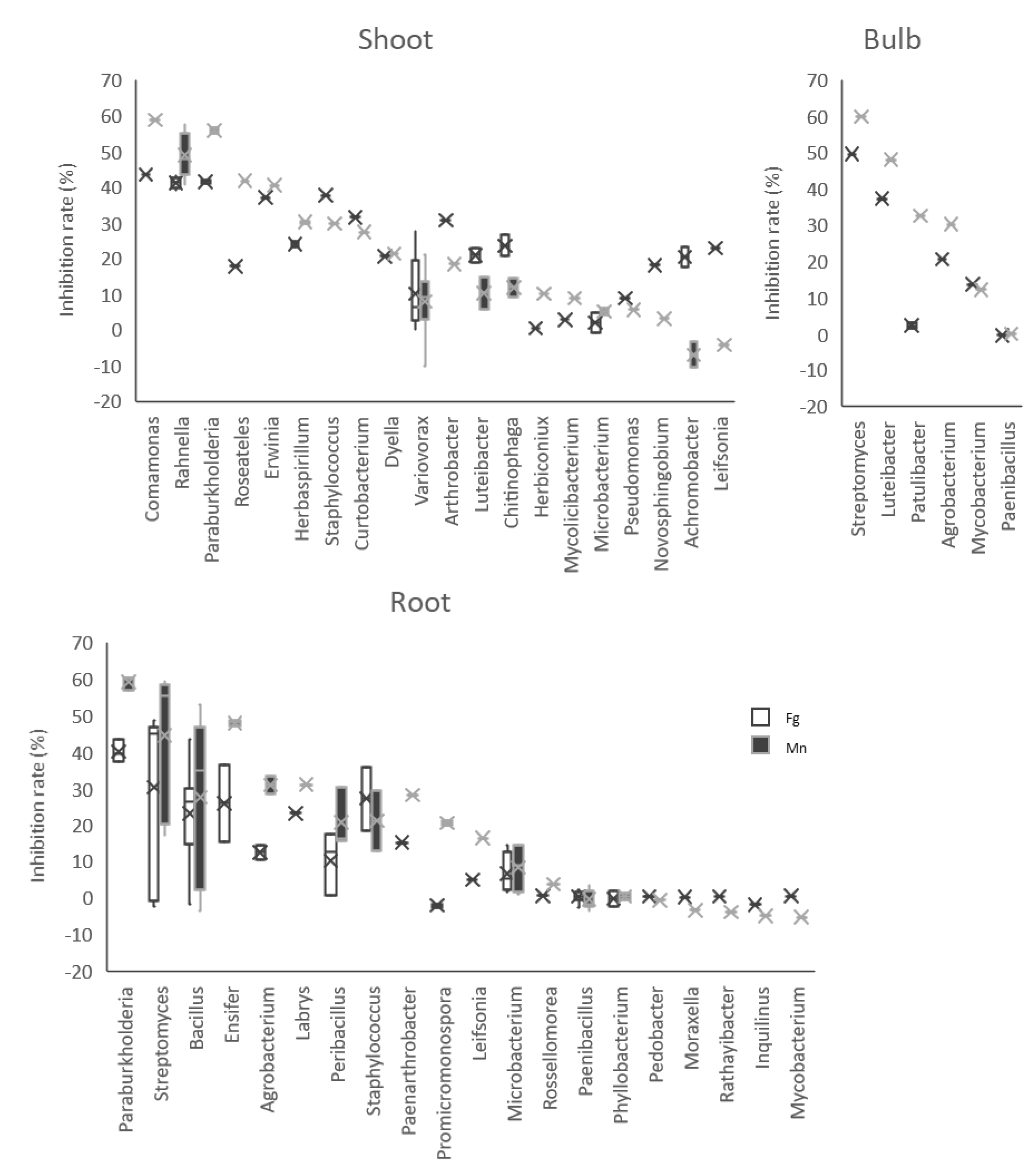
3.4. Antifungal Activities of the Isolates

To evaluate the capabilities of bacterial isolates which reacted with Dragendorff’s reagent to inhibit F. graminearum and M. nivale mycelial growth, in vitro dual-culture tests were performed (Table 1, Figure 6). For strains affiliate to the same genus, such as Rahnella, we chose to test only a few strains from the same taxon (Table S1). Moreover, the isolates which required more than three days for sufficient growth in the liquid culture were removed from the dual-culture test.
The antagonistic activities of the isolates against F. graminearum varied from 0% to 49.6% and differed between the bacterial group from shoot and root (Tukey–Kramer method, p = 0.00817) (Figure 7).
On the other hand, those against M. nivale had a variation from 10.6% to 60.4%, but no significant differences were related to the source of plant tissues. Among them, 24 isolates, indicated in a red square in Figure 8, showed more than 30% inhibition against F. graminearum and more than 40% against M. nivale. This group contained 16 Proteobacteria strains (5 Paraburkholderia, 1 Comamonas, 8 Rahnella, 1 Ensifer, 1 Luteibacter, 1 Erwinia), 5 Actinobacteria strains (Streptomyces) and 2 Firmicutes strains (Bacillus).
The bacterial genera with the highest antifungal activities were different depending on the origin of plant tissues. In the shoot group, three Proteobacteria genera, namely Comamonas, Rahnella and Paraburkholderia, had a higher inhibition rate than the others, while it was Streptomyces in the bulb. Paraburkholderia, Streptomyces and Bacillus had better antifungal activities in the root group (Figure 9). Interestingly, Paraburkholderia, which appeared in the shoot and root parts, showed higher antagonisms with less variation among the isolates.

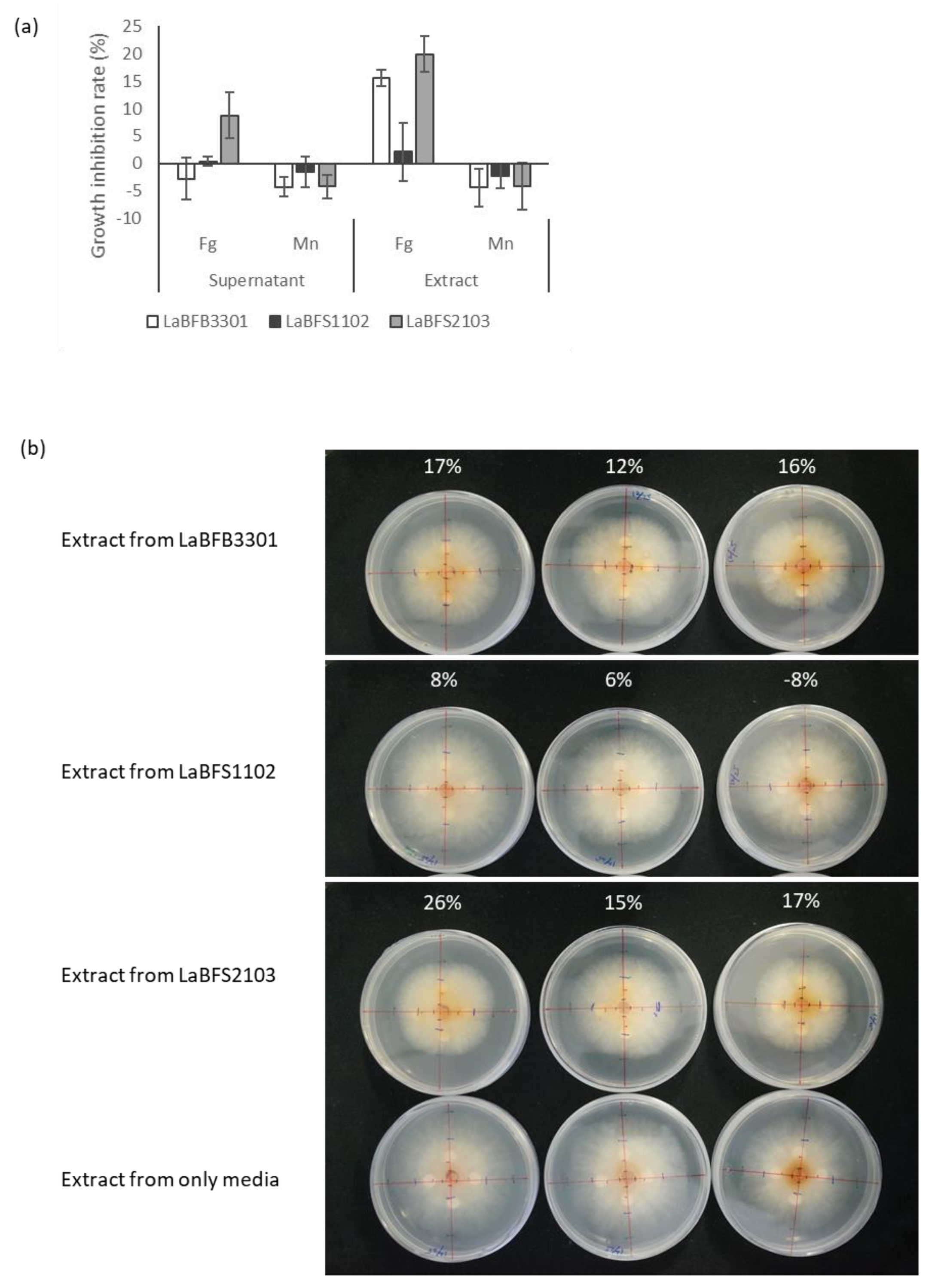
3.5. Antagonism Activities of Cell-Free Supernatant and Methanolic Extracts against F. graminearum

Among all the candidates, we selected three isolates, LaBFB3301 (N°2 Luteibacter rhizovicinus), LaBFS1102 (N°3 Paraburkholderia fungorum) and LaBFS2103 (N°16 Comamonas denitrificans), as our model strains. They rapidly grew in nutrient broth liquid culture, and their extracts reacted with Dragendorff’s reagent (Figure S2). The results obtained in diffusion assays of the cell-free supernatant of the culture broth or the methanolic extracts showed that the methanolic extracts from LaBFB3301 N°2 Luteibacter rhizovicinus and LaFS2103 N°16 Comamonas denitrificans delayed the growth of F. graminearum colonies on potato dextrose agar (PDA), with mean inhibition rates of 15.5% and 19.9%, respectively (Figure 10a,b). All the samples from LaBFS1102 N°3 Paraburkholderia fungorum had no or slight effects on the growth of the fungus (Figure 10a).

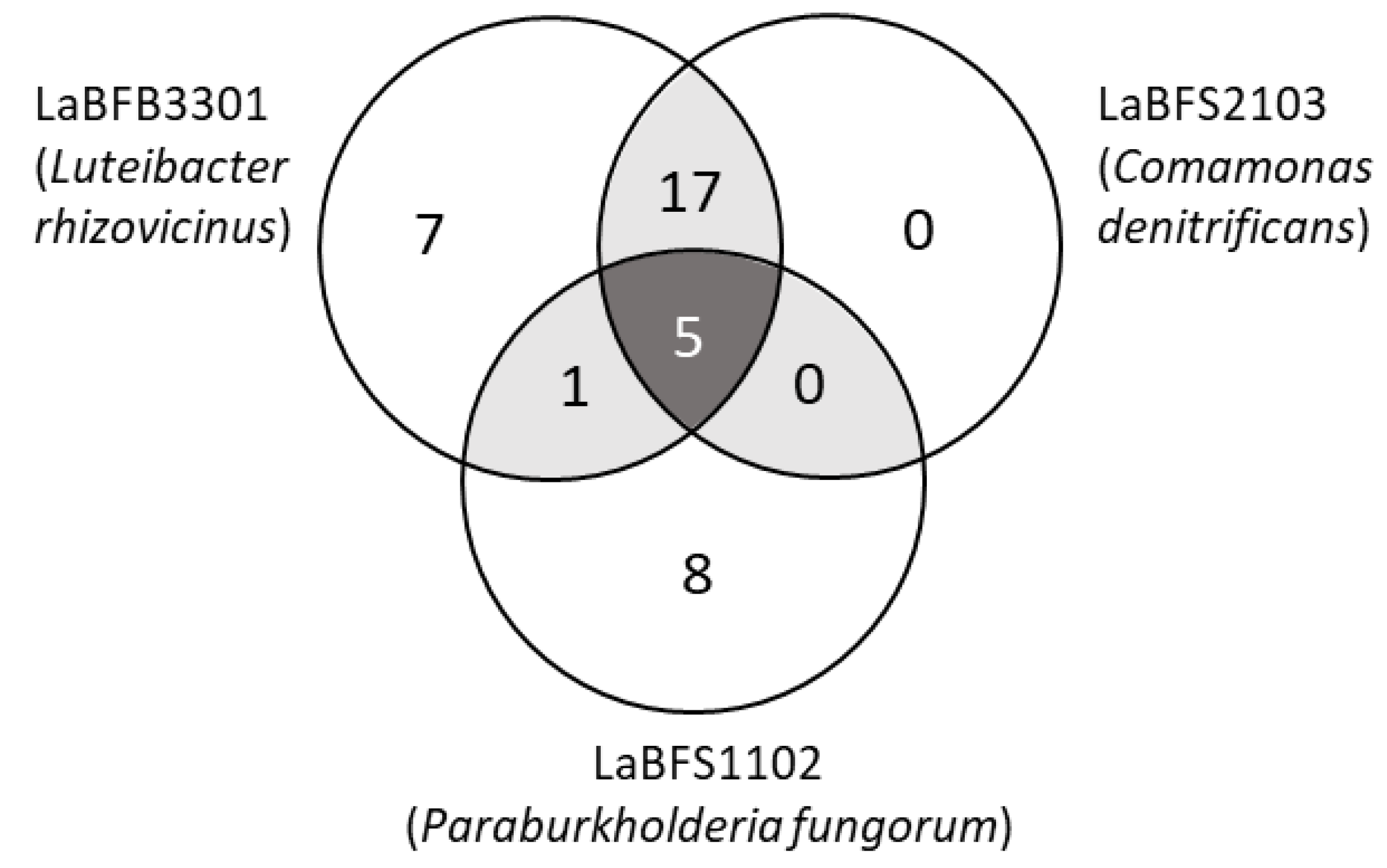
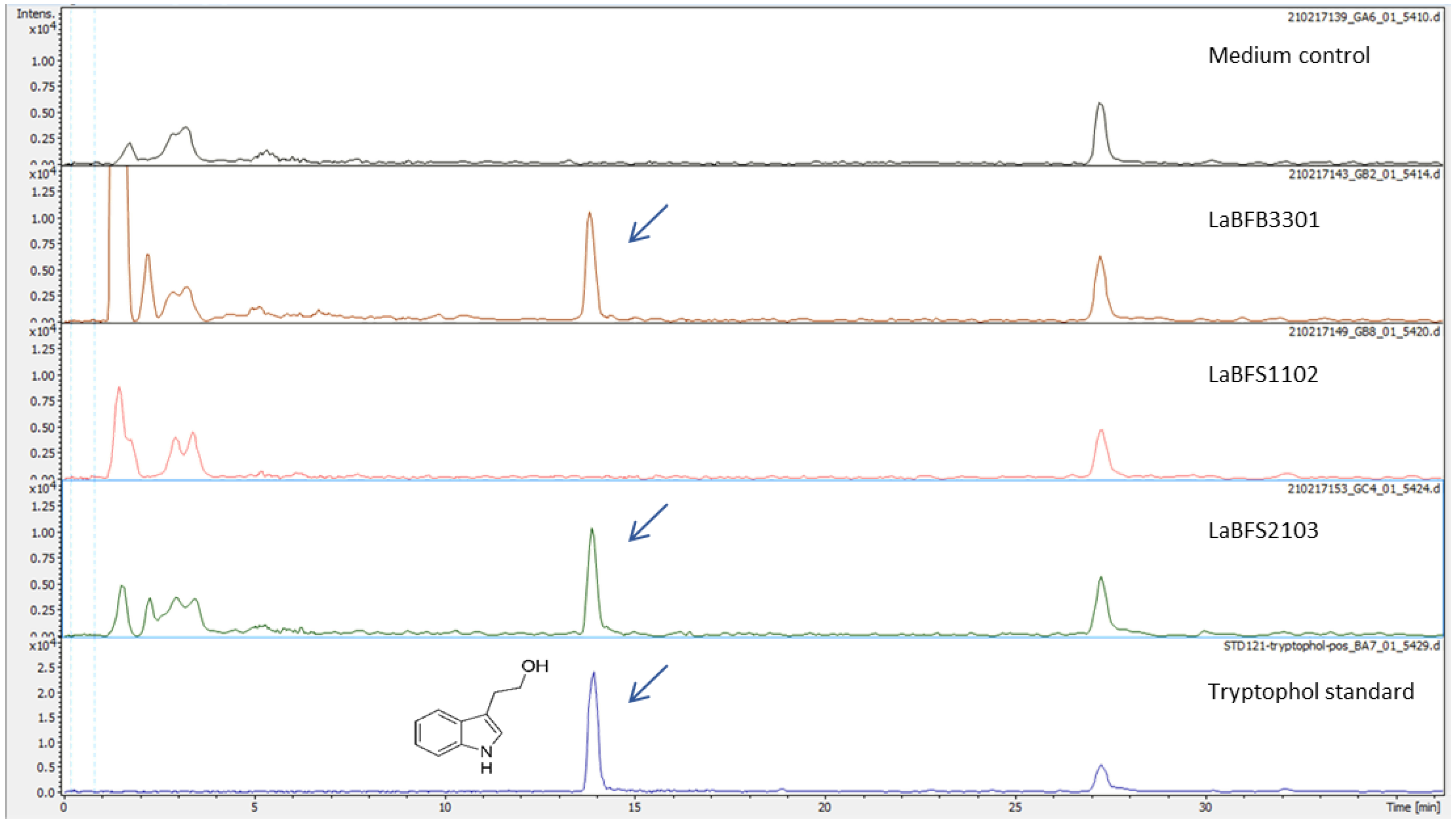
3.6. Untargeted LC-MS/MS Analysis

The extracts were analyzed for the identification of molecules using untargeted LC-MS/MS analysis. Liquid chromatography coupled with high-resolution mass spectrometry (LC-MS) analysis was performed on methanolic extracts of the three endophytic bacteria strains displaying signals on HPTLC for Dragendorff’s reagent (LaBFS1102, LaBFB3301, LaBFS2103) using the same analytical conditions as previously published [26]. LC-MS analysis performed in positive ion mode confirmed the absence of lycorine and galantamine in the methanolic extracts, as previously observed with the HPTLC analysis (Figure S3).
LC-MS profiling generated an extensive mass list corresponding to a wide variety of metabolites in the endophytic extracts from L. aestivum ‘Gravety giant’ bulb (LaBFB3301) and from L. aestivum ‘Gravety giant’ shoot (LaBFS1102, LaBFS2103), using HPLC-Q-Exactive-Orbitrap®. Using the Compound Discoverer software led to the identification of 38 peaks enriched in the methanolic extracts of the bacterial pellets (Figure 11, Table 2), thanks to MS/MS spectra comparison with online databases such as Mzcloud.
Seven, eight and zero compounds were specific to LaBFS1102, LaBFB3301 and LaBFS2103, respectively. Additionally, 17 compounds were shared commonly between LaBFB3301 and LaBFS2103, and only one compound (6,7-dihydro-5H-dibenzo diazepin-6-one) was shared between LaBFB3301 and LaBFS1102. Five molecules (adenosine 5′-monophosphate, palmitoyl ethanolamide, oleoyl ethanolamide, nicotinamide adenine dinucleotide, azobenzene) were identified in the three endophytic extracts.
In the 17 shared compounds between LaBFB3301 and LaBFS2103, we identified the indolic alkaloid tryptophol or l,3-(2-hydroxyethyl) indole, both by MS analysis (C10H11NO, [M + H]+ 162.0913) and by retention time (13.9 min) (Figure 12).
This result was confirmed using the commercial standard tryptophol. The same peaks and fragments were highlighted in all the triplicates of LaBFB3301 and LaBFS2103 samples (Figure S4). Tryptophol is a precursor of various indole alkaloids in plants and microorganisms [29]. The origin of tryptophol is closely related to the tryptophan metabolic pathway. However, the amino acid tryptophan (C11H12N2O2, [M + H]+ 205.0972) was not identified in the three endophytic bacteria strains. The peak of tryptophol was observed at 13.8–14.0 min with m/z 162.0913 [M + H]+, which had MS2 fragments with m/z 143, 130 and 115. Moreover, indole-3-acetic acid (IAA), which shares precursors of tryptophol and takes part in plant growth promotion effect, was not found in any samples, even compared with the standard compound.

4. Discussion

This research was conducted in order to investigate the diversity of endophytic culturable bacteria collected from three organs of L. aestivum ‘Gravity giant’ that were not cultured in sterile conditions. Different strains of endophytic bacteria were selected for the evaluation of their capabilities for producing alkaloids and displaying antagonistic effects against wheat fungal pathogens.
Endophytic bacteria and fungi have been isolated from many plant species, but only a few Amaryllidaceae plants have been studied thus far. The plants that have been investigated are Narcissus tazetta [25], Crinum macowanii [30,31,32] and Lycoris radiata [33]. The latest example was reported by Spina et al. [26], with in vitro bulblets of L. aestivum. In our study, the bulbs harbor a low number of cultivable bacterial strains, which is consistent with the observation of the few endophytic bacteria isolated from in vitro bulblets of L. aestivum [26]. This result could be associated with the accumulation of alkaloids such as galantamine, which is highest in the bulbs of Leucojum plants [34]. Moreover, it is known that in plant multiplication organs, bacterial loads are generally lower than in other parts of the plant. In contrast, the roots were found to contain the highest number of bacterial endophytes, probably explained by the contact of the roots with a large reservoir of bacteria in the soil [6,8,35].
The data collected in our study show that the dominant bacterial phyla in L. aestivum ‘Gravety giant’ among culturable endophytes are Proteobacteria (51.4%), Actinobacteria (23.9%), Firmicutes (22.5%) and Bacteroidetes (2.2%). A similar distribution was reported for Lycoris radiata [33] or Zea mays [36]. When focusing on the taxonomical distribution of endophytic bacteria throughout plant tissues, no genus was shared among the three different tissues we analyzed in Leucojum aestivum. This seems to be common, at least with Lycoris radiata, for which the analysis was performed among five different organs (flower, scape, leaf, bulb and root) [33]. This is an important point for further studies that aim to identify bacterial strains with some specific properties, and a relationship between plant metabolomic profiles and bacterial diversity might be established.
Amaryllidaceae plants seem to share a similar pattern of endophytic bacterial diversity. A survey carried out in seven different studies (including our study) showed that the most frequently isolated genera were Bacillus (5/7), followed by Acinetobacter, Novosphingobium and Pseudomonas (3/7) (Table S2). When going deeper in detail and when focusing on different plant tissues, we could highlight similarities but also some interesting differences. The leaf endophytic bacteria, Arthrobacter, Luteibacter and Pseudomonas, were common to other Amaryllidaceae plant leaf tissues [32,33]. Concerning roots, Bacillus and Paenibacillus genera were predominant in Leucojum aestivum as well as in Lycoris radiata [33]. The most interesting point is that Bacillus, mostly isolated from bulbs in previous studies, could not be highlighted in our Leucojum aestivum ‘Gravety giant’ bulbs. For example, rice seeds harbor mainly Bacillus during maturation [37], which was assumed because of the hardness and dormancy of the organ. In other studies, seed endophytes could colonize the next-generation seedlings as well as their rhizosphere [38]. In this study, we used the bulbs under germination, which can be considered as after the break of dormancy. The developmental stage of bulbs we used could be related to the absence of Bacillus endophytes.
It is known that endophytic microorganisms are able to synthesize a variety of specialized metabolites with application in agriculture and pharmaceutical industries [39]. Endophytic bacteria belonging to Bacillus and Streptomyces genera are well known for their metabolic capability to produce various bioactive compounds as well as their function of plant protection [40]. An endophytic strain of Paenibacillus amylolyticus was reported to produce dipicolinic acid with antibacterial activity [41] and Paraburkholderia fungorum was reported as an endophyte of rice, and it showed plant growth promotion in strawberry [42,43]. Chitinophaga ginsengisegetis was reported as a root endophyte in grapevine [44]. Rahnella aquatilis was also isolated from Crinum macowanii Baker [31], but it was suggested as a pathogen in onion bulb [45]. Luteibacter rhizovicinus was found in apple shoots, and displays plant growth promotion properties in barley [46]. The genus Variovorax is known for its metabolic diversity and for its plant growth-promoting function [47]. Staphylococcus is a well-known genus since some of its species are human pathogens, but this genus is also isolated as endophytes. Rebotiloe et al. [30] isolated bacterial endophytes from Crinum macowanii Baker, and three isolates among the total of five isolates were Staphylococcus species. They also observed moderate antibacterial activities from those strains.
In this work, the results showed some isolates displaying antagonism against wheat fungal pathogens. Interestingly, all the isolates belonging to Paraburkholderia fungorum that were isolated from the shoot and root tissues showed better and less various mycelial growth inhibition. In the genus Paraburkholderia, there is a beneficial endophytic strain named P. phytofirmans PsJN. This strain has the capability to promote plant growth and mitigate biotic/abiotic stress in a wide range of host plants by producing various compounds [48]. However, because the methanolic extracts and filtrate of the culture broth of this strain did not show antagonism for the same pathogens, it might be possible that the amounts of bioactive compounds were too low in those samples or that their production relies on induction by the presence of target microorganisms.
The chemical analysis of the methanolic extract of endophytic bacteria strains of shoot and bulbs showed no detection of Amaryllidaceae alkaloids such as galanthamine or lycorine. However, interestingly, two isolates of the endophytic bacteria, Luteibacter rhizovicinus and Commamonas denitrificans, contained the indolic alkaloid tryptophol. These two strains displayed an inhibitory effect against the growth of F. graminearum colonies, while Paraburkholderia fungorum, which did not synthesize tryptophol, showed no antagonism activity against this wheat fungal pathogen. This molecule is a precursor of different indole alkaloids in plants and microorganisms [29]. However, indole-3-acetic acid, which shared precursors of tryptophol and took part in the plant growth promotion effect, was not found in the three samples, even compared with the standard compound. On the other hand, IAA was produced by endophytic Luteibacter rhizobicinus, and the strain did not have harmful effects on plant growth [46]. Tryptophol itself has been found in plants and microorganisms and was also discovered as one of the quorum-sensing molecules in fungi. It might be possible that it is involved in the induction of cell apoptosis in specific fungi [49]. Tryptophol also has a growth promotion effect in roots and leaves of plants [29]. It is of interest to confirm if this compound relates to the observed antifungal activity and also to search its derivatives in methanolic extracts. Diketopiperazines having antimicrobial activities were detected in Comamonas testosteroni, isolated from nematodes [50].

5. Conclusions

This study showed for the first time that bacterial endophytic communities are different in bulbs, roots and shoots of L. aestivum ‘Gravety giant’. The indolic alkaloid tryptophol, detected only in the strains of Luteibacter rhizovivinus (LaBFB3301) and Comamonas denitrificans (LaBFS2103), showing an inhibitory effect against the growth of F. graminearum colonies, could be a potential agent to preserve plants against bioaggressors. In addition, it might be interesting to characterize the plant growth promotion effect of LaBFB3301 and LaBFS2103 strains in future research.

Supplementary Materials

The following supporting information can be downloaded at: https://www.mdpi.com/article/10.3390/microorganisms10102089/s1, Table S1: Bacterial isolates of Leucojum aestivum ‘Gravety giant’ endophytes. Table S2: Bacterial genera found as endophytes in the studies using Amaryllidaceae plants. Figure S1: (a) Sampled plants after washing with running tap water; (b) a segment of the bulb of plant 2. Figure S2: The plates of HPTLC of the 69 endophyte extracts after Dragendorff’s reagent spraying. Figure S3: Ion chromatograms of m/z 288.1266 ± 0.01 for lycorine and m/z 288.1564 ± 0.01 for galanthamine. Figure S4: Identification of tryptophol in the extract of LaBFB3301 and LaBFS2103.

Author Contributions

Conceptualization, Y.M., R.S., A.H., S.S.-D. and D.L.-M.; formal analysis, Y.M. and J.G.; investigation, Y.M. and R.S.; data curation, Y.M., R.S., A.H., S.S.-D. and D.L.-M.; writing—original draft preparation, Y.M., S.S.-D. and D.L.-M.; writing—review and editing, Y.M., R.S., A.H., S.S.-D. and D.L.-M.; funding acquisition, R.S., A.H., S.S.-D. and D.L.-M. All authors have read and agreed to the published version of the manuscript.

Funding

The study was funded by the ‘Impact Biomolecules’ project of the Lorraine Université d’Excellence (Investissements d’avenir—ANR-15-IDEX-04-LUE).

Acknowledgments

The authors acknowledge: the support of ‘Laboratoire Agronomie et Environnement, LAE UMR 1121′ and ‘Laboratoire Lorrain de Chimie Moléculaire_L2CM UMR 7053′ by the ‘Impact Biomolecules’ project of the Lorraine Université d’Excellence (Investissements d’avenir–ANR-15-IDEX-04-LUE). The authors acknowledge François Dupire and Romain Larbat for their assistance in LC/MS analysis. The authors are grateful to MassLor Plateform of L2CM and ‘Plateau d’Analyse Structurale et Métabolomique’, both at Lorraine University.

Conflicts of Interest

The authors declare no conflict of interest.

References

  1. Strobel, G.A. Endophytes as Sources of Bioactive Products. Microbes Infect. 2003, 5, 535–544. [Google Scholar] [CrossRef]
  2. Durán, P.; Thiergart, T.; Garrido-Oter, R.; Agler, M.; Kemen, E.; Schulze-Lefert, P.; Hacquard, S. Microbial Interkingdom Interactions in Roots Promote Arabidopsis Survival. Cell 2018, 175, 973–983.e14. [Google Scholar] [CrossRef] [PubMed]
  3. Brader, G.; Compant, S.; Mitter, B.; Trognitz, F.; Sessitsch, A. Metabolic Potential of Endophytic Bacteria. Curr. Opin. Biotechnol. 2014, 27, 30–37. [Google Scholar] [CrossRef] [PubMed] [Green Version]
  4. Rosenblueth, M.; Martínez-Romero, E. Bacterial Endophytes and Their Interactions with Hosts. Mol. Plant-Microbe Interact. 2006, 19, 827–837. [Google Scholar] [CrossRef] [PubMed] [Green Version]
  5. Bulgarelli, D.; Rott, M.; Schlaeppi, K.; van Themaat, E.V.L.; Ahmadinejad, N.; Assenza, F.; Rauf, P.; Huettel, B.; Reinhardt, R.; Schmelzer, E.; et al. Revealing Structure and Assembly Cues for Arabidopsis Root-Inhabiting Bacterial Microbiota. Nature 2012, 488, 91–95. [Google Scholar] [CrossRef] [PubMed]
  6. Lundberg, D.S.; Lebeis, S.L.; Paredes, S.H.; Yourstone, S.; Gehring, J.; Malfatti, S.; Tremblay, J.; Engelbrektson, A.; Kunin, V.; del Rio, T.G.; et al. Defining the Core Arabidopsis Thaliana Root Microbiome. Nature 2012, 488, 86–90. [Google Scholar] [CrossRef] [PubMed] [Green Version]
  7. Sturz, A.V.; Christie, B.R.; Nowak, J. Bacterial Endophytes: Potential Role in Developing Sustainable Systems of Crop Production. Crit. Rev. Plant Sci. 2000, 19, 1–30. [Google Scholar] [CrossRef]
  8. Bulgarelli, D.; Schlaeppi, K.; Spaepen, S.; van Themaat, E.V.L.; Schulze-Lefert, P. Structure and Functions of the Bacterial Microbiota of Plants. Annu. Rev. Plant Biol. 2013, 64, 807–838. [Google Scholar] [CrossRef] [Green Version]
  9. Sturz, A.V.; Christie, B.R.; Matheson, B.G.; Nowak, J. Biodiversity of Endophytic Bacteria Which Colonize Red Clover Nodules, Roots, Stems and Foliage and Their Influence on Host Growth. Biol. Fertil. Soils 1997, 25, 13–19. [Google Scholar] [CrossRef]
  10. Igarashi, Y. Screening of Novel Bioactive Compounds from Plant-Associated Actinomycetes. Actinomycetologica 2004, 18, 63–66. [Google Scholar] [CrossRef]
  11. Castillo, U.; Harper, J.K.; Strobel, G.A.; Sears, J.; Alesi, K.; Ford, E.; Lin, J.; Hunter, M.; Maranta, M.; Ge, H.; et al. Kakadumycins, Novel Antibiotics from Streptomyces Sp NRRL 30566, an Endophyte of Grevillea Pteridifolia. FEMS Microbiol. Lett. 2003, 224, 183–190. [Google Scholar] [CrossRef] [Green Version]
  12. Khiralla, A.; Spina, R.; Saliba, S.; Laurain-Mattar, D. Diversity of Natural Products of the Genera Curvularia and Bipolaris. Fungal Biol. Rev. 2019, 33, 101–122. [Google Scholar] [CrossRef]
  13. Jia, M.; Chen, L.; Xin, H.-L.; Zheng, C.-J.; Rahman, K.; Han, T.; Qin, L.-P. A Friendly Relationship between Endophytic Fungi and Medicinal Plants: A Systematic Review. Front. Microbiol. 2016, 7, 906. [Google Scholar] [CrossRef] [PubMed] [Green Version]
  14. Soujanya, K.N.; Siva, R.; Mohana Kumara, P.; Srimany, A.; Ravikanth, G.; Mulani, F.A.; Aarthy, T.; Thulasiram, H.V.; Santhoshkumar, T.R.; Nataraja, K.N.; et al. Camptothecin-Producing Endophytic Bacteria from Pyrenacantha Volubilis Hook. (Icacinaceae): A Possible Role of a Plasmid in the Production of Camptothecin. Phytomedicine 2017, 36, 160–167. [Google Scholar] [CrossRef]
  15. Alvin, A.; Miller, K.I.; Neilan, B.A. Exploring the Potential of Endophytes from Medicinal Plants as Sources of Antimycobacterial Compounds. Microbiol. Res. 2014, 169, 483–495. [Google Scholar] [CrossRef]
  16. Wu, W.; Chen, W.; Liu, S.; Wu, J.; Zhu, Y.; Qin, L.; Zhu, B. Beneficial Relationships Between Endophytic Bacteria and Medicinal Plants. Front. Plant Sci. 2021, 12, 646146. [Google Scholar] [CrossRef]
  17. Inahashi, Y.; Iwatsuki, M.; Ishiyama, A.; Matsumoto, A.; Hirose, T.; Oshita, J.; Sunazuka, T.; Panbangred, W.; Takahashi, Y.; Kaiser, M.; et al. Actinoallolides A-E, New Anti-Trypanosomal Macrolides, Produced by an Endophytic Actinomycete, Actinoallomurus Fulvus MK10-036. Org. Lett. 2015, 17, 864–867. [Google Scholar] [CrossRef]
  18. Miller, C.M.; Miller, R.V.; Garton-Kenny, D.; Redgrave, B.; Sears, J.; Condron, M.M.; Teplow, D.B.; Strobel, G.A. Ecomycins, Unique Antimycotics from Pseudomonas Viridiflava. J. Appl. Microbiol. 1998, 84, 937–944. [Google Scholar] [CrossRef] [Green Version]
  19. Kusari, S.; Lamshöft, M.; Kusari, P.; Gottfried, S.; Zühlke, S.; Louven, K.; Hentschel, U.; Kayser, O.; Spiteller, M. Endophytes Are Hidden Producers of Maytansine in Putterlickia Roots. J. Nat. Prod. 2014, 77, 2577–2584. [Google Scholar] [CrossRef] [Green Version]
  20. Shweta, S.; Bindu, J.H.; Raghu, J.; Suma, H.K.; Manjunatha, B.L.; Kumara, P.M.; Ravikanth, G.; Nataraja, K.N.; Ganeshaiah, K.N.; Uma Shaanker, R. Isolation of Endophytic Bacteria Producing the Anti-Cancer Alkaloid Camptothecine from Miquelia Dentata Bedd. (Icacinaceae). Phytomedicine 2013, 20, 913–917. [Google Scholar] [CrossRef]
  21. Chung, B.-S.; Aslam, Z.; Kim, S.-W.; Kim, G.-G.; Kang, H.-S.; Ahn, J.-W.; Chung, Y. A Bacterial Endophyte, Pseudomonas Brassicacearum YC5480, Isolated from the Root of Artemisia Sp. Producing Antifungal and Phytotoxic Compounds. Plant Pathol. J. 2008, 24, 461–468. [Google Scholar] [CrossRef]
  22. Agisha, V.N.; Kumar, A.; Eapen, S.J.; Sheoran, N.; Suseelabhai, R. Broad-Spectrum Antimicrobial Activity of Volatile Organic Compounds from Endophytic Pseudomonas Putida BP25 against Diverse Plant Pathogens. Biocontrol Sci. Technol. 2019, 29, 1069–1089. [Google Scholar] [CrossRef]
  23. Sheoran, N.; Valiya Nadakkakath, A.; Munjal, V.; Kundu, A.; Subaharan, K.; Venugopal, V.; Rajamma, S.; Eapen, S.J.; Kumar, A. Genetic Analysis of Plant Endophytic Pseudomonas Putida BP25 and Chemo-Profiling of Its Antimicrobial Volatile Organic Compounds. Microbiol. Res. 2015, 173, 66–78. [Google Scholar] [CrossRef]
  24. Jin, Z.; Yao, G. Amaryllidaceae and Sceletium Alkaloids. Nat. Prod. Rep. 2019, 36, 1462–1488. [Google Scholar] [CrossRef]
  25. Wang, W.-X.; Kusari, S.; Sezgin, S.; Lamshöft, M.; Kusari, P.; Kayser, O.; Spiteller, M. Hexacyclopeptides Secreted by an Endophytic Fungus Fusarium Solani N06 Act as Crosstalk Molecules in Narcissus Tazetta. Appl. Microbiol. Biotechnol. 2015, 99, 7651–7662. [Google Scholar] [CrossRef]
  26. Spina, R.; Saliba, S.; Dupire, F.; Ptak, A.; Hehn, A.; Piutti, S.; Poinsignon, S.; Leclerc, S.; Bouguet-Bonnet, S.; Laurain-Mattar, D. Molecular Identification of Endophytic Bacteria in Leucojum aestivum In Vitro Culture, NMR-Based Metabolomics Study and LC-MS Analysis Leading to Potential Amaryllidaceae Alkaloid Production. Int. J. Mol. Sci. 2021, 22, 1773. [Google Scholar] [CrossRef]
  27. Berkov, S.; Georgieva, L.; Kondakova, V.; Atanassov, A.; Viladomat, F.; Bastida, J.; Codina, C. Plant Sources of Galanthamine: Phytochemical and Biotechnological Aspects. Biotechnol. Biotechnol. Equip. 2009, 23, 1170–1176. [Google Scholar] [CrossRef]
  28. Munakata, Y.; Gavira, C.; Genestier, J.; Bourgaud, F.; Hehn, A.; Slezack-Deschaumes, S. Composition and Functional Comparison of Vetiver Root Endophytic Microbiota Originating from Different Geographic Locations That Show Antagonistic Activity towards Fusarium Graminearum. Microbiol. Res. 2021, 243, 126650. [Google Scholar] [CrossRef]
  29. Palmieri, A.; Petrini, M. Tryptophol and Derivatives: Natural Occurrence and Applications to the Synthesis of Bioactive Compounds. Nat. Prod. Rep. 2019, 36, 490–530. [Google Scholar] [CrossRef] [PubMed]
  30. Rebotiloe, F.M.; Eunice, U.-J.; Mahloro, H.S.-D. Isolation and Identification of Bacterial Endophytes from Crinum Macowanii Baker. Afr. J. Biotechnol. 2018, 17, 1040–1047. [Google Scholar] [CrossRef]
  31. Sebola, T.E.; Uche-Okereafor, N.C.; Tapfuma, K.I.; Mekuto, L.; Green, E.; Mavumengwana, V. Evaluating Antibacterial and Anticancer Activity of Crude Extracts of Bacterial Endophytes from Crinum Macowanii Baker Bulbs. Microbiologyopen 2019, 8, e914. [Google Scholar] [CrossRef] [PubMed] [Green Version]
  32. Sebola, T.E.; Uche-Okereafor, N.C.; Mekuto, L.; Makatini, M.M.; Green, E.; Mavumengwana, V. Antibacterial and Anticancer Activity and Untargeted Secondary Metabolite Profiling of Crude Bacterial Endophyte Extracts from Crinum Macowanii Baker Leaves. Int. J. Microbiol. 2020, 2020, 8839490. [Google Scholar] [CrossRef] [PubMed]
  33. Liu, Z.; Zhou, J.; Li, Y.; Wen, J.; Wang, R. Bacterial Endophytes from Lycoris Radiata Promote the Accumulation of Amaryllidaceae Alkaloids. Microbiol. Res. 2020, 239, 126501. [Google Scholar] [CrossRef]
  34. Diop, M.F.; Hehn, A.; Ptak, A.; Chrétien, F.; Doerper, S.; Gontier, E.; Bourgaud, F.; Henry, M.; Chapleur, Y.; Laurain-Mattar, D. Hairy Root and Tissue Cultures of Leucojum aestivum L.—Relationships to Galanthamine Content. Phytochem. Rev. 2007, 6, 137–141. [Google Scholar] [CrossRef]
  35. Afzal, I.; Shinwari, Z.K.; Sikandar, S.; Shahzad, S. Plant Beneficial Endophytic Bacteria: Mechanisms, Diversity, Host Range and Genetic Determinants. Microbiol. Res. 2019, 221, 36–49. [Google Scholar] [CrossRef]
  36. Correa-Galeote, D.; Bedmar, E.J.; Arone, G.J. Maize Endophytic Bacterial Diversity as Affected by Soil Cultivation History. Front. Microbiol. 2018, 9, 484. [Google Scholar] [CrossRef] [PubMed] [Green Version]
  37. Okunishi, S.; Sako, K.; Mano, H.; Imamura, A.; Morisaki, H. Bacterial Flora of Endophytes in the Maturing Seed of Cultivated Rice (Oryza Sativa). Microbes Environ. 2005, 20, 168–177. [Google Scholar] [CrossRef] [Green Version]
  38. Guo, J.; Ling, N.; Li, Y.; Li, K.; Ning, H.; Shen, Q.; Guo, S.; Vandenkoornhuyse, P. Seed-Borne, Endospheric and Rhizospheric Core Microbiota as Predictors of Plant Functional Traits across Rice Cultivars Are Dominated by Deterministic Processes. New Phytol. 2021, 230, 2047–2060. [Google Scholar] [CrossRef]
  39. Ryan, R.P.; Germaine, K.; Franks, A.; Ryan, D.J.; Dowling, D.N. Bacterial Endophytes: Recent Developments and Applications. FEMS Microbiol. Lett. 2008, 278, 1–9. [Google Scholar] [CrossRef] [Green Version]
  40. Ek-Ramos, M.J.; Gomez-Flores, R.; Orozco-Flores, A.A.; Rodríguez-Padilla, C.; González-Ochoa, G.; Tamez-Guerra, P. Bioactive Products From Plant-Endophytic Gram-Positive Bacteria. Front. Microbiol. 2019, 10, 463. [Google Scholar] [CrossRef] [PubMed]
  41. Anandan, K.; Vittal, R.R. Endophytic Paenibacillus Amylolyticus KMCLE06 Extracted Dipicolinic Acid as Antibacterial Agent Derived via Dipicolinic Acid Synthetase Gene. Curr. Microbiol. 2019, 76, 178–186. [Google Scholar] [CrossRef]
  42. Khan, M.M.A.; Haque, E.; Paul, N.C.; Khaleque, M.A.; Al-Garni, S.M.S.; Rahman, M.; Islam, M.T. Enhancement of Growth and Grain Yield of Rice in Nutrient Deficient Soils by Rice Probiotic Bacteria. Rice Sci. 2017, 24, 264–273. [Google Scholar] [CrossRef]
  43. Rahman, M.; Sabir, A.A.; Mukta, J.A.; Khan, M.M.A.; Mohi-Ud-Din, M.; Miah, M.G.; Rahman, M.; Islam, M.T. Plant Probiotic Bacteria Bacillus and Paraburkholderia Improve Growth, Yield and Content of Antioxidants in Strawberry Fruit. Sci. Rep. 2018, 8, 2504. [Google Scholar] [CrossRef] [Green Version]
  44. Marasco, R.; Rolli, E.; Fusi, M.; Cherif, A.; Abou-Hadid, A.; El-Bahairy, U.; Borin, S.; Sorlini, C.; Daffonchio, D. Plant Growth Promotion Potential Is Equally Represented in Diverse Grapevine Root-Associated Bacterial Communities from Different Biopedoclimatic Environments. Biomed Res. Int. 2013, 2013, 491091. [Google Scholar] [CrossRef] [Green Version]
  45. Asselin, J.E.; Eikemo, H.; Perminow, J.; Nordskog, B.; Brurberg, M.B.; Beer, S.V. Rahnella Spp. Are Commonly Isolated from Onion (Allium Cepa) Bulbs and Are Weakly Pathogenic. J. Appl. Microbiol. 2019, 127, 812–824. [Google Scholar] [CrossRef]
  46. Guglielmetti, S.; Basilico, R.; Taverniti, V.; Arioli, S.; Piagnani, C.; Bernacchi, A. Luteibacter Rhizovicinus MIMR1 Promotes Root Development in Barley (Hordeum Vulgare L.) under Laboratory Conditions. World J. Microbiol. Biotechnol. 2013, 29, 2025–2032. [Google Scholar] [CrossRef] [PubMed]
  47. Sun, S.-L.; Yang, W.-L.; Fang, W.-W.; Zhao, Y.-X.; Guo, L.; Dai, Y.-J. The Plant Growth-Promoting Rhizobacterium Variovorax Boronicumulans CGMCC 4969 Regulates the Level of Indole-3-Acetic Acid Synthesized from Indole-3-Acetonitrile. Appl. Environ. Microbiol. 2018, 84, e00298-18. [Google Scholar] [CrossRef] [Green Version]
  48. Esmaeel, Q.; Miotto, L.; Rondeau, M.; Leclère, V.; Clément, C.; Jacquard, C.; Sanchez, L.; Barka, E.A. Paraburkholderia Phytofirmans PsJN-Plants Interaction: From Perception to the Induced Mechanisms. Front. Microbiol. 2018, 9, 2093. [Google Scholar] [CrossRef] [PubMed]
  49. Singkum, P.; Muangkaew, W.; Suwanmanee, S.; Pumeesat, P.; Wongsuk, T.; Luplertlop, N. Suppression of the Pathogenicity of Candida Albicans by the Quorum-Sensing Molecules Farnesol and Tryptophol. J. Gen. Appl. Microbiol. 2020, 65, 277–283. [Google Scholar] [CrossRef] [Green Version]
  50. Nishanth Kumar, S.; Mohandas, C.; Nambisan, B. Purification, Structural Elucidation and Bioactivity of Tryptophan Containing Diketopiperazines, from Comamonas Testosteroni Associated with a Rhabditid Entomopathogenic Nematode against Major Human-Pathogenic Bacteria. Peptides 2014, 53, 48–58. [Google Scholar] [CrossRef] [PubMed]
Figure 1. A used plant (plant 2) (A) and the number of colonies on TSA medium from 1 g of each fresh material after 20 days (B). CFU was tested with the Tukey–Kramer test, and the same letters (a, b) have no significant difference with p < 0.05.
Microorganisms 10 02089 g001
Figure 2. Taxonomical composition of the bacterial collection isolated from L. aestivum ‘Gravety giant’. (A) Phylum Actinobacteria, (B) Phylum Bacteroidetes, (C) Phylum Firmicutes, (D) Phylum Proteobacteria.
Microorganisms 10 02089 g002aMicroorganisms 10 02089 g002b
Figure 3. Principal coordinate analysis (PCoA) with Bray–Curtis distance of the L. aestivum ‘Gravety giant’ bacterial collections at the genus level.
Microorganisms 10 02089 g003
Figure 4. Relative abundance of each phylum in the bacterial collection of the different plant tissues.
Microorganisms 10 02089 g004
Figure 5. Venn diagram of shared and unique bacterial genera in L. aestivum ‘Gravity giant’.
Microorganisms 10 02089 g005
Figure 6. Dual-culture test of representative strains.
Microorganisms 10 02089 g006
Figure 7. Antifungal activities against F. graminearum differed between the groups of shoot and root bacterial collections, but there was no difference in the antifungal activities for M. nivale. Different letters (a, b) show significant difference with p < 0.05 by the Tukey–Kramer method.
Microorganisms 10 02089 g007
Figure 8. Correlations between antifungal activities against F. graminearum and M. nivale, colored by taxonomical affiliation at the phylum level.
Microorganisms 10 02089 g008
Figure 9. Growth inhibition rate against F. graminearum (Fg) and M. nivale (Mn) by genus of bacterial isolates in three different tissues: shoot, bulb and root.
Microorganisms 10 02089 g009
Figure 10. Growth inhibition test of cell-free supernatant and methanolic extracts of three endophytic bacteria, LaBFB3301, LaBFS1102 and LaBFS2103 against mycelial growth of F. graminearum (Fg) and M. nivale (Mn) after 3-day culture. (a) Mean growth inhibition rate with SE as error bars; (b) cultured plates of F. graminearum on day 3. The above number represents the growth inhibition rate.
Microorganisms 10 02089 g010
Figure 11. Venn diagram of shared and unique peaks of metabolites enriched and identified in the methanolic extract of the three strains LaBFB 3301, LaBFS1102 and LaBFS2103, using HPLC-Q-Exactive-Orbitrap®.
Microorganisms 10 02089 g011
Figure 12. Ion chromatogram of m/z 162.0913 ± 0.02 for tryptophol found in the methanolic extracts of LaBFB3301 and LaBFS2103.
Microorganisms 10 02089 g012
Table 1. List of strains of which extract (possibly) reacted to Dragendorff’s reagent.
Source TissuesStrainGenBank AccessionBlast Top HitE ValueInhibition Rate vs. Fg (Mean +/− SE)Inhibition Rate vs. Mn (Mean +/− SE)Dragendorff
ShootLaBFS1102OL307020Paraburkholderia fungorum
strain LMG 16225
041.91 ± 0.1956.69 ± 0.12Yes
LaBFS1106OL307021Patulibacter ginsengiterrae
strain P4–5
0--Yes
LaBFS1107OL307022Chitinophaga ginsengisegetis
strain Gsoil 040
026.57 ± 0.6714.54 ± 2.3Yes
LaBFS1112OL307023Variovorax ginsengisoli
strain Gsoil 3165
00 ± 0.29.15 ± 0.76Yes
LaBFS1201OL307024Staphylococcus warneri
strain AW 25
037.71 ± 0.5729.73 ± 3.06Yes
LaBFS1208 Mycolicibacterium setense
strain CIP 109395
1 × 10−1512.81 ± 0.748.87 ± 0.82Yes
LaBFS1307OL307027Microbacterium luteolum
strain IFO 15074
04.79 ± 0.954.17 ± 1.21Yes
LaBFS2103OL307031Comamonas denitrificans
strain 123
043.51 ± 0.0958.82 ± 1.19Yes
LaBFS2201 Microbacterium arthrosphaerae strain CC-VM-Y7 × 10−55−0.8 ± 0.066.17 ± 0.16Yes
LaBFS3105OL307062Variovorax paradoxus
strain NBRC 15149
03.4 ± 0.237.54 ± 2.97Yes
LaBFS3313OL307078Arthrobacter oryzae
strain KV-651
030.73 ± 0.9618.46 ± 3.11Not performed
BulbLaBFB1102OL307013Paenibacillus amylolyticus
strain JCM 9906
0−0.4 ± 0.120 ± 0Yes
LaBFB1103OL307014Streptomyces canescens
strain DSM 40001
049.59 ± 0.7959.85 ± 0.82Not performed
LaBFB1301OL307015Patulibacter ginsengiterrae
strain P4–5
02.99 ± 1.0432.61 ± 0.36Yes
LaBFB1302OL307016Patulibacter ginsengiterrae
strain P4–5
01.6 ± 0.3532.62 ± 0.91Yes
LaBFB3301OL307019Luteibacter rhizovicinus
strain LJ96
037.32 ± 0.248.06 ± 1.01Yes
The growth inhibition rates against F. graminearum and M. nivale are also shown in the right columns (means +/− SE).
Table 2. List of metabolites detected in the methanolic extracts of the three strains LaBFB3301, LaBFS1102 and LaBFS2103, using UHPLC-Q-Exactive-Orbitrap, combined with the identification using the software, Compound Discoverer (Thermo).
Enrichment in the Samples
(Compared with Medium Control)
Compound NameFormulaMolecular WeightError (ppm)RT (Min)
LaBFB3301 uniqueN2,1-Diphenyl-6-imino-1,6-dihydro-1,3,5-triazine-2,4-diamineC15H14N6278.12676 7.16
O-Arachidonoyl ethanolamineC22H37NO2347.282310.3433.43
L(-)-CarnitineC7H15NO3161.105210.101.64
AdenineC5H5N5135.054520.181.84
2′-O-MethyladenosineC11H15N5O4281.112520.413.11
BetulinC30H50O2442.381070.0235.83
R-Palmitoyl-(2-methyl) ethanolamideC19H39NO2313.29806 27.45
LaBFS1102 unique16-Heptadecyne-1,2,4-triolC17H32O3284.235240.3332.86
Ethyl oleateC20H38O2310.287240.1938.96
Palmitelaidic acid methyl esterC17H32O2268.2403 34.74
GuanineC5H5N5O151.049330.532.66
Ethyl palmitoleateC18H34O2282.256020.4935.39
cis-12-Octadecenoic acid methyl esterC19H36O2296.271710.6136.45
3-amino-2-phenyl-2H-pyrazolo [4,3-c]pyridine-4,6-diolC12H10N4O2242.08040.1016.44
1,4:3,6-Dianhydro-2-(benzoylamino)-2,5-dideoxy-5-{[4-(3-fluorophenyl)-2-pyrimidinyl]amino}-L-iditolC23H21FN4O3420.15888 30.35
Shared peaks between LaBFB3301 and LaBFS12036,7-dihydro-5H-dibenzo[d,f][1,3]diazepin-6-oneC13H10N2O210.07936 20.64
Shared peaks between LaBFB3301 and LaBFS2103L-HistidineC6H9N3O2155.069420.521.52
S-AdenosylhomocysteineC14H20N6O5S384.121490.261.85
Linoleoyl EthanolamideC20H37NO2323.282371.1432.46
PropionylcarnitineC10H19NO4217.13150.421.97
Stearoyl ethanolamideC20H41NO2327.313650.2434.99
N6-Acetyl-L-lysineC8H16N2O3188.116040.321.80
3-(2-Hydroxyethyl)indoleC10H11NO161.084020.5013.90
9(Z),11(E),13(E)-Octadecatrienoic Acid methyl esterC19H32O2292.240430.6835.25
Cytidine 5′-diphosphocholineC14H26N4O11P2488.107140.331.83
HypoxanthineC5H4N4O136.038450.452.10
L-Glutathione oxidizedC20H32N6O12S2612.151520.72195
Imidazolelactic acidC6H8N2O3156.053440.331.81
UDP-N-acetylglucosamineC17H27N3O17P2607.081580.312.40
11(Z),14(Z),17(Z)-Eicosatrienoic acidC20H34O2306.255940.1935.05
Υ-GlutamylcysteineC8H14N2O5S250.062350.032.01
BiotinC10H16N2O3S244.08820.1510.72
Ergosterol peroxideC28H44O3428.329010.0834.95
Shared peaks among the three strainsAdenosine 5′-monophosphateC10H14N5O7P347.063010.221.88
Palmitoyl ethanolamideC18H37NO2299.282520.3033.11
Oleoyl ethanolamideC20H39NO2325.298050.0533.69
Nicotinamide adenine dinucleotide (NAD+)C21H27N7O14P2663.108930.291.86
AzobenzeneC12H10N2182.084340.3210.46
Publisher’s Note: MDPI stays neutral with regard to jurisdictional claims in published maps and institutional affiliations.

