Article

Extracellular c-di-GMP Plays a Role in Biofilm Formation and Dispersion of Campylobacter jejuni

by
Bassam A. Elgamoudi
,
Kirstie S. Starr
and
Victoria Korolik
*
Institute for Glycomics, Griffith University, Gold Coast, QLD 4222, Australia
*
Author to whom correspondence should be addressed.
Microorganisms 2022, 10(10), 2030; https://doi.org/10.3390/microorganisms10102030
Submission received: 21 September 2022 / Revised: 8 October 2022 / Accepted: 12 October 2022 / Published: 14 October 2022

Abstract

:
Cyclic diguanosine monophosphate (c-diGMP) is a ubiquitous second messenger involved in the regulation of many signalling systems in bacteria, including motility and biofilm formation. Recently, it has been reported that c-di-GMP was detected in C. jejuni DRH212; however, the presence and the role of c-di-GMP in other C. jejuni strains are unknown. Here, we investigated extracellular c-di-GMP as an environmental signal that potentially triggers biofilm formation in C. jejuni NCTC 11168 using a crystal violet-based assay, motility-based plate assay, RT-PCR and confocal laser scanning microscopy (CLSM). We found that, in presence of extracellular c-di-GMP, the biofilm formation was significantly reduced (>50%) and biofilm dispersion enhanced (up to 60%) with no effect on growth. In addition, the presence of extracellular c-di-GMP promoted chemotactic motility, inhibited the adherence of C. jejuni NCTC 11168-O to Caco-2 cells and upregulated the expression of Cj1198 (luxS, encoding quarum sensing pathway component, autoinducer-2), as well as chemotaxis genes Cj0284c (cheA) and Cj0448c (tlp6). Unexpectedly, the expression of Cj0643 (cbrR), containing a GGDEF-like domain and recently identified as a potential diguanylate cyclase gene, required for the synthesis of c-di-GMP, was not affected. Our findings suggest that extracellular c-di-GMP could be involved in C. jejuni gene regulation, sensing and biofilm dispersion.

1. Introduction

Campylobacter jejuni is the most common foodborne pathogen which commonly causes bacterial diarrheal illness worldwide [1,2]. The common symptoms of C. jejuni gastroenteritis include mild or severe bloody diarrhoea, with abdominal pain and high fever, that frequently requires hospitalisation and antimicrobial treatment. C. jejuni can also cause post-infection complications such as meningitis, urinary tract infections and immune-mediated neuropathies (i.e., Guillian Barré Syndrome) [3,4]. Campylobacter spp. can be detected in water reservoirs, as commensals in the gastrointestinal tract of many animals including birds, and as virulent pathogens in humans. Animal reservoirs, including domestic and wild animals, are an important link in the chain of transmission of infectious pathogens to humans [5,6,7,8]. Contaminated animal food products, poultry, in particular, are considered to be a major source of bacteria causing human campylobacteriosis [9,10]. Moreover, Campylobacter spp (i.e., C. jejuni) display intrinsic resistance to many antimicrobial agents [11,12]. There is increasing resistance of Campylobacter spp. to many frequently prescribed antibiotics against campylobacters (i.e., erythromycin, and fluoroquinolones). Campylobacters are now listed on the WHO priority pathogens list for development of new antibiotics [13,14,15]. These pathogens have several potential survival mechanisms such as stress responses, viable but nonculturable state (VBNC), stationary phase survival mechanisms, and, importantly, biofilm formation [16,17,18,19,20]. Campylobacter species have reported to be capable of forming a mono-species biofilm and can integrate into a pre-existing biofilm [21,22,23,24,25]. Biofilms have been found to be involved in the transmission of campylobacter disease through multi-microbial species communities, which can be as part of the normal microbiota in animal intestines [7,26,27]. Consequently, biofilms are becoming recognised as a contributing factor of C. jejuni transmission through the food chain [8,28]. For example, the role of biofilm formation of C. jejuni in antibiotics resistance has been previously demonstrated where antimicrobial resistance gene transferred between C. jejuni strains within biofilms [12,15,29].
One of the strategies for combating bacterial biofilms is the anti-biofilm compounds, natural or synthetic, which have antimicrobial and chemo-preventive properties [30,31]. Some of those compounds have anti-quorum sensing properties or the ability to modify cyclic di-GMP (c-di-GMP), a universal bacterial second sensory messenger [32]. Here, we focus on c-di-GMP which regulates many cellular biological processes such as biofilm formation, motility, cell cycle, and virulence factors [33]. Generally, low and high levels of intracellular c-di-GMP regulate the transition between motile (planktonic) and sessile states which contributes to slow growth and control biofilm formation [34,35,36,37,38]. Two enzymes that commonly regulate the intracellular c-di-GMP level, a diguanylate cyclase (DGCs) which synthesizes c-di-GMP by converting two GTP molecules to c-di-GMP, and the phosphodiesterase (PDE) enzyme, that degrades c-di-GMP into 5′-phosphoguanylyl-(3′-5′)-guanosine (pGpG) and/or GMP [39]. c-di-GMP had been implicated in all stages of biofilm formation from attachment and proliferation, including dispersal. It also plays a role in the production of extracellular polymeric substances (EPS), as components involved in biofilm assembly, which mainly composed of polysaccharides (or known as exopolysaccharides), extracellular DNA (eDNA), protein adhesins, and [37,38,40,41,42]. Synthetic c-di-GMP has been shown to have an inhibitory effect on biofilm formation of Staphylococcus mutans and methicillin-resistant Staphylococcus aureus (MRSA) [43,44,45], whereas adding an extracellular c-di-GMP at high concentrations suppressed the biofilm formation [46]. Those studies suggested that using c-di-GMP in combination with other antimicrobial agents, may be a promising novel approach to treating or inhibiting biofilms [47]. Recently, the c-di-GMP signalling system has been reported in C. jejuni DRH212 [48]; however, the presence and the role of c-di-GMP in other C. jejuni strains, particularly C. jejuni NCTC 11168, are still unknown.
In this work, we demonstrate that c-di-GMP (extracellular c-di-GMP) inhibits and disperses biofilms formed by C. jejuni strains, and it appeared to promote chemotactic motility. Furthermore, the presence of c-di-GMP inhibited the adherence of C. jejuni NCTC 11168 to Caco-2 cells. Interestingly, RT-PCR showed that the presence of c-di-GMP upregulated Cj1198 (luxS, a quorum sensing signal (autoinducer-2)), as well as chemotaxis genes Cj0284c (CheA) and Cj0448c (tlp6). Unexpectedly, the presence of extracellular c-di-GMP has not altered on gene expression of Cj0643 (cbrR, Campylobacter bile resistance regulator). CbrR is the only gene that contains a GGDEF domain in C. jejuni NCTC 11168, which has been identified as a potential diguanylate cyclase that synthesizes c-di-GMP. Our findings suggest that c-di-GMP could be involved in C. jejuni biofilm dispersion.

2. Materials and Methods

2.1. C. jejuni Strains and Growth Conditions

Bacterial strains used in this study were C. jejuni NCTC 11168-O, C. jejuni 81116 (courtesy of Prof. D. G. Newell, Central Veterinary Laboratories, Surrey, UK), and C. jejuni 81-176 (courtesy of Prof. C. Szymanski, University of Alberta, Edmonton, AB, Canada) [49]. Cells were grown microaerobically (85% N2, 10% CO2 and 5% O2) at 42 °C on Mueller-Hinton agar/broth (MHA/MHB) (Thermo Fisher Scientific Inc., Sydney, Australia) and in Mueller-Hinton broth (MHB), which was supplemented with Trimethoprim (2.5 μg mL−1) and Vancomycin (10 μg mL−1) (TV) (Sigma Aldrich, St. Louis, MO, USA).

2.2. Chemicals Used in This Study

Cyclic diguanylate monophosphate (c-di-GMP) compounds provide by InvivoGen (San Diego, CA, USA).

2.3. Biofilm Formation and Dispersion Assays

The assays were performed as previously described [50]. Briefly, overnight cultures of C. jejuni strains were diluted to an OD600 (optical density at 600 nm) of 0.05, and 2 mL of cell suspension were dispensed into 24-wells plates (Geiner Bio-One, Monroe, NC, USA). Extracellular c-di-GMP (ex-c-di-GMP) at different concentrations (50–400 µM) were added directly to the culture in the wells and incubated at 42 °C under microaerobic conditions for 48 h. For dispersion assay, C. jejuni cells were grown as described above, except no ex-c-di-GMP was added. Then, PBS containing the appropriate concentration of ex-c-di-GMP (50–400 µM) was added to the wells and plates were incubated for a further 24 h. Concentration of dispersed cells in the aqueous phase was measured (OD600) spectrophotometrically, and the remaining biofilm was assessed by crystal violet (CV) staining. After that, the plates were gently rinsed with water, dried at 55 °C for 30 min and stained using the modified CV staining method as described previously [51]. The percentage of biofilm inhibition and dispersion (%) was calculated as described by [50,52,53]. Data are presented as mean ± standard errors.

2.4. Swarm Assay

Cell suspensions of overnight cultures of C. jejuni were diluted with MHB to an absorbance of 0.1 at OD600. Then, 20 μL aliquots (untreated) and mixing with 200 µM of ex-c-di-GMP were spotted in the centre of swarm plates (0.35% MHA) and incubated at 42 °C under microaerophilic conditions. The plates were photographed at 24 h intervals for up to 96 h.

2.5. Effect of Extracellular c-di-GMP on C. jejuni Growth

C. jejuni cells were grown onto MHA plates supplemented with antibiotics (TV) overnight at 42 °C under microaerobic conditions. These plate cultures were used to inoculate 5 mL MHB-TV starter cultures which were then incubated with shaking (50 rpm) overnight microaerobically. The OD600 of each starter culture was measured and the cultures were then diluted accordingly in fresh MHB-TV to give a final OD600 of 0.03. Triplicate cultures of 5 mL in 15 mL tubes for untreated (control) and treated (with 200 µM of ex-c-di-GMP) samples were prepared and placed under microaerobic conditions shaking at 50 rpm. The OD600 was measured at 0, 2, 6, 10, 12, 18 and 24 h using a spectrophotometer.

2.6. Auto-Aggregation Assay

The ability of bacteria to auto-aggregate was assessed as described by [21,48]. Briefly, C. jejuni cells were harvested from overnight MHA plates, washed with PBS and resuspended in PBS to OD600 0.8. To determine percentage auto-aggregation, two millilitres of bacterial suspension were placed into glass culture tubes with MHB and incubated at room temperature and monitored the decrease in OD600 at different times (0, 2 and 24 h). The auto-aggregation percentage was calculated as follows: 100 × [1 – (ODfinal/ODinitial)] [54].

2.7. Adherence Assay with Cultured Caco-2 Cells In Vitro

Adherence rate was calculated against the size of the inoculum, (~109 bacterial cells per human cell in confluent cell culture) in the presence and absence of ex-c-di-GMP. The assay was performed with Caco-2 cells as previously described [55]. Briefly, Caco-2 cells were grown to confluence in a 24-well plate using the minimal essential medium (MEM, Gibco Laboratories, Grand Island, NY, USA) supplemented with 10% foetal bovine serum. Adherence assay was performed with a starting inoculum of ∼109 bacteria per well. Ex-c-di-GMP competition was pre-incubating C. jejuni with 200 µM of ex-c-di-GMP. Experiments were repeated using biological replicates.

2.8. RT-PCR Analysis

The RT-PCR was performed as described previously in [50]. Briefly, C. jejuni 11168-O cells grown overnight at 42 °C with shaking at 50 rpm, under microaerobic conditions. Cells were centrifuged at 4000 rpm for 15 min to collect the pellets and then suspended in MHB and adjusted OD600 to 1 (~3 × 10⁹ cells/mL) and subsequently challenged with 200 µM of ex-c-di-GMP for 3-h. The bacterial survival was confirmed by viable cell counts after 3 h. The cells were centrifugated at 4000 rpm for 15 min and pellets were used for RNA extraction by Rneasy kit according to the manufacturer’s protocol (Bioline, Eveleigh NSW, Australia) [56]. To synthesise cDNA, 0.5 μg of extracted RNA was reverse transcribed via Omniscript®® Reverse TranscriptionKit according to the manufacturer’s protocol (Qiagen, Clayton, VIC, Australia). Real-time cycler conditions were 20 s at 95 °C, 3 s at 95 °C followed by 30 s at 60 °C with 40 cycles. All primers used in the present study are listed in (Table S1), which have ordered from (Sigma Aldrich, St. Louis, MO, USA). Clone Manager (version 9) (Scientific and Educational Software, Westminster, CO, USA) was used to design primers to amplify 100-bp fragments. Amplification plots and melt curves were analysed by Applied Biosystems 7500 Fast real-time PCR system. Samples were normalised using gyrA gene as a housekeeping gene [56,57,58]. All the samples, including controls, were analysed in triplicate and each experiment was biological replicates of each sample. Relative n-fold changes in the transcription of the examined genes between the treated and non-treated samples were calculated as previously described by [50].

2.9. Confocal Laser Scanning Microscopy

Biofilm preparation was previously described in [50]. C. jejuni cells, overnight cultures, were diluted to an OD600 of 0.05, and 3 mL of each sample was placed into 6-well plate containing a glass coverslip to form the biofilm (Geiner Bio-One, Monroe, NC USA) as described above in dispersion assay. The plates were then incubated at 42 °C microaerobically for 48 h. Following incubation, MH broth was removed and replaced with PBS containing 200 µM ex-c-di-GMP, and the plates were incubated for a further 3 h and then gently washed. The coverslips were fixed using 5% formaldehyde solution for 1 h at room temperature, gently washed and stained with two fluorescent dyes as follows: (I) Calcofluor White (CFW) (Sigma Aldrich, St. Louis, MO, USA), which labels polysaccharides by binding to N-acetylglucosamine and sialic acid residues, at 25 μM final concentration for 20 min [59,60]; (II) coverslips were then mounted on slides under coverslips in DAPI (Sigma Aldrich, USA), containing 4′,6-diamidino-2-phenylindole (DAPI), which labels DNA [50]. All samples were examined by CLSM (Nikon A1R+, Griffith University, Gold Coast, QLD, Australia), and images were processed using ImageJ analysis software version 1.5i (National Institutes of Health, Bethesda, MD, USA).

2.10. Statistical Analysis

The statistical significance of data generated in this study was determined using an unpaired two-tailed Student’s t-test, which was carried out in GraphPad Prism (GraphPad Software, San Diego, CA, USA). p ≤ 0.05 was considered statistically significant.

3. Results

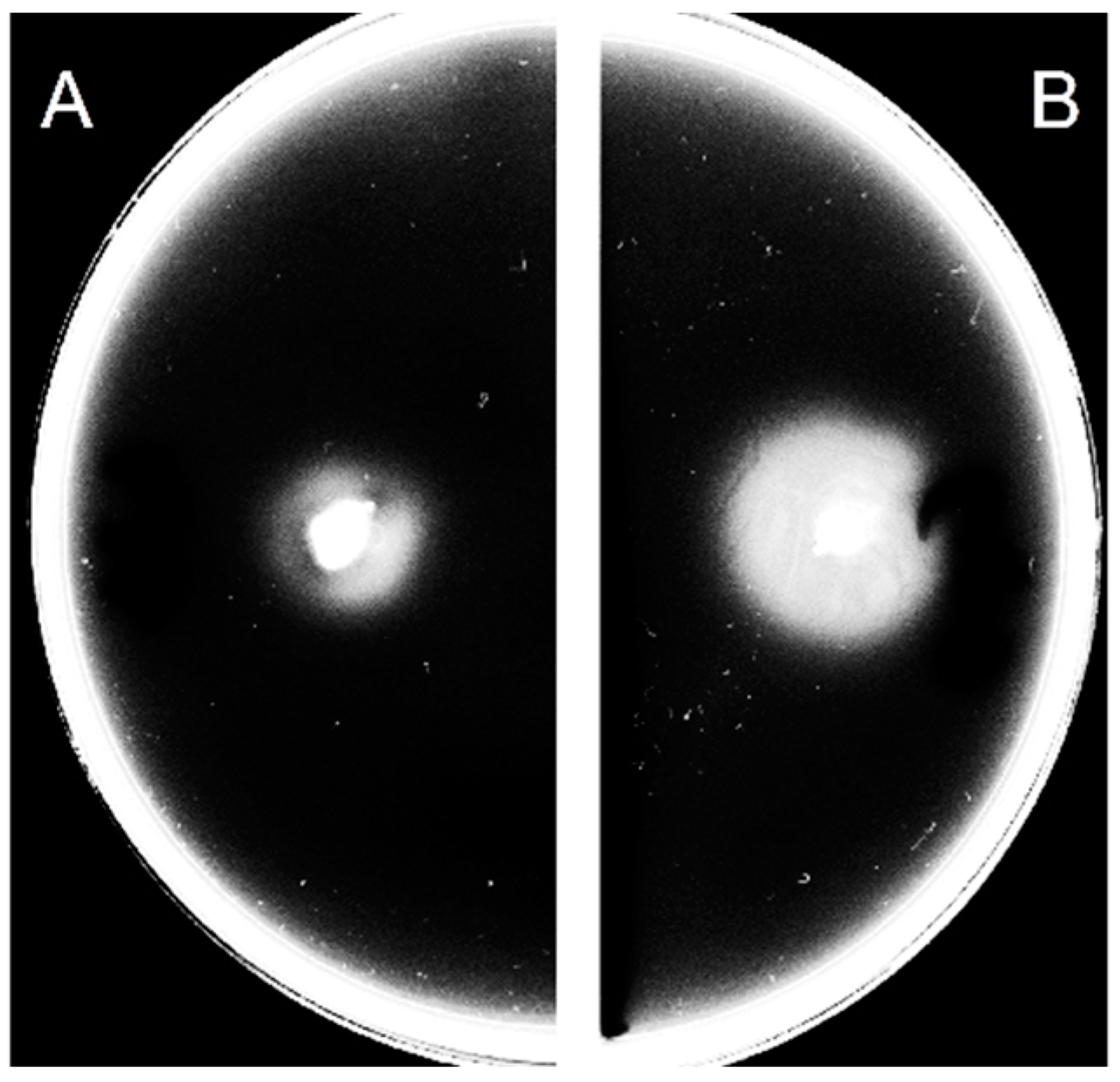
3.1. Extracellular c-di-GMP Affects the Motility of C. jejuni

The examination of the relationship between c-di-GMP and the swarming motility of wild-type C. jejuni 11168-O showed that the presence of 200 µM ex-c-di-GMP reduced the ability of C. jejuni 11168-O to swarm on soft agar (Figure 1). Ex-c-di-GMP did not affect the growth of C. jejuni 11168-O (Figure S1).

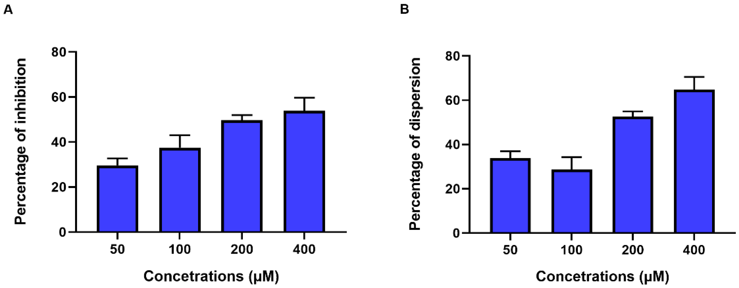
3.2. Extracellular c-di-GMP Affects Biofilm Formation by C. jejuni

In order to investigate the effect of ex-c-di-GMP on biofilm formation, different concentrations of ex-c-di-GMP (50–400 µM) were tested for their inhibitory effect on C. jejuni biofilm by the inhibition assay. The presence of ex-c-di-GMP had an significant inhibitory effect (p < 0.001, Table S2) on biofilm formation by C. jejuni 11168-O in a dose-dependent manner (Figure 2A) where 400 µM reduced biofilm formation by 48% (Figure 2A). Furthermore, the dispersion assay was used to determine the effect on the dispersion of a formed biofilm. Ex-c-di-GMP was able to disrupt existing biofilm, where 400 µM ex-c-di-GMP disrupted formed biofilm to greater than 60% extent (Figure 2B and Figure S2).

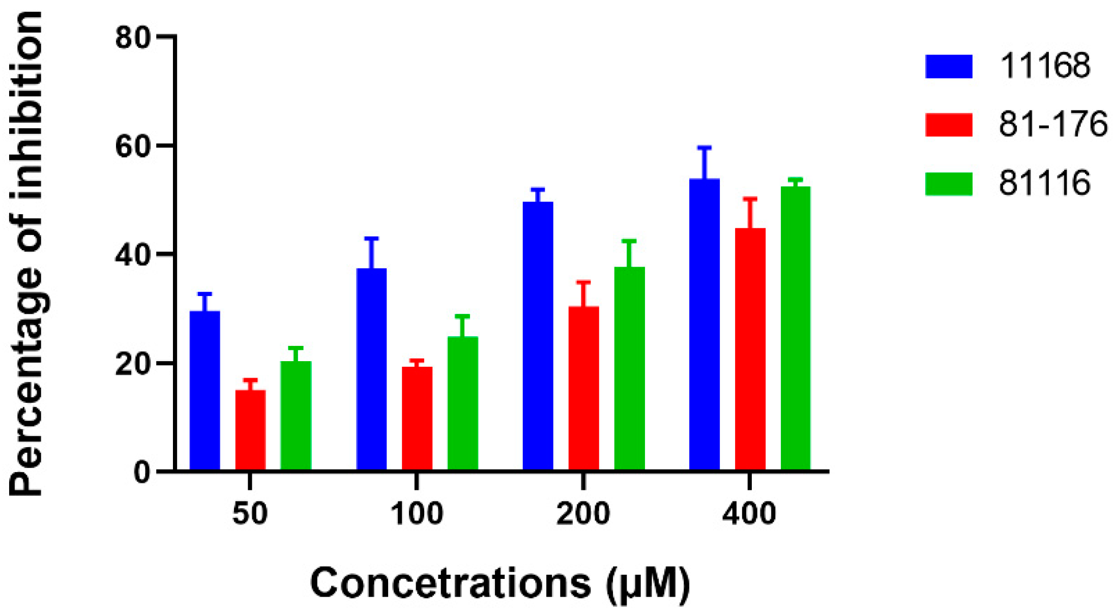
3.3. Extracellular c-di-GMP Affects Biofilm Formation by Different Campylobacter Strains

In order to determine if ex-c-di-GMP affected C. jejuni in a strain-specific manner, C. jejuni 11168-O, C. jejuni 81-176, and C. jejuni 81116, were used to demonstrate general inhibitory effect of ex-c-di-GMP on biofilm formation (Figure 3, Table S2).

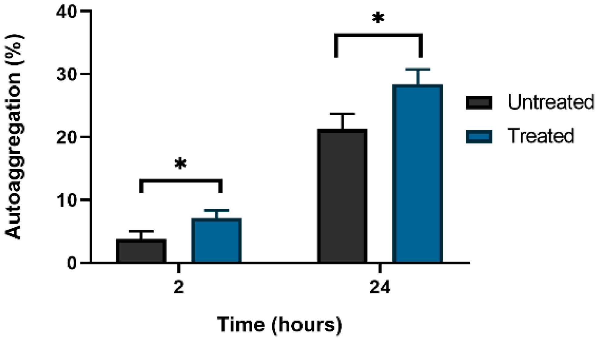
3.4. Extracellular c-di-GMP Treatment Effects Auto-Aggregation of C. jejuni

Untreated and cultures treated with ex-c-di-GMP were visually examined for visible cell–cell clumping and aggregation. Next, 200 μM ex-c-di-GMP-treated culture of C. jejuni 11168-O displayed significantly (p = 0.029 and 0.022, respectively) less auto-aggregation, compared to the untreated culture (Figure 4).

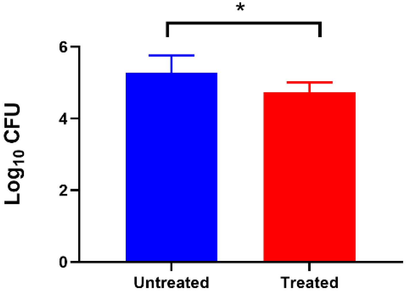
3.5. Effect of Extracellular c-di-GMP on Adhesion of C. jejuni to Caco-2 Cell Lines

To understand the effect of ex-c-di-GMP on C. jejuni 11168-O adhesion ability, we performed a cell adherence assay using polarised mammalian cultured Caco-2 cells in the presence and absence of 200 µM ex-c-di-GMP. As shown in Figure 5, a significant reduction (p < 0.05) was observed in adherence of C. jejuni 11168-O cells treated with 200 µM Ex-c-di-GMP, as compared with untreated cells to Caco-2 cells.

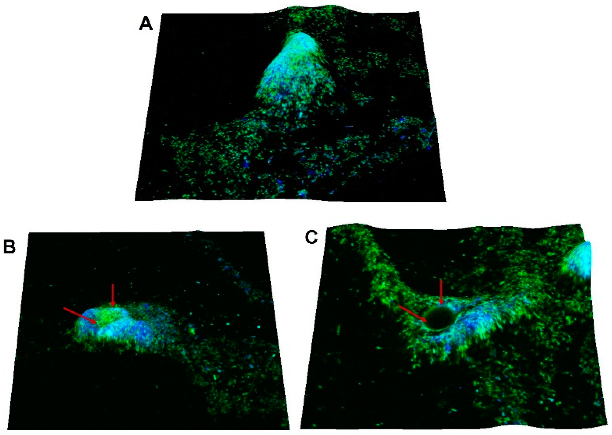
3.6. Dispersal Effect of Extracellular c-di-GMP on C. jejuni Biofilm Can Be Visualized

In order to observe the dispersal effect of ex-c-di-GMP on formed biofilm of C. jejuni 11168-O, the mature biofilm of C. jejuni 11168-O was treated with ex-c-di-GMP for 3 h (Figure 6 and Figure S3) and then visualized using CLSM. The biofilm architecture showed monolayers of cells covered with the extracellular matrix, likely to be EPS [61]. The treatment of mature biofilm with ex-c-di-GMP appeared to reduce previously formed structures, and the formation of central void within the biofilm structure typical of the biofilm dispersal could be observed.

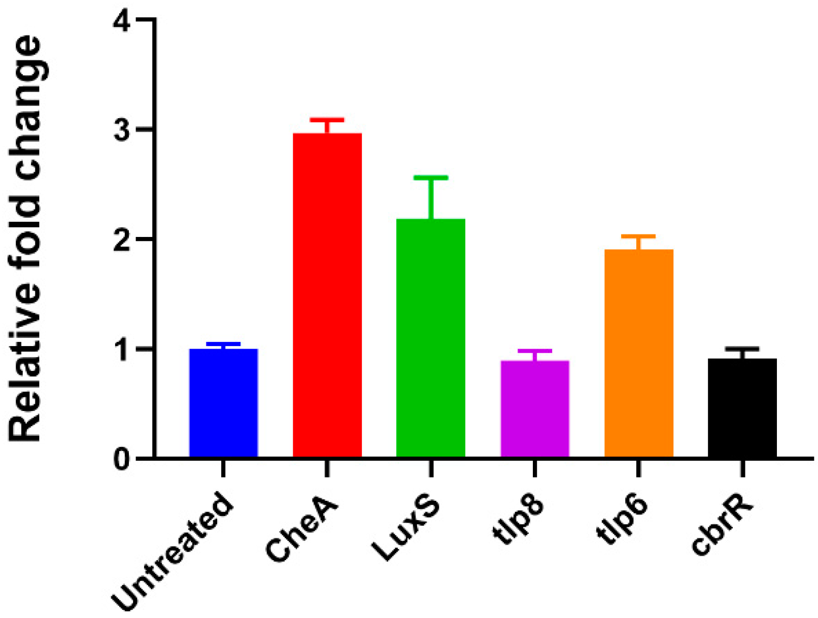
3.7. Extracellular c-di-GMP Dysregulates Specific Gene Expression

To elucidate the mechanism of action of ex-c-di-GMP, its effect on the expression levels of genes previously shown to be dysregulated during biofilm formation [62,63,64,65,66] was examined. The expression of genes involved in chemotactic motility and quorum sensing in C.jejuni 11168-O (cheA, tlp6, tlp8 and luxS) were examined in the presence and absence of ex-c-di-GMP by RT-PCR. CbrR gene as the only gene in the C. jejuni genome encoding GGDEF-like motif, reported to be potentially involved in c-di-GMP synthesis [48] was also included. The expression of, tlp6, LuxS and CheA was upregulated in presence of 200 µM ex-c-di-GMP by 1.9 to 3-fold (Figure 7); however, no significant effect on the expression of tlp8 was observed. Surprisingly, no changes in gene expression of cbrR have been noted.

4. Discussion

c-di-GMP signalling system is involved in the regulation of biofilm development stages from attachment to dispersal, where cells revert from the sessile lifestyle to planktonic and vice versa [26,38,67,68]. Interestingly, introduction of extracellular c-di-GMP (ex-c-di-GMP) has been shown to inhibit biofilm formation in some bacterial species including S. aureus and S. mutans [43,44,46,47]. Here, we demonstrate that extraneous c-di-GMP can reduce the formation C. jejuni biofilms as well as disperse formed biofilms in a dose-dependent manner. Ex-c-di-GMP at concentrations of ≥50 µM was able to significantly trigger the disassembly of the formed biofilms. This is in agreement with previously reported observations of significant inhibitory effect of ex-c-di-GMP on biofilm formation by S. aureus and S. mutans [44,46,47].
Since our data show that ex-c-di-GMP treatment inhibited C. jejuni biofilms, we hypothesised that ex-c-di-GMP may also affect established biofilms. Indeed, ex-c-di-GMP treatment of formed C. jejuni biofilms led to a reduction of biofilm development by up to 60. Moreover, while the inhibitory effect of ex-c-di-GMP was observed for all strains tested, the extent of inhibition was strain-dependent. Furthermore, microscopic analysis showed that when the formed biofilm was treated with ex-c-di-GMP, formation of central voids within the biofilm could be observed. This is similar to the reports of void formation with hollow cavities, containing swimming bacterial cells (seeding dispersal cells), within P. aeruginosa biofilm during biofilm dispersion [69,70,71].
However, it is still unclear how the ex-c-di-GMP acts to inhibit C. jejuni biofilm or how it enters C. jejuni cells. One of the possibilities is that C. jejuni senses this external compound as a part of the quorum sensing pathway where it might trigger a signalling cascade, modulating gene expression. Here, we have shown that luxS, a quorum sensing pathway gene, present in many bacterial species and responsible for the production of autoinducer-2 (AI-2) [72,73,74,75,76], was upregulated in C. jejuni in presence of ex-c-di-GMP. Moreover, AI-2 can mediate auto-aggregation and has been linked to pathogenicity, drug resistance, and host immune evasion [77]. Auto-aggregation has been identified as one of the first steps of biofilm formation (cell–cell attachment) [77,78]. This is generally mediated by surface proteins or carbohydrates, particularly exopolysaccharides (i.e., poly-N-acetylglucosamine), which can act as auto-agglutinins [79]. We found that C. jejuni cells treated with 200 μM ex-c-di-GMP show reduced aggregation, similar to that reported for S. aureus [46].
AI-2 had also been shown to influence chemotaxis and swarming motility [80,81,82]. Here, we found that chemotaxis associated genes cheA and tlp6 are upregulated in presence of ex-c-di-GMP, in agreement with previous report where these genes were shown to be upregulated within formed biofilms [83]. Changes in levels of CheA, in particular, are not surprising as it ultimately controls the directional rotation of the flagellar motor by changing its own phosphorylation levels, and thereby triggering changes in response regulators in the chemotaxis pathway [84]. Motility and flagellar gene expression in C. jejuni are indeed affected by luxS mutation Jeon et al. [85], where the mutation reduces expression of flagellin gene, flaA by 43% leading to reduction in motility. Here, we have shown that addition of ex-c-di-GMP affected C. jejuni swarming motility and enhanced the swarm zone achieved by the wild type cells, potentially as the result of overexpression of luxS. The change in motility in presence of ex-c-di-GMP could potentially reflect the transition between planktonic and biofilm states.
The ability of c-di-GMP to enter the Campylobacter cells can be reasonably postulated, as it has been reported to be able to enter eukaryotic cells [86]. Moreover, some microorganisms are known to secrete c-di-AMP or c-di-GMP as signalling molecules, such as Bacillus subtilis, Listeria monocytogenes and amoeba Dictyostelium discoideum [87,88,89]. For example, B. subtilis is able to secret c-di-AMP which impacts biofilm formation and plant root colonization [35,87].
Such messengers can also be detected by the innate immune system and can activate the expression of interferon genes and thus act as pathogen-associated molecular patterns (PAMPs) during infection [90,91]. Indeed, ex-c-di-GMP had been suggested to be a novel immunostimulatory agent that can modulate the host immune response, as was demonstrated by its ability to inhibit S. aureus infection in a mouse model [44]. This is consistent with our data, showing adherence of C. jejuni cells to the Caco-2 epithelial cells is significantly decreased after treatment with 200 μM ex-c-di-GMP, further suggesting that ex-c-di-GMP can potentially inhibit biofilm formation of biotic surfaces. However, the molecular bases for ex c-di-GMP inhibition of C. jejuni epithelial cell adherence are yet to be elucidated.
It is also possible that ex-c-di-GMP enters the cells by binding to the diguanylate cyclase (DGCs) which synthesizes c-di-GMP. Recently, the c-di-GMP signalling system has been reported in C. jejuni DRH212 [48], where cbrR gene was proposed to be a negative regulator of FlaA expression and motility. However, the presence and the role of c-di-GMP in other C. jejuni strains are still unknown, and comparative gene expression between biofilm and planktonic of C. jejuni 11168-O cells showed no differences in cbrR expression [66]. Similarly, no differences in expression of cbrR expression before and after treatment with ex-c-di-GMP have been observed in this study. It is possible, however, that this gene is constitutively expressed due to its potential role in bile resistance.

5. Conclusions

Together, these data show that ex-c-di-GMP inhibits the initial formation of biofilms and induces dispersion of formed biofilms of C. jejuni. It is possible to speculate that the bacterial cells that may not produce c-di-GMP, can nevertheless sense extraneously available molecules, such as QS molecules, during the pathogen-host–environment interactions. This study highlights the importance ex-c-di-GMP in regulating biofilm formation and dispersion by C. jejuni and indicates that the influence of external regulators in the life cycle of this organism, may need to be closely examined. Furthermore, the sensitivity of C. jejuni biofilms to ex-c-di-GMP makes them a promising drug target for controlling biofilm formation by using c-di-GMP analogues or similar small molecules [68].

Supplementary Materials

The following supporting information can be downloaded at: https://www.mdpi.com/article/10.3390/microorganisms10102030/s1, Figure S1: The growth rate of C. jejuni with and without 200 μM ex-c-di-GMP. Figure S2: Dispersion of C. jejuni 11168-O biofilms in the presence of ex-c-di-GMP at different concentrations. Figure S3: CLSM images of biofilm dispersion associated with ex-c-di-GMP. Table S1: Primer sets were used in this study. Table S2: Effect of ex-c-di-GMP on biofilm formation by C. jejuni strains.

Author Contributions

V.K. and B.A.E. conceived and designed the study; B.A.E. and K.S.S. performed the experiments; V.K. and B.A.E. analysed the data and prepared the manuscript. All authors reviewed the manuscript. All authors have read and agreed to the published version of the manuscript.

Funding

This project was partially supported by Griffith University, Institute for Glycomics.

Institutional Review Board Statement

Not applicable.

Informed Consent Statement

Not applicable.

Data Availability Statement

All the data is included in the article and the supplementary material.

Conflicts of Interest

Authors declare that there is no conflict of interest regarding the publication of this article.

References

  1. Buzby, J.C.; Allos, B.M.; Roberts, T. The economic burden of Campylobacter-associated Guillain-Barré syndrome. J. Infect. Dis 1997, 176, S192–S197. [Google Scholar] [CrossRef] [Green Version]
  2. World Health Organization. The Global View of Campylobacteriosis: Report of an Expert Consultation; WHO Newsletters; World Health Organization: Utrecht, The Netherlands, 2013.
  3. Salloway, S.; Mermel, L.; Seamans, M.; Aspinall, G.; Shin, J.N.; Kurjanczyk, L.; Penner, J. Miller-Fisher syndrome associated with Campylobacter jejuni bearing lipopolysaccharide molecules that mimic human ganglioside GD3. Infect. Immun. 1996, 64, 2945–2949. [Google Scholar] [CrossRef] [PubMed] [Green Version]
  4. van den Berg, B.; Walgaard, C.; Drenthen, J.; Fokke, C.; Jacobs, B.C.; van Doorn, P.A. Guillain-Barre syndrome: Pathogenesis, diagnosis, treatment and prognosis. Nat. Rev. Neurol. 2014, 10, 469–482. [Google Scholar] [CrossRef] [PubMed]
  5. Whiley, H.; van den Akker, B.; Giglio, S.; Bentham, R. The role of environmental reservoirs in human campylobacteriosis. Int. J. Environ. Res. Public Health 2013, 10, 5886–5907. [Google Scholar] [CrossRef] [Green Version]
  6. Ellis-Iversen, J.; Ridley, A.; Morris, V.; Sowa, A.; Harris, J.; Atterbury, R.; Sparks, N.; Allen, V. Persistent environmental reservoirs on farms as risk factors for Campylobacter in commercial poultry. Epidemiol. Infect. 2012, 140, 916–924. [Google Scholar] [CrossRef] [PubMed]
  7. Carrascosa, C.; Raheem, D.; Ramos, F.; Saraiva, A.; Raposo, A. Microbial biofilms in the food industry—A comprehensive review. Int. J. Environ. Res. Public Health 2021, 18, 2014. [Google Scholar] [CrossRef] [PubMed]
  8. Bronowski, C.; James, C.E.; Winstanley, C. Role of environmental survival in transmission of Campylobacter jejuni. FEMS Microbiol. Lett. 2014, 356, 8–19. [Google Scholar] [CrossRef] [PubMed] [Green Version]
  9. Backert, S.; Tegtmeyer, N.; Cróinín, T.Ó.; Boehm, M.; Heimesaat, M.M. Human campylobacteriosis. In Campylobacter; Elsevier: Amsterdam, The Netherlands, 2017; pp. 1–25. [Google Scholar]
  10. Igwaran, A.; Okoh, A.I. Human campylobacteriosis: A public health concern of global importance. Heliyon 2019, 5, e02814. [Google Scholar] [CrossRef] [PubMed]
  11. Luangtongkum, T.; Jeon, B.; Han, J.; Plummer, P.; Logue, C.M.; Zhang, Q. Antibiotic Resistance in Campylobacter: Emergence, Transmission and Persistence. Future Microbiol. 2009, 4, 189–200. [Google Scholar] [PubMed] [Green Version]
  12. Ma, L.; Konkel, M.E.; Lu, X. Antimicrobial resistance gene transfer from Campylobacter jejuni in mono-and dual-species biofilms. Appl. Environ. Microbiol. 2021, 87, e00659-21. [Google Scholar] [CrossRef] [PubMed]
  13. Smith, J.L.; Fratamico, P.M. Fluoroquinolone resistance in Campylobacter. J. Food Prot. 2010, 73, 1141–1152. [Google Scholar] [CrossRef] [PubMed]
  14. World Health Organization. WHO Publishes List of Bacteria for Which New Antibiotics Are Urgently Needed; WHO Newsletters; World Health Organization: Geneva, Switzerland, 2017.
  15. García-Sánchez, L.; Melero, B.; Jaime, I.; Rossi, M.; Ortega, I.; Rovira, J. Biofilm formation, virulence and antimicrobial resistance of different Campylobacter jejuni isolates from a poultry slaughterhouse. Food Microbiol. 2019, 83, 193–199. [Google Scholar] [CrossRef] [PubMed]
  16. Magajna, B.A.; Schraft, H. Campylobacter jejuni biofilm cells become viable but non-culturable (VBNC) in low nutrient conditions at 4 C more quickly than their planktonic counterparts. Food Control 2015, 50, 45–50. [Google Scholar] [CrossRef]
  17. Lv, R.; Wang, K.; Feng, J.; Heeney, D.D.; Liu, D.; Lu, X. Detection and quantification of viable but non-culturable Campylobacter jejuni. Front. Microbiol. 2020, 10, 2920. [Google Scholar] [CrossRef] [PubMed]
  18. Pitkänen, T. Review of Campylobacter spp. in drinking and environmental waters. J. Microbiol. Methods 2013, 95, 39–47. [Google Scholar] [CrossRef] [PubMed]
  19. Stetsenko, V.; Efimochkina, N.; Pichugina, T. Growth and persistence of Campylobacter jejuni in foodstuffs. Bull. Exp. Biol. Med. 2019, 166, 759–765. [Google Scholar] [CrossRef]
  20. Kim, S.-H.; Chelliah, R.; Ramakrishnan, S.R.; Perumal, A.S.; Bang, W.-S.; Rubab, M.; Daliri, E.B.-M.; Barathikannan, K.; Elahi, F.; Park, E. Review on stress tolerance in Campylobacter jejuni. Front. Cell. Infect. Microbiol. 2020, 10, 596570. [Google Scholar] [CrossRef] [PubMed]
  21. Reuter, M.; Mallett, A.; Pearson, B.M.; van Vliet, A.H. Biofilm formation by Campylobacter jejuni is increased under aerobic conditions. Appl. Environ. Microbiol. 2010, 76, 2122–2128. [Google Scholar] [CrossRef] [Green Version]
  22. Hanning, I.; Jarquin, R.; Slavik, M. Campylobacter jejuni as a secondary colonizer of poultry biofilms. J. Appl. Microbiol. 2008, 105, 1199–1208. [Google Scholar] [CrossRef] [PubMed]
  23. Hanning, I.; Donoghue, D.; Jarquin, R.; Kumar, G.; Aguiar, V.; Metcalf, J.; Reyes-Herrera, I.; Slavik, M. Campylobacter biofilm phenotype exhibits reduced colonization potential in young chickens and altered in vitro virulence. Poult. Sci. 2009, 88, 1102–1107. [Google Scholar] [CrossRef] [PubMed]
  24. Alary, M.; Nadeau, D. An outbreak of Campylobacter enteritis associated with a community water supply. Can. J. Public Health. 1989, 81, 268–271. [Google Scholar]
  25. Sung, K.; Khan, S. Biofilm development by Campylobacter Jejuni. Biofilms Food Environ. 2015, 24, 29–50. [Google Scholar]
  26. Ica, T.; Caner, V.; Istanbullu, O.; Nguyen, H.D.; Ahmed, B.; Call, D.R.; Beyenal, H. Characterization of mono- and mixed-culture Campylobacter jejuni biofilms. Appl. Environ. Microbiol 2012, 78, 1033–1038. [Google Scholar] [CrossRef] [Green Version]
  27. Zhong, X.; Wu, Q.; Zhang, J.; Ma, Z.; Wang, J.; Nie, X.; Ding, Y.; Xue, L.; Chen, M.; Wu, S. Campylobacter jejuni biofilm formation under aerobic conditions and inhibition by ZnO nanoparticles. Front. Microbiol. 2020, 11, 207. [Google Scholar] [CrossRef]
  28. Joshua, G.P.; Guthrie-Irons, C.; Karlyshev, A.; Wren, B. Biofilm formation in Campylobacter jejuni. Microbiology 2006, 152, 387–396. [Google Scholar] [CrossRef] [PubMed] [Green Version]
  29. Iovine, N.M. Resistance mechanisms in Campylobacter jejuni. Virulence 2013, 4, 230–240. [Google Scholar] [CrossRef] [Green Version]
  30. Mishra, R.; Panda, A.K.; De Mandal, S.; Shakeel, M.; Bisht, S.S.; Khan, J. Natural anti-biofilm agents: Strategies to control biofilm-forming pathogens. Front. Microbiol. 2020, 11, 2640. [Google Scholar] [CrossRef] [PubMed]
  31. Elgamoudi, B.A.; Korolik, V. Campylobacter biofilms: Potential of natural compounds to disrupt Campylobacter jejuni transmission. Int. J. Mol. Sci. 2021, 22, 12159. [Google Scholar] [CrossRef] [PubMed]
  32. Roy, R.; Tiwari, M.; Donelli, G.; Tiwari, V. Strategies for combating bacterial biofilms: A focus on anti-biofilm agents and their mechanisms of action. Virulence 2018, 9, 522–554. [Google Scholar] [CrossRef] [PubMed] [Green Version]
  33. Jenal, U.; Reinders, A.; Lori, C. Cyclic di-GMP: Second messenger extraordinaire. Nat. Rev. Microbiol. 2017, 15, 271–284. [Google Scholar] [CrossRef] [PubMed] [Green Version]
  34. Jones, C.J.; Utada, A.; Davis, K.R.; Thongsomboon, W.; Sanchez, D.Z.; Banakar, V.; Cegelski, L.; Wong, G.C.; Yildiz, F.H. C-di-GMP regulates motile to sessile transition by modulating MshA pili biogenesis and near-surface motility behavior in Vibrio cholerae. PLoS Pathog. 2015, 11, e1005068. [Google Scholar] [CrossRef] [PubMed]
  35. Chen, L.H.; Koseoglu, V.K.; Guvener, Z.T.; Myers-Morales, T.; Reed, J.M.; D’Orazio, S.E.; Miller, K.W.; Gomelsky, M. Cyclic di-GMP-dependent signaling pathways in the pathogenic Firmicute Listeria monocytogenes. PLoS Pathog. 2014, 10, e1004301. [Google Scholar] [CrossRef] [PubMed]
  36. Kalia, D.; Merey, G.; Nakayama, S.; Zheng, Y.; Zhou, J.; Luo, Y.; Guo, M.; Roembke, B.T.; Sintim, H.O. Nucleotide, c-di-GMP, c-di-AMP, cGMP, cAMP, (p)ppGpp signaling in bacteria and implications in pathogenesis. Chem. Soc. Rev. 2013, 42, 305–341. [Google Scholar] [CrossRef] [PubMed]
  37. Romling, U.; Galperin, M.Y.; Gomelsky, M. Cyclic di-GMP: The first 25 years of a universal bacterial second messenger. Microbiol. Mol. Biol. Rev. 2013, 77, 1–52. [Google Scholar] [CrossRef] [PubMed] [Green Version]
  38. Chua, S.L.; Liu, Y.; Yam, J.K.; Chen, Y.; Vejborg, R.M.; Tan, B.G.; Kjelleberg, S.; Tolker-Nielsen, T.; Givskov, M.; Yang, L. Dispersed cells represent a distinct stage in the transition from bacterial biofilm to planktonic lifestyles. Nat. Commun. 2014, 5, 4462. [Google Scholar] [CrossRef] [PubMed]
  39. Ghosh, A.; Jayaraman, N.; Chatterji, D. Small-molecule inhibition of bacterial biofilm. ACS Omega 2020, 5, 3108–3115. [Google Scholar] [CrossRef]
  40. Poulin, M.B.; Kuperman, L.L. Regulation of biofilm exopolysaccharide production by cyclic di-guanosine monophosphate. Front. Microbiol. 2021, 12, 730980. [Google Scholar] [CrossRef] [PubMed]
  41. Reichhardt, C.; Parsek, M.R. Confocal laser scanning microscopy for analysis of Pseudomonas aeruginosa biofilm architecture and matrix localization. Front. Microbiol. 2019, 10, 677. [Google Scholar] [CrossRef] [PubMed] [Green Version]
  42. Gomelsky, M.; Galperin, M.Y. Bacterial second messengers, cGMP and c-di-GMP, in a quest for regulatory dominance. EMBO J. 2013, 32, 2421–2423. [Google Scholar] [CrossRef] [Green Version]
  43. Brouillette, E.; Hyodo, M.; Hayakawa, Y.; Karaolis, D.K.; Malouin, F. 3′,5′-cyclic diguanylic acid reduces the virulence of biofilm-forming Staphylococcus aureus strains in a mouse model of mastitis infection. Antimicrob. Agents Chemother. 2005, 49, 3109–3113. [Google Scholar] [CrossRef] [PubMed] [Green Version]
  44. Karaolis, D.K.; Means, T.K.; Yang, D.; Takahashi, M.; Yoshimura, T.; Muraille, E.; Philpott, D.; Schroeder, J.T.; Hyodo, M.; Hayakawa, Y. Bacterial c-di-GMP is an immunostimulatory molecule. J. Immunol. 2007, 178, 2171–2181. [Google Scholar] [CrossRef] [PubMed] [Green Version]
  45. Wolfe, A.J.; Visick, K.L. Get the message out: Cyclic-Di-GMP regulates multiple levels of flagellum-based motility. J. Bacteriol. 2008, 190, 463–475. [Google Scholar] [CrossRef] [PubMed]
  46. David, K.R.K.; Rashid, M.H.; Chythanya, R.; Luo, W.; Hyodo, M.; Hayakawa, Y. c-di-GMP (3′-5′-Cyclic Diguanylic Acid) inhibits Staphylococcus aureus cell-cell interactions and biofilm formation. Antimicrob. Agents Chemother. 2005, 49, 1029–1038. [Google Scholar]
  47. Yan, W.; Qu, T.; Zhao, H.; Su, L.; Yu, Q.; Gao, J.; Wu, B. The effect of c-di-GMP (3’-5’-cyclic diguanylic acid) on the biofilm formation and adherence of Streptococcus mutans. Microbiol. Res. 2010, 165, 87–96. [Google Scholar] [CrossRef] [PubMed]
  48. Cox, C.A.; Bogacz, M.; El Abbar, F.M.; Browning, D.D.; Hsueh, B.Y.; Waters, C.M.; Lee, V.T.; Thompson, S.A. The Campylobacter jejuni response regulator and Cyclic-Di-GMP binding CbrR is a novel regulator of flagellar motility. Microorganisms 2021, 10, 86. [Google Scholar] [CrossRef] [PubMed]
  49. Day; Hartley-Tassell, L.E.; Shewell, L.K.; King, R.M.; Tram, G.; Day, S.K.; Semchenko, E.A.; Korolik, V. Variation of chemosensory receptor content of Campylobacter jejuni strains and modulation of receptor gene expression under different in vivo and in vitro growth conditions. BMC Microbiol. 2012, 12, 128. [Google Scholar]
  50. Elgamoudi, B.A.; Taha, T.; Korolik, V. Inhibition of Campylobacter jejuni biofilm formation by D-amino acids. Antibiotics 2020, 9, 836. [Google Scholar] [CrossRef] [PubMed]
  51. Tram, G.; Korolik, V.; Day, C.J. MBDS solvent: An improved method for assessment of biofilms. Adv. Microbiol. 2013, 3, 5. [Google Scholar] [CrossRef] [Green Version]
  52. Singh, N.; Patil, A.; Prabhune, A.; Goel, G. Inhibition of quorum-sensing-mediated biofilm formation in Cronobacter sakazakii strains. Microbiology 2016, 162, 1708–1714. [Google Scholar] [CrossRef] [PubMed]
  53. Bai, J.-R.; Wu, Y.-P.; Elena, G.; Zhong, K.; Gao, H. Insight into the effect of quinic acid on biofilm formed by Staphylococcus aureus. RSC Adv. 2019, 9, 3938–3945. [Google Scholar] [CrossRef] [Green Version]
  54. Sorroche, F.G.; Spesia, M.B.; Zorreguieta, Á.; Giordano, W. A positive correlation between bacterial autoaggregation and biofilm formation in native Sinorhizobium meliloti isolates from Argentina. Appl. Environ. Microbiol. 2012, 78, 4092–4101. [Google Scholar] [CrossRef] [Green Version]
  55. Day, C.J.; King, R.M.; Shewell, L.K.; Tram, G.; Najnin, T.; Hartley-Tassell, L.E.; Wilson, J.C.; Fleetwood, A.D.; Zhulin, I.B.; Korolik, V. A direct-sensing galactose chemoreceptor recently evolved in invasive strains of Campylobacter jejuni. Nat. Commun. 2016, 7, 13206. [Google Scholar] [CrossRef] [Green Version]
  56. Xie, Y.; He, Y.; Irwin, P.L.; Jin, T.; Shi, X. Antibacterial activity and mechanism of action of zinc oxide nanoparticles against Campylobacter jejuni. Appl. Environ. Microbiol. 2011, 77, 2325–2331. [Google Scholar] [CrossRef] [Green Version]
  57. Yan, X. Omics, Microbial Modeling and Technologies for Foodborne Pathogens; DEStech Publications, Inc.: Lancaster, PA, USA, 2012. [Google Scholar]
  58. Wösten, M.M.; Wagenaar, J.A.; van Putten, J.P. The FlgS/FlgR two-component signal transduction system regulates the fla regulon in Campylobacter jejuni. J. Biol. Chem. 2004, 279, 16214–16222. [Google Scholar] [CrossRef]
  59. Drozd, M.; Chandrashekhar, K.; Rajashekara, G. Polyphosphate-mediated modulation of Campylobacter jejuni biofilm growth and stability. Virulence 2014, 5, 680–690. [Google Scholar] [CrossRef] [Green Version]
  60. McLennan, M.K.; Ringoir, D.D.; Frirdich, E.; Svensson, S.L.; Wells, D.H.; Jarrell, H.; Szymanski, C.M.; Gaynor, E.C. Campylobacter jejuni biofilms up-regulated in the absence of the stringent response utilize a calcofluor white-reactive polysaccharide. J. Bacteriol. 2008, 190, 1097–1107. [Google Scholar] [CrossRef] [Green Version]
  61. Hou, J.; Veeregowda, D.H.; van de Belt-Gritter, B.; Busscher, H.J.; van der Mei, H.C. Extracellular polymeric matrix production and relaxation under fluid shear and mechanical pressure in Staphylococcus aureus biofilms. Appl. Environ. Microbiol. 2018, 84, e01516-17. [Google Scholar] [CrossRef] [Green Version]
  62. Zhang, L.; Li, S.; Liu, X.; Wang, Z.; Jiang, M.; Wang, R.; Xie, L.; Liu, Q.; Xie, X.; Shang, D. Sensing of autoinducer-2 by functionally distinct receptors in prokaryotes. Nat. Commun. 2020, 11, 5371. [Google Scholar] [CrossRef]
  63. Cloak, O.M.; Solow, B.T.; Briggs, C.E.; Chen, C.-Y.; Fratamico, P.M. Quorum sensing and production of autoinducer-2 in Campylobacter spp., Escherichia coli O157: H7, and Salmonella enterica serovar Typhimurium in foods. Appl. Environ. Microbiol. 2002, 68, 4666–4671. [Google Scholar] [CrossRef] [Green Version]
  64. Bezek, K.; Kurinčič, M.; Knauder, E.; Klančnik, A.; Raspor, P.; Bucar, F.; Smole Možina, S. Attenuation of adhesion, biofilm formation and quorum sensing of Campylobacter jejuni by Euodia ruticarpa. Phytother Res. 2016, 30, 1527–1532. [Google Scholar] [CrossRef]
  65. Tram, G.; Klare, W.P.; Cain, J.A.; Mourad, B.; Cordwell, S.J.; Day, C.J.; Korolik, V. Assigning a role for chemosensory signal transduction in Campylobacter jejuni biofilms using a combined omics approach. Sci. Rep. 2019, 10, 6829. [Google Scholar] [CrossRef] [Green Version]
  66. Tram, G.; Klare, W.P.; Cain, J.A.; Mourad, B.; Cordwell, S.J.; Korolik, V.; Day, C.J. RNA sequencing data sets identifying differentially expressed transcripts during Campylobacter jejuni biofilm formation. Microbiol. Resour. Announc. 2020, 9, e00982-19. [Google Scholar] [CrossRef] [Green Version]
  67. Rumbaugh, K.P.; Sauer, K. Biofilm dispersion. Nat. Rev. Microbiol. 2020, 18, 571–586. [Google Scholar] [CrossRef]
  68. Sintim, H.O.; Smith, J.A.; Wang, J.; Nakayama, S.; Yan, L. Paradigm shift in discovering next-generation anti-infective agents: Targeting quorum sensing, c-di-GMP signaling and biofilm formation in bacteria with small molecules. Future Med. Chem. 2010, 2, 1005–1035. [Google Scholar] [CrossRef]
  69. Kaplan, J.B. Biofilm dispersal: Mechanisms, clinical implications, and potential therapeutic uses. J. Dent. Res. 2010, 89, 205–218. [Google Scholar] [CrossRef] [Green Version]
  70. Ma, L.; Conover, M.; Lu, H.; Parsek, M.R.; Bayles, K.; Wozniak, D.J. Assembly and development of the Pseudomonas aeruginosa biofilm matrix. PLoS Pathog. 2009, 5, e1000354. [Google Scholar] [CrossRef] [Green Version]
  71. Goodwine, J.; Gil, J.; Doiron, A.; Valdes, J.; Solis, M.; Higa, A.; Davis, S.; Sauer, K. Pyruvate-depleting conditions induce biofilm dispersion and enhance the efficacy of antibiotics in killing biofilms in vitro and in vivo. Sci. Rep. 2019, 9, 3763. [Google Scholar] [CrossRef] [Green Version]
  72. Tereň, M.; Shagieva, E.; Vondrakova, L.; Viktorova, J.; Svarcova, V.; Demnerova, K.; Michova, H.T. Mutagenic strategies against luxS gene affect the early stage of biofilm formation of Campylobacter jejuni. J. Appl. Genet. 2021, 63, 145–157. [Google Scholar] [CrossRef]
  73. Püning, C.; Su, Y.; Lu, X.; Gölz, G. Molecular mechanisms of Campylobacter biofilm formation and quorum sensing. Fight. Campylobacter Infect. 2021, 431, 293–319. [Google Scholar] [CrossRef]
  74. Solano, C.; Echeverz, M.; Lasa, I. Biofilm dispersion and quorum sensing. Curr. Opin. Microbiol. 2014, 18, 96–104. [Google Scholar] [CrossRef] [Green Version]
  75. Waters, C.M.; Lu, W.; Rabinowitz, J.D.; Bassler, B.L. Quorum sensing controls biofilm formation in Vibrio cholerae through modulation of Cyclic Di-GMP levels and repression of vpsT. J. Bacteriol. 2008, 190, 2527–2536. [Google Scholar] [CrossRef] [Green Version]
  76. Elvers, K.T.; Park, S.F. Quorum sensing in Campylobacter jejuni: Detection of a luxS encoded signalling molecule. Microbiology 2002, 148, 1475–1481. [Google Scholar] [CrossRef] [Green Version]
  77. Nwoko, E.-s.Q.; Okeke, I.N. Bacteria autoaggregation: How and why bacteria stick together. Biochem. Soc. Trans. 2021, 49, 1147–1157. [Google Scholar] [CrossRef]
  78. Trunk, T.; Khalil, H.S.; Leo, J.C. Bacterial autoaggregation. AIMS Microbiol. 2018, 4, 140. [Google Scholar] [CrossRef]
  79. Formosa-Dague, C.; Feuillie, C.; Beaussart, A.; Derclaye, S.; Kucharikova, S.; Lasa, I.; Van Dijck, P.; Dufrene, Y.F. Sticky matrix: Adhesion mechanism of the staphylococcal polysaccharide intercellular adhesin. ACS Nano 2016, 10, 3443–3452. [Google Scholar] [CrossRef]
  80. Laganenka, L.; Colin, R.; Sourjik, V. Chemotaxis towards autoinducer 2 mediates autoaggregation in Escherichia coli. Nat. Commun. 2016, 7, 1–11. [Google Scholar] [CrossRef] [Green Version]
  81. Ramić, D.; Klančnik, A.; Možina, S.S.; Dogsa, I. Elucidation of the AI-2 communication system in the food-borne pathogen Campylobacter jejuni by whole-cell-based biosensor quantification. Biosens. Bioelectron. 2022, 30, 114439. [Google Scholar] [CrossRef]
  82. Song, S.; Wood, T.K. The primary physiological roles of autoinducer 2 in Escherichia coli are chemotaxis and biofilm formation. Microorganisms 2021, 9, 386. [Google Scholar] [CrossRef]
  83. Xu, L.; Xin, L.; Zeng, Y.; Yam, J.K.H.; Ding, Y.; Venkataramani, P.; Cheang, Q.W.; Yang, X.; Tang, X.; Zhang, L.-H. A cyclic di-GMP–binding adaptor protein interacts with a chemotaxis methyltransferase to control flagellar motor switching. Sci. Signal. 2016, 9, ra102. [Google Scholar] [CrossRef]
  84. Korolik, V. The role of chemotaxis during Campylobacter jejuni colonisation and pathogenesis. Curr. Opin. Microbiol. 2019, 47, 32–37. [Google Scholar] [CrossRef]
  85. Jeon, B.; Itoh, K.; Misawa, N.; Ryu, S. Effects of quorum sensing on flaA transcription and autoagglutination in Campylobacter jejuni. Microbiol. Immunol. 2003, 47, 833–839. [Google Scholar] [CrossRef]
  86. Steinberger, O.; Lapidot, Z.; Ben-Ishai, Z.; Amikam, D. Elevated expression of the CD4 receptor and cell cycle arrest are induced in Jurkat cells by treatment with the novel cyclic dinucleotide 3′, 5′-cyclic diguanylic acid. FEBS Lett. 1999, 444, 125–129. [Google Scholar] [CrossRef] [Green Version]
  87. Townsley, L.; Yannarell, S.M.; Huynh, T.N.; Woodward, J.J.; Shank, E.A. Cyclic di-AMP acts as an extracellular signal that impacts Bacillus subtilis biofilm formation and plant attachment. mBio 2018, 9, e00341-18. [Google Scholar] [CrossRef] [Green Version]
  88. Unterholzner, L. Beyond sensing DNA: A role for cGAS in the detection of extracellular cyclic di-nucleotides. EMBO Rep. 2019, 20, e47970. [Google Scholar] [CrossRef]
  89. Chen, Z.-h.; Schaap, P. Secreted cyclic di-GMP induces stalk cell differentiation in the eukaryote Dictyostelium discoideum. J. Bacteriol. 2016, 198, 27–31. [Google Scholar] [CrossRef] [Green Version]
  90. Schaap, P. Cyclic di-nucleotide signaling enters the eukaryote domain. IUBMB Life 2013, 65, 897–903. [Google Scholar] [CrossRef]
  91. Amarante-Mendes, G.P.; Adjemian, S.; Branco, L.M.; Zanetti, L.C.; Weinlich, R.; Bortoluci, K.R. Pattern recognition receptors and the host cell death molecular machinery. Front. Immunol. 2018, 9, 2379. [Google Scholar] [CrossRef]
Figure 1. The swarming motility of C. jejuni 11168-O following 24 h incubation. (A) Wild type cells. (B) Wild type cells mixed with 200 µM of ex-c-di-GMP.
Microorganisms 10 02030 g001
Figure 2. Inhibition and dispersion of C. jejuni 11168-O biofilms in the presence of ex-c-di-GMP at different concentrations. (A). Inhibition of biofilm formation by different concentrations of ex-c-di-GMP. (B) Dispersion of the existing biofilm induced by different concentrations of ex-c-di-GMP. Data are presented as mean ± standard error (3 or more biological repeats) of the percentage of inhibition (normalized to untreated control).
Microorganisms 10 02030 g002
Figure 3. Inhibition of biofilm formation by different C. jejuni strains in the presence of 50–400 µM ex-c-di-GMP. C. jejuni 11168-O (11168), C. jejuni 81-176 (81-176), and C. jejuni 81116 (81116). Data are presented as mean ± standard error (3 or more biological repeats) of the percentage of inhibition (normalized to untreated control).
Microorganisms 10 02030 g003
Figure 4. Percentage of C. jejuni auto-aggregation following ex-c-di-GMP treatment. Data are presented as mean ± standard errors (3 or more biological repeats). The asterisk (*) indicates a statistically significant difference compared to the untreated control (p < 0.05).
Microorganisms 10 02030 g004
Figure 5. Effect of ex-c-di-GMP on adhesion of C. jejuni to Caco-2 cell. Inhibition of cell adhesion in the presence of 200 µM ex-c-di-GMP. Data are presented as mean ± standard error (3 or more biological repeats) and were analysed using two-tailed Student’s t-test. The asterisk (*) indicates a statistically significant difference compared to the untreated control (p < 0.05).
Microorganisms 10 02030 g005
Figure 6. Void formation indicative of biofilm dispersion after exposure C. jejuni 11168-O biofilm to ex-c-di-GMP. (A) Representative CLSM images of biofilm grown for 48 h untreated (control). (B,C) Biofilm after exposure to 200 µM ex-c-di-GMP. Calcofluor White (CFW) (green, polysaccharides) and DAPI (blue, DNA). Red arrow indicates central hollowing of microcolony (Scale bar = 10 µm).
Microorganisms 10 02030 g006
Figure 7. Analysis of the relative expression of genes in the presence of 200 µM ex-c-di-GMP. The expression of tlp6, tlp8, luxS cheA and cbrR genes relative to housekeeping gene gryA after incubation of C. jejuni 11168-O cells with 200 µM ex-c-di-GMP for 3 h.
Microorganisms 10 02030 g007
Publisher’s Note: MDPI stays neutral with regard to jurisdictional claims in published maps and institutional affiliations.

