Article

Mitigation of Expression of Virulence Genes in Legionella pneumophila Internalized in the Free-Living Amoeba Willaertia magna C2c Maky

by
Rayane Mouh Mameri
1,
Jacques Bodennec
2,
Laurent Bezin
2 and
Sandrine Demanèche
1,*
1
R&D Department, Amoéba, 69680 Chassieu, France
2
Lyon Neuroscience Research Center CRNL UMR5292 U1028, University of Lyon, Univ Lyon 1, CNRS, Inserm, 69500 Bron, France
*
Author to whom correspondence should be addressed.
Pathogens 2020, 9(6), 447; https://doi.org/10.3390/pathogens9060447
Submission received: 14 May 2020 / Revised: 29 May 2020 / Accepted: 3 June 2020 / Published: 5 June 2020
(This article belongs to the Special Issue Legionella Contamination in Water Environment)

Abstract

:
Legionella pneumophila is a human pathogen responsible for a severe form of pneumonia named Legionnaire disease. Its natural habitat is aquatic environments, being in a free state or intracellular parasites of free-living amoebae, such as Acanthamoeba castellanii. This pathogen is able to replicate within some amoebae. Willaertia magna C2c Maky, a non-pathogenic amoeba, was previously demonstrated to resist to L. pneumophila and even to be able to eliminate the L. pneumophila strains Philadelphia, Lens, and Paris. Here, we studied the induction of seven virulence genes of three L. pneumophila strains (Paris, Philadelphia, and Lens) within W. magna C2c Maky in comparison within A. castellanii and with the gene expression level of L. pneumophila strains alone used as controls. We defined a gene expression-based virulence index to compare easily and without bias the transcript levels in different conditions and demonstrated that W. magna C2c Maky did not increase the virulence of L. pneumophila strains in contrast to A. castellanii. These results confirmed the non-permissiveness of W. magna C2c Maky toward L. pneumophila strains.

1. Introduction

The genus Legionella includes more than 60 species, but human infections that progress to a severe pneumonia, known as Legionnaires’ disease, are most often caused by Legionella pneumophila [1,2]. This Gram-negative bacterium especially affects immunocompromised individuals after inhalation of Legionella-contaminated aerosols. L. pneumophila invades and replicates within alveolar macrophages and epithelial cells of the lungs, inducing a severe respiratory infection [1,3].
L. pneumophila is ubiquitous in natural, artificial, and industrial aquatic environments and is mostly nested in intracellular hosts, such as free-living amoebae (FLA). In 1980, Rowbotham first demonstrated the intracellular multiplication of L. pneumophila within Acanthamoeba spp. and Naegleria spp. [4]. Following this study, several reports described the replication of Legionella isolates from clinical samples within protozoa isolated from the presumed source of infection. Intracellular growth within protozoa was shown to increase the ability of L. pneumophila to infect human monocytes and to resist to chemical disinfectants, biocides, and antibiotics [5,6,7]. Inhalation of legionellae packaged in amoebae is associated with the induction of more severe clinical cases of legionellosis. The speculated link between the homing of legionella in amoebae and the increased virulence of legionella is supported by the publication of a mouse model of co-inhalation of L. pneumophila and Hartmannella vermiformis. It was shown to significantly enhance the intrapulmonary growth of L. pneumophila, with a greater mortality than that observed from inhalation of legionellae alone [8]. The intra-amoebae growth was demonstrated to enhance the ability of L. pneumophila to infect epithelial cells (100- to 1000-fold), murine macrophages (10- to 100-fold), human monocytes (100- to 1000-fold), and Acanthamoeba castellanii (10- to 100-fold) [9]. Moreover, L. pneumophila grown in A. castellanii displays enhanced infection in monocytes compared to buffered charcoal yeast extract (BCYE)-grown bacteria [10]. Within FLA and human macrophages, L. pneumophila cells are able to reroute the phagosome thanks to a functional Dot/Icm type 4 secretion system (T4SS) and the approximately 300 proteins it secretes and to induce the biogenesis of a legionella-containing vacuole (LCV) [11,12]. Bacterial replication occurs in LCV, evades fusion with lysosomes, and associates intimately with the host endoplasmic reticulum (ER), inducing the lysis of the host cells. The induction of apoptosis in the host cell is induced by a type IV-like secretion machinery [13]. The internalization of L. pneumophila into host cells, such as FLA, promotes not only its ability to survive and multiply but also to acquire and increase its virulence [14]. The L. pneumophila Dot/Icm-secreted effector PlcC/CegC1 was demonstrated to promote virulence [15], and the translocated Dot/Icm type IVB secretion system effector SdhA was demonstrated to be of crucial importance in infection processes [16].
Genes of the T4SS system have also been identified, with several other genes, as being responsible for the increased virulence in L. pneumophila once internalized by FLA, such as A. castellanii and Vermamoeba vermiformis [17]. In two studies from NJ Ashbolt’s laboratory, about 30 transcripts of genes involved in bacterial metabolism, replication, and virulence have been investigated using reverse transcription quantitative polymerase chain reaction (RT-qPCR) in L. pneumophila Philadelphia after exposure to CuO nanoparticles (CuO-NPs) or synthetic gray water (Gw) for a period ranging from 3 to 48 h [18,19].
Here, we investigated the expression level by RT-qPCR of 7 genes (htpX, icmE, lirR, ccmF, gacA, tatB, and lvrE) that we have shown to be expressed by all three reference strains, L. pneumophila Paris, Philadelphia, and Lens, after co-incubation with two FLA: A. castellanii known to be permissive to the legionella multiplication [10], and Willaertia magna C2c Maky considered as non-permissive to the legionella multiplication [20]. We also evaluated transcript levels of these genes in L. pneumophila cultivated alone and harvested at the end of the growth exponential phase corresponding to the end of the replicative phase and the beginning of the virulence phase. The aim of this work was to evaluate the evolution of virulence of L. pneumophila strains after internalization into a non-permissive amoeba, such as W. magna C2c Maky. To facilitate the interpretation and comparison of gene expression, a GENE EXPRESSION-based index was developed.

2. Results

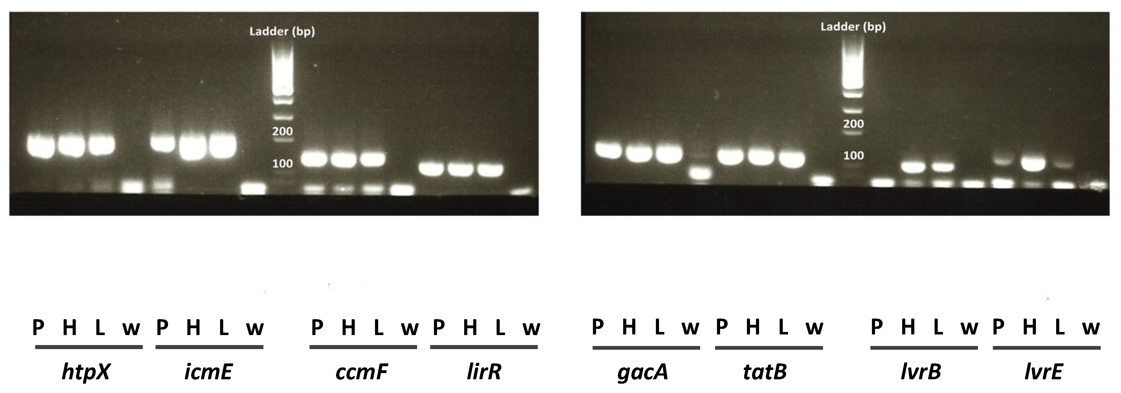
2.1. Virulence Gene Selection

Virulence genes of interest were selected based on previous studies [18,19] that investigated the potential increase in virulence gene expression in L. pneumophila Philadelphia exposed to CuO-NPs or synthetic Gw (Table S1). As our objective was to assess virulence gene expression in L. pneumophila internalized for three days in amoebae, we focused on genes whose expression was increased after 24–48 h of environmental exposure to CuO-NPs (dotA, enhC, htpX, icmE, and pvcA) or synthetic Gw (lirR, ccmF, gacA, tatB, lvrB, and lvrE). We excluded the genes cegC1 and sidF displaying conflicting variations between the two environmental conditions (Table S1).
Among the 11 genes selected, we were not able to amplify pvcA cDNA in L. pneumophila Philadelphia using published primers [19]. We thus ran qPCR for the other 10 genes, using the primers listed in Table 1.
As expected, these primers allowed high quality amplification of the targeted cDNAs in L. pneumophila Philadelphia cultured for three days in BCYE plates, with the exception of dotA and enhC cDNAs. Indeed, the amplification of these two amplicons did not reach satisfactory criteria for either the melt curve analysis or the agarose gel electrophoresis of the end products, for the three L. pneumophila strains. We thus excluded these two genes from the final analysis. The primers used for the eight remaining genes produced good amplifications, with end products of the expected size (Figure 1). However, the expression of lvrB in L. pneumophila Paris was not detected in all conditions tested. It is possible that the lvrB gene differs in L. pneumophila Paris from the two other strains, in regions recognized by the primers used. To avoid any bias when comparing the three strains, lvrB was not included in the final list of the seven selected transcripts.

2.2. Validation of rpsL as a Non-Acceptable Housekeeping Gene, in Which Transcript Level Should Be Stable between the Different Tested Conditions

While real-time PCR is a quantitative method with high sensitivity and great reproducibility, to measure targeted DNAs over a wide range of concentrations, the RT of biological sample RNAs is a reaction that is difficult to calibrate. To normalize differences in RT efficiency across samples, the transcript level of a gene, mostly a housekeeping gene, is used as an internal control, whose expression is supposed to be invariant between various treatments or conditions. The ribosomal gene rpsL is a housekeeping gene commonly used for phylogenic analysis of Legionella [21] and has been used to normalize the reverse transcription of RNA extracted from L. pneumophila exposed to Cuo-NPs nanoparticles or synthetic Gw [18,19]. An alternative way to normalize the RT reaction between the different samples is to use a known quantity of synthetic RNA, added directly to the reaction mix, thereby allowing the same number of copies of this synthetic RNA to be added across all samples. As an external control, we used the so-called synthetic non-homologous standard mRNA (SmRNA) [22], of which we ensured that the primers used for the amplification of its cDNA do not amplify DNA sequences resulting from the RT of the endogenous RNAs of L. pneumophila.
Thus, using SmRNA as an external control, the expression of rpsL could be examined by RT-qPCR like any other gene. Using a graphical representation with an ordinate axis on a linear scale, we show that there is a high variability in the rpsL mRNA level, in particular in L. pneumophila Paris and L. pneumophila Lens (Figure S1). In a logarithmic representation of the ordinate axis, one can note that the rpsL mRNA level was almost identical between the three Legionella strains under the T′0 control condition. In addition, the rpsL mRNA level in the L. pneumophila Philadelphia strain remained unchanged under the various conditions tested. On the other hand, the maintenance for three days of the incubation of the L. pneumophila Paris and L. pneumophila Lens strains in the liquid medium (3D-FREE) caused an increase in the rpsL mRNA level, which was significant compared to the T′0 control in L. pneumophila Lens. The internalization of L. pneumophila Paris and L. pneumophila Lens in A. castellanii was also followed by a strong and significant increase in the rpsL mRNA level, not only compared to the T′0 condition but also compared to the T3D-FREE condition (A. castellanii only). Finally, the internalization of the three strains in W. magna C2c Maky did not lead to a significant increase in the rpsL mRNA level (Figure 2).
Given that rpsL is not a gene whose expression is invariant under the conditions tested, we did not use it for the normalization of the RT reaction and instead used the SmRNA for all of the targeted genes.

2.3. Definition of a Gene Expression-Based Virulence Index

Studies aimed at investigating virulence gene expression usually analyze each gene separately, making it difficult to draw clear conclusions, especially when expression increases for some genes, decreases for others, and finally remains stable for the latest.
Since qPCR makes it possible to quantify cDNA copies in a sample, we eluded the above-mentioned issue by defining for each sample a virulence index, which is the sum of all virulence cDNAs quantified by qPCR.
However, in the calculation of the virulence index, we paid attention to avoid masking important variations for genes expressed at low levels in basal conditions by genes initially expressed at high levels. To this end, for each transcript, the cDNA copy number contained in a given sample has been expressed as a percentage of the averaged copy number measured in all samples.

2.4. Comparison of the Virulence Index of three L. pneumophila Strains

Firstly, we evaluated the “Gene Expression-based” virulence index of three strains of L. pneumophila after coincubation for three days within two amoebic species: W. magna C2c Maky and A. castellanii, (Figure 3A), based on the measurements of the transcript levels of the seven virulence genes selected: ccmF (Figure S2), gacA (Figure S3), htpX (Figure S4), icmE (Figure S5), lirR (Figure S6), lvrE (Figure S7), and tatB (Figure S8). These measures were performed after three days of coculture when the amount of intracellular Legionella was not significantly different between W. magna C2c Maky and A. castellanii.
Secondly, after Day 3, every condition (L. pneumophila alone, L. pneumophila strains co-incubated with W. magna C2c Maky, and L. pneumophila strains co-incubated with A. castellanii) was seeded on BCYE plates for three additional days (until Day 6) to remove all amoeba traces, because amoebae are not able to survive on a BCYE plate, and to evaluate the fate of the virulence of L. pneumophila after their release from amoebae. Once L. pneumophila had grown on the BCYE plates, bacteria were harvested, and the virulence index was evaluated (Figure 3B) as above, based on the measurements of the transcript levels of the seven virulence genes selected: ccmF (Figure S9A), gacA (Figure S9B), htpX (Figure S10A), icmE (Figure S10B), lirR (Figure S11A), lvrE (Figure S11B), and tatB (Figure S12). As expected, the transcript levels varied differently between the different genes selected, making it difficult to draw a clear conclusion (Figures S2–S12).
A virulence index was thus calculated for each condition, and an ANOVA 2 revealed for Steps 1 and 2 (Figure S13A,B) that the three L. pneumophila strains behaved in the same way under all tested conditions. Their results were then pooled and averaged (Figure 3).
ANOVA 2 also revealed a statistical difference between culture conditions (p < 0.0001), and post-hoc analysis (Tukey’s HSD test) showed a significant increase of the virulence genes after the internalization of L. pneumophila in A. castellanii compared to the controls containing L. pneumophila alone at Day 0 and Day 3 (Figure 3A). Conversely, a tendency (not significant) to decrease the level of virulence of the three L. pneumophila strains within W. magna C2c Maky was observed when compared to L. pneumophila alone (control) at Day 0 and Day 3 (Figure 3A). L. pneumophila strains internalized within W. magna C2c Maky exhibit a virulence index that is not statistically different from the controls at T’0 and T3D, demonstrating that W. magna C2c Maky did not increase the virulence of L. pneumophila strains in contrast to A. castellanii.

3. Discussion

These experiments brought evidence of a different behavior between L. pneumophila strains internalized by W. magna C2c Maky and strains internalized by A. castellanii. Indeed, the expression of virulence genes is reduced in L. pneumophila internalized into W. magna C2c Maky cells, while it is significantly increased in L. pneumophila internalized into A. castellanii.
These results confirm the non-permissiveness observed in W. magna C2c Maky towards L. pneumophila. Unlike A. castellanii, W. magna C2c Maky is able to internalize and digest L. pneumophila by phagocytosis [20,23]. These data suggest that L. pneumophila strains were unable to use their T4SS system to deflect the cellular machinery of W. magna C2c Maky. As a consequence, the phago-lysosomal fusion could happen and LCV could not be created, leading to the intracellular destruction of bacteria.
Considering FLA known to be permissive to L. pneumophila multiplication, such as A. castellanii, Buse et al. reported that the Dot/Icm T4SS system was responsible for the translocation of a large number of Legionella effectors in the host cells to promote infectivity [18]. These translocated effector genes are located in a hypervariable region of the L. pneumophila genome, and the plasminogen activator homologue of L. pneumophila, was strongly involved in A. castellanii intracellular growth [24,25]. The virulence associated genes lvrB and lvrE were also shown to be involved in the T4SS [21,26]. Another effector of the Dot/Icm substrate, SidF, was shown to be involved in the inhibition of programmed cell death in the host and anchoring of binding effectors to bacterial phagosomes [27,28]. However, Legionella vir homologues were shown to be not required for intracellular replication in amoeba or macrophage [21].
Surprisingly, L. pneumophila strains cultivated alone in liquid medium for three days and then incubated for three additional days on BCYE plates exhibit a great increase of the virulence index (Figure 3B), demonstrating an induction of the expression of virulence genes. ANOVA 2 revealed a statistical difference between culture conditions (p < 0.0001). Post-hoc analysis (Tukey’s HSD test) showed a tendency of the virulence index to increase from Day 0 to Day 3 under the control conditions (not significant, p = 0.898) and a clear statistical difference from other conditions (Figure 3B), i.e., the control at Day 6, and the L. pneumophila strains internalized within W. magna C2c Maky and A. castellanii at Day 6 (p = 0.023). No statistical difference was observed at Day 6 between the control condition and the L. pneumophila strains co-cultivated with both amoebae. It seems that cultivation on BCYE plates erased the virulence index acquired during internalization in amoebae to confer a virulence index specific to BCYE culturing. Bacteria cultivated on BCYE plates were shown to be virulent, even if some isolates transiently lost their flagella. Indeed, some isolates characterized as aflagellate when harvested from BCYE agar were shown to be able to multiply in amoebae, and flagella were subsequently detectable by immunologic methods [29]. Moreover, Nowicki et al. have concluded that infection with aerosols of L. pneumophila coming from cultures on BCYE plates causes mortality in guinea pigs, showing that BCYE-cultivated Legionella are indeed virulent [30]. Consequently, the virulence index can only be assessed in conditions of liquid culture without sub-culturing on BCYE plates.
To conclude, we developed a tool that can be named the “GENE EXPRESSION-based Virulence Index” based on the quantitative measurement of cDNAs of 7 virulence genes (ccmF, gacA, htpX, icmE, lirR, lvrE, and tatB) using a calibrated RT and real-time PCR. Virulence genes were expressed at low levels in the three strains of L. pneumophila in the absence of amoebae, making it possible to determine a “GENE EXPRESSION-based Virulence Index” both at T0 and after 3 days in a liquid medium.
L. pneumophila strains followed a similar trend, since the “GENE EXPRESSION-based Virulence Index” between the three strains was not statistically different. After internalization in W. magna C2c Maky, there was no increase in the “GENE EXPRESSION-based Virulence Index” compared to the control conditions, since no statistical difference between the index determined after internalization in W. magna C2c Maky and the index determined in control L. pneumophila was observed for the three L. pneumophila strains. A huge and significant increase in “GENE EXPRESSION-based Virulence Index” was observed in the three tested L. pneumophila strains internalized in A. castellanii compared to W. magna C2c Maky, with a fold difference between the two amoebae reaching 9.4 (Virulence Index was 185 ± 44 in WILL condition and 1745 ± 257 in ACANTH condition). These data confirmed that W. magna C2c Maky cells resist Legionella strains, are able to phagocyte Legionella strains, and do not increase their virulence after internalization.
This index synthesized the expression of all the genes in one data for each condition, rendering the comparison easier. Moreover, a particular attention was brought to the genes that were expressed at low level by expressing the cDNA copy number contained in a given sample in percent of the averaged copy number measured in all samples. This GENE EXPRESSION-based Virulence Index could be used in many studies that focus on the variation of gene expression in a given function (here virulence, elsewhere inflammation, development, cell death, etc.) where many transcripts are analyzed. When different genes associated with the same function are compiled or integrated in the form of an index, it makes it easier to conclude about the overall evolution of these genes, a conclusion often made difficult when the individual variations do not always point in the same direction.

4. Materials and Methods

4.1. L. pneumophila Cultures

The three strains of L. pneumophila serogroup 1, Lens CIP 108 286, Paris CIP 107 629T, and Philadelphia ATCC 33152, were cultured at 36 °C for 3 days on buffered charcoal yeast extract (BCYE) agar plates (Thermo Fisher Scientific, Dardilly, France) before coculture experiments. Quantification of L. pneumophila was performed via Real-time qPCR using Biorad kits (Biorad, Hercules, CA, USA). Briefly, aliquots of cell suspension were sampled, and genomic DNA was extracted using the AquadienTM DNA extraction kit. L. pneumophila was quantified using the iQ-CheckTM L. pneumophila Real-time qPCR kit and a CFX96 Biorad thermocycler. The data were analyzed using the Bio-Rad CFX Manager Industrial Diagnostic Edition software (version 2.2). The whole procedure was designed for quantification of L. pneumophila, including intra-amoebic bacteria, and has been validated by AFNOR (Agence Française de Normalisation, the French standardization authority).

4.2. FLA Culture

The amoebae used in this study were W. magna C2c Maky ATCC PTA-7824 and A. castellanii ATCC 30010. Amoebae were grown at 30 °C for 3 days using adhesion culture on CF4 with serum casein glucose yeast extract medium (SCGYEM) [31]. Amoeba cells were maintained in the exponential growth phase by subculturing every 3 days. Quantification of amoeba populations was performed using 0.1 mL of each aliquot utilizing a hemocytometer cell counting chamber method with Trypan blue.

4.3. Coculture of L. pneumophila with Amoebae

Tubes containing 5 mL of Peptone Yeast Extract Glucose Broth (PYG) [32] were seeded with 5 × 10⁵ W. magna C2c Maky or A. castellanii cells. At Day 0, the different strains of L. pneumophila, grown on BCYE plates, were suspended in sterile distilled water at 2.5 × 10⁸ cells/mL, and inoculated into the amoebic cultures at a multiplicity of infection (MOI) of 50. Low-speed centrifugation (30 min at 1000× g) was used to initiate physical interaction between bacteria and amoebae, and then incubated at 36 °C for 1 h. To eliminate extracellular legionella, cocultures were treated for 2 h at 30 °C with 0.5 mg/mL of Penicillin and Streptomycin. Cells were then washed twice by, first, centrifuging assay tubes for 10 min at 1000× g, second, removing the supernatant, and, finally, adding 5 mL of fresh PYG medium preheated at room temperature. After the second wash, cocultures were incubated at 30 °C in PYG medium for 3 days (Figure S13A).
In a second step, coculture solutions were submitted to a mechanical cell lysis using a FastPrep®-24 instrument (MP Biomedicals, Illkirch-Graffenstaden, France) for 2 × 30 s at a speed of 5.0 in order to release legionella from the amoebae. Afterward, 100 µL of each treated coculture were deposited onto BCYE plates and incubated for 3 days at 36 °C (Figure S13B).
Control conditions consisted in legionella cultured in PYG medium in the absence of amoebae (FREE condition) for 3 days; for the second step, 100 µL of the latter culture were deposited onto BCYE plates for 3 additional days (Day 6), as described above.

4.4. Preservation of RNA Samples

Free legionella and cocultures (amoebae with internalized legionella) in liquid medium, or legionella colonies on BCYE plates were collected, rinsed in sterile osmosed water, and centrifuged for 5 min at 4 °C at 6000× g. The TRIzol® Max™ Bacterial RNA Isolation Kit (Thermo Fisher Scientific, Lyon, France) was used to improve the isolation of intact total RNA. The kit utilizes both the Max™ Bacterial Enhancement Reagent and TRIzol® Reagent to inactivate endogenous RNases and promote protein denaturing, improving RNA quality and integrity. After removal of the supernatant, 200 µL of pre-heated (95 °C) Max Bacterial Enhancement Reagent buffer was added on the pelleted cells and incubated for 5 min at 95 °C. Afterward, 1 mL of TRIzol Reagent was added, and the tubes were frozen in dry ice before being stored at −80 °C.

4.5. Total RNA Extraction

Biological material in TRIzol® was used for total RNA extraction according to the manufacturer’s instruction and MIQE guidelines [33]. Total nucleic acid extracted was treated with Turbo DNA-free DNAse (Thermo Fisher Scientific, Lyon, France) as recommended by the manufacturer, to remove genomic DNA. Total RNA was then transferred into a nuclease-free tube and stored at −80 °C after the concentration was measured using a Biodrop spectrophotometer (Biodrop, Cambridge, UK).

4.6. Calibrated Reverse Transcription (cRT)

Briefly, 180 ng of purified total RNAs were used for cRT using random hexamers and Multiscribe™ reverse trancriptase in the presence of RNAse inhibitor (Thermo Fisher Scientific). A synthetic external and non-homologous Standard RNA (SmRNA) was used to control the quality of the RT and to normalize the reverse transcription of mRNAs of biological samples [34]. At the end of the RT, cDNAs were stored at −20 °C until further use. It should be noted that the Multiscribe® reverse transcriptase was not added in the RT master mix as recommended by the manufacturer. Indeed, thanks to the data obtained for the SmRNA, we discerned that the enzyme was rapidly altered in the master mix, thus significantly affecting the efficiency of reverse transcription across samples. This problem was resolved by preparing a master mix without the enzyme and then by adding the enzyme directly in each individual reaction tube.

4.7. Quantitative PCR (qPCR)

qPCR was performed using a Rotorgene Q (Qiagen, Courtaboeuf, France) and the Rotor-Gene SYBR Green PCR kit (Qiagen). All qPCR assays were run under the following conditions: 95 °C for 5 min, followed by 40 cycles of 5 s at 95 °C and 20–45 s (depending on the size of the amplicons) at 60 °C. The melting curve analysis was performed to evaluate the specificity of the DNA amplified during the PCR. The cycle of quantification Cq was determined at the intersection between the threshold line and the amplification curves when data were displayed as semi-logarithmic representation of the accumulated fluorescence versus cycle number. Cq was then transformed into a number of cDNA copies, according to standard curves composed of standards with known copies of cDNAs (1 to 108 copies). These standard curves reliably indicated the method’s detection sensitivity to targeted cDNAs (1 copy detected in 25% of cases) and the linear range of the quantification (from 10 to 108 copies). Primer sequences, the slope of the standard curve established between the Cq and the initial cDNA concentration, as well as the efficiency of the qPCR of each targeted cDNAs, are presented in Table 1.

4.8. Calculation of the Virulence Index

As mentioned above (Section 2.3), the cDNA copy number contained in a given sample has been expressed in percent of the averaged copy number measured in all samples: 36 samples in each series of Task-1 (Lp Paris + Lp Philadelphia + Lp Lens) and 30 samples in Task-2 (Lp Paris + Lp Lens). Thus, at the end of each experiment series, and for each gene, the averaged “balanced” cDNA copy number calculated from all samples equaled 100. Finally, for a given sample, Gene expression-based Virulence Index was calculated as follows:
Virulence Index   for   sample   A = n = 1 7 ( cDNA   copy   nbr   for   gene ( n ) )   in   sample   A   ×   100 average   cDNA   copy   nbr   for   gene ( n )   in   all   samples

4.9. Data and Statistical Analysis

For each group of the first step, experiments were replicated once with n = 3 in each replicate, and the data obtained in each replicate were pooled; therefore, n = 6 in each group. For the second step, experiments were performed once with n = 3 in each group. For each group, data are expressed as mean ± SD of either the number of copies of the different cDNAs analyzed or the “gene expression”-based virulence index. Statistical significance for within-group comparisons was calculated by a two-way analysis of variance (ANOVA 2) with Tukey’s post hoc test, using XLSTAT Software (version 19.4). The p value of 0.05 defined the significance cut-off.
When the p value for Factor 1 was below 0.05, meaning that differences between L. pneumophila strains were not statistically different, data for L. pneumophila strains were pooled together with “n = n L. pneumophila Paris + n L. pneumophila Philadelphia + n L. pneumophila Lens.”
For the first step, Factor 1 equals “L. pneumophila strain (Paris, Philadelphia, and Lens)” and Factor 2 equals “culture condition (T′0, T3D-FREE, T3D-WILL, T3D-ACANTH).” For the virulence index, because ANOVA 2 showed no statistical difference between the three strains (p = 0.832), values for the three strains were pooled together (n = 18).
For the second step, Factor 1 equals “L. pneumophila strain (Paris and Lens)” and Factor 2 equals “culture condition (T′0, T3D-FREE, T6D-FREE, T6D-WILL, T6D-ACANTH).” Strain Philadelphia was not analyzed in this second step because after internalization into both amoebae, it did not grow on BCYE plates in 2 out of 3 samples; because n = 1 for this condition, data from strain Philadelphia were not considered for the statistical analysis. Because ANOVA 2 showed no statistical difference between the two other strains (p = 0.566), values for both strains were pooled together (n = 6).

Supplementary Materials

The following are available online at https://www.mdpi.com/2076-0817/9/6/447/s1, Figure S1: Level of rspL transcript in the different conditions, expressed as the number of copies in 106 L. pneumophila (Lp) ± SD, Figure S2: ccmF cDNA copy number measured in 106 L. pneumophila for each strain and each culture condition, Figure S3: gacA cDNA copy number measured in 106 L. pneumophila for each strain and each culture condition, Figure S4: htpX cDNA copy number measured in 106 L. pneumophila for each strain and each culture condition, Figure S5: icmE cDNA copy number measured in 106 L. pneumophila for each strain and each culture condition, Figure S6: lirR cDNA copy number measured in 106 L. pneumophila for each strain and each culture condition, Figure S7: lvrE cDNA copy number measured in 106 L. pneumophila for each strain and each culture condition, Figure S8: tatB cDNA copy number measured in 106 L. pneumophila for each strain and each culture condition, Figure S9: ccmF and gacA cDNA copy number measured in 106 L. pneumophila for each strain and each culture condition, Figure S10: htpX and icmE cDNA copy number measured in 106 L. pneumophila for each strain and each culture condition, Figure S11: lirR and lvrE cDNA copy number measured in 106 L. pneumophila for each strain and each culture condition, Figure S12: tatB cDNA copy number measured in 106 L. pneumophila for each strain and each culture condition, Figure S13: Synoptic diagram of the experiments, Table S1: Fold changes of gene expression after 3-48h exposure of L. pneumophila Philadelphia to CuO nanoparticles (CuO-NPs) or synthetic Gray water (Gw).

Author Contributions

Conceptualization, R.M.M., J.B. and L.B.; methodology, R.M.M.; formal analysis, L.B.; writing—original draft preparation; R.M.M., S.D., and L.B.; writing—review and editing, S.D. and L.B.; supervision, L.B.; L.B. and S.D. contributed equally to the manuscript. All authors have read and agreed to the published version of the manuscript.

Funding

This research received no external funding.

Acknowledgments

The authors are grateful to Florian Lanet, Béatrice Georges and Florian Bénitière for their technical assistance in RT-qPCR and to Benjamin Quelard for his technical assistance for the coculture experiments

Conflicts of Interest

J.B. is the co-founder of Amoeba SA.

References

  1. Cunha, C.B.; Cunha, B.A. Legionnaire’s Disease Since Philadelphia. Infect. Dis. Clin. N. Am. 2017, 31, 1–5. [Google Scholar] [CrossRef] [PubMed]
  2. Khodr, A.; Kay, E.; Gomez-Valero, L.; Ginevra, C.; Doublet, P.; Buchrieser, C.; Jarraud, S. Molecular epidemiology, phylogeny and evolution of Legionella. Infect. Genet. Evol. 2016, 43, 108–122. [Google Scholar] [CrossRef] [PubMed]
  3. Rossier, O.; Starkenburg, S.R.; Cianciotto, N.P. Legionella pneumophila Type II Protein Secretion Promotes Virulence in the A/J Mouse Model of Legionnaires’ Disease Pneumonia. Infect. Immun. 2004, 72, 310–321. [Google Scholar] [CrossRef] [PubMed] [Green Version]
  4. Rowbotham, T.J. Preliminary report on the pathogenicity of Legionella pneumophila for freshwater and soil amoebae. J. Clin. Pathol. 1980, 33, 1179–1183. [Google Scholar] [CrossRef] [Green Version]
  5. Balczun, C.; Scheid, P. Free-Living Amoebae as Hosts for and Vectors of Intracellular Microorganisms with Public Health Significance. Viruses 2017, 9, 65. [Google Scholar] [CrossRef] [PubMed]
  6. Berjeaud, J.-M.; Chevalier, S.; Schlusselhuber, M.; Portier, E.; Loiseau, C.; Aucher, W.; Lesouhaitier, O.; Verdon, J. Legionella pneumophila: The Paradox of a Highly Sensitive Opportunistic Waterborne Pathogen Able to Persist in the Environment. Front. Microbiol. 2016, 7, 21660. [Google Scholar] [CrossRef] [PubMed] [Green Version]
  7. Borella, P.; Guerrieri, E.; Marchesi, I.; Bondi, M.; Messi, P. Water ecology of Legionella and protozoan: Environmental and public health perspectives. Biotechnol. Annu. Rev. 2005, 11, 355–380. [Google Scholar] [CrossRef]
  8. Neumeister, B.; Reiff, G.; Faigle, M.; Dietz, K.; Northoff, H.; Lang, F. Influence of Acanthamoeba castellanii on Intracellular Growth of Different Legionella Species in Human Monocytes. Appl. Environ. Microbiol. 2000, 66, 914–919. [Google Scholar] [CrossRef] [Green Version]
  9. Cirillo, J.D.; Falkow, S.; Tompkins, L.S. Growth of Legionella pneumophila in Acanthamoeba castellanii enhances invasion. Infect. Immun. 1994, 62, 3254–3261. [Google Scholar] [CrossRef] [Green Version]
  10. Cirillo, J.D.; Cirillo, S.L.G.; Yan, L.; Bermudez, L.E.; Falkow, S.; Tompkins, L.S. Intracellular Growth in Acanthamoeba castellanii Affects Monocyte Entry Mechanisms and Enhances Virulence of Legionella pneumophila. Infect. Immun. 1999, 67, 4427–4434. [Google Scholar] [CrossRef] [Green Version]
  11. Michard, C.; Doublet, P. Post-translational modifications are key players of the Legionella pneumophila infection strategy. Front. Microbiol. 2015, 6, 87. [Google Scholar] [CrossRef] [PubMed]
  12. Prashar, A.; Terebiznik, M.R. Legionella pneumophila: Homeward bound away from the phagosome. Curr. Opin. Microbiol. 2015, 23, 86–93. [Google Scholar] [CrossRef] [PubMed]
  13. Zink, S.; Pedersen, L.; Cianciotto, N.P.; Abu Kwaik, Y. The Dot/Icm Type IV Secretion System of Legionella pneumophila Is Essential for the Induction of Apoptosis in Human Macrophages. Infect. Immun. 2002, 70, 1657–1663. [Google Scholar] [CrossRef] [PubMed] [Green Version]
  14. Gomes, T.S.; Gjiknuri, J.; Magnet, A.; Vaccaro, L.; Ollero, D.; Izquierdo, F.; Fenoy, S.; Hurtado, C.; Del Águila, C. The Influence of Acanthamoeba–Legionella Interaction in the Virulence of Two Different Legionella Species. Front. Microbiol. 2018, 9, 2962. [Google Scholar] [CrossRef] [PubMed] [Green Version]
  15. Aurass, P.; Schlegel, M.; Metwally, O.; Harding, C.R.; Schroeder, G.; Frankel, G.; Flieger, A. The Legionella pneumophila Dot/Icm-secreted Effector PlcC/CegC1 Together with PlcA and PlcB Promotes Virulence and Belongs to a Novel Zinc Metallophospholipase C Family Present in Bacteria and Fungi. J. Boil. Chem. 2013, 288, 11080–11092. [Google Scholar] [CrossRef] [PubMed] [Green Version]
  16. Harding, C.R.; Stoneham, C.; Schuelein, R.; Newton, H.J.; Oates, C.V.; Hartland, E.L.; Schroeder, G.; Frankel, G. The Dot/Icm Effector SdhA Is Necessary for Virulence of Legionella pneumophila in Galleria mellonella and A/J Mice. Infect. Immun. 2013, 81, 2598–2605. [Google Scholar] [CrossRef] [PubMed] [Green Version]
  17. Fontana, M.F.; Banga, S.; Barry, K.; Shen, X.; Tan, Y.; Luo, Z.-Q.; E Vance, R. Secreted Bacterial Effectors That Inhibit Host Protein Synthesis Are Critical for Induction of the Innate Immune Response to Virulent Legionella pneumophila. PLoS Pathog. 2011, 7, e1001289. [Google Scholar] [CrossRef]
  18. Buse, H.Y.; Lu, J.; Ashbolt, N.J. Exposure to Synthetic Gray Water Inhibits Amoeba Encystation and Alters Expression of Legionella pneumophila Virulence Genes. Appl. Environ. Microbiol. 2015, 81, 630–639. [Google Scholar] [CrossRef] [Green Version]
  19. Lu, J.; Struewing, I.; Buse, H.Y.; Kou, J.; Shuman, H.A.; Faucher, S.P.; Ashbolt, N.J. Legionella pneumophila Transcriptional Response following Exposure to CuO Nanoparticles. Appl. Environ. Microbiol. 2013, 79, 2713–2720. [Google Scholar] [CrossRef] [Green Version]
  20. Hasni, I.; Jarry, A.; Quelard, B.; Carlino, A.; Eberst, J.-B.; Abbe, O.; Demanèche, S. Intracellular Behaviour of Three Legionella pneumophila Strains within Three Amoeba Strains, Including Willaertia magna C2c Maky. Pathogens 2020, 9, 105. [Google Scholar] [CrossRef] [Green Version]
  21. Schroeder, G.; Petty, N.K.; Mousnier, A.; Harding, C.R.; Vogrin, A.J.; Wee, B.; Fry, N.K.; Harrison, T.G.; Newton, H.J.; Thomson, N.R.; et al. Legionella pneumophila Strain 130b Possesses a Unique Combination of Type IV Secretion Systems and Novel Dot/Icm Secretion System Effector Proteins. J. Bacteriol. 2010, 192, 6001–6016. [Google Scholar] [CrossRef] [PubMed] [Green Version]
  22. Xuereb, B.; Bezin, L.; Chaumot, A.; Budzinski, H.; Augagneur, S.; Tutundjian, R.; Garric, J.; Geffard, O. Vitellogenin-like gene expression in freshwater amphipod Gammarus fossarum (Koch, 1835): Functional characterization in females and potential for use as an endocrine disruption biomarker in males. Ecotoxicology 2011, 20, 1286–1299. [Google Scholar] [CrossRef] [PubMed]
  23. Dey, R.; Bodennec, J.; Mameri, M.O.; Pernin, P. Free-living freshwater amoebae differ in their susceptibility to the pathogenic bacteriumLegionella pneumophila. FEMS Microbiol. Lett. 2009, 290, 10–17. [Google Scholar] [CrossRef] [PubMed] [Green Version]
  24. Vranckx, L.; De Buck, E.; Anné, J.; Lammertyn, E. Legionella pneumophila exhibits plasminogen activator activity. Microbiology 2007, 153, 3757–3765. [Google Scholar] [CrossRef] [Green Version]
  25. Zusman, T.; Degtyar, E.; Segal, G. Identification of a Hypervariable Region Containing New Legionella pneumophila Icm/Dot Translocated Substrates by Using the Conserved icmQ Regulatory Signature. Infect. Immun. 2008, 76, 4581–4591. [Google Scholar] [CrossRef] [Green Version]
  26. Cianciotto, N.P. Many substrates and functions of type II secretion: Lessons learned fromLegionella pneumophila. Futur. Microbiol. 2009, 4, 797–805. [Google Scholar] [CrossRef] [Green Version]
  27. Banga, S.; Gao, P.; Shen, X.; Fiscus, V.; Zong, W.-X.; Chen, L.; Luo, Z.-Q. Legionella pneumophila inhibits macrophage apoptosis by targeting pro-death members of the Bcl2 protein family. Proc. Natl. Acad. Sci. USA 2007, 104, 5121–5126. [Google Scholar] [CrossRef] [Green Version]
  28. Hsu, F.; Zhu, W.; Brennan, L.; Tao, L.; Luo, Z.-Q.; Mao, Y. Structural basis for substrate recognition by a unique Legionella phosphoinositide phosphatase. Proc. Natl. Acad. Sci. USA 2012, 109, 13567–13572. [Google Scholar] [CrossRef] [Green Version]
  29. Bosshardt, S.C.; Benson, R.F.; Fields, B.S. Flagella are a positive predictor for virulence inLegionella. Microb. Pathog. 1997, 23, 107–112. [Google Scholar] [CrossRef]
  30. Nowicki, M.; Bornstein, N.; Paucod, J.; Binder, P.; Fleurette, J. Effect of Culture Medium on Morphology and Virulence of Legionella pneumophila Serogroup 1. Zent. Bakteriol. Mikrobiol. Hyg. 1987, 264, 167–177. [Google Scholar] [CrossRef]
  31. De Jonckheere, J. Use of an axenic medium for differentiation between pathogenic and nonpathogenic Naegleria fowleri isolates. Appl. Environ. Microbiol. 1977, 33, 751–757. [Google Scholar] [CrossRef] [PubMed] [Green Version]
  32. Laskowski-Arce, M.A.; Orth, K. Acanthamoeba castellanii Promotes the Survival of Vibrio parahaemolyticus. Appl. Environ. Microbiol. 2008, 74, 7183–7188. [Google Scholar] [CrossRef] [PubMed] [Green Version]
  33. Bustin, S.; Benes, V.; Garson, J.A.; Hellemans, J.; Huggett, J.F.; Kubista, M.; Mueller, R.; Nolan, T.; Pfaffl, M.W.; Shipley, G.L.; et al. The MIQE Guidelines: Minimum Information for Publication of Quantitative Real-Time PCR Experiments. Clin. Chem. 2009, 55, 611–622. [Google Scholar] [CrossRef] [PubMed] [Green Version]
  34. Bezin, L.; Morales, A. Method of Calibration of Reverse Transcription Using a Synthetic Messenger RNA (SmRNA) as an Internal Control. Patent WO20044092414, 28 October 2004. [Google Scholar]
Figure 1. Amplicon size verification on 2% agarose gel for the three L. pneumophila strains. bp: base pair; P: strain Paris; H: strain Philadelphia; L: strain Lens; w: water. The ladder is the MassRuler Low Range DNA Ladder (Thermo Fisher Scientific, Lyon, France SM0383); the 100 and 200 bp bands are indicated.
Pathogens 09 00447 g001
Figure 2. Level of rpsL transcript in the different conditions, expressed as the number of copies in 1 × 10⁶ L. pneumophila (Lp) ± SD and displayed using a log scale. Abbreviations: FREE: L. pneumophila strains alone; WILL: L. pneumophila strains cocultured with W. magna C2c Maky; ACANTH: L. pneumophila strains cocultured with A. castellanii; T′0: reference transcript level; 3D: transcript level after 3 days. ANOVA 2: Factor 1: “L. pneumophila strain”, p < 0.0001; Factor 2: “culture conditioned”, p < 0.0001; Interaction Factor 1 × Factor 2, p < 0.0001.
Pathogens 09 00447 g002
Figure 3. Measurement of the virulence index. (A) After 3 days in liquid medium; (B) after 3 days in liquid medium plus 3 days on buffered charcoal yeast extract (BCYE) plates. FREE: L. pneumophila strains alone; WILL: L. pneumophila strains cocultured with W. magna C2c Maky; ACANTH: L. pneumophila strains cocultured with A. castellanii; T′0: reference virulence index; T3D: virulence index after 3 days; T6D: virulence index after 6 days, 3 days in liquid medium, and 3 days on BCYE plates.
Pathogens 09 00447 g003
Table 1. The sequences of primer pairs used for qPCR amplification, the slope “a” of the calibration curve: Cq = a × Log[DNA]i + b and the efficiency “E” of the qPCR are given for the targeted cDNAs.
Gene IDSize (bp)Forward Primer (5′->3′)Reverse Primer (5′->3′)aE
ccmF149TGA ATA CAC AGG GCC GTG ATC TGAACT GGT TTC TAC TTT CCC TGC CCA−3.3691.98
dotA81CTG AGA TGG ATA GGT GGT AGT CTCT TAC TCT ACC TTT GGC TTC CTC−3.4731.94
enhC438AAT GCT TTG TAT GCC CTC GGCAT ATC AGC GCT TTG GCC ATC−3.4011.97
gacA120TTT AAA CGA CGC GTC ACT TCC CACTGC AGA TGC TGA AAG TGG TGA GCA−3.3861.97
htpX196ATT GAC TCT CAT GGT TGC CGT GCTAGC CAT GTA TTC TCT GGT TCG GCT−3.3302.00
icmE200GCT CAA ATC AAA GCT GCT CAG GCACCT GCG TTT GCT AAA TCC GCA TCA−3.3312.00
lirR124CCA TGC TTA ATG CTC TCT ACC AGGG TTG CTC CGC AAT TAA AC−3.5411.92
lvrB99CAT TGG TGT ACT CTC GGT CTT CAGC ACC ATG CAG AGC ATA C−3.3851.97
lvrE128CCG TAA CAA GTG GGT GAT TCTCAT TGC CCA ACA AAC CAT AGA C−3.3302.00
rpsL132GAA AGC CTC GTG TGG ACG TACAA CCT TAC GCA TAG CTG AGT TA−3.3401.99
tatB115ATT GTG TTT GGG CCA TCA AAGCAT TGA GTT GTT GCT GCC AAA−3.4841.94

