Article

Transcriptome Analysis of Campylobacter jejuni and Campylobacter coli during Cold Stress

1
Department of Biological Science, The University of Tulsa, Tulsa, OK 74104, USA
2
Genes and Human Disease Research Program, Oklahoma Medical Research Foundation, Oklahoma City, OK 73104, USA
*
Author to whom correspondence should be addressed.
Pathogens 2023, 12(7), 960; https://doi.org/10.3390/pathogens12070960
Submission received: 26 May 2023 / Revised: 5 July 2023 / Accepted: 13 July 2023 / Published: 21 July 2023
(This article belongs to the Special Issue Pathogens in 2023)

Abstract

:
Campylobacter spp. are known to cause campylobacteriosis, a bacterial disease that remains a public health threat. Campylobacter spp. are prevalent in retail meat and liver products, and the prolonged survival of Campylobacter in the low temperatures needed for storage is a challenge for food safety. In this study, RNA-seq was used for the analysis of the C. coli HC2-48 (Cc48) and C. jejuni OD2-67 (Cj67) transcriptomes at 4 °C in a nutrient-rich medium (chicken juice, CJ) and Mueller–Hinton broth (MHB) for 0 h, 0.5 h, 24 h and 48 h. Differentially expressed genes (DEGs) involved in flagellar assembly were highly impacted by low temperatures (4 °C) in C. coli HC2-48, whereas genes related to the ribosome and ribonucleoprotein complex were modulated for C. jejuni OD2-67 at 4 °C. Most of the DEGs in cells grown at 4 °C in the two medium formulations were not significantly expressed at different incubation times. Although more DEGs were observed in CJ as compared to MHB in both Campylobacter strains, the absence of common genes expressed at all incubation times indicates that the food matrix environment is not the sole determinant of differential expression in Campylobacter spp. at low temperatures.

1. Introduction

Campylobacter spp. are microaerobic, thermophilic bacteria (optimal temperature ~42 °C) that lack cold-shock-response genes; despite this limitation, Campylobacter strains survive at the low temperatures used for food storage conditions [1]. The microenvironment has a profound influence on the survival of Campylobacter [2]; for example, a nutrient-rich environment containing meat or liver juice facilitates the survival of Campylobacter strains at low temperatures and may contribute to the high frequency of Campylobacter spp. in retail meats and liver products during slaughter, processing and storage [1,3]. We previously reported the high prevalence of C. jejuni and C. coli strains in retail liver, chicken, pig and beef products [4,5,6]. Aerotolerance and co-contaminants such as Staphylococcus aureus also improve Campylobacter survival in adverse environments including aerobic conditions and low temperatures [7,8].
A prior study reported that Campylobacter survival at low temperatures is presumably an active process where changes occur in lipids, oligosaccharides and polysaccharides [9]. In contrast, another report suggested that adaptation to low temperatures is a passive mechanism where various genes (e.g., clpB, trxC, perR) and two-component regulatory systems (RacRS) were essential for survival in a nutrient-rich or minimal medium [10]. Other genes that might contribute to Campylobacter survival at low temperatures include sodB, luxS and genes related to motility, chemotaxis, energy production/conversion, amino acid transport/metabolism and lipid transport/metabolism [9,11,12,13]. Furthermore, the acquisition of cryoprotectant molecules might also contribute to survival at low temperatures [10,13].
It is well-established that microorganisms cope with environmental adversity by altering gene expression. Global changes in Campylobacter gene expression were influenced by medium, days of incubation, temperature and atmospheric conditions [9,12]. Several microarray studies documented changes in the C. jejuni transcriptome at low temperatures [9,11,12,13]; however, only one report used RNA-seq [11], which is a more sensitive and accurate approach than the microarray analysis for genomic expression studies [14]. The global response of C. coli strains to low temperatures remains unclear, although a prior report suggested that Campylobacter species respond differentially to environmental stress [15].
In the current study, RNA-seq was used to explore gene expression of C. jejuni and C. coli during cold stress. The transcriptomes of C. coli and C. jejuni strains were analyzed at 4 °C in chicken juice (CJ) and Mueller Hinton Broth (MHB) at 0 h, 0.5 h, 24 h and 48 h of incubation and compared to controls that were incubated in MHB at 42 °C. Food matrix environments and incubation times affected the transcriptomes of Campylobacter strains during cold stress. Meanwhile, transcriptomic analysis indicated that cold shock responses might differ between Campylobacter spfecies.

2. Methodology

2.1. Preparation of Retail Meat and Liver Juices

Retail chicken was used as a food model, and MHB was used as a medium control. CJ was prepared as described previously [1]. Briefly, frozen, whole chickens were purchased from retail stores, thawed overnight at room temperature, and juices were isolated as described in [1]. The absence of contaminants in CJ was confirmed by subculturing in Mueller Hinton agar (MHA) supplemented with 5% laked horse blood [1]. Sterile CJ was stored at −20 °C until needed for experiments. Frozen CJ was thawed at 4 °C overnight, and 7 mL aliquots were dispensed up to the rim of polystyrene culture tubes (5 mL); CJ was then stored at 4 °C until it was used for survival assays. Freshly prepared MHB was used as a control and maintained at 4 °C in culture tubes.

2.2. Bacterial Strains and Growth Conditions

Two Campylobacter strains, C. jejuni OD2-67 (Cj67) and C. coli HC2-48 (Cc48), were used in this study; these two strains were previously isolated, characterized and sequenced in our laboratory [4,6,16,17]. Campylobacter strains were cultured from frozen stocks (−70 °C) in MHA containing 5% laked horse blood and supplemented with antibiotics (Bolton selective supplement, Himedia). Strains were grown at 42 °C in microaerobic conditions (CampyGenTM 3.5L, Thermo Scientific, Waltham, MA, USA) for 48 h, subcultured into freshly prepared MHB (75 mL) for 16 h at 42 °C in microaerobic conditions and harvested during log phase as described below.

2.3. Preparation of Cells for RNA Isolation

Bacterial cells (log phase cultures) were pelleted at 6000 rpm for 10 min and suspended in freshly prepared MHB to an OD600~0.5. Prepared bacterial suspensions were maintained at 42 °C in microaerobic conditions for 2 h. For control samples at 42 °C, Cc48_42 (C. coli HC2-48) and Cj67_42 (C. jejuni OD2-67), cells (150 µL bacterial suspensions) were collected from cultures incubated at 42 °C in microaerobic condition. Control samples were then immediately mixed with TRI reagent (Zymo Research, Irvine, CA, USA) (700 µL) and maintained at −70 °C until RNA was isolated.
For experiments documenting survival at 4 °C, bacterial cell suspensions (0.7 mL) maintained at 42 °C in microaerobic condition for 2 h were added to 7 mL pre-chilled (4 °C) MHB or CJ and mixed. Inoculated medium (350 µL) was then removed and mixed with three volumes TRI reagent and designated as the 0 h sample for C. coli HC2-48 (Cc48_MHB_0h and Cc48_CJ_0h) and C. jejuni OD2-67 (Cj67_MHB_0h and Cj67_CJ_0h). Sample names were assigned to include strain name (Cc48 or Cj67)_medium (MHB or CJ)_time of incubation at 4 °C (0 h, 0.5 h, 24 h or 48 h). Remaining inoculated media were then incubated at 4 °C. A similar approach was used to collect samples at 0.5 h (Cc48_MHB_0.5h, Cc48_CJ_0.5h, Cj67_MHB_0.5h and Cj67_CJ_0.5h), 24 h (Cc48_MHB_24h, Cc48_CJ_24h, Cj67_MHB_24h and Cj67_CJ_24h) and 48 h (Cc48_MHB_48h, Cc48_CJ_48h, Cj67_MHB_48h and Cj67_CJ_48h) (Table S1). Collected samples were mixed with TRI reagent and stored at −70 °C until needed for RNA isolation. All experiments were carried out in triplicate.

2.4. RNA Extraction

Frozen samples were allowed to reach room temperature, and cells were disrupted with vigorous vortexing. Total RNA was extracted with the Directzol RNA isolation kit (Zymo Research, Irvine, CA, USA) and TRI reagent as recommended by the manufacturer. On-column DNA digestion was performed using instructions provided with the Directzol RNA isolation kit followed by an additional two-step DNase treatment (TURBO DNA-free Kit, Invitrogen, Vilnius, Lithuania) to eliminate any contaminating DNA. Total RNA was quantified with the Nanodrop spectrophotometer and the Qubit™ RNA HS Assay Kit (Invitrogen, Eugene, OR, USA). Absence of genomic DNA contamination in RNA samples was confirmed by PCR with primers of housekeeping genes glyA and aspA as described previously [7]. The quality of RNA was checked by denaturing RNA electrophoresis in agarose gels [18]. RNA samples (5–10 µg) were treated with the Ribominus Transcriptome Isolation Kit (bacteria) (Invitrogen, Carlsbad, CA, USA) for removal of ribosomal RNA and re-analyzed by denaturing RNA electrophoresis. Prior to preparation of cDNA libraries, triplicate samples from replicated biological experiments were mixed together in equal concentration.

2.5. cDNA Library Preparation, Sequencing and Expression Analysis

Approximately 100 ng of purified mRNA samples were used as starting material for cDNA libraries using the Illumina TrueSeq Stranded mRNA Library Prep kit as recommended (Illumina, San Diego, CA, USA) with minor modifications. The procedure for selective enrichment of mRNA was skipped, and cDNA libraries were quantified with the Qubit dsDNA HS Assay kit. The quality of cDNA libraries was determined using the Agilent High Sensitivity DNA kit and Agilent Bioanalyzer 2100 (Agilent, Santa Clara, CA, USA). Prepared cDNA libraries were sent to service provider (Quick Biology Inc., Monrovia, CA, USA) for sequencing in the Illumina HiSeq 4000 platform with a read length of 2 × 150 bp (paired-end run). All the original sequence reads for this experiment were deposited in GenBank and are accessible in the Sequence Read Archive (SRA) database within Bio project id: PRJNA828109.
RNA sequences were analyzed with the CLC Genomics Workbench v. 20 as described previously [19]. Raw sequence reads were trimmed to remove adapter sequences and ambiguous reads as needed, and all reads <10 bp were discarded. Genomic sequences of C. jejuni OD2-67 (NZ_CP014744, NZ_CP014745 and NZ_CP014746) and C. coli HC2-48 (NZ_CP013034 and NZ_CP013035) were downloaded from Ref Seq (https://www.ncbi.nlm.nih.gov/refseq, accessed on 9 March 2020) and used as references. Sequence reads that mapped to rRNAs from bacteria and chicken were removed from downstream analyses. Read counts were normalized and differential expression was conducted in the CLC Genomic Workbench v. 20 with default settings. All noncoding sequences, pseudogenes, and frameshifted genes were excluded from analysis. Differentially expressed genes (DEGs) with fold-change values ≥ 1.5 or ≤−1.5 and false discovery rates (FDR) < 0.05 were considered significant. Heatmaps were created using the ComplexHeatmap package (Bioconductor), and Venn diagrams were created with Venny 2.1 (https://bioinfogp.cnb.csic.es/tools/venny, accessed on 9 March 2020). Genome-wide functional annotation of C. jejuni OD2-67 and C. coli HC2-48 was carried out with the noncurated eggNOG v. 5.0 database [20] (http://eggnog-mapper.embl.de, accessed on 9 March 2020) with e-value of 0.001, seed ortholog score of 80 and both query and subject cover cutoff value of 60%. Gene annotation and gene enrichment analysis for common DEGs were conducted using STRING v. 11 (https://string-db.org, accessed on 9 March 2020) with default settings. Schematic representations of expression levels in different functional groups were executed with the Circos Table viewer (http://mkweb.bcgsc.ca/tableviewer, accessed on 9 March 2020).
Nucleotide sequence similarity analyses (BlastN, BLAST Atlas) were conducted with genomes of C. coli HC2-48 and C. jejuni OD2-67 using files retrieved from RefSeq in the Gview server (https://server.gview.ca, accessed on 9 March 2020); the settings included e-values of 1e-10, alignment length cutoff value of 100 bp and identity cutoff values of 80%. Other genes with similar names and functions were included as orthologous genes.

2.6. Validation of Differential Gene Expression by qRT-PCR

RNA samples from biological experiments (triplicates) were extracted as described above for RNA isolation. The primers used for target genes and endogenous controls (ilvC and slyD) are listed in Table S2. Quantitative real-time PCR (qRT-PCR) was conducted using the QuantiTect SYBR Green RT-PCR kit (Qiagen) in MicroAmp Fast 96-well reaction plates (Applied Biosystems) as described in [7]. One-step qRT-PCR cycles were executed using the StepOne Real-Time PCR System (Applied Biosystems). Relative quantification of gene expression was conducted using the 2−∆∆Ct method [21], and statistical analysis was performed using GraphPad Prism v. 9 (https://www.graphpad.com/scientific-software/prism, accessed on 9 March 2020).

3. Results

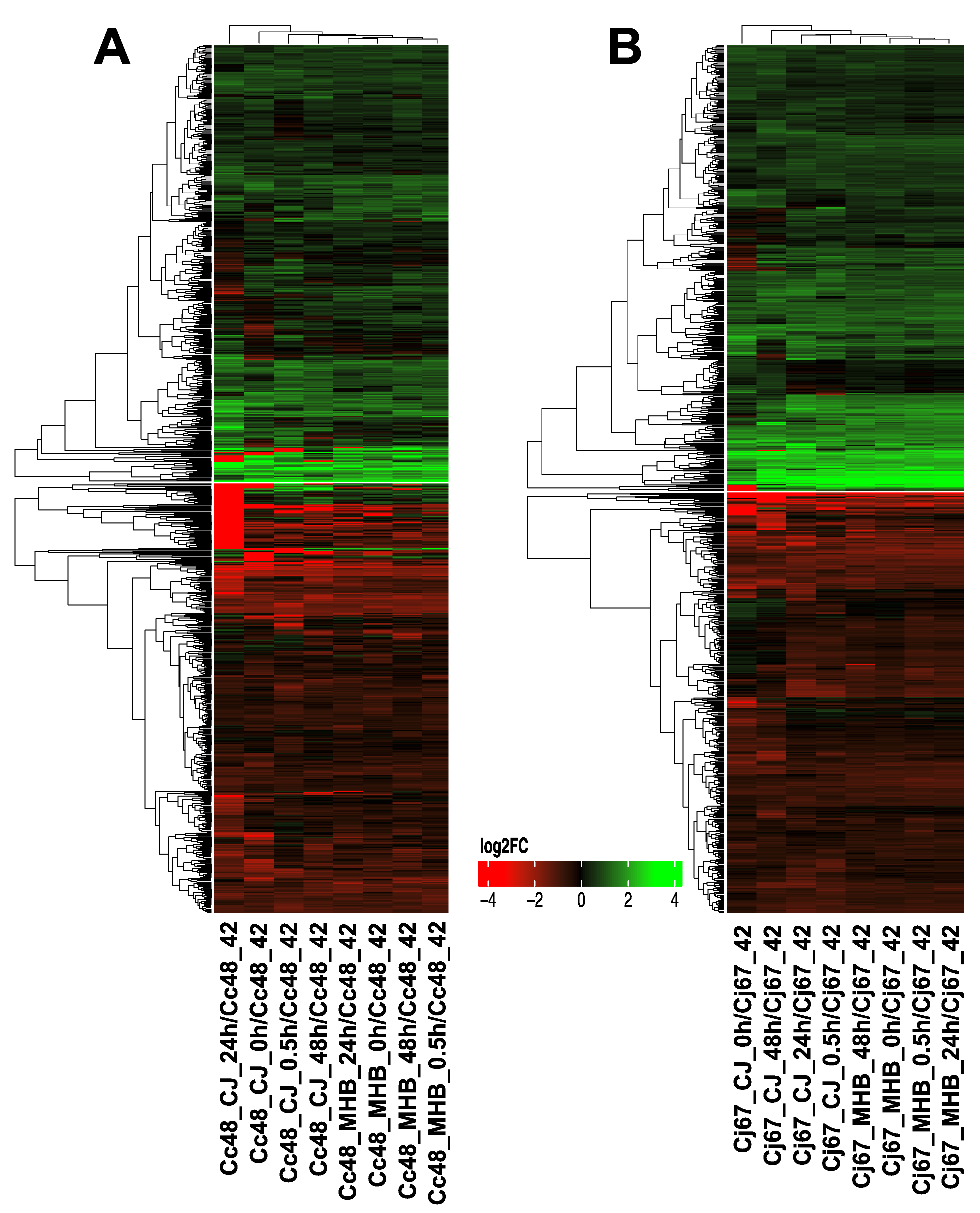
3.1. Overview of RNA-Seq

Raw sequence reads were trimmed for adapters using the CLC Genomic Workbench v. 20 with default settings; all ambiguous reads and reads less than 10 bp were discarded. Among the trimmed sequence reads from microaerobic incubation of C. coli HC2-48 (sample Cc48_42) and C. jejuni OD2-67 (Cj67_42) at 42 °C, approximately 97% mapped to reference sequences. Similarly, over 93% of sequence reads from MHB samples of C. coli HC2-48 (Cc48_MHB_0h, Cc48_MHB_0.5h, Cc48_MHB_24h and Cc48_MHB_48h) and C. jejuni OD2-67 (Cj67_MHB_0h, Cj67_MHB_0.5h, Cj67_MHB_24h and Cj67_MHB_48h) mapped to reference sequences. However, only 17% to 39.14% of sequence reads from samples incubated in CJ (C. coli samples Cc48_CJ_0h, Cc48_CJ_0.5h, Cc48_CJ_24h and Cc48_CJ_48h; C. jejuni samples Cj67_CJ_0h, Cj67_CJ_0.5h, Cj67_CJ_24h and Cj67_CJ_48h) were mapped to reference sequences. Details regarding mapping percentages of individual samples are listed in Table S1.

3.2. Influence of Temperature on Gene Expression (4 °C vs. 42 °C)

From a total of 1651 analyzed genes from C. coli HC2-48, 831 were differentially expressed at 4 °C vs. 42 °C (fold-change ≥ 1.5 or ≤−1.5 fold; FDR < 0.05) in MHB and/or CJ at one or more sampling times (0 h, 0.5 h (30 min), 24 h, 48 h). Similarly, 808 genes out of 1774 were differentially expressed at 4 °C vs. 42 °C in C. jejuni OD2-67 in MHB and/or CJ at one or more different sampling times. Details regarding the number of genes upregulated or downregulated at the four incubation times in MHB and CJ are shown in Table 1 and Figure 1.
C. coli HC2-48: Only 49 genes in HC2-48 showed significant levels of differential expression in both MHB and CJ at all incubation times (Table 2 and Table S3). The 13 upregulated DEGs included ppa (inorganic diphosphatase), petC (cytochrome C1), rplM, rplQ, rpsI (ribosomal proteins) and the lipolysaccharide (LPS) gene, lptF (LptF/LptG family permease). The 36 downregulated genes included ten associated with flagella assembly (flhB, fliD, flgL, flgG, flaG, fliS, flgH, flgB, flgF, flgK, flgM, flgN). Meanwhile, one hundred twenty-six genes were differentially expressed in MHB at 4 °C vs. 42 °C in all incubation times (Table S4), whereas sixty-five genes were differentially expressed in CJ at 4 °C vs. 42 °C in all four incubation times (Table S5).
Six genes were significantly upregulated in MHB but not in CJ at all incubation times. These DEGs included the following: lptD, LPS assembly protein; tolA, TonB C-terminal domain-containing protein; murE, UDP-N-acetylmuramoyl-L-alanyl-D-glutamate-2, 6-diaminopimelate ligase; AR446_RS05150, hypothetical protein; AR446_RS04310, DUF342- domain-containing protein; and lptA, LPS periplasmic transport protein. Two genes, rpmL (50S ribosomal protein L35) and shlA (filamentous hemagglutinin N-terminal-domain-containing protein) were downregulated in MHB but not in CJ. However, no genes were identified that were significantly expressed in CJ but not in MHB.
C. jejuni OD2-67: Fifty eight genes showed significant differential expression in MHB and CJ at 4 °C vs. 42 °C in all incubation times (Table 3 and Table S6). In MHB, 195 genes were differentially expressed at 4 °C vs. 42 °C in all incubation times (Tables S7 and S8), and 71 genes were significantly expressed in all CJ samples (Table S9). Genes that were expressed in MHB but not in CJ included four upregulated genes (pgpA, cmeR, tssJ and macB1) and four downregulated genes (ndk, corC, A0W68_RS03455 (HAD-IB family hydrolase) and A0W68_RS06390 (hypothetical protein)). Two genes were significantly expressed in CJ but not in MHB; these included upregulated corA (magnesium/cobalt transporter) and downregulated A0W68_RS09110 (NYN-domain-containing protein).

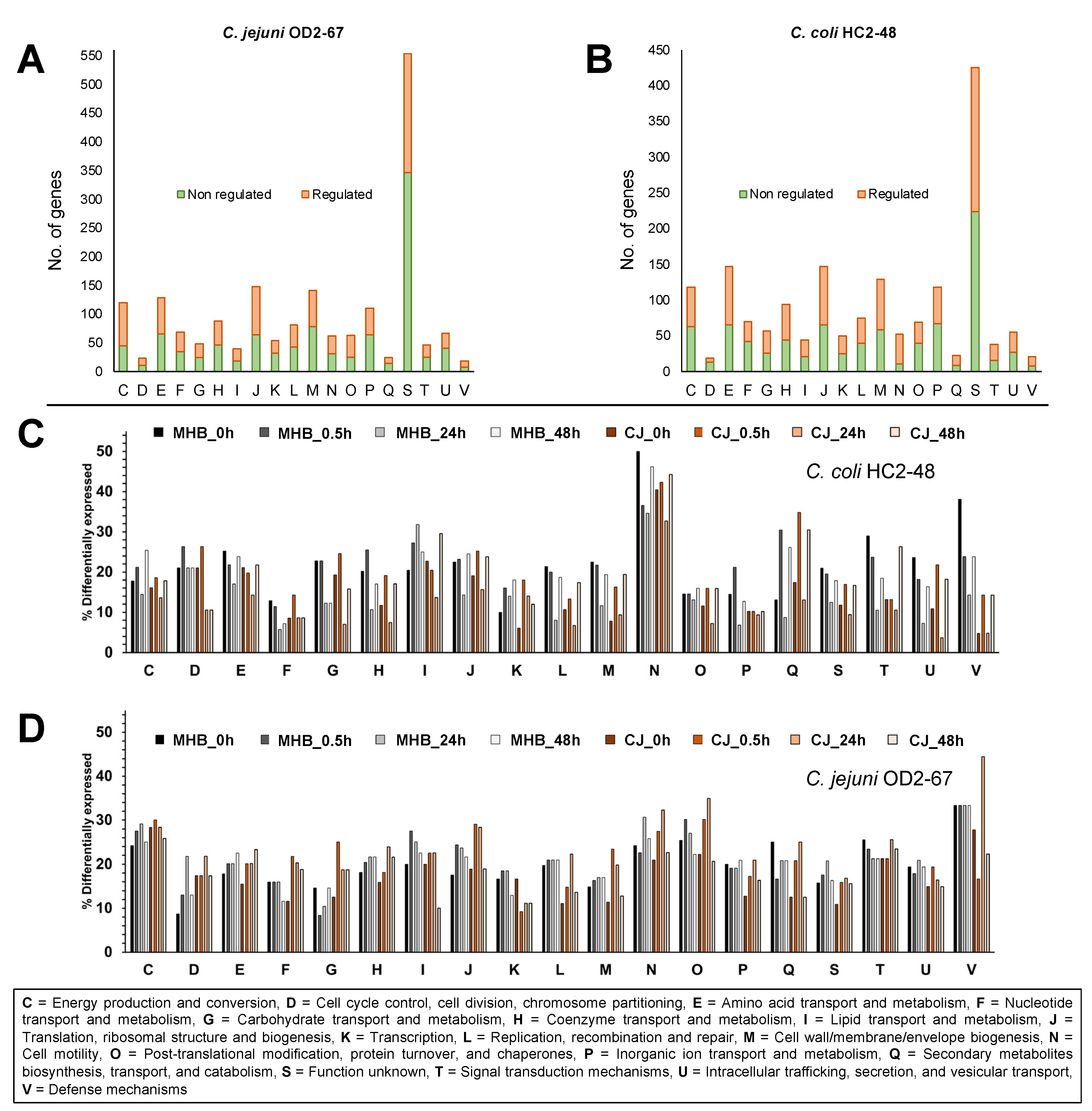
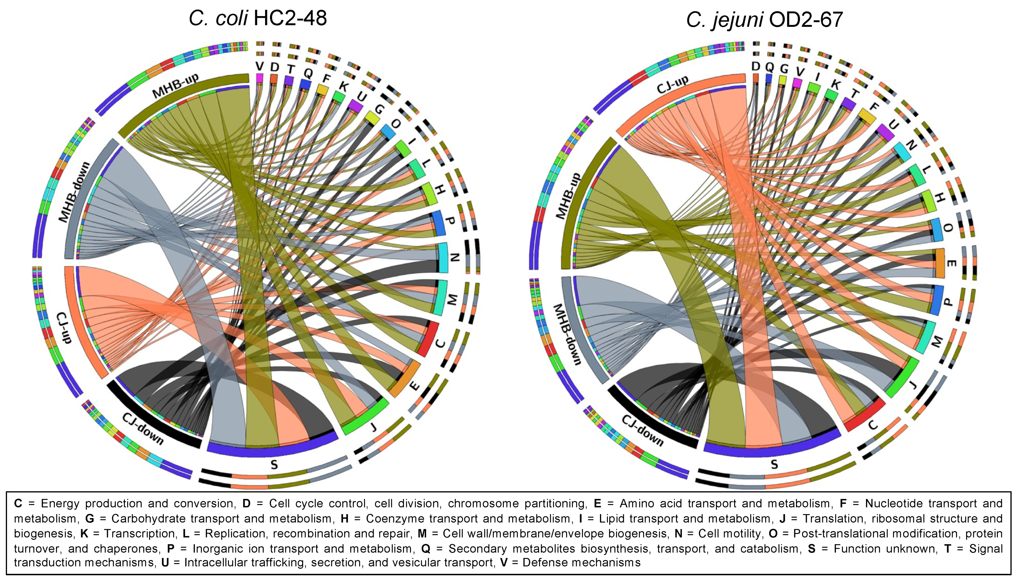
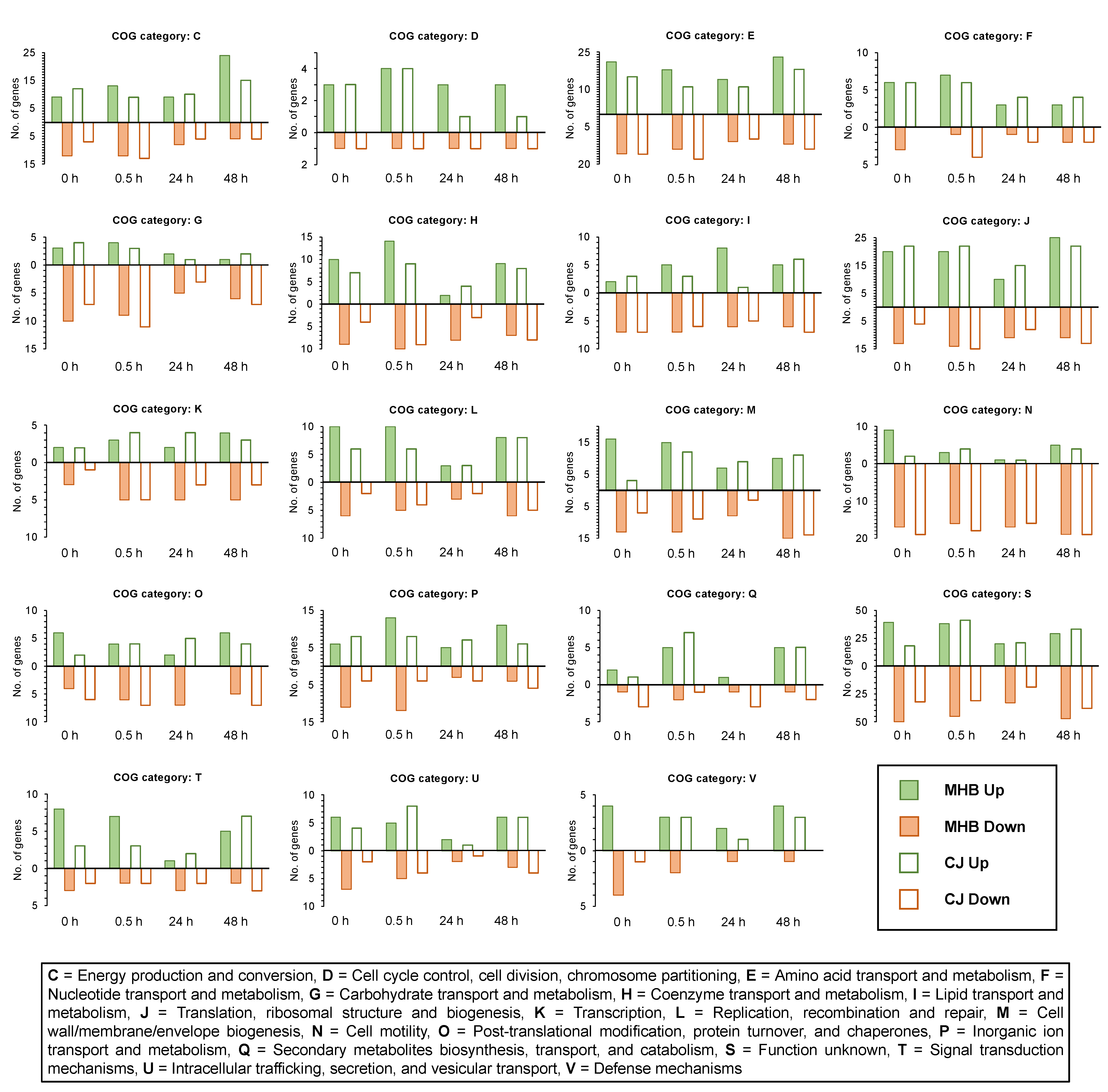
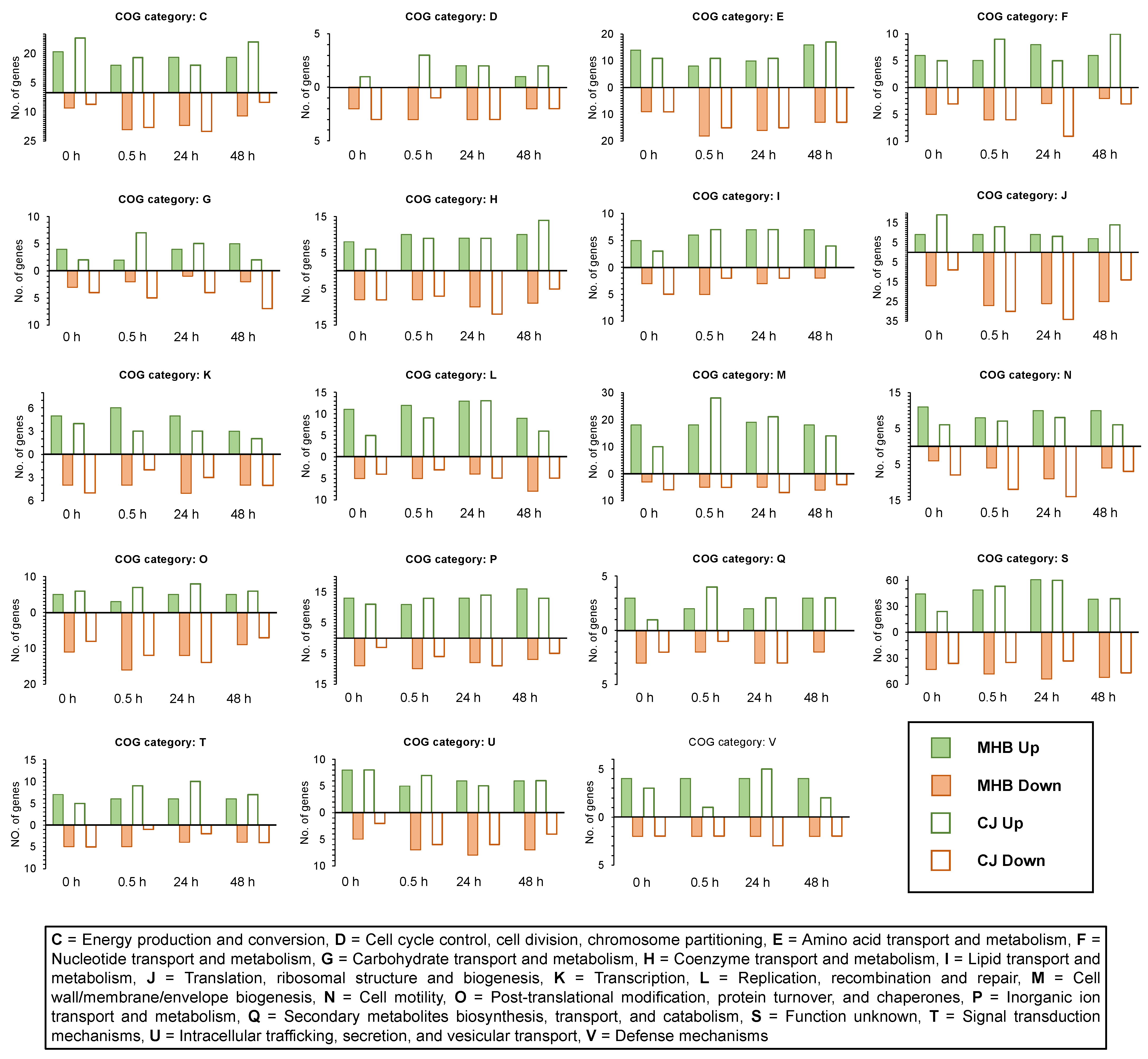
3.3. Functional Group Assignments of DEGs

Among DEGs expressed (4 °C vs. 42 °C) at one or more incubation times, 10.83% of C. coli HC2-48 and 25.62% of C. jejuni OD2-67 genes were assigned to the ‘unknown function’ category (Figure 2A,B, group S).
The highest percentage of differential expression in C. coli HC2-48 was observed for cell motility (group N), and this group assignment was consistent for all incubation times and medium formulations (Figure 2C). For C. jejuni OD2-67, genes from energy production/conversion, cell motility, post-translational modification/chaperones and defense mechanisms (groups C, N, O and V, respectively) were differentially expressed at 4 °C in both media (Figure 2D).
In general, larger fold-change values were observed for upregulated C. coli HC2-48 genes in both media than for downregulated genes; however, exceptions were genes in the following functional groups: N (cell motility), I (lipid transport/metabolism), O (post-translational modification/chaperones) and G (carbohydrate transport/metabolism) (Figure 3 and Figure 4). Accumulative expression values (fold-change) for the genes related to amino acid transport/metabolism (group E) in C. coli were greater for upregulated genes than downregulated genes in MHB, but it was found to be the opposite in CJ. In C. jejuni OD2-67, downregulated genes in functional group J (translation/ribosome structure, O (post-translational modification, chaperones) and D (cell cycle control, cell division, chromosome partitioning) had larger fold-changes values than upregulated genes (Figure 3 and Figure 5). However, accumulative fold-change values of upregulated genes in functional groups C, M, P, H, L, U, F, T, I, V and Q for both media were higher than downregulated genes from respective functional groups (Figure 3 and Figure 5).
Among the DEGs of C. coli HC2-48 found in both media (CJ and MHB) and at all incubation times, gene enrichment analysis in STRING found significant enrichment (FDR < 0.05) in the KEGG pathway for flagellar assembly (https://www.genome.jp/dbget-bin/www_bget?ko02040, accessed on 9 March 2020). Although C. jejuni OD2-67 lacked a KEGG pathway that was enriched for all incubation times and media, Gene Ontology analysis showed that the ribosome/ribonucleoprotein complex was significantly enriched (FDR < 0.05) for all sampling times in MHB.

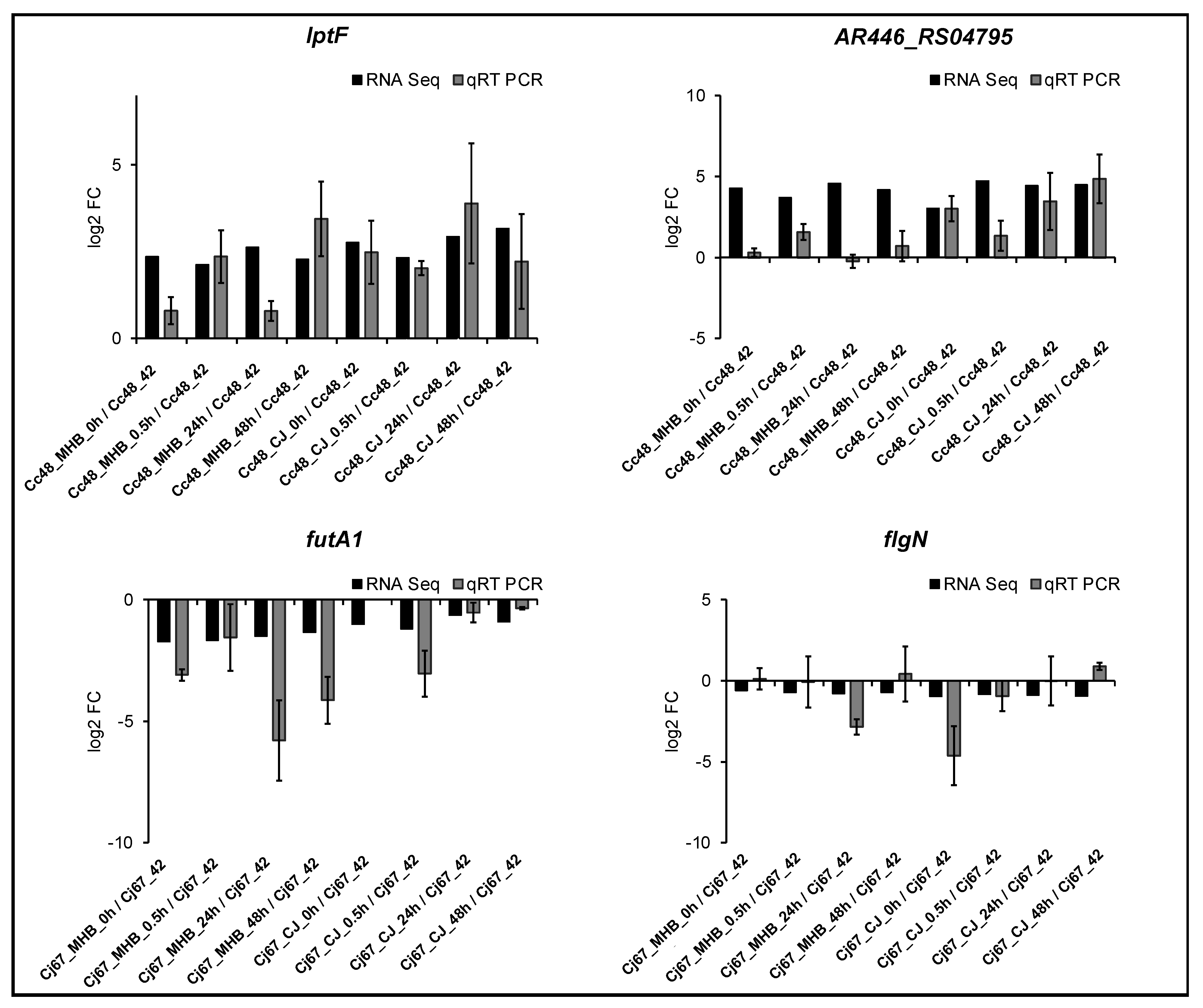
3.4. Validation of RNA-Seq Data by qRT-PCR

Four DEGs with distinct expression patterns were used to confirm RNA-seq data by qRT-PCR. Two upregulated genes, namely lptF and AR446_RS04795, were chosen to validate RNA-seq results for C. coli HC2-48, and the downregulated genes futA1 and flgN were used to validate results for C. jejuni OD2-67. The expression profiles obtained by qRT-PCR were consistent with results obtained with RNA-seq, indicating that the RNA-seq data are reliable (Figure 6).

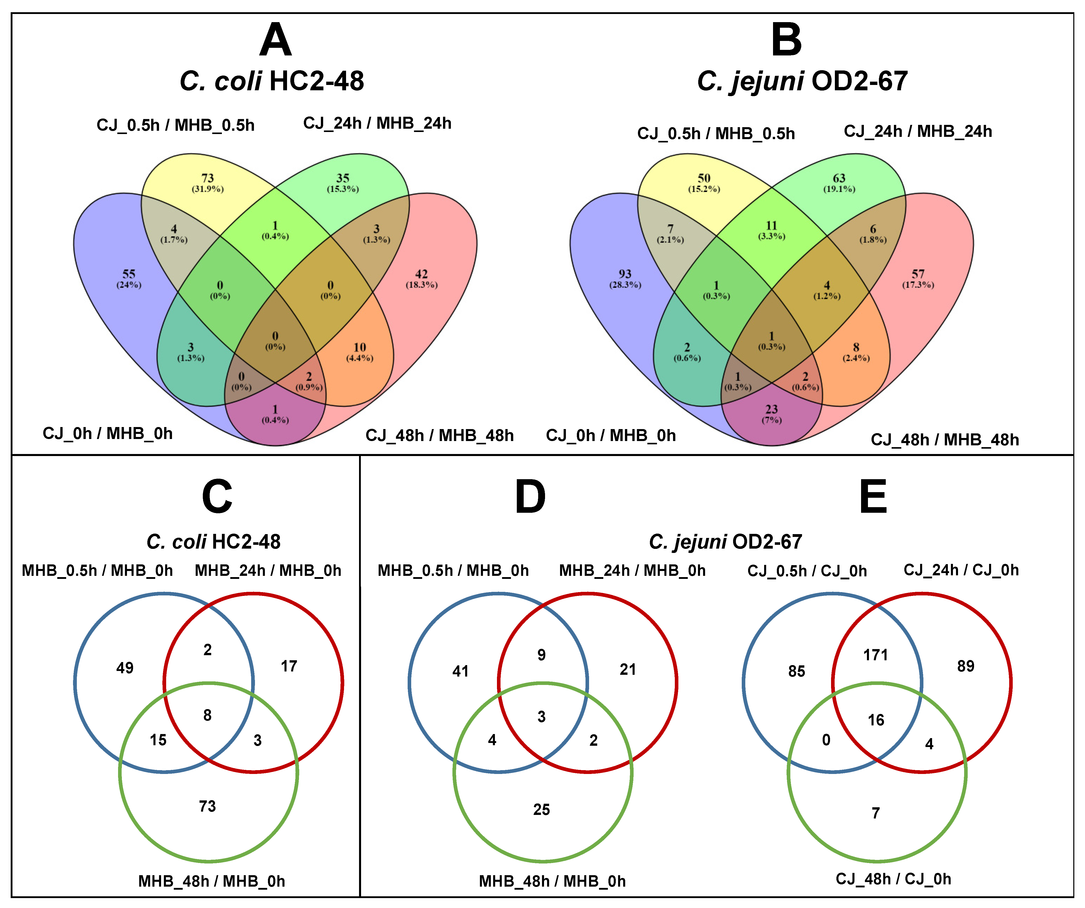
3.5. Common DEGs in C. coli and C. jejuni (4 °C vs. 42 °C)

Analysis of RNA-seq data indicated that 1386 and 1385 genes from C. coli HC2-48 and C. jejuni OD2-67, respectively, were shared orthologs. However, only 11 genes had fold-change values with FDR < 0.05 at all incubation times and in both media and strains (Table 4 and Table S10), but only two genes, fliS and flgN, were differentially expressed with significant fold change (fold-change values ≥ 1.5 or ≤−1.5 and FDR < 0.05 for both strains at all data points in both media.
Common DEGs with significance differences (fold-change ≤−1.5 fold) in CJ included downregulated hemE, flhB, flaG, fliS and flgN at all incubation time points. Three genes including mug (DNA-deoxyinosine glycosylase), amaA (AI-2E family transporter) and AR446_RS04310/A0W68_RS04275 (DUF342-domain-containing protein) were upregulated in MHB for both strains at all times. Common downregulated genes for the two strains in MHB included cysK (cysteine synthase A), rpmI, cfa (class I SAM-dependent methyltransferase) and the flagellar genes fliS, flgG and flgN.
Forty-one genes exhibited a positive fold change in expression (significant or nonsignificant) in C. coli HC2-48 but had negative fold-changes values (significant or nonsignificant) in C. jejuni OD2-67. This group included genes involved in amino acid transport and metabolism (e.g., ilvF, ilvK, hisD, hisF, cysE, pfs, dapE) and translation and ribosomal structure (rimP, thrS, rpiL, mnmE, leuS, pnp, tyrS, tsaD, rpsD, rpsC, rplW). Fifty-eight genes exhibited positive fold-change values for C. jejuni OD2-67 but negative fold-change values for C. coli HC2-48. This group included genes for amino acid transport and metabolism (aroQ, aroB, proA, proC, sstT) and genes involved in cell wall and membrane biogenesis (lpxB, kpsC, murC, rfaD, galU, murB).

3.6. Influence of Medium Formulation on Gene Expression (CJ vs. MHB)

Although most genes in the two Campylobacter spp. had similar expression patterns (Figure 7), some genes were differentially expressed in the different medium formulations (CJ vs. MHB) (Figure 8). In C. coli HC2-48, 229 DEGs were significantly expressed in CJ vs. MHB at one or more sampling times (Cc48_CJ_0h/Cc48_MHB_0h, Cc48_CJ_0.5h/Cc48_MHB_0.5h, Cc48_CJ_24h/Cc48_MHB_24h and Cc48_CJ_48h/Cc48_MHB_48h). Among these DEGs, no gene was significantly expressed for all sampling times, but 31 genes had positive fold-change values in CJ vs. MHB (Tables S11 and S12), and 23 genes had negative fold-change values in CJ vs. MHB at all sampling times (Table S13). Among the orthologs, torD (molecular chaperone TorD family protein) was upregulated (at one or more sampling times) for both C. jejuni and C. coli in CJ when compared to MHB at 4 °C. Similarly, katA (catalase) was downregulated (at one or more sampling times) for both species in CJ as compared to MHB at 4 °C.
In C. jejuni OD2-67, 329 DEGs showed significant expression values in CJ at one or more incubation times when compared to MHB at 4 °C (Cj67_CJ_0/h/Cj67_MHB_0h, Cj67_CJ_0.5h/Cj67_MHB_0.5h, Cj67_CJ_24h/Cj67_MHB_24h and Cj67_CJ_48h/Cj67_MHB_48h). Only one gene A0W68_RS09275 (EexN family lipoprotein) had significant expression levels (downregulated) at all incubation times (Table S14). Excluding common genes, 45 DEGs had positive fold-change values (significant or nonsignificant) for CJ vs. MHB at all incubation times (Table S15); similarly, 55 DEGs had negative fold-change values (significant or nonsignificant) for CJ vs. MHB at all incubation times (Table S16).

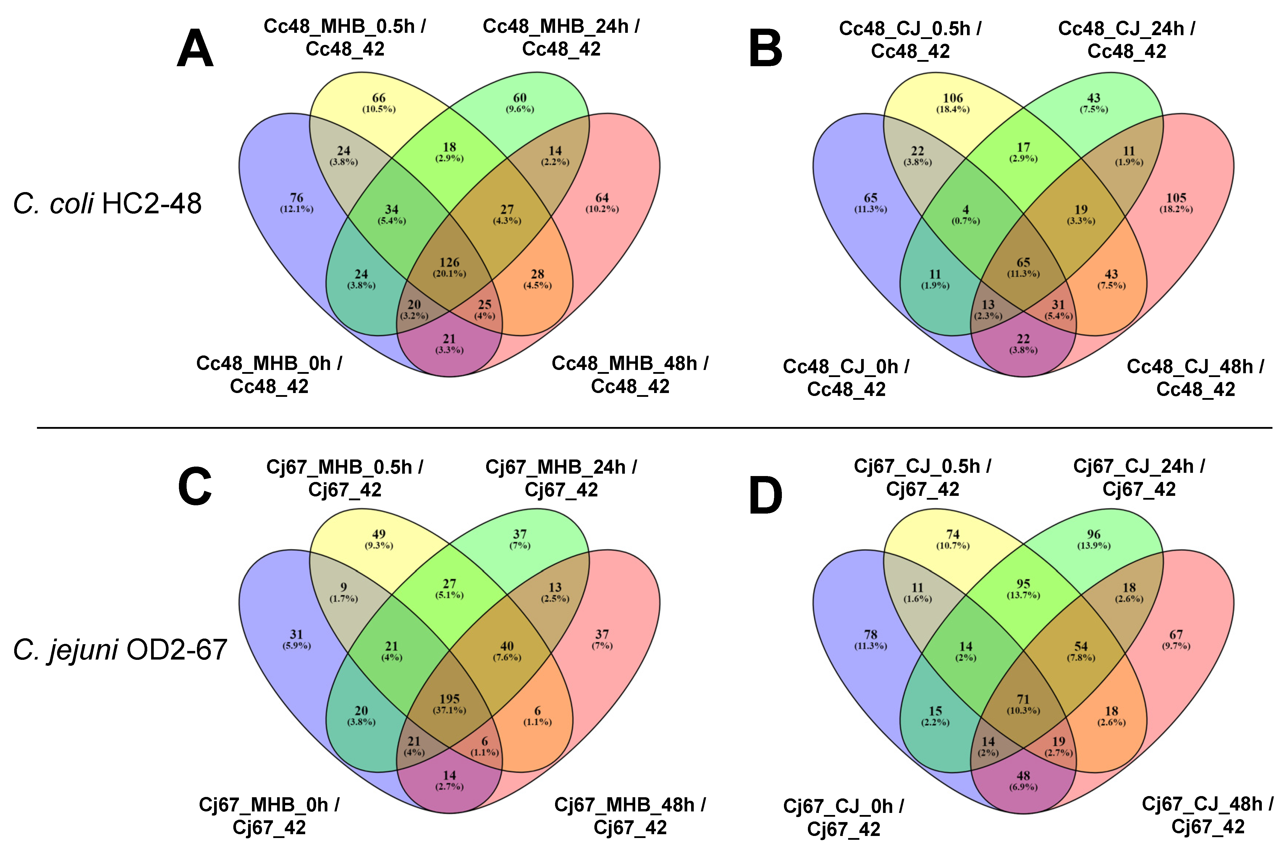
3.7. Influence of Incubation Time on Gene Expression (0.5 h vs. 0 h, 24 h vs. 0 h and 48 h vs. 0 h)

MHB medium and time of incubation: For C. coli HC2-48, genes were differentially expressed in MHB at 0.5 h (Cc48_MHB_0.5h), 24 h (Cc48_MHB_24h) and 48 h (Cc48_MHB_48h) at 4 °C compared to 0 h (Cc48_MHB_0h). Seventy-four DEGs (fifty-one upregulated, twenty-three downregulated), thirty (eighteen upregulated, twelve downregulated) and ninety-nine (forty-nine upregulated, fifty downregulated) were identified for Cc48_MHB_0.5h/Cc48_MHB_0h, Cc48_MHB_24h/Cc48_MHB_0h and Cc48_MHB_48h/Cc48_MHB_0h, respectively. Only eight genes were differentially regulated at three incubation times (0.5 h, 24 h and 48 h) compared to 0 h at 4 °C in MHB (Tables S17 and S18). Downregulated genes at 0.5 h included dnaJ, purH, cfrA, ribosomal genes (rplS, rpmH, rpmJ), flagellar genes (flgJ, flaG) and hspR (heat shock protein). Upregulated DEGs included genes involved in coenzyme transport/metabolism (mqnA, cca, hemA, ispA, coaD, mob), translation/ribosomal structure/biogenesis (prmA, def, fmt), transport (AR446_RS06920, AR446_RS07750, mdtL, mdtJ, AR446_RS04345, AR446_RS08130) and multidrug ABC transporter permease (AR446_RS00345, AR446_RS02400). At 24 h and 4 °C, maf, metE, ttcA, rub, virB11, nuoE and hyaE were upregulated; downregulated genes included radA and clpX. Various genes related energy production and conversion (nuoJ, nuoN, hydD, lldP, napH, dsbD, nuoL), nucleotide metabolism (purB, pyrB, pyrF), ribosomal structure (rpsC, rpsE, rpsS, prmA) and transport (gltJ, tcyB, tctB, dcuA, matE) were upregulated at 48 h and 4 °C. However, many genes were downregulated at 48 h and 4 °C as compared to 0 h, including those involved in amino acid transport/metabolism (gmhB, pepF, argC, proB), ribosomal structure (gatC, yqeV, rplS, rpmH, rpmJ, rpsP, rpsT) and heat shock (hspR).
For C. jejuni OD2-67 incubated in MHB at 4 °C, 57 genes were differentially expressed at 0.5 h (40 upregulated, 17 downregulated), 35 at 24 h (26 upregulated, 9 downregulated) and 34 at 48 h (12 upregulated, 22 downregulated) as compared to 0 h. Meanwhile, only three DEGs (two upregulated, one downregulated) showed significant fold-change values at 0.5 h, 24 h and 48 h as compared to 0 h in MHB (Figure 8, Table S19); these were upregulated genes A0W68_RS01005 (hypothetical protein) and A0W68_RS09275 (EexN family lipoprotein) and the downregulated gene A0W68_RS02685 (hypothetical protein). At 0.5 h, genes encoding hypothetical proteins, translation-related proteins (ffmJ, yabO and rpmJ), and inorganic ion transporters (A0W68_RS00285, A0W68_RS07265 and modB) were upregulated. Genes encoding threonine synthase (thrC), lipid transport and metabolism (plsC, pssA), and ribosomal protein RplT were downregulated at 0.5 h. A0W68_RS04835 (putative membrane protein), A0W68_RS06415 (exopolyphosphatase), lpxH (UDP-2,3-diacylglucosamine diphosphatase), hypA and vgrG (type VI secretion system) were among the upregulated genes at 24 h as compared to 0 h. Downregulated genes at 24 h included ldH (L-lactate dehydrogenase), rplT and A0W68_RS01575 (TonB-dependent receptor). At 48 h, leuA and metF (amino acid transport/metabolism, tonB (energy transducer TonB) and moaC and ropZ were upregulated. Genes that were downregulated at 48 h in MHB included ldH, virB11, torD, tssE and many genes encoding hypothetical proteins.
CJ medium and time of incubation: In C. coli HC2-48, 61 DEGs (21 upregulated, 40 downregulated) were identified at 0.5 h (Cc48_CJ_0.5h/Cc48_CJ_0h), one upregulated gene was identified at 24 h (Cc48_CJ_24h/Cc48_CJ_0h), and no genes showed significant upregulation at 48 h (Cc48_CJ_48h/Cc48_CJ_0h) (Table S20). Gene fliK was the only upregulated gene at both 0.5 hand 24 h. Upregulated genes at 0.5 included sdaC, glnH, pgi, acpP, efp, rim, cmeD, djlA, radA, uraH and virB4. Downregulated DEGs at 0.5 included genes related to energy production and conversion (aldA, fdhA, hydA, oorA, mdh, frdA, lutB), amino acid transport/metabolism (leuA), nucleotide transport/metabolism (atpD, guaB, sucC), carbohydrate metabolism (uxaA, glmM, eno, fbaA) and translation (rluD, proS, fmt, rplO, rplC, rplD, rplP, rplV, rpsD, rpsJ, rpsK).
For C. jejuni OD2-67, differential expression of genes in CJ was observed at 0.5 h (Cj67_CJ_0.5h), 24 h (Cj67_CJ_24h) and 48 h (Cj67_CJ_48h) as compared to 0 h (Cj67_CJ_0h). Two hundred seventy-two DEGs (one hundred fifteen upregulated, one hundred fifty-seven downregulated) were identified at 0.5 h, two hundred eighty DEGs (one hundred fourteen upregulated and one hundred sixty-six downregulated) at 24 h and twenty-seven DEGs (eleven upregulated, sixteen downregulated) at 48 h (Table S21). Excluding common genes in Table S22, 61 more genes were upregulated (Table S23) and 110 genes were downregulated (Table S24) at 0.5 hand 24 h.

4. Discussion

Campylobacter strains (C. coli HC2-48 and C. jejuni OD2-67) were isolated from retail liver products {C. coli HC2-48 (retail beef liver) and C. jejuni OD2-67 (retail chicken liver)} [4,6]. Meanwhile, C. coli HC2-48 had been identified as an aerotolerant strain (could survive up to 12 h of aerobic incubation), but C. jejuni OD2-67 was found to be aero-sensitive [7]. Both strains harbored chromosome of about 1.7 Mb size, but C. coli HC2-48 carried one plasmid (pCCDM1) [17] and C. jejuni OD2-67 possessed two plasmids pCJDM67L and pCJDM67S [16]. The megaplasmid pCJDM67L (117 Kb) found in C. jejuni OD2-67 harbored genes encoding tetracycline resistance and core genes of type VI secretion system [16].
Prolonged survival of Campylobacter species at lower temperatures is influenced by multiple factors including nutrient availability, environment and inherent characteristics of the strain [1,2,22,23]. A nutrient-rich environment such as CJ provides a nutritional and protective environment that enhances Campylobacter survival at lower temperatures [1]. In a previous study in our laboratory, both Campylobacter strains (C. coli HC2-48 and C. jejuni OD2-67) used in this study could not produce colonies after two days of incubation in MHB medium at 4 °C but could survive up to fourteen days or more in other tested media (chicken liver juice, beef liver juice, chicken juice and beef juice) at 4 °C [1]. However, Campylobacter strains can also survive for long periods in nutrient-poor environment despite losing cultivability [11]. Transcriptomic analysis of these bacteria during lower temperatures provides insight into their survival mechanisms and is a better approach for studying gene expression in variable environments [24]. Furthermore, changes in the incubation period, medium composition, temperature and atmospheric conditions alters expression in Campylobacter genomes [9,12,13]. In the current study, RNA-seq revealed that a large number of genes in two Campylobacter spp. were impacted by temperature fluctuations (from 42 °C to 4 °C). At 4 °C, medium composition (CJ vs. MHB) and incubation time also altered the Campylobacter transcriptome.
In this study, relatively few orthologous genes were differentially expressed in both Campylobacter spp. when data were compared for temperature, medium formulation and incubation time. For example, our results show that mreB, which is involved in bacterial cell shape, was upregulated for C. coli but downregulated for C. jejuni. Temperature fluctuations are known to differentially impact Campylobacter gene expression; for example, the transcriptomes of C. coli and C. lari showed considerable variability in response to heat stress [15].
The oxidative stress response is reportedly a component of the Campylobacter response to cold shock [10,11,25], and the oxidative stress response genes katA and sodB, were previously upregulated at low temperatures in C. jejuni [25]. A previous study demonstrated that trxB, which encodes thioredoxin-disulfide reductase, had a potential role in the response of C. jejuni to oxidative stress [26]; however, in the current study, trxB was downregulated for both C. jejuni and C. coli in CJ and MHB media at all incubation times. Although msrP was downregulated in C. jejuni OD2-67 at all incubation times and in both media, it was either downregulated or unaltered in C. coli HC2-48. MsrP helps to repair oxidized proteins in the bacterial envelope during oxidative stress [27], and the oxidative stress response during cold shock might vary with the Campylobacter strain, medium and time of incubation. Although temperature fluctuations were closely monitored in this study, the exposure of bacterial samples to atmospheric oxygen during sample processing might cause in discrepancies in the oxidative stress response and gene expression. The iron concentration in media also influences the expression of genes involved in oxidative stress; for example, katA, cj1386, ahpC and trxB were repressed by iron [28]. Likewise, iron ions also reported to play role in the formation of oxygen radicals mediated by Fenton reaction in the bacterial cells [29,30,31,32,33]. Thus, variation in the iron content of CJ might have impacted katA expression in the current study. Interestingly, upregulation of fur (transcriptional repressor) was found in MHB for C. coli HC2-48 {Cc48_MHB_(0.5 h, 24 h, 48 h) vs. Cc48_MHB_0h} and in CJ for C. jejuni OD2-67 {Cj67_CJ_(0.5 h, 24 h, 48 h) vs. Cj67_CJ_0h} and might explain the reduced expression of genes related to oxidative stress and iron acquisition [28,34]. Meanwhile, perR, a transcriptional regulator that also plays role in oxidative stress and iron metabolism, was found upregulated in CJ for C. coli HC2-48 at 0.5 h {for both Cc48_CJ_0.5h/Cc48_42 and Cc48_CJ_0.5h/Cc48_MHB_0.5h) [28,34].
ClpB (AAA family ATPase) functions in various stress responses (oxidative, heat shock, starvation) and virulence in bacteria [2,35]. In our study, clpB was significantly downregulated in both Campylobacter spp. at low temperatures as compared to 42 °C in CJ and MHB. In a previous report, clpB expression in C. jejuni was induced by heat shock and repressed at low temperatures in MHB [13]. It was proposed that heat shock proteins such as ClpB, GroEL, GrpE, HrcA, CbpA, HspR, DnaK and GroES might not be required for survival at lower temperatures [13]. In the current study, these genes were generally downregulated or unaltered for the two Campylobacter strains. Hence, downregulation of heat shock response proteins might be common for Campylobacter spp. at temperatures lower than the optimum for growth.
A number of flagellar genes (flaG, fliS, flgG, flgP, flgN) were downregulated in the two Campylobacter spp. in both CJ and MHB and at all incubation times. A few flagellar genes (e.g., fliH, fliN and fliW) were upregulated or unaltered in all experiments; interestingly, fliK, fliW and fliY were upregulated, but flhB and flgC were downregulated in CJ vs. MHB at one or more incubation times in C. coli HC2-48. For C. jejuni OD2-67, flgM was downregulated in CJ vs. MHB at 0 and 48 h. Downregulation of genes encoding flagellar proteins at suboptimal temperatures was previously reported for C. jejuni and may help the bacterium to conserve energy during adverse environmental conditions [13]. Another study reported upregulation of flg and downregulation of fli genes in C. jejuni at low temperatures [9]. In the current study, a greater number of flagellar genes was impacted in C. coli vs. C. jejuni; furthermore, variability in the expression of flagellar genes might be caused by variation in Campylobacter strains and medium formulations [9,11,13].
Suboptimal temperatures impacted the expression of Campylobacter genes that function in translation and ribosome structure [13]. In the current study, the expression of genes with roles in translation and ribosomal structure/biogenesis was highly represented in both C. coli and C. jejuni. A large proportion of translation/ribosome genes was upregulated in C. coli HC2-48 but downregulated in C. jejuni OD2-67 (Figure 3). Maintenance of cellular function during cold temperatures is essential and preserves the functionality of translational machinery [13], and the expression of these genes varies among Campylobacter spp.
Genes for energy production and conservation were highly upregulated in both Campylobacter strains in this study; however, genes with significant expression in both strains at all incubation times and all medium formulations were limited. For example, ppa and petC encoding inorganic diphosphatase and cytochrome C1, respectively, were common upregulated genes for C. coli HC2-48 at 4 °C at all incubation times; however, ppa was unaltered, and petC was generally downregulated in C. jejuni OD2-67. Other genes including atpB, ccpA (cytochrome-c peroxidase), hdrC (FAD-binding oxidoreductase), lldP (L-lactate permease), nuoA (NADH quinone oxidoreductase, subunit 3) and nuoM (NADH quinone oxidoreductase, subunit M) were significantly upregulated in C. jejuni OD2-67 and were generally unaltered or significantly upregulated in C. coli HC2-48. These discrepancies show the interspecific variation in the transcriptome of Campylobacter spp. with respect to genes involved in energy production and conversion. In a prior report, the upregulation of lldP and other genes involved in macromolecule transport in C. jejuni was proposed to help the bacterium to acquire cryoprotectants that enhance survival at low temperatures [13]. In this study, DEGs related to amino acid transport and metabolism were less frequent and mostly downregulated. In contrast, genes involved in cell wall/membrane/envelope biogenesis were generally upregulated in the current study, which supports previous reports indicating that modifications in cell membrane structure might function in the cold-shock response [9,11,13].
Multiple genes encoding the type VI secretion system (T6SS) were upregulated in C. jejuni OD2-67 at 4 °C in CJ and MHB at all incubation times. A previous study reported that the T6SS enhanced the oxidative stress response, host colonization and virulence of Campylobacter strains [36,37]. Among the genes related to twin arginine translocation (TAT) system and Sec dependent pathways, tatC was generally upregulated in C. coli HC2-48, whereas secE and secF were upregulated or unaltered, and secG was downregulated or unaltered. Hence, it seems probable that translocation/secretion systems are important in C. coli HC2-48 at 4 °C. Meanwhile, secY was upregulated and secE was downregulated at one or more time points in C. jejuni OD2-67. No significant change in expression was observed for tat genes in C. jejuni OD2-67.
In this study, many Campylobacter genes were differentially expressed in the two medium formulations at 4 °C. However, a previous report using microarrays identified only eight genes with significantly different expression in CJ as compared to brain heart infusion (BHI) medium at 5 °C [12]. This discrepancy might be attributed to the higher sensitivity of RNA-seq as compared to microarray analysis [28]. Despite the high number of DEGs identified in the present study, we did not identify a common gene in C. coli HC2-48 that was differentially expressed in CJ vs. MHB medium at all incubation times. Only one gene A0W68_RS09275 (EexN family lipoprotein) was significantly expressed (downregulated) at all incubation times for C. jejuni OD2-67 and was plasmid-encoded. Hence, medium composition is unlikely a defining factor in differential expression of Campylobacter genes at low temperatures for prolonged time periods. In a previous report, luxS was upregulated in CJ vs. BHI and was proposed to function in adaptation to the CJ environment [12]. In the current study, luxS had a positive-fold-change value for all incubation times in C. coli HC2-48 but was only significantly upregulated at 48 h; in contrast, luxS expression was not differentially regulated in C. jejuni OD2-67. A gene related to iron transport (A0W68_RS08550, FTR1 family iron permease) was significantly upregulated for all incubation times except 48 h in C. jejuni OD2-67 cultivated in CJ. At one or more incubation times, a gene torD (cytoplasmic chaperone TorD) was differentially regulated in CJ vs. MHB in both Campylobacter strains; it was upregulated at 4 °C when compared to 42 °C in C. coli HC2-48 but downregulated for C. jejuni OD2-67 in both medium formulations. TorD reportedly functions in maturation of prokaryotic molybdoenzyme TorA [38], which is involved in electron transfer during anaerobic respiration [39].
A previous study identified various essential genes in C. jejuni for survival at low temperatures in nutrient-rich and nutrient-poor media [10]. Among the identified genes, trxC was described as having a fundamental role in the oxidative stress response [10]. In the current study, trxC was generally downregulated at 4 °C as compared to 42 °C in the two Campylobacter spp. and in both CJ and MHB. Two-component signal transduction systems have also been described as essential for survival at low temperatures [10]. In the two Campylobacter strains used in this study, the DccRS two-component system was notable, and dccS was significantly upregulated at most incubation times and in both media at 4 °C. Other upregulated genes in one or both Campylobacter strains included czcD (cation transporter), hisC (histidinol-phosphate transaminase) and purN (phosphoribosylglycinamide formyltransferase).
Differential expression of various genes happened immediately after the temperature shifted from 42 °C to 4 °C; furthermore, some genes continued to be expressed at low temperatures, potentially due to differences in medium composition. In CJ, the number of significantly expressed genes decreased as time of incubation increased for C. coli HC2-48. Similarly, fewer genes were differentially expressed at 48 h (Cj67_CJ_48h/Cj67_CJ_0h) in C. jejuni OD2-67. However, unlike samples in CJ, a rapid decline in DEGs was not observed in MHB. No distinctive trends were observed for differences in expression with incubation time in the two media; for example, genes involved in motility and translation (ribosomal proteins) that were differentially expressed at 4 °C vs. 42 °C were not differentially expressed at other incubation times when compared to 0 h. Consequently, the expression of flagellar genes might not revert to original levels as suggested previously [13].
Many of the DEGs related to energy production/conversion in C. coli HC2-48 in MHB were upregulated after 0 h, which supports the contention that these functions are critical to survival at low temperatures [9,13]. Upregulation of maf and downregulation of ftsW indicate potential disruption of cell division since Maf inhibits septum formation and FtsW helps to recruit the transpeptidase FtsI to the division site [40,41]. Downregulation of AR446_RS01805 (RNA pyrophosphoryl hydrolase) with respect to incubation time indicates that lower temperatures might alter degradosome activity and increase mRNA stability. RNA pyrophosphoryl hydrolase functions in mRNA degradation [42], and inactivation of the degradosome during stress has been reported [43].
In summary, RNA-seq revealed that a large number of genes in two Campylobacter spp. were impacted by temperature (4 °C vs. 42 °C). Genes related to cellular motility and the ribosome were impacted by 4 °C in C. coli HC2-48 and C. jejuni OD2-67, respectively. Variations in gene expression were observed as a function of incubation time, but a complete reversal of gene expression was not observed during prolonged incubation (48 h) at 4 °C. Although multiple genes were significantly expressed in CJ when compared to expression in MHB, no gene for C. coli HC2-48 and only one gene for C. jejuni OD2-67 was found significantly expressed at all incubation times. Hence, food matrix composition is not the sole determining factor for differential gene expression in Campylobacter spp. at low temperature.

Supplementary Materials

The following supporting information can be downloaded at: https://www.mdpi.com/article/10.3390/pathogens12070960/s1.

Author Contributions

A.B.K. and M.K.F.; experimental procedures, A.B.K.; transcriptomic data analysis, A.B.K. and B.K. All authors have read and agreed to the published version of the manuscript.

Funding

This research received no external funding.

Institutional Review Board Statement

Not applicable.

Informed Consent Statement

Not applicable.

Data Availability Statement

All the original sequence reads for this experiment accessible in the Sequence Read Archive (SRA) database within Bio project id: PRJNA828109 (https://www.ncbi.nlm.nih.gov/sra?linkname=bioproject_sra_all&from_uid=828109, accessed on 9 March 2020).

Acknowledgments

The authors would like to acknowledge financial support from the Research Office of The University of Tulsa (Tulsa, OK, USA) for granting Mohamed K. Fakhr a Faculty Research Grant.

Conflicts of Interest

The authors declare no conflict of interest.

References

  1. Karki, A.B.; Wells, H.; Fakhr, M.K. Retail Liver Juices Enhance the Survivability of Campylobacter jejuni and Campylobacter coli at Low Temperatures. Sci. Rep. 2019, 9, 2733. [Google Scholar] [CrossRef] [Green Version]
  2. Bronowski, C.; James, C.E.; Winstanley, C. Role of Environmental Survival in Transmission of Campylobacter jejuni. FEMS Microbiol. Lett. 2014, 356, 8–19. [Google Scholar] [CrossRef] [PubMed] [Green Version]
  3. Birk, T.; Ingmer, H.; Andersen, M.T.; Jørgensen, K.; Brøndsted, L. Chicken Juice, a Food-Based Model System Suitable to Study Survival of Campylobacter jejuni. Lett. Appl. Microbiol. 2004, 38, 66–71. [Google Scholar] [CrossRef]
  4. Noormohamed, A.; Fakhr, M.K. A Higher Prevalence Rate of Campylobacter in Retail Beef Livers Compared to Other Beef and Pork Meat Cuts. Int. J. Environ. Res. Public Health 2013, 10, 2058–2068. [Google Scholar] [CrossRef] [Green Version]
  5. Noormohamed, A.; Fakhr, M.K. Prevalence and Antimicrobial Susceptibility of Campylobacter spp. in Oklahoma Conventional and Organic Retail Poultry. Open Microbiol. J. 2014, 8, 130–137. [Google Scholar] [CrossRef] [Green Version]
  6. Noormohamed, A.; Fakhr, M.K. Incidence and Antimicrobial Resistance Profiling of Campylobacter in Retail Chicken Livers and Gizzards. Foodborne Pathog. Dis. 2012, 9, 617–624. [Google Scholar] [CrossRef]
  7. Karki, A.B.; Marasini, D.; Oakey, C.K.; Mar, K.; Fakhr, M.K. Campylobacter coli from Retail Liver and Meat Products Is More Aerotolerant than Campylobacter jejuni. Front. Microbiol. 2018, 9, 2951. [Google Scholar] [CrossRef] [PubMed]
  8. Oh, E.; Andrews, K.J.; McMullen, L.M.; Jeon, B. Tolerance to Stress Conditions Associated with Food Safety in Campylobacter jejuni Strains Isolated from Retail Raw Chicken. Sci. Rep. 2019, 9, 11915. [Google Scholar] [CrossRef] [Green Version]
  9. Moen, B.; Oust, A.; Langsrud, Ø.; Dorrell, N.; Marsden, G.L.; Hinds, J.; Kohler, A.; Wren, B.W.; Rudi, K. Explorative Multifactor Approach for Investigating Global Survival Mechanisms of Campylobacter jejuni under Environmental Conditions. Appl. Environ. Microbiol. 2005, 71, 2086–2094. [Google Scholar] [CrossRef] [PubMed] [Green Version]
  10. De Vries, S.P.W.; Gupta, S.; Baig, A.; Wright, E.; Wedley, A.; Jensen, A.N.; Lora, L.L.; Humphrey, S.; Skovgard, H.; MacLeod, K.; et al. Genome-Wide Fitness Analyses of the Foodborne Pathogen Campylobacter jejuni in In Vitro and In Vivo Models. Sci. Rep. 2017, 7, 1251. [Google Scholar] [CrossRef] [Green Version]
  11. Bronowski, C.; Mustafa, K.; Goodhead, I.; James, C.E.; Nelson, C.; Lucaci, A.; Wigley, P.; Humphrey, T.J.; Williams, N.J.; Winstanley, C. Campylobacter jejuni Transcriptome Changes during Loss of Culturability in Water. PLoS ONE 2017, 12, e0188936. [Google Scholar] [CrossRef] [PubMed] [Green Version]
  12. Ligowska, M.; Cohn, M.T.; Stabler, R.A.; Wren, B.W.; Brøndsted, L. Effect of Chicken Meat Environment on Gene Expression of Campylobacter jejuni and Its Relevance to Survival in Food. Int. J. Food Microbiol. 2011, 145, S111–S115. [Google Scholar] [CrossRef] [PubMed]
  13. Stintzi, A.; Whitworth, L. Investigation of the Campylobacter jejuni Cold-Shock Response by Global Transcript Profiling. Genome Lett. 2003, 2, 18–27. [Google Scholar] [CrossRef]
  14. Butcher, J.; Stintzi, A. The Transcriptional Landscape of Campylobacter jejuni under Iron Replete and Iron Limited Growth Conditions. PLoS ONE 2013, 8, e79475. [Google Scholar] [CrossRef] [PubMed] [Green Version]
  15. Riedel, C.; Förstner, K.U.; Püning, C.; Alter, T.; Sharma, C.M.; Gölz, G. Differences in the Transcriptomic Response of Campylobacter coli and Campylobacter Lari to Heat Stress. Front. Microbiol. 2020, 11, 523. [Google Scholar] [CrossRef]
  16. Marasini, D.; Fakhr, M.K. Complete Genome Sequences of Campylobacter jejuni Strains OD267 and WP2202 Isolated from Retail Chicken Livers and Gizzards Reveal the Presence of Novel 116-Kilobase and 119-Kilobase Megaplasmids with Type VI Secretion Systems. Genome Announc. 2016, 4, e01060-16. [Google Scholar] [CrossRef] [Green Version]
  17. Marasini, D.; Fakhr, M.K. Complete Genome Sequences of the Plasmid-Bearing Campylobacter coli Strains HC2-48, CF2-75, and CO2-160 Isolated from Retail Beef Liver. Genome Announc. 2016, 4, e01004-16. [Google Scholar] [CrossRef] [Green Version]
  18. Masek, T.; Vopalensky, V.; Suchomelova, P.; Pospisek, M. Denaturing RNA Electrophoresis in TAE Agarose Gels. Anal. Biochem. 2005, 336, 46–50. [Google Scholar] [CrossRef]
  19. Carroll, R.K.; Weiss, A.; Shaw, L.N. Rna-Sequencing of Staphylococcus Aureus Messenger RNA. In Methods in Molecular Biology; Humana Press Inc.: New York, NY, USA, 2016; Volume 1373, pp. 131–141. [Google Scholar]
  20. Huerta-Cepas, J.; Szklarczyk, D.; Heller, D.; Hernández-Plaza, A.; Forslund, S.K.; Cook, H.; Mende, D.R.; Letunic, I.; Rattei, T.; Jensen, L.J.; et al. EggNOG 5.0: A Hierarchical, Functionally and Phylogenetically Annotated Orthology Resource Based on 5090 Organisms and 2502 Viruses. Nucleic Acids Res. 2019, 47, D309–D314. [Google Scholar] [CrossRef] [Green Version]
  21. Pfaffl, M.W. Quantification Strategies in Real-Time PCR. In A-Z of Quantitative PCR; International University Line.: La Jolla, CA, USA, 2004; Volume 29, pp. 87–112. [Google Scholar]
  22. Boysen, L.; Knøchel, S.; Rosenquist, H. Survival of Campylobacter jejuni in Different Gas Mixtures. FEMS Microbiol. Lett. 2007, 266, 152–157. [Google Scholar] [CrossRef] [Green Version]
  23. Oh, E.; McMullen, L.M.; Chui, L.; Jeon, B. Differential Survival of Hyper-Aerotolerant Campylobacter jejuni under Different Gas Conditions. Front. Microbiol. 2017, 8, 954. [Google Scholar] [CrossRef] [PubMed] [Green Version]
  24. Haas, B.J.; Chin, M.; Nusbaum, C.; Birren, B.W.; Livny, J. How Deep Is Deep Enough for RNA-Seq Profiling of Bacterial Transcriptomes? BMC Genom. 2012, 13, 734. [Google Scholar] [CrossRef] [PubMed] [Green Version]
  25. Bang, D.D.; Madsen, M. Cytolethal Distending Toxins of Campylobacter jejuni: Genetics, Structure, Mode of Action, Epidemiology, and the Role of CDT in Campylobacter Pathogenesis. Genome Lett. 2003, 2, 73–82. [Google Scholar] [CrossRef]
  26. Palyada, K.; Sun, Y.Q.; Flint, A.; Butcher, J.; Naikare, H.; Stintzi, A. Characterization of the Oxidative Stress Stimulon and PerR Regulon of Campylobacter jejuni. BMC Genom. 2009, 10, 481. [Google Scholar] [CrossRef] [PubMed] [Green Version]
  27. Gennaris, A.; Ezraty, B.; Henry, C.; Agrebi, R.; Vergnes, A.; Oheix, E.; Bos, J.; Leverrier, P.; Espinosa, L.; Szewczyk, J.; et al. Repairing Oxidized Proteins in the Bacterial Envelope Using Respiratory Chain Electrons. Nature 2015, 528, 409–412. [Google Scholar] [CrossRef] [Green Version]
  28. Butcher, J.; Handley, R.A.; van Vliet, A.H.M.; Stintzi, A. Refined Analysis of the Campylobacter jejuni Iron-Dependent/Independent Fur- and PerR-Transcriptomes. BMC Genom. 2015, 16, 498. [Google Scholar] [CrossRef] [Green Version]
  29. Imlay, J.A. Pathways of Oxidative Damage. Annu. Rev. Microbiol. 2003, 57, 395–418. [Google Scholar] [CrossRef]
  30. Anjem, A.; Imlay, J.A. Mononuclear Iron Enzymes Are Primary Targets of Hydrogen Peroxide Stress. J. Biol. Chem. 2012, 287, 15544–15556. [Google Scholar] [CrossRef] [Green Version]
  31. Gu, M.; Imlay, J.A. Superoxide Poisons Mononuclear Iron Enzymes by Causing Mismetallation. Mol. Microbiol. 2013, 89, 123–134. [Google Scholar] [CrossRef] [Green Version]
  32. Imlay, J.A. Cellular Defenses against Superoxide and Hydrogen Peroxide. Annu. Rev. Biochem. 2008, 77, 755–776. [Google Scholar] [CrossRef] [Green Version]
  33. Flint, A.; Stintzi, A.; Saraiva, L.M. Oxidative and Nitrosative Stress Defences of Helicobacter and Campylobacter Species That Counteract Mammalian Immunity. FEMS Microbiol. Rev. 2016, 40, 938–960. [Google Scholar] [CrossRef] [PubMed] [Green Version]
  34. Holmes, K.; Mulholland, F.; Pearson, B.M.; Pin, C.; McNicholl-Kennedy, J.; Ketley, J.M.; Wells, J.M. Campylobacter jejuni Gene Expression in Response to Iron Limitation and the Role of Fur. Microbiology 2005, 151, 243–257. [Google Scholar] [CrossRef] [PubMed]
  35. Alam, A.; Bröms, J.E.; Kumar, R.; Sjöstedt, A. The Role of ClpB in Bacterial Stress Responses and Virulence. Front. Mol. Biosci. 2021, 8, 668910. [Google Scholar] [CrossRef]
  36. Liaw, J.; Hong, G.; Davies, C.; Elmi, A.; Sima, F.; Stratakos, A.; Stef, L.; Pet, I.; Hachani, A.; Corcionivoschi, N.; et al. The Campylobacter jejuni Type VI Secretion System Enhances the Oxidative Stress Response and Host Colonization. Front. Microbiol. 2019, 10, 2864. [Google Scholar] [CrossRef]
  37. Marasini, D.; Karki, A.B.; Bryant, J.M.; Sheaff, R.J.; Fakhr, M.K. Molecular Characterization of Megaplasmids Encoding the Type VI Secretion System in Campylobacter jejuni Isolated from Chicken Livers and Gizzards. Sci. Rep. 2020, 10, 12514. [Google Scholar] [CrossRef]
  38. Ilbert, M.; Méjean, V.; Giudici-Orticoni, M.T.; Samama, J.P.; Iobbi-Nivol, C. Involvement of a Mate Chaperone (TorD) in the Maturation Pathway of Molybdoenzyme TorA. J. Biol. Chem. 2003, 278, 28787–28792. [Google Scholar] [CrossRef] [PubMed] [Green Version]
  39. Sellars, M.J.; Hall, S.J.; Kelly, D.J. Growth of Campylobacter jejuni Supported by Respiration of Fumarate, Nitrate, Nitrite, Trimethylamine-N-Oxide, or Dimethyl Sulfoxide Requires Oxygen. J. Bacteriol. 2002, 184, 4187–4196. [Google Scholar] [CrossRef] [PubMed] [Green Version]
  40. Mercer, K.L.N.; Weiss, D.S. The Escherichia Coli Cell Division Protein FtsW Is Required to Recruit Its Cognate Transpeptidase, FtsI (PBP3), to the Division Site. J. Bacteriol. 2002, 184, 904–912. [Google Scholar] [CrossRef] [Green Version]
  41. Hamoen, L.W. Cell Division Blockage: But This Time by a Surprisingly Conserved Protein. Mol. Microbiol. 2011, 81, 1–3. [Google Scholar] [CrossRef]
  42. Richards, J.; Liu, Q.; Pellegrini, O.; Celesnik, H.; Yao, S.; Bechhofer, D.H.; Condon, C.; Belasco, J.G. An RNA Pyrophosphohydrolase Triggers 5′-Exonucleolytic Degradation of MRNA in Bacillus Subtilis. Mol. Cell 2011, 43, 940–949. [Google Scholar] [CrossRef] [PubMed] [Green Version]
  43. Vargas-Blanco, D.A.; Shell, S.S. Regulation of MRNA Stability During Bacterial Stress Responses. Front. Microbiol. 2020, 11, 2111. [Google Scholar] [CrossRef] [PubMed]
Figure 1. Campylobacter genes with significantly different expression values (fold-change ≥ 1.5 or ≤−1.5 fold and FDR < 0.05) at 4 °C vs. 42 °C. C. coli HC2-48 (A,B) and C. jejuni OD2-67 (C,D) were incubated in MHB (A,C) and CJ (B,D). {Sample name for samples incubated at 4 °C: strain(Cc48 or Cj67)_medium (MHB or CJ)_incubation time at 4 °C (0 h or 0.5 h or 24 h or 48 h), sample name for control sample incubated at 42 °C: strain(Cc48 or Cj67)_incubation temperature (42)}.
Pathogens 12 00960 g001
Figure 2. Schematic representation of significantly regulated and nonregulated genes (for 4 °C vs. 42 °C) at one or more incubation times or medium formulations. Numbers of genes are shown for C. coli HC2-48 (A) and C. jejuni OD2-67 (B). The percentage of DEGs in each functional group is shown for sampling time and media for C. coli HC2-48 (C) and C. jejuni OD2-67 (D). Sample names represent medium (MHB or CJ) and sampling time (0 h, 0.5 h, 24 h or 48 h) for each Campylobacter strain. COG functional groups are described below panel (D).
Pathogens 12 00960 g002
Figure 3. Differential expression of genes (for 4 °C vs. 42 °C) in 19 COG functional groups. Log2 fold-change values are shown for genes with significant expression at one or more incubation times or media.
Pathogens 12 00960 g003
Figure 4. Numbers of significantly upregulated and downregulated DEGs in C. coli HC2-48 incubated at 4 °C and four different incubation times in MHB and CJ as compared to expression at 42 °C in MHB. The COG functional group assignments are listed below the figure.
Pathogens 12 00960 g004
Figure 5. Number of significantly upregulated or downregulated DEGs in C. jejuni OD2-67 at 4 °C for four different times in MHB and CJ and compared to expression at 42 °C in MHB. The COG functional group assignments are listed below the figure.
Pathogens 12 00960 g005
Figure 6. qRT-PCR validation of RNA-seq results. Two genes that were upregulated in RNA-seq data, lptF and AR446_RS04795, were used to follow expression in C. coli HC2-48, and two downregulated genes, futA1 and flgN, were used to validate RNA-seq data for C. jejuni OD2-67. Samples were incubated in MHB or CJ medium at different incubation times (0 h, 0.5 h, 24 h and 48 h), and expression was compared to control strains cultivated in MHB at 42 °C with microaeration. Triplicate biological replicates were used for qRT-PCR analysis, and error bars represent the standard error of means (mean ± SEM).
Pathogens 12 00960 g006
Figure 7. Heatmap showing DEGs with significant fold-change values at one or more sampling times or medium formulations of (A) C. coli HC2-48 and (B) C. jejuni OD2-67. Sample designations are provided below the heatmaps. Each sample was compared to expression levels in the control (MHB at 42 °C, with microaerobic conditions).
Pathogens 12 00960 g007
Figure 8. Differentially expressed genes in CJ with significant fold-change values (≥1.5 or ≤−1.5, FDR < 0.05) at different incubation times at 4 °C as compared to expression in MHB (A,B). Venn diagrams (C,D) showing DEGs with significant expression at different incubation times in MHB as compared to expression in MHB at 0 h. DEGs at different incubation times in CJ as compared to expression in CJ at 0 h (E). Panels: (A,C) C. coli HC2-48, (B,D,E) C. jejuni OD2-67. Sample names for each strain include medium (MHB or CJ)_incubation time at 4 °C (0 h or 0.5 h or 24 h or 48 h).
Pathogens 12 00960 g008
Table 1. Overview of the C. coli HC2-48 (Cc48) and C. jejuni OD2-67 (Cj67) transcriptomes at 4 °C {(Cc48 or Cj67)_(MHB or CJ)_(0h or 0.5 h or 24 h or 48 h)} vs. 42 °C (Cc48_42 or Cj67_42). Genes showing ≥1.5 or ≤−1.5 fold-change and FDR < 0.05 were considered significant. Campylobacter strains were incubated in MHB and CJ.
MediumMHBCJ
Time of Incubation at 4 °C0 h0.5 h24 h48 h0 h0.5 h24 h48 h
C. coli HC2-48, Total genes analyzed = 1651
Number of regulated genes350348323325233307183309
Upregulated (%)49.14%52.01%45.82%54.46%49.79%51.47%53.55%50.81%
Downregulated (%)50.85%47.99%54.18%45.54%50.21%48.53%46.45%49.19%
C. jejuni OD2-67, Total genes analyzed = 1774
Number of regulated genes317353374332270356377309
Upregulated (%)56.15%47.03%51.87%50.90%56.67%57.02%50.93%57.93%
Downregulated (%)43.85%52.97%48.13%49.10%43.33%42.98%49.07%42.07%
Table 2. Common genes of C. coli HC2-48 showing significant changes in expression at all incubation times (0 h, 0.5 h, 24 h and 48 h) and in both MHB and CJ media at 4 °C as compared to the control (microaerobic conditions in MHB at 42 °C).
Functional GroupUpregulated GenesDownregulated Genes
Energy production/conversionppa/inorganic diphosphatase
petC (AR446_RS02380)/cytochrome c1
prpC (AR446_RS06780)/citrate synthase/methylcitrate synthase
Cell cycle control, cell division,
chromosome partitioning
mreB/rod shape-determining proteinpseA/pseudaminic acid biosynthesis protein PseA
Amino acid transport/metabolism hisH/imidazole glycerol phosphate synthase subunit HisH
dapF (AR446_RS00670)/diaminopimelate epimerase
cysK cysteine synthase A
pseC/UDP-4-amino-4, 6-dideoxy-N-acetyl-beta-L-altrosamine transaminase
ycaD (AR446_RS04050)/MFS transporter
glnH (AR446_RS04215)/transporter-substrate-binding-domain-containing protein
Nucleotide transport/metabolismpurl/phosphoribosylformylglycinamidine synthase subunit PurLdut (AR446_RS01050)/dUTP diphosphatase
Carbohydrate transport/metabolism pseB/UDP-N-acetylglucosamine 4,6-dehydratase (inverting) #
Coenzyme transport/metabolism hemE (AR446_RS02015)/uroporphyrinogen decarboxylase
Lipid transport/metabolism cdsA (AR446_RS01495)/phosphatidate cytidylyltransferase
prpE (AR446_RS06790)/propionyl-CoA synthetase
dxr/1-deoxy-D-xylulose-5-phosphate reductoisomerase
acs acetate—CoA ligase
Translation, ribosomal structure/
biogenesis
rplM/50S ribosomal protein L13
rplQ/50S ribosomal protein L17
rpsI/30S ribosomal protein S9
hisS (AR446_RS04490)/histidine--tRNA ligase
mnmE tRNA uridine-5 carboxymethylaminomethyl(34) synthesis GTPase MnmE
tet(O) tetracycline resistance ribosomal protection protein Tet(O)
TranscriptionrpoA (AR446_RS00265)/DNA-directed RNA polymerase subunit alpha
Cell wall/membrane/envelope
biogenesis
rlpA/septal ring lytic transglycosylase RlpA family protein
Cell motility flhB/flagellar biosynthesis protein FlhB
fliD/flagellar filament capping protein FliD
flgL/flagellar hook-associated protein FlgL
flgG/flagellar basal-body rod protein FlgG
flaG (AR446_RS05375)/flagellar protein FlaG
fliS/flagellar export chaperone FliS
flgH/flagellar basal body L-ring protein FlgH
flgB/flagellar basal body rod protein FlgB
flgF (AR446_RS04770)/flagellar hook-basal body protein
flgK/flagellar hook-associated protein FlgK
flgM (AR446_RS00985) */flagellar biosynthesis anti-sigma factor FlgM
flgN (AR446_RS00980) */flagellar protein FlgN
Secondary metabolites biosynthesis/transport/catabolism paaI (AR446_RS03420)/PaaI family thioesterase
Function unknownAR446_RS07500/hypothetical protein
lptF (AR446_RS04170)/LptF/LptG family permease
cj1450 (AR446_RS01055)/hypothetical protein
actP (AR446_RS08050)/cation acetate symporter
AR446_RS00045/hypothetical protein
AR446_RS07515/hypothetical protein
AR446_RS08285/hypothetical protein
AR446_RS01490/hypothetical protein
rny/ribonuclease Y
* Annotated within ‘function unknown’ category in eggNOG vs. 5.0. # Also annotated in cell wall/membrane/envelope biogenesis.
Table 3. DEGs in C. jejuni OD2-67 that were significantly expressed in MHB and CJ at 4 °C and at all incubation times (0 h, 0.5 h, 24 h and 48 h) as compared to the control (microaerobic conditions in MHB at 42 °C).
Functional GroupUpregulated GenesDownregulated Genes
Energy production/ conversionnuoA/NAD(P)H-quinone oxidoreductase subunit 3 lldP/L-lactate permease
nuoM/NADH-quinone oxidoreductase subunit M
atpB/F0F1 ATP synthase subunit A
ccpA/cytochrome-c peroxidase
hdrC/FAD-binding oxidoreductase
aldA/aldehyde dehydrogenase
msrP/protein-methionine-sulfoxide reductase catalytic subunit MsrP
Amino acid transport/ metabolismaroQ/type II 3-dehydroquinate dehydratasemap/type I methionyl aminopeptidase
dapA/4-hydroxy-tetrahydrodipicolinate synthase
leuA/2-isopropylmalate synthase
trpE * (A0W68_RS01665)/anthranilate synthase component I family protein
Coenzyme transport/metabolismPcm/protein-L-isoaspartate (D-aspartate)O-methyltransferase
Translation, ribosomal structure/biogenesisridA (A0W68_RS03730)/RidA family proteinrim/16S rRNA processing protein RimM
rpsM/30S ribosomal protein S13
rpsK/30S ribosomal protein S11
TranscriptionA0W68_RS06530/response regulator transcription factor
Replication, recombination, repairdnaN (A0W68_RS00010)/DNA polymerase III subunit beta
hup (A0W68_RS04770)/HU family DNA-binding protein
Cell wall/membrane/envelope biogenesispseF/pseudaminic acid cytidylyltransferase
A0W68_RS0595/hypothetical protein
lspA (A0W68_RS01740)/lipoprotein signal peptidase
cfa (A0W68_RS06140)/class I SAM-dependent methyltransferase
Cell motilityflip/flagellar type III secretion system pore protein FliP
methyl accepting chemotaxis proteins (A0W68_RS08070, A0W68_RS00090)
fliS/flagellar export chaperone FliS
flgN */flagellar protein FlgN
Post-translational modification, protein turnover and chaperonesclpP/ATP-dependent Clp endopeptidase proteolytic subunit ClpPgrpE/nucleotide exchange factor GrpE
cbpA (A0W68_RS06375)/DnaJ family protein
Inorganic ion transport and metabolismSodium dependent transporter (A0W68_RS04880, A0W68_RS02830)
ctf/non-heme ferritin
futA1 (A0W68_RS00880)/Fe(3+) ABC transporter substrate-binding protein
Secondary metabolites biosynthesis/transport/catabolismfahA (A0W68_RS00100)/fumarylacetoacetate hydrolase family protein
Function unknownA0W68_RS04155/hypothetical protein
arsP /organoarsenical efflux permease ArsP
pseE1 (A0W68_RS07185)/motility associated factor glycosyltransferase family protein
yccA (A0W68_RS01145)/Bax inhibitor-1/YccA family protein
A0W68_RS01955/SPOR-domain-containing protein
A0W68_RS02215/DUF374-domain-containing protein
A0W68_RS07500/hypothetical protein
A0W68_RS01890/hypothetical protein
A0W68_RS02435/YkgJ family cysteine cluster protein
ciaC/invasion antigen CiaC
uup (A0W68_RS04650)/ABC-F family ATP-binding-cassette-domain-containing protein
A0W68_RS04080/cupin-domain-containing protein
A0W68_RS07830/LysR family transcriptional regulator
dba/disulfide bond formation protein Dba
A0W68_RS00330/membrane protein), A0W68_RS06720/hypothetical protein
Intracellular trafficking, secretion and vesicular transportdctA (A0W68_RS06185)/cation:dicarboxylase symporter family transporteryajC/preprotein translocase subunit YajC
Defense mechanism macB (A0W68_RS02860)/ABC transporter permease
yokD (A0W68_RS06985)/aminoglycoside N(3)-acetyltransferase
* Annotated within ‘function unknown’ category in eggNOG vs. 5.0.
Table 4. Genes having fold-change values with FDR < 0.05 in both C. coli HC2-48 and C. jejuni OD2-67 at all incubation times (0 h, 0.5 h, 24 h and 48 h) in MHB and CJ at 4 °C as compared to the control (microaerobic condition in MHB at 42 °C). Only fliS and flgN had significant expression (fold-change values ≥ 1.5 or ≤ −1.5 and FDR < 0.05) in both strains at all sampling times in both media.
Functional GroupUpregulated GenesDownregulated Genes
Energy production/conversion trxB/thioredoxin-disulfide reductase
Cell cycle control, cell division,
chromosome partitioning
mreB #/rod shape-determining protein
Coenzyme transport/ metabolism hemE/uroporphyrinogen decarboxylase
Translation, ribosomal structure/biogenesisrplM/50S ribosomal protein L13
Cell motility flaG/flagellar protein FlaG
fliS/flagellar export chaperone FliS
flgG/flagellar basal-body rod protein FlgG
flgP */flagellar assembly lipoprotein FlgP
flgN */flagellar protein FlgN
Post translational modification, chaperones clpB/AAA family ATPase
Function unknown AR446_RS06165/A0W68_RS01890/hypothetical protein
* Annotated as Function Unknown by eggNOG 5.0. # Upregulated for C. coli HC2-48 but downregulated for C. jejuni OD2-67.
Disclaimer/Publisher’s Note: The statements, opinions and data contained in all publications are solely those of the individual author(s) and contributor(s) and not of MDPI and/or the editor(s). MDPI and/or the editor(s) disclaim responsibility for any injury to people or property resulting from any ideas, methods, instructions or products referred to in the content.

