Article

Dose-Dependent Inhibitory Effect of Probiotic Lactobacillus plantarum on Streptococcus mutans-Candida albicans Cross-Kingdom Microorganisms

1
Eastman Institute for Oral Health, University of Rochester Medical Center, Rochester, NY 14642, USA
2
School of Stomatology, Henan University, Zhengzhou 450046, China
3
Department of Biostatistics and Computational Biology, University of Rochester Medical Center, Rochester, NY 14642, USA
4
School of Arts and Science, University of Rochester, Rochester, NY 14627, USA
*
Authors to whom correspondence should be addressed.
These authors contributed equally to this work.
Pathogens 2023, 12(6), 848; https://doi.org/10.3390/pathogens12060848
Submission received: 9 May 2023 / Revised: 2 June 2023 / Accepted: 15 June 2023 / Published: 20 June 2023
(This article belongs to the Special Issue Advanced Research on the Streptococcus mutans)

Abstract

:
Dental caries is one of the most common chronic diseases worldwide. Streptococcus mutans and Candida albicans are two major pathogens associated with dental caries. Several recent studies revealed that Lactobacillus plantarum inhibits S. mutans and C. albicans in biofilms and in a rodent model of dental caries. The aim of this study was to investigate the dose-dependent effect of L. plantarum against S. mutans and C. albicans in a planktonic model that simulated a high-caries-risk clinical condition. Mono-, dual-, and multi-species models were utilized, with five doses of L. plantarum (ranging from 1.0 × 10⁴ to 1.0 × 10⁸ CFU/mL). Real-time PCR was used to assess the expression of the virulence genes of C. albicans and S. mutans and the genes of L. plantarum. Student’s t-tests and one-way ANOVA, followed by post hoc tests, were employed to compare the cell viability and gene expression among groups. A dose-dependent inhibition on C. albicans and S. mutans was observed with increased dosages of L. plantarum. L. plantarum at 108 CFU/mL demonstrated the highest antibacterial and antifungal inhibitory effect in the dual- and multi-species models. Specifically, at 20 h, the growth of C. albicans and S. mutans was suppressed by 1.5 and 5 logs, respectively (p < 0.05). The antifungal and antibacterial effects were attenuated in lower doses of L. plantarum (104–107 CFU/mL). The expression of C. albicans HWP1 and ECE 1 genes and S. mutans lacC and lacG genes were significantly downregulated with an added 108 CFU/mL of L. plantarum (p < 0.05). The addition of 108 CFU/mL L. plantarum further inhibited the hyphae or pseudohyphae formation of C. albicans. In summary, L. plantarum demonstrated dose-dependent antifungal and antibacterial effects against C. albicans and S. mutans. L. plantarum emerged as a promising candidate for the creation of novel antimicrobial probiotic products targeting dental caries prevention. Further research is warranted to identify the functional metabolites produced by L. plantarum at different dosages when interacting with C. albicans and S. mutans.

1. Introduction

Dental caries is one of the most common chronic diseases worldwide [1]. It is a microbial, sugar-driven, complex, dynamic disease characterized by the phasic demineralization of dental hard tissues [2]. Caries development is associated with fermentable sugar, host susceptibility, cariogenic microflora, and other environmental factors [3,4]. Streptococcus mutans is considered as the main culprit for dental caries, as it is acidogenic and aciduric and has the capability of assembling an extracellular matrix that is essential for dental biofilms (plaque) formation [5].
Candida albicans, an opportunistic fungal pathogen, is considered as the main cause of oral candidiasis [6]. A recent systematic review revealed that children with oral C. albicans are more likely to have early childhood caries than children who do not have oral C. albicans [7]. Rodent models further demonstrated that C. albicans can be cariogenic [8] and play a critical role in root caries [9]. In mixed-species biofilms, C. albicans promoted S. mutans accumulation and extracellular matrix formation [10,11,12,13]. In vitro, S. mutans and C. albicans’s symbiotic relationship increased the virulence of plaque biofilms and resulted in more severe caries on smooth surfaces [11].
Dental caries is traditionally controlled by mechanical non-specific oral biofilm removals, such as tooth brushing and dental flossing. In addition, chlorhexidine is often used as an antibacterial and antifungal agent in caries prophylaxis [14]. Recent studies adopted an antifungal approach for caries risk reduction [15,16]. However, the long-term efficacy of such chemical intervention remains unclear. An ecological approach for caries prevention has emerged and is considered a more desirable strategy, compared to chemical interventions [17].
Probiotics are live microorganisms that benefit the host’s health when given in sufficient amounts [18]. Many probiotics are sold as dietary supplements. Some foods are rich in probiotics, such as Greek yogurt, kefir, and kimchi. Probiotic bacteria have been used to preserve health quality for decades. Lactobacillus is one of the most commonly used probiotics in commercial products [19]. Of particular interest is the use of probiotic Lactobacillus in caries prevention and treatment. Probiotic lactobacilli can produce lactic acid, peroxide, and bacteriocin, which inhibit the growth of potential pathogens [20].
L. plantarum is a facultative heterofermentative Gram-positive bacterium that is frequently used to ferment dairy products including cheese, kefir, and fermented meats and beverages [21]. According to a recent study, children without C. albicans infection had dental plaque L. plantarum levels that were three times greater than those with C. albicans infection [22]. This study suggests the potential of L. plantarum as an antifungal and antibacterial probiotic. Several recent studies revealed that L. plantarum inhibits the growth of S. mutans and C. albicans in biofilms and in rodents [23,24,25,26,27,28,29,30,31]. However, the optimal dosage of L. plantarum for the inhibition of these microorganisms has yet to be determined. The aim of this experiment in vitro is to assess the effect of various doses of L. plantarum against S. mutans and C. albicans in planktonic models that simulated high-caries-risk clinical conditions. This study expands our understanding of the dosage-dependent nature of the antimicrobial and antifungal aspects of L. plantarum, using the dual-species model comprising S. mutans and C. albicans, and provides insights into the development of novel antimicrobial and antifungal probiotic products in the context of, but not limited to, dental caries prevention.

2. Materials and Methods

2.1. Bacterial Strains and Starter Preparation

C. albicans SC5314, S. mutans UA159, L. plantarum ATCC 14917 were used in this study. C. albicans, S. mutans, and L. plantarum that were recovered from frozen stock were added into YPD agar (BD Difco™, San Jose, CA, USA, 242720), blood agar (TSA with sheep blood, Thermo Scientifific™, Waltham, MA, USA, R01202), and MRS agar (BD Difco™, 288210), respectively, and grown for 48 h. Next, 3-5 colonies were incubated in 10 mL of broth overnight (5% CO2, 37 °C). C. albicans, S. mutans, and L. plantarum were grown overnight in YPD broth (BD Difco™, 242820), TSBYE broth (3% Tryptic Soy, 0.5% Yeast Extract Broth, BD Bacto™ 286220 and Gibco™ 212750) with 1% glucose, and MRS broth (BD Difco™, 288130), respectively. To reach the mid-exponential phase with desirable optical density, 0.5 mL of the overnight starter were transferred into individual glass tubes with broth and cultured for about 4 h on the second day. Based on the standard curves for these three strains, when morning starters reached the target OD, the represented concentration of C. albicans was 106 CFU/mL, S. mutans was 108 CFU/mL, and L. plantarum was 109 CFU/mL. The morning starters were then serial dilution into starting concentration for the planktonic models described below [28,32].

2.2. Planktonic Model

The starting concentration for microorganisms was 103 CFU/mL for C. albicans, 105 CFU/mL for S. mutans, and 104-108 CFU/mL for L. plantarum. C. albicans (103 CFU/mL) and S. mutans (105 CFU/mL) were used in this model to mimic a high-caries-risk clinical condition [33]. The highest inoculation level of L. plantarum (108 CFU/mL) corresponded to the lower dosage of probiotics utilized in commercial probiotic products (109–1012 CFU/mL as a single dosage).
Mono-species, dual-species, and multi-species models were used to assess the interaction amongst C. albicans, S. mutans, and different doses of L. plantarum (104–108 CFU/mL). The experimental design of the study is shown as a schematic in Figure S1. The planktonic models consist of three classes: mono-species, dual-species, and multi-species. For the mono-species models, C. albicans, S. mutans, or one of the five dosages of L. plantarum (104-108 CFU/mL) were incubated in 10 mL of TSBYE broth with 1% glucose for 20 h (5% CO2, 37°C). For the dual-species models, either C. albicans or S. mutans was co-cultured with one of the various doses of L. plantarum (104–108 CFU/mL) for 20 h under the same conditions. For the multi-species models, C. albicans, S. mutans, and one of the different doses of L. plantarum (104–108 CFU/mL) were cultivated for 20 h under the same circumstances. The colony-forming unit per milliliter (CFU/mL) and pH value were measured at selected time points for each model.
Inhibition of C. albicans hyphae/pseudohyphae formation was evaluated by observing the 20 h culture medium under a light microscope (Olympus BX43, 214, Tokyo, Japan) with a 100X oil objective (Olympus UPlanFL N 100X, Tokyo, Japan). Then, 20 µL of culture medium was placed on the glass slide and viewed without staining.

2.3. Quantitative Real-Time Reverse Transcription Polymerase Chain Reaction (qRT-PCR)

Quantitative real-time reverse transcription polymerase chain reaction assay (qRT-PCR) was conducted to validate particular genes related to S. mutans, C. albicans, and L. plantarum virulence factors or viability. The specific genes of interest and primers used in this study are shown in Tables S1 and S2. First, RNAs were collected and extracted from 4 mL culture media at 6 and 20 h. Then, 0.2 μg of purified RNA were used to synthesize complementary DNAs (cDNAs) with an iScript cDNA Synthesis Kit (Bio-Rad Laboratories, Inc., Hercules, CA, USA)). Negative controls and the resultant cDNA were quantitatively amplified using Applied Biosystems™ PowerTrack™ SYBR Green Master Mix and a QuantStudio™ 3 Real-Time PCR System (Thermo Fisher Scientific, Wilmington, DE, USA). Each 20 μL of PCR reaction comprised cDNA template, 10 μM of each primer, and 2× SYBR-Green mix (SYBR-Green and Taq DNA Polymerase). Three replicates were set up, and relative gene expression was determined using the comparative ΔΔCt method [34]. Unique core genes of C. albicans, S. mutans, and L. plantarum were utilized as the housekeeping genes for gene expression comparisons: ACT1 for C. albicans, gyrA for S. mutans genes, and ropB for L. plantarum.

2.4. Statistical Analysis

To compare the abundance of S. mutans, C. albicans, and L. plantarum in planktonic models, the CFU/mL values were first converted into natural log values before analysis. Zero values were retained as zero. Normality tests were initially conducted for measurements of pH value, converted CFU/mL value, and 2−ΔΔCT at selected time points. When data were normally distributed, the difference between groups were examined using Student’s t-test for two groups and one-way ANOVA followed by a post hoc test for more than two groups. Nevertheless, when data were not normally distributed, the Mann–Whitney U test was used to compare the results of the two groups, whereas the Kruskal–Wallis test was used to compare the results for more than two groups. Tests of statistical significance were two-sided with a significance level of p < 0.05. All analyses were performed in SPSS Version 24 (SPSS Statistics for Windows, Version 24.0; IBM, Armonk, NY, USA).

3. Results

3.1. Dose-Dependent Inhibition of L. plantarum on C. albicans in Dual- and Multi-Species Conditions

Compared to individually grown C. albicans, 108 CFU/mL of L. plantarum significantly reduced the growth of C. albicans by 1 log at 6 and 20 h (Figure 1A). Furthermore, 108 CFU/mL of L. plantarum had a stronger inhibition of C. albicans in the multi-species condition when S. mutans was included; the growth of C. albicans was inhibited by 1.5 logs at 20 h (p < 0.05) (Figure 1B).

3.2. Dose-Dependent Inhibition of L. plantarum on S. mutans in Dual- and Multi-Species Conditions

Compared to S. mutans that was grown alone, 108 CFU/mL of L. plantarum significantly inhibited the growth of S. mutans by 2 logs at 6 h and 5 logs at 20 h in the dual-species model. However, 107 CFU/mL of L. plantarum only inhibited the growth of S. mutans by 1 log at 20 h (Figure 1C). Unexpectedly, the lower doses of L. plantarum (104–106 CFU/mL) promoted the growth of S. mutans in the dual-species model (Figure 1C) and in the multi-species condition when C. albicans was included (Figure 1D, p < 0.05). Concurrent with this finding, the viable cells of L. plantarum in 104–106 CFU/mL conditions were significantly reduced at 20 h (Figure 1G,H).

3.3. Cross-Kingdom Competition between L. plantarum, S. mutans, and C. albicans

The growth of L. plantarum in the mono-species condition during the initial 6 h was positively associated with the starting concentration. However, at 20 h, only the condition with an inoculation concentration at 108 CFU/mL reached an ending concentration of 109 CFU/mL, while the groups that had an inoculation concentration between 104–107 CFU/mL of L. plantarum only reached a final concentration of 108 CFU/mL (Figure 1E).
Next, we assessed the growth of L. plantarum in the dual-species condition when L. plantarum grew with either S. mutans or C. albicans and in the multi-species condition when L. plantarum grew with S. mutans and C. albicans. When separately interacting with S. mutans and C. albicans (dual-species model, Figure 1F,G), the competitive capability of L. plantarum was significantly attenuated at the lower starting concentrations of 104 and 105 CFU/mL. The growth of L. plantarum at 104–106 CFU/mL was significantly impeded by the presence of C. albicans and S. mutans in the multi-species model, while the growth patterns of higher doses of L. plantarum at 107 and 108 CFU/mL were not influenced (Figure 1H).

3.4. Dose-Dependent Ecological Shift in Multi-Species Conditions

The growth competition between C. albicans, S. mutans, and L. plantarum in the multi-species condition is shown in Figure 2. Overall, L. plantarum at a dose of 108 CFU/mL was able to retain its dominance when competing with C. albicans and S. mutans in the multi-species model (Figure 2A). When the starting dose of L. plantarum was lowered to 107 CFU/mL, the level of L. plantarum was reduced by 37% at 6 h. However, subsequently, L. plantarum regained its dominance in the microbial community at 20 h (Figure 2B). When the starting dose of L. plantarum was lower than 107 CFU/mL, S. mutans took over and became the dominant species at 20 h (Figure 2C–E). A similar scenario was seen in the dual-species condition when S. mutans was present (Figure S2F–J).

3.5. Dose-Dependent Effect of L. plantarum on pH Drop in Mono-, Dual-, and Multi-Species Conditions

In general, the pH of the culture medium decreased faster and to a lower value when a higher dose of L. plantarum was added. The pH of the culture medium decreased over time and reached a nadir at 20 h for all groups in the four models. The reduction in pH was dose-dependent in the mono-species model (104–108 CFU/mL L. plantarum). The pH value decreased to 3.7 in the 108 CFU/mL L. plantarum group at 20 h (Figure S3A). The addition of C. albicans had a negligible effect on the culture medium pH, compared to L. plantarum alone. In other words, the trend of pH decline was nearly identical (Figure S3B). The pH in all groups significantly dropped when incubated with S. mutans at 20 h. For the 108 CFU/mL L. plantarum + S. mutans model, the pH was found to be the lowest at 3.7 (Figure S3C). Similarly, the multi-species model (Figure S3D) showed the same pH drop trend as the S. mutans + L. plantarum dual-species model.

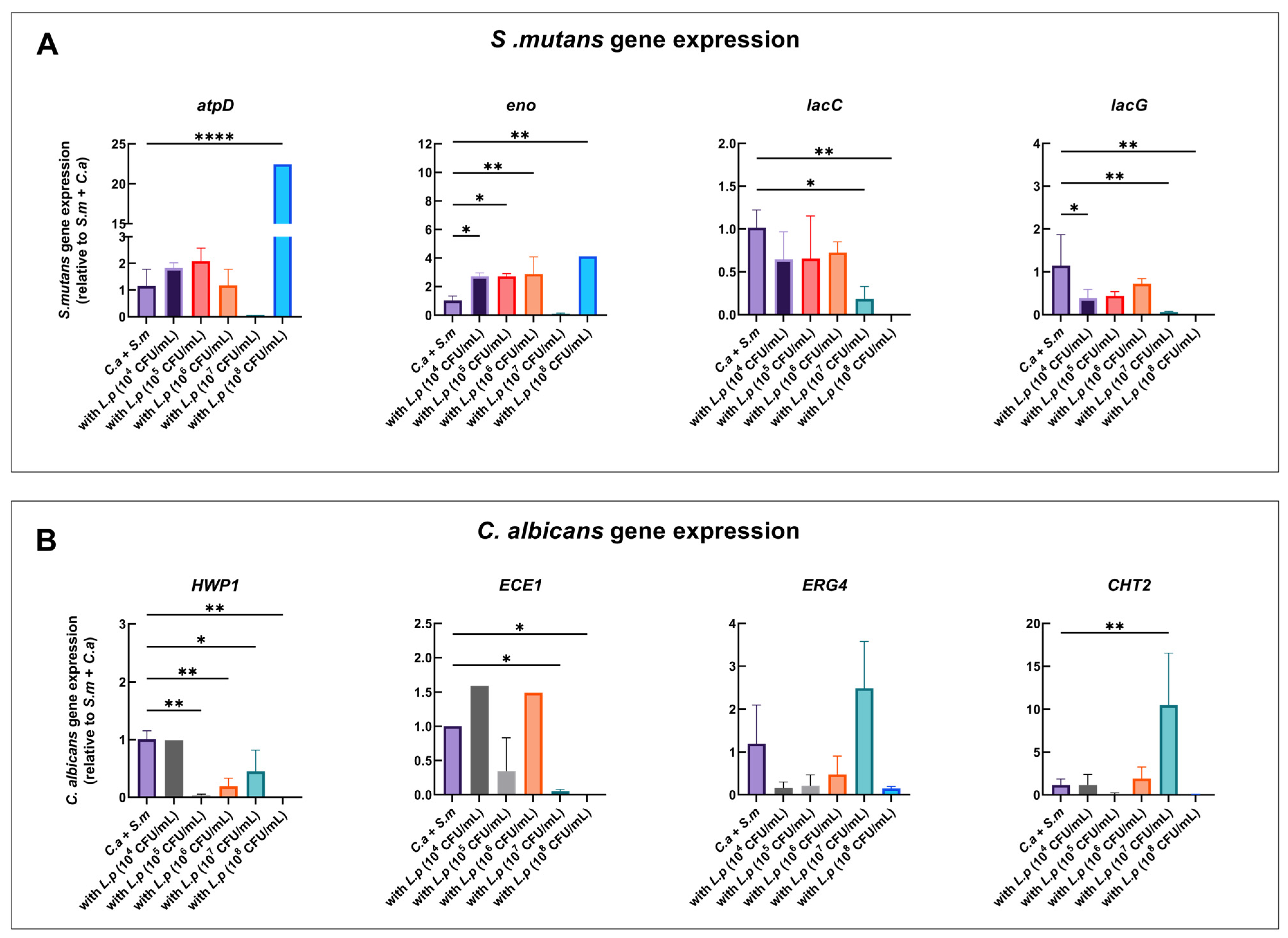
3.6. Inhibition of S. mutans Virulence Genes by L. plantarum

To evaluate the differential gene expression between the control and the condition with added L. plantarum, qRT-PCR was conducted at 6 and 20 h (Figure S4 and Figure 3).
At 20 h, S. mutans atpD and eno genes were significantly upregulated with L. plantarum at 108 CFU/mL in the multi-species model in comparison to the control group (C. albicans + S. mutans). lacC and lacG were significantly downregulated with L. plantarum at 107 CFU/mL, but they could not be detected with L. plantarum at 108 CFU/mL (Figure 3A).

3.7. Inhibition of C. albicans Virulence Genes by L. plantarum

For C. albicans, both the HWP1 and ECE1 genes were downregulated by 99.9% with L. plantarum at 108 CFU/mL in the multi-species model in comparison to the control group (C. albicans and S. mutans). HWP1 and ECE1 was also significantly downregulated with L. plantarum at 107 CFU/mL. CHT2 was upregulated with L. plantarum at 107 CFU/mL. CHT2 expressions increased by 10.48-fold. In addition, the virulence genes HWP1, ECE1, ERG4, and CHT2 were observed to be the lowest with L. plantarum at 108 CFU/mL (Figure 3B).
Figure 3. Effect of L. plantarum on the expression of C. albicans and S. mutans genes in multi-species model at 20 h. qRT-PCR was performed for S. mutans and C. albicans genes of interest for mixed-species culture at 20 h. S. mutans (A) and C. albicans (B) gene expression ratios are shown, and the comparison is relative to S. mutans and C. albicans dual-species. p values were determined by one-way ANOVA with post hoc test. * p < 0.05, ** p < 0.01, **** p < 0.0001.
Pathogens 12 00848 g003

3.8. Dose-Dependent Gene Expressions by L. plantarum in Mono-Species Model

Plantaricin are antimicrobial peptides produced by L. plantarum. The expression of plantaricin-related genes plnA and plnN exhibited dose-dependent effects in the L. plantarum mono-species model. Specifically, plnA and plnN were significantly upregulated with L. plantarum at 107 and 108 CFU/mL. For instance, plnA was 23.5-fold and 58.8-fold higher and plnN was 61.6-fold and 109.4-fold higher with L. plantarum at 107 and 108 CFU/mL, respectively, than with L. plantarum at 104 CFU/mL (Figure 4).

3.9. Dose-Dependent Gene Expressions by L. plantarum in Multi-Species Model

In the multi-species model where L. plantarum was co-cultured with S. mutans and C. albicans, significant downregulation of plnA (82%) and plnN (49.2%) was observed, compared to the mono-species model with L. plantarum at 108 CFU/mL (Figure 5E,J). However, the plnA and plnN genes were significantly upregulated with L. plantarum at 107 CFU/mL in the multi-species model, compared to the L. plantarum mono-species model (Figure 5D,I).
In the multi-species model, the highest expressions for the plnA and plnN genes were observed with L. plantarum at 107 CFU/mL (Figure S5).
Figure 4. Dose-related expression of L. plantarum gene in mono-species model. qRT-PCR was performed for L. plantarum genes of interest for mono-species culture at 20 h. L. plantarum gene expression ratio is shown, and the comparison is relative to 104 CFU/mL L. plantarum mono-species group. p values were determined by one-way ANOVA with post hoc test. * p < 0.05, *** p < 0.001, **** p < 0.0001.
Pathogens 12 00848 g004
Figure 5. Expression of L. plantarum genes when interacting with S. mutans and C. albicans in multi-species model at 20 h. qRT-PCR was performed for L. plantarum genes of interest for mixed-species culture at 20 h. L. plantarum gene expression ratio is shown (AO), and the comparison is relative to L. plantarum mono-species group. p values were determined by t-test. * p < 0.05, ** p < 0.01, *** p < 0.001, **** p < 0.0001.
Pathogens 12 00848 g005

3.10. Inhibition of C. albicans Hyphae/Pseudohyphae Formation

Inhibition of C. albicans hyphae or pseudohyphae formation was assessed by observing the 20 h culture medium under a light microscope. Figure 6 shows that the addition of 108 CFU/mL L. plantarum inhibited the growth of C. albicans and the transition from yeast to hyphae or pseudohyphae form, compared to other groups.

4. Discussion

The findings of the present study indicate that the probiotic L. plantarum inhibited the growth of C. albicans and S. mutans in a dose-dependent manner in the dual- and multi-species planktonic models. L. plantarum at 108 CFU/mL significantly inhibited the growth of both C. albicans and S. mutans while its own growth pattern was unaffected. L. plantarum at doses lower than 107 CFU/mL had no significant effect on the growth of C. albicans, but L. plantarum promoted the growth of S. mutans.
One possible explanation for the effects of L. plantarum against C. albicans and S. mutans is its ability to produce organic acids. L. plantarum strains have the capacity to synthesize diverse organic acids, which were reported in a previous study to exhibit antimicrobial and antifungal properties [35,36]. Lactic acid and acetic acid are the two primary organic acids produced by these strains [35,37].
C. albicans exhibits acidogenic and aciduric properties [38] with the ability to produce acids in low pH environments, albeit less efficiently than lactobacilli [39]. The inhibitory effect of L. plantarum on C. albicans is consistent with an earlier report [28], but the current study shows that such an effect is only evident with L. plantarum at doses higher than 107 CFU/mL. Though the medium pH only dropped to a level below 4.0 with L. plantarum at 108 CFU/mL in the multi-species model, the pH levels could not explain the antifungal effect, as C. albicans could thrive in pH 3.0 [40]. One possible explanation is that L. plantarum produces acetic acid [37], which is cytotoxic against C albicans [36].
L. plantarum produces lactic acid that is the primary end-product of carbohydrate fermentation [41]. A recent study indicated that lactic acid plays a dominant role in the L. plantarum cell-free supernatant against S. mutans [42]. In addition to organic acids, L. plantarum can produce the antimicrobial substance plantaricin. Plantaricin (pln) loci are associated with plantaricin production. Each locus has around 25 genes covering a length of DNA, grouped into 5–6 operons [43]. The regulatory operons (plnABCD) involved in plantaricin production encode different proteins. The plnA gene encodes inducing peptides that regulate the transcription of the other operons [44]. In addition, the plnN gene encodes a putative prebacteriocin. An increased expression of genes involved in plantaricin production was observed in low pH conditions [27]. We found that the expressions of plnA and plnN genes increased with increasing doses of L. plantarum in the mono-species model. These genes were downregulated in the multi-species model with L. plantarum at 108 CFU/mL. A likely explanation is that that quantity of viable cells of S. mutans became very low in this model, and the needs for L. plantarum to produce plantaricin had decreased.
The genes associated with the virulence of C. albicans were altered in the dual- and multi-species models containing L. plantarum. Both the HWP1 and ECE1 genes were downregulated at 20 h with L. plantarum at 108 CFU/mL. The HWP1 gene encoded the hyphal wall protein 1 [45], and the ECE1 gene encoded the extent of cell elongation 1 [46]. The HWP1 gene encoded the hyphal wall protein 1 that works as an adhesin and is critical for the integrity of cell-to-cell adhesions in biofilms [47]. HWP1-deficient strains of C. albicans were less capable of inducing systemic candidiasis in mice and were unable to form persistent attachments to human epithelial cells [47,48]. In addition, ECE1 was tightly associated with yeast-to-hyphae transition [49]. Hyphal growth was significantly inhibited with L. plantarum at 108 CFU/mL in the multi-species model. These results were consistent with other research, which found that acidic pH repressed the yeast-to-filamentous transition [50]. In addition, ERG4 and CHT2 were only significantly upregulated in the 107 CFU/mL of L. plantarum multi-species groups. ERG4 encoded sterolC-24 reductase and was related to antifungal resistance. The CHT2 gene encoded the chitinase 2 precursor related to yeast cell wall chitin remodeling [51]. The upregulation of ERG4 and CHT2 genes resulted in increasing resistance to stress and surviving in the competitive interactions for C. albicans. This may explain why C. albicans can thrive with L. plantarum at 108 CFU/mL in the multi-species model.
The genes associated with the virulence of S. mutans were also altered in the dual- and multi-species models containing L. plantarum. The atpD gene was upregulated with L. plantarum at 108 CFU/mL in the multi-species group, compared to the dual-species control group (C. albicans + S. mutans) at 20 h. This was consistent with the previous study [27]; a probable explanation is that S. mutans cells may use most of their energy to maintain internal pH homeostasis under acidic circumstances. This physiological process was associated with the atpD gene [52]. The acidogenic properties of S. mutans were inhibited in the presence of 108 and 107 CFU/mL of L. plantarum, as indicated by the downregulation of the lacC and lacG genes.
With regard to the research methods, some limitations need to be acknowledged. Firstly, a limitation of this study is that a planktonic model was employed. Biofilm and animal models, together with potential clinic studies, need to be carried out to assess the effect of L. plantarum on caries prevention. Secondly, our study introduced glucose as the sugar challenge in the planktonic model, so future biofilm and animal studies should assess the condition when other types of carbohydrates such as sucrose are tested in the multi-species interaction. Thirdly, qRT-PCR was employed as a critical methodology in this study. However, it should be noted that RNA sequencing might offer more precision in data acquisition and analysis.

5. Conclusions

In summary, L. plantarum demonstrated dose-dependent antifungal and antibacterial effects against C. albicans and S. mutans. L. plantarum emerged as a promising candidate for the creation of novel antimicrobial probiotic products targeting dental caries prevention. Further research is warranted to identify the functional metabolites produced by L. plantarum at different dosages when interacting with C. albicans and S. mutans.

Supplementary Materials

The following supporting information can be downloaded at https://www.mdpi.com/article/10.3390/pathogens12060848/s1. Figure S1: Schematic study design; Figure S2: Changes in species composition in dual species; Figure S3: pH in culture media; Figure S4: Effect of L. plantarum on the expression of C. albicans and S. mutans genes in multi-species model at six hours; Figure S5: Dose-related expression of L. plantarum gene in multi-species model at twenty hours; Figure S6: Dose-dependent inhibition of C. albicans hyphae formation by L. plantarum gene in multi-species model at ×20 magnification; Table S1: Gene of interest; Table S2: Primers used in qRT-PCR.

Author Contributions

Conceptualization, J.B., Y.R. and J.X.; data curation, J.B., X.H. and Y.Z.; formal analysis, J.B., X.H., Y.Z., T.T.W. and X.L.; funding acquisition, J.X.; investigation, J.B., X.H., Y.Z., T.T.W. and G.M.; methodology, J.B., X.H., Y.Z. and J.X.; project administration, Y.R. and J.X.; resources, Y.R. and J.X.; supervision, Y.R. and J.X.; validation, J.B., X.H. and X.L.; writing—original draft, J.B.; writing—review and editing, J.B., Y.Z., Y.R. and J.X. All authors have read and agreed to the published version of the manuscript.

Funding

This research was funded by NIH/NIDCR K23DE027412 (PI: J.X.) and R01DE031025 (PI: J.X.). The funding agencies had no role in the study design, data collection, analyses, decision to publish, or preparation of the manuscript.

Institutional Review Board Statement

Not applicable.

Informed Consent Statement

Not applicable.

Data Availability Statement

All data generated or analyzed during this study are included in this article. Further enquiries can be directed to the corresponding author.

Conflicts of Interest

The authors declare no conflict of interest.

References

  1. Bernabe, E.; Marcenes, W.; Hernandez, C.R.; Bailey, J.; Abreu, L.G.; Alipour, V.; Amini, S.; Arabloo, J.; Arefi, Z.; Arora, A.; et al. Global, Regional, and National Levels and Trends in Burden of Oral Conditions from 1990 to 2017: A Systematic Analysis for the Global Burden of Disease 2017 Study. J. Dent. Res. 2020, 99, 362–373. [Google Scholar] [CrossRef] [PubMed] [Green Version]
  2. Pitts, N.B.; Zero, D.T.; Marsh, P.D.; Ekstrand, K.; Weintraub, J.A.; Ramos-Gomez, F.; Tagami, J.; Twetman, S.; Tsakos, G.; Ismail, A. Dental caries. Nat. Rev. Dis. Prim. 2017, 3, 17030. [Google Scholar] [CrossRef] [PubMed] [Green Version]
  3. Paes Leme, A.F.; Koo, H.; Bellato, C.M.; Bedi, G.; Cury, J.A. The role of sucrose in cariogenic dental biofilm formation—New insight. J. Dent. Res. 2006, 85, 878–887. [Google Scholar] [CrossRef]
  4. Lamont, R.J.; Koo, H.; Hajishengallis, G. The oral microbiota: Dynamic communities and host interactions. Nat. Rev. Microbiol. 2018, 16, 745–759. [Google Scholar] [CrossRef]
  5. Bowen, W.H.; Burne, R.A.; Wu, H.; Koo, H. Oral Biofilms: Pathogens, Matrix, and Polymicrobial Interactions in Microenvironments. Trends Microbiol. 2018, 26, 229–242. [Google Scholar] [CrossRef] [PubMed]
  6. Budtz-Jörgensen, E. Etiology, pathogenesis, therapy, and prophylaxis of oral yeast infections. Acta Odontol. Scand. 1990, 48, 61–69. [Google Scholar] [CrossRef]
  7. Xiao, J.; Huang, X.; Alkhers, N.; Alzamil, H.; Alzoubi, S.; Wu, T.T.; Castillo, D.A.; Campbell, F.; Davis, J.; Herzog, K.; et al. Candida albicans and Early Childhood Caries: A Systematic Review and Meta-Analysis. Caries Res. 2018, 52, 102–112. [Google Scholar] [CrossRef] [Green Version]
  8. Klinke, T.; Guggenheim, B.; Klimm, W.; Thurnheer, T. Dental caries in rats associated with Candida albicans. Caries Res. 2011, 45, 100–106. [Google Scholar] [CrossRef] [Green Version]
  9. Du, Q.; Ren, B.; He, J.; Peng, X.; Guo, Q.; Zheng, L.; Li, J.; Dai, H.; Chen, V.; Zhang, L.; et al. Candida albicans promotes tooth decay by inducing oral microbial dysbiosis. ISME J. 2021, 15, 894–908. [Google Scholar] [CrossRef]
  10. Hwang, G.; Marsh, G.; Gao, L.; Waugh, R.; Koo, H. Binding Force Dynamics of Streptococcus mutans-glucosyltransferase B to Candida albicans. J. Dent. Res. 2015, 94, 1310–1317. [Google Scholar] [CrossRef] [Green Version]
  11. Falsetta, M.L.; Klein, M.I.; Colonne, P.M.; Scott-Anne, K.; Gregoire, S.; Pai, C.-H.; Gonzalez-Begne, M.; Watson, G.; Krysan, D.J.; Bowen, W.H.; et al. Symbiotic Relationship between Streptococcus mutans and Candida albicans Synergizes Virulence of Plaque Biofilms In Vivo. Infect. Immun. 2014, 82, 1968–1981. [Google Scholar] [CrossRef] [Green Version]
  12. Metwalli, K.H.; Khan, S.A.; Krom, B.P.; Jabra-Rizk, M.A. Streptococcus mutans, Candida albicans, and the human mouth: A sticky situation. PLoS Pathog. 2013, 9, e1003616. [Google Scholar] [CrossRef] [PubMed] [Green Version]
  13. Gregoire, S.; Xiao, J.; Silva, B.B.; Gonzalez, I.; Agidi, P.S.; Klein, M.I.; Ambatipudi, K.S.; Rosalen, P.L.; Bauserman, R.; Waugh, R.E.; et al. Role of glucosyltransferase B in interactions of Candida albicans with Streptococcus mutans and with an experimental pellicle on hydroxyapatite surfaces. Appl. Environ. Microbiol. 2011, 77, 6357–6367. [Google Scholar] [CrossRef] [Green Version]
  14. Shino, B.; Peedikayil, F.C.; Jaiprakash, S.R.; Ahmed Bijapur, G.; Kottayi, S.; Jose, D. Comparison of Antimicrobial Activity of Chlorhexidine, Coconut Oil, Probiotics, and Ketoconazole on Candida albicans Isolated in Children with Early Childhood Caries: An In Vitro Study. Scientifica 2016, 2016, 7061587. [Google Scholar] [CrossRef] [PubMed] [Green Version]
  15. Aljaffary, M.; Jang, H.; Alomeir, N.; Zeng, Y.; Alkhars, N.; Vasani, S.; Almulhim, A.; Wu, T.T.; Quataert, S.; Bruno, J.; et al. Effects of Nystatin oral rinse on oral Candida species and Streptococcus mutans among healthy adults. Clin. Oral Investig. 2023. [Google Scholar] [CrossRef] [PubMed]
  16. Alomeir, N.; Zeng, Y.; Fadaak, A.; Wu, T.T.; Malmstrom, H.; Xiao, J. Effect of Nystatin on Candida albicans-Streptococcus mutans duo-species biofilms. Arch. Oral Biol. 2023, 145, 105582. [Google Scholar] [CrossRef]
  17. Philip, N.; Suneja, B.; Walsh, L.J. Ecological Approaches to Dental Caries Prevention: Paradigm Shift or Shibboleth? Caries Res. 2018, 52, 153–165. [Google Scholar] [CrossRef]
  18. FAOD; WHO. Evaluation of health and nutritional properties of powder milk and live lactic acid bacteria. In Food and Agriculture Organization of the United Nations and World Health Organization Expert Consultation Report, Cordoba, Argentina; World Health Organization; Food and Agriculture Organization of the United Nations: Rome, Italy, 2001. [Google Scholar]
  19. Markowiak, P.; Śliżewska, K. Effects of Probiotics, Prebiotics, and Synbiotics on Human Health. Nutrients 2017, 9, 1021. [Google Scholar] [CrossRef]
  20. Klaenhammer, T.R. Genetics of bacteriocins produced by lactic acid bacteria. FEMS Microbiol. Rev. 1993, 12, 39–85. [Google Scholar] [CrossRef]
  21. Jung, S.; Lee, J.-H. Characterization of transcriptional response of Lactobacillus plantarum under acidic conditions provides insight into bacterial adaptation in fermentative environments. Sci. Rep. 2020, 10, 19203. [Google Scholar] [CrossRef]
  22. Xiao, J.; Grier, A.; Faustoferri, R.C.; Alzoubi, S.; Gill, A.L.; Feng, C.; Liu, Y.; Quivey, R.G.; Kopycka-Kedzierawski, D.T.; Koo, H.; et al. Association between Oral Candida and Bacteriome in Children with Severe ECC. J. Dent. Res. 2018, 97, 1468–1476. [Google Scholar] [CrossRef] [PubMed]
  23. Zhang, Q.; Qin, S.; Xu, X.; Zhao, J.; Zhang, H.; Liu, Z.; Chen, W. Inhibitory Effect of Lactobacillus plantarum CCFM8724 towards Streptococcus mutans- and Candida albicans-Induced Caries in Rats. Oxidative Med. Cell. Longev. 2020, 2020, 4345804. [Google Scholar] [CrossRef] [PubMed]
  24. Zhang, G.; Lu, M.; Liu, R.; Tian, Y.; Vu, V.H.; Li, Y.; Liu, B.; Kushmaro, A.; Li, Y.; Sun, Q. Inhibition of Biofilm Formation and Virulence by K41 Isolated From Traditional Sichuan Pickles. Front. Microbiol. 2020, 11, 774. [Google Scholar] [CrossRef] [PubMed]
  25. Srivastava, N.; Ellepola, K.; Venkiteswaran, N.; Chai, L.Y.A.; Ohshima, T.; Seneviratne, C.J. Lactobacillus Plantarum 108 Inhibits Streptococcus mutans and Candida albicans Mixed-Species Biofilm Formation. Antibiotics 2020, 9, 478. [Google Scholar] [CrossRef] [PubMed]
  26. Kim, A.R.; Ahn, K.B.; Yun, C.-H.; Park, O.-J.; Perinpanayagam, H.; Yoo, Y.-J.; Kum, K.-Y.; Han, S.H. Lactobacillus plantarum Lipoteichoic Acid Inhibits Oral Multispecies Biofilm. J. Endod. 2019, 45, 310–315. [Google Scholar] [CrossRef]
  27. Wasfi, R.; Abd El-Rahman, O.A.; Zafer, M.M.; Ashour, H.M. Probiotic Lactobacillus sp. inhibit growth, biofilm formation and gene expression of caries-inducing Streptococcus mutans. J. Cell. Mol. Med. 2018, 22, 1972–1983. [Google Scholar] [CrossRef] [PubMed] [Green Version]
  28. Zeng, Y.; Fadaak, A.; Alomeir, N.; Wu, T.T.; Rustchenko, E.; Qing, S.; Bao, J.; Gilbert, C.; Xiao, J. Lactobacillus plantarum Disrupts S. mutansC. albicans Cross-Kingdom Biofilms. Front. Cell. Infect. Microbiol. 2022, 12, 872012. [Google Scholar] [CrossRef]
  29. Hasslöf, P.; Hedberg, M.; Twetman, S.; Stecksén-Blicks, C. Growth inhibition of oral mutans streptococci and candida by commercial probiotic lactobacilli—An in vitro study. BMC Oral Health 2010, 10, 18. [Google Scholar] [CrossRef] [PubMed] [Green Version]
  30. Qiuxiang, Q.S.X.W.Z. Inhibitory effect of Lactobacillus plantarum CCFM8724 on caries-causing dual biofilms. Food Ferment. Ind. 2020, 46, 127–132. [Google Scholar]
  31. Zeng, Y.; Fadaak, A.; Alomeir, N.; Wu, Y.; Wu, T.T.; Qing, S.; Xiao, J. Effect of Probiotic Lactobacillus plantarum on Streptococcus mutans and Candida albicans Clinical Isolates from Children with Early Childhood Caries. Int. J. Mol. Sci. 2023, 24, 2991. [Google Scholar] [CrossRef]
  32. Huang, X.; Bao, J.; Zeng, Y.; Meng, G.; Lu, X.; Wu, T.T.; Ren, Y.; Xiao, J. Anti-cariogenic Properties of Lactobacillus plantarum in the Utilization of Galacto-Oligosaccharide. Nutrients 2023, 15, 2017. [Google Scholar] [CrossRef]
  33. Alkhars, N.; Zeng, Y.; Alomeir, N.; Al Jallad, N.; Wu, T.T.; Aboelmagd, S.; Youssef, M.; Jang, H.; Fogarty, C.; Xiao, J. Oral Candida Predicts Streptococcus mutans Emergence in Underserved US Infants. J. Dent. Res. 2022, 101, 54–62. [Google Scholar] [CrossRef]
  34. Schmittgen, T.D.; Livak, K.J. Analyzing real-time PCR data by the comparative C(T) method. Nat. Protoc. 2008, 3, 1101–1108. [Google Scholar] [CrossRef] [PubMed]
  35. Hu, C.H.; Ren, L.Q.; Zhou, Y.; Ye, B.C. Characterization of antimicrobial activity of three Lactobacillus plantarum strains isolated from Chinese traditional dairy food. Food Sci. Nutr. 2019, 7, 1997–2005. [Google Scholar] [CrossRef] [PubMed] [Green Version]
  36. Lourenço, A.; Pedro, N.A.; Salazar, S.B.; Mira, N.P. Effect of Acetic Acid and Lactic Acid at Low pH in Growth and Azole Resistance of Candida albicans and Candida glabrata. Front. Microbiol. 2018, 9, 3265. [Google Scholar] [CrossRef] [Green Version]
  37. Zalán, Z.; Hudáček, J.; Štětina, J.; Chumchalová, J.; Halász, A. Production of organic acids by Lactobacillus strains in three different media. Eur. Food Res. Technol. 2010, 230, 395–404. [Google Scholar] [CrossRef]
  38. Fakhruddin, K.S.; Perera Samaranayake, L.; Egusa, H.; Ngo, H.C.; Pesee, S. Profuse diversity and acidogenicity of the candida-biome of deep carious lesions of Severe Early Childhood Caries (S-ECC). J. Oral Microbiol. 2021, 13, 1964277. [Google Scholar] [CrossRef]
  39. Klinke, T.; Kneist, S.; De Soet, J.J.; Kuhlisch, E.; Mauersberger, S.; Förster, A.; Klimm, W. Acid Production by Oral Strains of Candida albicans and Lactobacilli. Caries Res. 2009, 43, 83–91. [Google Scholar] [CrossRef] [Green Version]
  40. Pereira, D.; Seneviratne, C.J.; Koga-Ito, C.Y.; Samaranayake, L.P. Is the oral fungal pathogen Candida albicans a cariogen? Oral Dis. 2018, 24, 518–526. [Google Scholar] [CrossRef]
  41. Felis, G.E.; Dellaglio, F. Taxonomy of Lactobacilli and Bifidobacteria. Curr. Issues Intest. Microbiol. 2007, 8, 44–61. [Google Scholar]
  42. Liang, J.; Zhou, Y.; Tang, G.; Wu, R.; Lin, H. Exploration of the Main Antibiofilm Substance of Lactobacillus plantarum ATCC 14917 and Its Effect against Streptococcus mutans. Int. J. Mol. Sci. 2023, 24, 1986. [Google Scholar] [CrossRef] [PubMed]
  43. Diep, D.B.; Straume, D.; Kjos, M.; Torres, C.; Nes, I.F. An overview of the mosaic bacteriocin pln loci from Lactobacillus plantarum. Peptides 2009, 30, 1562–1574. [Google Scholar] [CrossRef] [PubMed]
  44. Diep, D.B.; Myhre, R.; Johnsborg, O.; Aakra, A.; Nes, I.F. Inducible bacteriocin production in Lactobacillus is regulated by differential expression of the pln operons and by two antagonizing response regulators, the activity of which is enhanced upon phosphorylation. Mol. Microbiol. 2003, 47, 483–494. [Google Scholar] [CrossRef] [PubMed]
  45. Staab, J.F.; Sundstrom, P. Genetic organization and sequence analysis of the hypha-specific cell wall protein gene HWP1 of Candida albicans. Yeast 1998, 14, 681–686. [Google Scholar] [CrossRef]
  46. Birse, C.E.; Irwin, M.Y.; Fonzi, W.A.; Sypherd, P.S. Cloning and characterization of ECE1, a gene expressed in association with cell elongation of the dimorphic pathogen Candida albicans. Infect. Immun. 1993, 61, 3648–3655. [Google Scholar] [CrossRef] [Green Version]
  47. Sharkey, L.L.; McNemar, M.D.; Saporito-Irwin, S.M.; Sypherd, P.S.; Fonzi, W.A. HWP1 functions in the morphological development of Candida albicans downstream of EFG1, TUP1, and RBF1. J. Bacteriol. 1999, 181, 5273–5279. [Google Scholar] [CrossRef] [PubMed] [Green Version]
  48. Staab, J.F.; Bradway, S.D.; Fidel, P.L.; Sundstrom, P. Adhesive and mammalian transglutaminase substrate properties of Candida albicans Hwp1. Science 1999, 283, 1535–1538. [Google Scholar] [CrossRef]
  49. Martin, R.; Albrecht-Eckardt, D.; Brunke, S.; Hube, B.; Hünniger, K.; Kurzai, O. A core filamentation response network in Candida albicans is restricted to eight genes. PLoS ONE 2013, 8, e58613. [Google Scholar] [CrossRef] [Green Version]
  50. Du, H.; Huang, G. Environmental pH adaption and morphological transitions in Candida albicans. Curr. Genet. 2016, 62, 283–286. [Google Scholar] [CrossRef]
  51. Nobile, C.J.; Solis, N.; Myers, C.L.; Fay, A.J.; Deneault, J.-S.; Nantel, A.; Mitchell, A.P.; Filler, S.G. Candida albicans transcription factor Rim101 mediates pathogenic interactions through cell wall functions. Cell. Microbiol. 2008, 10, 2180–2196. [Google Scholar] [CrossRef] [Green Version]
  52. Gong, Y.; Tian, X.-L.; Sutherland, T.; Sisson, G.; Mai, J.; Ling, J.; Li, Y.-H. Global transcriptional analysis of acid-inducible genes in Streptococcus mutans: Multiple two-component systems involved in acid adaptation. Microbiology 2009, 155, 3322–3332. [Google Scholar] [CrossRef] [PubMed] [Green Version]
Figure 1. Dynamic changes in the viable cells of C. albicans, S. mutans, and L. plantarum in mono-, dual-, and multi-species models. (A,B) C. albicans viable cells in dual- and multi-species conditions. (C,D) S. mutans viable cells in dual- and multi-species conditions. (E) L. plantarum viable cells in mono-species condition. (F) L. plantarum viable cells in C. albicans presence dual-species condition. (G) L. plantarum viable cells in S. mutans presence dual-species condition. (H) L. plantarum viable cells in multi-species condition. Overall, dose-dependent antimicrobial and antifungal effects were seen for L. plantarum; 108 CFU/mL of L. plantarum showed inhibition on the growth of C. albicans and S. mutans. The dotted line represents the control groups in each model.
Pathogens 12 00848 g001
Figure 2. Changes in species composition in multi-species. The composition of each microorganism in multi-species condition is shown. (AE) The composition of C. albicans, S. mutans, and L. plantarum in multi-species condition.
Pathogens 12 00848 g002
Figure 6. Dose-dependent inhibition of C. albicans hyphae formation by L. plantarum gene in multi-species model at × 100 magnification. (A) S. mutans and C. albicans grown in dual-species model at 20 h. (BF) S. mutans and C. albicans grown with addition of various doses of L. plantarum at 20 h. The addition of 108 CFU/mL L. plantarum inhibited the growth of C. albicans and the transition from yeast to hyphae or pseudohyphae form. These are representative images of multiple fields of view.
Pathogens 12 00848 g006
Disclaimer/Publisher’s Note: The statements, opinions and data contained in all publications are solely those of the individual author(s) and contributor(s) and not of MDPI and/or the editor(s). MDPI and/or the editor(s) disclaim responsibility for any injury to people or property resulting from any ideas, methods, instructions or products referred to in the content.

