
What about the Cytoskeletal and Related Proteins of Tapeworms in the Host’s Immune Response? An Integrative Overview
 and
and
Abstract
:1. Introduction
Tapeworm Overview
2. Cytoskeletal Proteins Identified in Tapeworms
2.1. Microfilaments and Muscular System
2.1.1. Actin
2.1.2. Myosin and Paramyosin
2.1.3. Tropomyosin
2.2. Microtubules
2.2.1. Tubulin
2.2.2. Dyneins
2.3. Intermediate Filaments
2.4. Septins
2.5. Other Cytoskeleton Proteins
3. Expression and Function of Cytoskeletal Proteins in Tapeworms
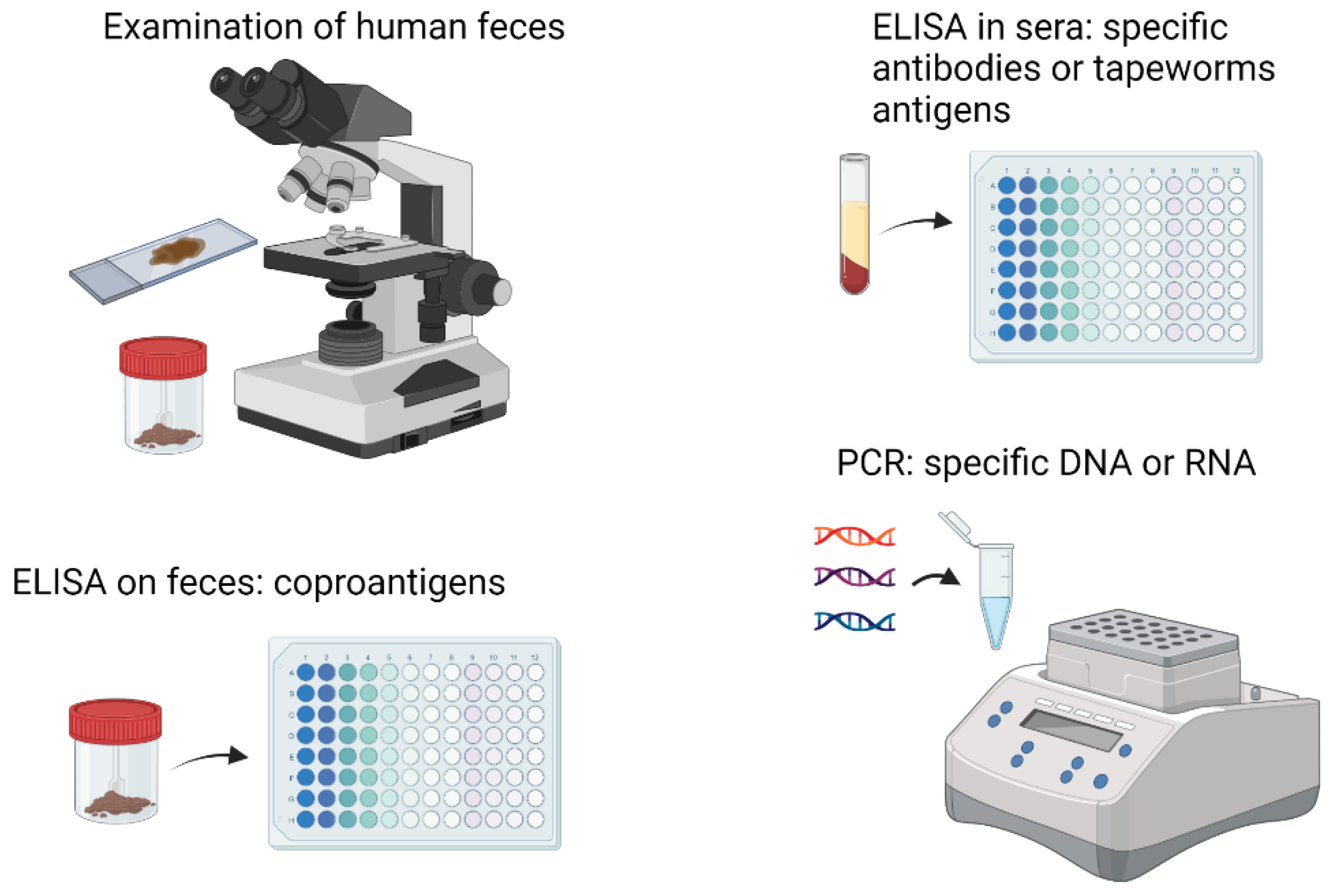
4. Diagnosis of Tapeworm Diseases and the Cytoskeletal Proteins
5. Cytoskeletal Tapeworm Proteins in Vaccine-Development
6. Cytoskeletal Proteins as Targets of Cestocidal Drugs
7. Conclusions
Author Contributions
Funding
Institutional Review Board Statement
Informed Consent Statement
Data Availability Statement
Acknowledgments
Conflicts of Interest
References
- Martínez-González, J.D.J.; Guevara-Flores, A.; del Arenal Mena, I.P. Evolutionary Adaptations of Parasitic Flatworms to Different Oxygen Tensions. Antioxidants 2022, 11, 1102. [Google Scholar] [CrossRef]
- Bobes, R.J.; Fragoso, G.; Fleury, A.; García-varela, M.; Sciutto, E.; Larralde, C.; Laclette, J.P. molecular epidemiology and perspectives on the research of taeniid parasites with special emphasis on Taenia solium. Infect. Genet. Evol. 2014, 23, 150–160. [Google Scholar] [CrossRef]
- Budke, C.M.; White, A.C.; Garcia, H.H. Zoonotic Larval Cestode Infections: Neglected, Neglected Tropical Diseases? PLoS Negl. Trop. Dis. 2009, 3, e319. [Google Scholar] [CrossRef] [Green Version]
- Torgerson, P.R.; Devleesschauwer, B.; Praet, N.; Speybroeck, N.; Willingham, A.L.; Kasuga, F.; Rokni, M.B.; Zhou, X.-N.; Fèvre, E.M.; Sripa, B.; et al. World Health Organization Estimates of the Global and Regional Disease Burden of 11 foodborne parasitic diseases, 2010: A Data Synthesis. PLoS Med. 2015, 12, e1001920. [Google Scholar] [CrossRef] [Green Version]
- Dalton, J.P.; Skelly, P.; Halton, D.W. Role of the tegument and gut in nutrient uptake by parasitic platyhelminths 1. J. Zool. 2004, 82, 211–232. [Google Scholar] [CrossRef]
- McKay, D.M. The immune response to and immunomodulation by Hymenolepis diminuta. Parasitology 2010, 137, 385–394. [Google Scholar] [CrossRef]
- Parfrey, L.W.; Jirků, M.; Šíma, R.; Jalovecká, M.; Sak, B.; Grigore, K.; Pomajbíková, K.J. A benign helminth alters the host immune system and the gut microbiota in a rat model system. PLoS ONE 2017, 12, e0182205. [Google Scholar]
- Baily, G.; Garcia, H.H. Other Cestode Infections: Intestinal Cestodes, Cysticercosis, Other Larval Cestode Infections, Manson’s Tropical Diseases, 23rd ed.; Elsevier: Amsterdam, The Netherlands, 2014; pp. 820–832.e1. [Google Scholar]
- Waeschenbach, A.; Webster, B.L.; Littlewood, D.T.J. Adding resolution to ordinal level relationships of tapeworms (Platyhelminthes: Cestoda) with large fragments of mtDNA. Mol. Phylogenet Evol. 2012, 63, 834–847. [Google Scholar] [CrossRef] [PubMed]
- Caira, J.N.; Reyda, F.B. Eucestoda (true tapeworms). In Marine Parasitology; Rhode, K., Ed.; CSIRO Publishing: Collingwood, Australia, 2005; pp. 92–104. [Google Scholar]
- Littlewood, D.T.J.; Rohde, K.; Bray, R.A.; Herniou, E.A. Phylogeny of the Platyhelminthes and the evolution of parasitism. Biol. J. Linn. Soc. 1999, 68, 257–287. [Google Scholar] [CrossRef]
- Webb, C.; Cabada, M.M. Intestinal cestodes. Curr. Opin. Infect. Dis. 2017, 30, 504–510. [Google Scholar] [CrossRef] [PubMed]
- Ito, A. Immunology in Cestode Infections. Encycl. Immunobiol. 2016, 4, 159–165. [Google Scholar] [CrossRef]
- Heyneman, D.; Baron, S. Cestodes. Med. Microbiol. 1996, 89, 79–81. [Google Scholar]
- Hoberg, E.P. Phylogeny of Taenia: Species definitions and origins of human parasites. Parasitol. Int. 2006, 55, S23–S30. [Google Scholar] [CrossRef] [PubMed] [Green Version]
- Cheng, T. General Parasitology, 2nd ed.; Elsevier: Amsterdam, The Netherlands, 1986; ISBN 9780323140102. [Google Scholar]
- Soulsby, E.J.L. The Physiology of Biochemistry of Cestodes; Smyth, J.D., McManus, D.P., Eds.; Cambridge University Press: Cambridge, UK, 1989; p. 398. ISBN 0-521-35557-5. [Google Scholar]
- Jones, M.K. Structure and diversity of cestode epithelia. Int. J. Parasitol. 1998, 28, 913–923. [Google Scholar] [CrossRef] [PubMed]
- Dorsey, C.; Cousin, C. Cyton II: A subtegumental cell type in the cercaria of Schistosoma mansoni. J. Morphol. 1995, 224, 233–240. [Google Scholar] [CrossRef]
- Gobert, G.N.; Stenzel, D.J.; McManus, D.P.; Jones, M.K. The ultrastructural architecture of the adult Schistosoma japonicum tegument. Int. J. Parasitol. 2003, 33, 1561–1575. [Google Scholar] [CrossRef] [PubMed]
- Berenguer, J.G. Manual de Parasitología: Morfología y Biología de los Parásitos de Interés Sanitario; Edicions Universitat Barcelona: Barcelona, Spain, 2007; ISBN 8447531414. [Google Scholar]
- Rozario, T.; Newmark, P.A. A confocal microscopy-based atlas of tissue architecture in the tapeworm Hymenolepis diminuta. Exp. Parasitol. 2015, 158, 31–41. [Google Scholar] [CrossRef] [PubMed] [Green Version]
- Flisser, A.; Craig, P.S.; Ito, A. Cysticercosis and Taeniosis: Taenia solium, Taenia saginata and Taenia asiatica; Oxford University Press: Oxford, UK, 2011. [Google Scholar]
- Larralde, C.; de Aluja, A.S. Cisticercosis: Guía para Profesionales de la Salud. J. Parasitol. 2007, 93, 975–976. [Google Scholar]
- Smith, J.K.; Esch, W. Growth and Development of larval Taenia crassiceps (cestoda). I. Aneuplody in the anomalous ORF strain. Int. J. Parasitol. 1972, 2, 261–263. [Google Scholar] [CrossRef]
- Willms, K.; Zurabian, R. Taenia crassiceps: In vivo and in vitro models. Parasitology 2010, 137, 335–346. [Google Scholar] [CrossRef]
- Thompson, R.C. Neglected zoonotic helminthes Hymenolepis nana, Echinococcus canadensis and Ancylostoma ceylanicum. Clin. Microbol Infect. 2015, 21, 426–432. [Google Scholar] [CrossRef] [Green Version]
- Colville, J.L.; Berryhill, D.L. Tapeworms (Cestodes), Handbook of Zoonoses; Mosby Elsevier: St. Louis, MI, USA, 2007; pp. 182–192. [Google Scholar]
- Tsai, I.J.; Zarowiecki, M.; Holroyd, N.; Garciarrubio, A.; Sanchez-Flores, A.; Brooks, K.L.; Tracey, A.; Bobes, R.J.; Fragoso, G.; Sciutto, E.; et al. The genomes of four tapeworm species reveal adaptations to parasitism. Nature 2013, 496, 57–63. [Google Scholar] [CrossRef] [PubMed] [Green Version]
- Bobes, R.J.; Estrada, K.; Rios-Valencia, D.G.; Calderón-Gallegos, A.; de la Torre, P.; Carrero, J.C.; Sanchez-Flores, A.; Laclette, J.P. The Genomes of Two Strains of Taenia crassiceps the Animal Model for the Study of Human Cysticercosis. Front. Cell. Infect. Microbiol. 2022, 12, 551. [Google Scholar] [CrossRef] [PubMed]
- Cruz-Rivera, M.; Reyes-Torres, A.; Reynoso-Ducoing, O.; Flisser, A.; Ambrosio, J.R. Comparison of biochemical and immunochemical properties of myosin II in taeniid parasites. Cell. Biol. Int. 2006, 30, 598–602. [Google Scholar] [CrossRef] [PubMed]
- Świderski, Z.; Miquel, J.; Młocicki, D.; Georgiev, B.B.; Eira, C.; Grytner-Zięcina, B.; Feliu, C. Post-embryonic development and ultrastructural characteristics of the polycephalic larva of Taenia parva Baer, 1926 (Cyclophyllidea, Taeniidae). Acta Parasitol. 2007, 52, 31–50. [Google Scholar] [CrossRef]
- Valverde-Islas, L.E.; Arrangoiz, E.; Vega, E.; Robert, L.; Villanueva, R.; Reynoso-Ducoing, O.; Willms, K.; Zepeda-Rodríguez, A.; Fortoul, T.I.; Ambrosio, J.R. Visualization and 3D reconstruction of flame cells of Taenia solium (cestoda). PLoS ONE 2011, 6, e14754. [Google Scholar] [CrossRef] [PubMed] [Green Version]
- Reynoso-Ducoing, O.; Valverde-Islas, L.; Paredes-Salomon, C.; Pérez-Reyes, A.; Landa, A.; Robert, L.; Mendoza, G.; Ambrosio, J.R. Analysis of the expression of cytoskeletal proteins of Taenia crassiceps ORF strain cysticerci (Cestoda). Parasitol. Res. 2014, 113, 1955–1969. [Google Scholar] [CrossRef] [PubMed]
- Ampe, C.; van Troys, M. Mammalian Actins: Isoform-Specific Functions and Diseases. Handb. Exp. Pharmacol. 2017, 235, 1–37. [Google Scholar]
- Vandekerckhove, J.; Weber, K. At least six different actins are expressed in a higher mammal: An analysis based on the amino acid sequence of the amino-terminal tryptic peptide. J. Mol. Biol. 1978, 126, 783–802. [Google Scholar] [CrossRef]
- Doherty, G.J.; McMahon, H.T. Mediation, modulation, and consequences of membrane-cytoskeleton interactions. Annu. Rev. Biophys. 2008, 37, 65–95. [Google Scholar] [CrossRef] [Green Version]
- Campos, A.; Bernard, P.; Fauconnier, A.; Landa, A.; Gómez, E.; Hernández, R.; Willms, K.; Laclette, J.P. Cloning and sequencing of two actin genes from Taenia solium (Cestoda). Mol. Biochem. Parasitol. 1990, 40, 87–93. [Google Scholar] [CrossRef] [PubMed]
- Pan, W.; Shen, Y.; Han, X.; Wang, Y.; Liu, H.; Jiang, Y.; Zhang, Y.; Wang, Y.; Xu, Y.; Cao, J.; et al. Transcriptome Profiles of the Protoscoleces of Echinococcus granulosus Reveal that Excretory-Secretory Products Are Essential to Metabolic Adaptation. PLoS Negl. Trop. Dis. 2014, 8, e3392. [Google Scholar] [CrossRef] [Green Version]
- Ambrosio, J.R.; Valverde-Islas, L.; Nava-Castro, K.E.; Palacios-Arreola, M.I.; Ostoa-Saloma, P.; Reynoso-Ducoing, O.; Escobedo, G.; Ruíz-Rosado, A.; Dominguez-Ramírez, L.; Morales-Montor, J. Androgens Exert a Cysticidal Effect upon Taenia crassiceps by Disrupting Flame Cell Morphology and Function. PLoS ONE 2015, 10, e0127928. [Google Scholar] [CrossRef] [Green Version]
- La-Rocca, S.; Farias, J.; Chalar, C.; Kun, A.E.; Fernandez, V. Echinococcus granulosus: Insights into the protoscolex F-actin cytoskeleton. Acta Trop. 2019, 199, 105122. [Google Scholar] [CrossRef] [PubMed]
- Wahlberg, M.H. The distribution of F-actin during the development of Diphyllobothrium dendriticum (Cestoda). Cell. Tissue Res. 1998, 291, 561–570. [Google Scholar] [CrossRef]
- Koziol, U.; Costábile, A.; Domínguez, M.F.; Iriarte, A.; Alvite, G.; Kun, A.; Castillo, E. Developmental expression of high molecular weight tropomyosin isoforms in Mesocestoides corti. Mol. Biochem. Parasitol. 2011, 175, 181–191. [Google Scholar] [CrossRef] [PubMed]
- Craig, R.; Woodhead, J.L. Structure and function of myosin filaments. Curr. Opin. Struct. Biol. 2006, 16, 204–212. [Google Scholar] [CrossRef]
- Feng, Z.; Okada, S.; Cai, G.; Zhou, B.; Bi, E. Myosin-II Heavy Chain and Formin Mediate the Targeting of Myosin Essential Light Chain to the Division Site Before and During Cytokinesis. Mol. Biol. Cell. 2015, 26, 1211–1224. [Google Scholar] [CrossRef]
- Ambrosio, J.; Cruz-Rivera, M.; Allan, J.; Morán, E.; Ersfeld, K.; Flisser, A. Identification and partial characterization of a myosin-like protein from cysticerci and adults of Taenia solium using a monoclonal antibody. Parasitology 1997, 114 Pt 6, 545–553. [Google Scholar]
- Maroto, M.; Arredondo, J.J.; San Roman, M.; Marco, R.; Cervera, M. Analysis of the paramyosin/miniparamyosin gene. Miniparamyosin is an independently transcribed, distinct paramyosin isoform, widely distributed in invertebrates. J. Biol. Chem. 1995, 270, 4375–4382. [Google Scholar] [CrossRef] [Green Version]
- Landa, A.; Laclette, J.P.; Nicholson-Weller, A.; Shoemaker, C.B. cDNA cloning and recombinant expression of collagen-binding and complement inhibitor activity of Taenia solium paramyosin (AgB). Mol. Biochem. Parasitol. 1993, 60, 343–347. [Google Scholar] [CrossRef] [PubMed]
- Ferrer, E.; Moyano, E.; Benitez, L.; González, L.M.; Bryce, D.; Foster-Cuevas, M.; Dávila, I.; Cortéz, M.M.; Harrison, L.J.S.; Parkhouse, R.M.E.; et al. Cloning and characterization of Taenia saginata paramyosin cDNA. Parasitol. Res. 2003, 91, 60–67. [Google Scholar] [CrossRef] [PubMed]
- Chemale, G.; van Rossum, A.J.; Jefferies, J.R.; Barrett, J.; Brophy, P.M.; Ferreira, H.B.; Zaha, A. Proteomic analysis of the larval stage of the parasite Echinococcus granulosus: Causative agent of cystic hydatid disease. Proteomics 2003, 3, 1633–1636. [Google Scholar] [CrossRef]
- Epstein, H.F.; Miller, D.M.; Ortiz, I.; Berliner, G.C. Myosin and paramyosin are organized about a newly identified core structure. JCB 1985, 100, 904–915. [Google Scholar] [CrossRef] [PubMed] [Green Version]
- Solís, C.F.; Vázquez-Talavera, J.; Laclette, J.P. Toward development of a Taenia solium paramyosin-based vaccine against porcine cysticercosis. Gac. Med. Mex. 2004, 140, 129–138. [Google Scholar]
- Laclette, J.P.; Landa, A.; Arcos, L.; Willms, K.; Davis, A.E.; Shoemaker, C.B. Paramyosin is the Schistosoma mansoni (Trematoda) homologue of antigen B from Taenia solium (Cestoda). Mol. Biochem. Parasitol. 1991, 44, 287–295. [Google Scholar] [CrossRef]
- Młocicki, D.; Sulima, A.; Bień, J.; Näreaho, A.; Zawistowska-Deniziak, A.; Basałaj, K.; Sałamatin, R.; Conn, D.B.; Savijoki, K. Immunoproteomics and Surfaceomics of the Adult Tapeworm Hymenolepis diminuta. Front. Immunol. 2018, 9, 2487. [Google Scholar] [CrossRef] [Green Version]
- Eyayu, T.; Zeleke, A.J.; Worku, L. Current status and future prospects of protein vaccine candidates against Schistosoma mansoni infection. Parasite Epidemiol. Control. 2020, 11, e00176. [Google Scholar] [CrossRef]
- Wang, C.L.; Coluccio, L.M. New insights into the regulation of the actin cytoskeleton by tropomyosin. Int. Rev. Cell. Mol. Biol. 2010, 281, 91–128. [Google Scholar]
- Brown, J.H.; Zhou, Z.; Reshetnikova, L.; Robinson, H.; Yammani, R.D.; Tobacman Cohen, C. Structure of the mid-region of tropomyosin: Bending and binding sites for actin. Proc. Natl. Acad. Sci. USA 2005, 102, 18878–18883. [Google Scholar] [CrossRef] [Green Version]
- Gunning, P.; O’Neill, G.; Hardeman, E. Tropomyosin-based regulation of the actin cytoskeleton in time and space. Physiol. Rev. 2008, 88, 1–35. [Google Scholar] [CrossRef] [PubMed] [Green Version]
- Khaitlina, S.Y. Tropomyosin as a Regulator of Actin Dynamics. Int. Rev. Cell. Mol. Biol. 2015, 318, 255–291. [Google Scholar] [PubMed]
- Alvite, G.; Esteves, A. Echinococcus granulosus tropomyosin isoforms: From gene structure to expression analysis. Gene 2009, 433, 40–49. [Google Scholar] [CrossRef]
- Samereier, M.; Meyer, I.; Koonce, M.P.; Gräf, R. Live Cell-Imaging Techniques for Analyses of Microtubules in Dictyostelium; Academic Press: Cambridge, MA, USA, 2010. [Google Scholar]
- Nogales, E. Structural insights into microtubule function. Annu. Rev. Biophys. Biomol. Struct. 2001, 30, 397–420. [Google Scholar] [CrossRef]
- Song, Y.; Brady, S.T. Post-translational modifications of tubulin: Pathways to functional diversity of microtubules. Trends Cell. Biol. 2014, 25, 125–136. [Google Scholar] [CrossRef] [PubMed] [Green Version]
- Kim, D.W.; Yoo, W.; Lee, M.R.; Yang, H.W.; Kim, Y.J.; Cho, S.H.; Lee, W.J.; Ju, J.W. Transcriptome sequencing and analysis of the zoonotic parasite Spirometra erinacei spargana (plerocercoids). Parasit. Vectors 2014, 7, 368. [Google Scholar] [CrossRef] [PubMed] [Green Version]
- Verhey, K.J.; Gaertig, J. The tubulin code. Cell. Cycle 2007, 6, 62152–62160. [Google Scholar] [CrossRef] [PubMed]
- Binarová, P.; Tuszynski, J. Tubulin: Structure, Functions and Roles in Disease. Cells 2019, 8, 1294. [Google Scholar] [CrossRef] [Green Version]
- Bera, A.; Gupta, M.L. Microtubules in Microorganisms: How Tubulin Isotypes Contribute to Diverse Cytoskeletal Functions. Front. Cell. Dev. Biol. 2022, 10, 1391. [Google Scholar] [CrossRef]
- Wloga, D.; Gaertig, J. Post-translational modifications of microtubules. J. Cell. Sci. 2010, 124, 3447–3455. [Google Scholar] [CrossRef] [Green Version]
- Koziol, U.; Rauschendorfer, T.; Zanon Rodríguez, L.; Krohne, G.; Brehm, K. The unique stem cell sysem of the immortal larva of the human parasite Echinococcus multilocularis. Evodevo 2014, 5, 10. [Google Scholar] [CrossRef] [Green Version]
- Laclette, J.P.; Guerra, G.; Zetina, C. Inhibition of tubulin polymerization by mebendazole. Biochem. Biophys. Res. Commun. 1980, 92, 417–423. [Google Scholar] [CrossRef]
- Márquez-Navarro, A.; Pérez-Reyes, A.; Zepeda-Rodríguez, A.; Reynoso-Ducoing, O.; Hernández-Campos, A.; Hernández-Luis, F.; Castillo, R.; Yépez-Mulia, L.; Ambrosio, J.R. RCB20, an experimental benzimidazole derivative, affects tubulin expression and induces gross anatomical changes in Taenia crassiceps cysticerci. Parasitol. Res. 2013, 112, 2215–2226. [Google Scholar] [CrossRef]
- Roberts, A.J.; Kon, T.; Knight, P.J.; Sutoh, K.; Burgess, S.A. Functions and mechanics of dynein motor proteins. Nat. Rev. Mol. Cell. Biol. 2013, 102, 2033–2041. [Google Scholar] [CrossRef] [PubMed] [Green Version]
- Zheng, H.; Zhang, W.; Zhang, L.; Zhang, Z.; Li, J.; Lu, G.; Zhu, Y.; Wang, Y.; Huang, Y.; Liu, J.; et al. The genome of the hydatid tapeworm Echinococcus granulosus. Nature 2013, 45, 1168–1175. [Google Scholar] [CrossRef] [PubMed] [Green Version]
- Protasio, A.V.; Tsai, I.J.; Babbage, A.; Nichol, S.; Hunt, M.; Aslett, M.A.; de Silva, N.; Velarde, G.S.; Anderson, T.J.C.; Clark, R.C.; et al. A systematically improved high quality genome and transcriptome of the human blood fluke Schistosoma mansoni. PLoS Negl. Trop. Dis. 2012, 6, 1455. [Google Scholar] [CrossRef] [PubMed]
- Luo, F.; Yin, M.; Mo, X.; Sun, C.; Wu, Q.; Zhu, B.; Xiang, M.; Wang, J.; Wang, Y.; Li, J.; et al. An improved genome assembly of the fluke Schistosoma japonicum. PLoS Negl. Trop. Dis. 2019, 13, e0007612. [Google Scholar] [CrossRef] [PubMed] [Green Version]
- Huang, Y.; Zhou, Z.; Hu, X.; Wei, Q.; Xu, J.; Wu, Z.; Yu, X. A novel tegumental protein 31.8 kDa of Clonorchis sinensis: Sequence analysis, expression, and immunolocalization. Parasitol. Res. 2007, 102, 77–81. [Google Scholar] [CrossRef]
- Zhang, L.H.; McManus, D.P.; Sunderland, P.; Lu, X.M.; Ye, J.J.; Loukas, A.; Jones, M.K. The cellular distribution and stage-specific expression of two dynein light chains from the human blood fluke Schistosoma japonicum. Int. J. Biochem. Cell. Biol. 2005, 37, 1511–1524. [Google Scholar] [CrossRef]
- Yang, W.; Jones, M.K.; Fan, J.; Hughes-Stamm, S.R.; McManus, D.P. Characterisation of a family of Schistosoma japonicum proteins related to dynein light chains. Biochim. Biophys. Acta 1999, 1432, 13–26. [Google Scholar] [CrossRef]
- Jones, M.K.; Gobert, G.N.; Zhang, L.; Sunderland, P.; McManus, D.P. The cytoskeleton and motor proteins of human schistosomes and their roles in surface maintenance and host–parasite interactions. BioEssays 2004, 26, 752–765. [Google Scholar] [CrossRef] [PubMed]
- Shi, Y.; Yu, K.; Liang, A.; Huang, Y.; Ou, F.; Wei, H.; Wan, X.; Yang, Y.; Zhang, W.; Jiang, Z. Identification and Analysis of the Tegument Protein and Excretory-Secretory Products of the Carcinogenic Liver Fluke Clonorchis sinensis. Front. Microbiol. 2020, 11, 555730. [Google Scholar] [CrossRef]
- Pazour, G.J.; Wilkerson, C.G.; Witman, G.B. A dynein light chain is essential for the retrograde particle movement of intraflagellar transport (IFT). J. Cell. Biol. 1998, 141, 979–992. [Google Scholar] [CrossRef]
- Reck-Peterson, S.L.; Redwine, W.B.; Vale, R.D.; Carter, A.P. The cytoplasmic dynein transport machinery and its many cargoes. Nat. Rev. Mol. Cell. Biol. 2018, 19, 382–398. [Google Scholar] [CrossRef] [Green Version]
- Ambrosio, J.; Landa, A.; Merchant, M.T.; Laclette, J.P. Protein uptake by cysticerci of Taenia crassiceps. Arch. Med. Res. 1994, 25, 325–330. [Google Scholar]
- Coulombe, P.A.; Wong, P. Cytoplasmic intermediate filaments revealed as dynamic and multipurpose scaffolds. Nat. Cell. Biol. 2004, 6, 699–706. [Google Scholar] [CrossRef]
- Etienne-Manneville, S. Cytoplasmic Intermediate Filaments in Cell Biology. Annu. Rev. Cell. Dev. Biol. 2018, 6, 1–28. [Google Scholar] [CrossRef] [PubMed]
- Fuchs, E.; Weber, K. Intermediate Filaments: Structure, Dynamics, Function and Disease. Annu. Rev. Biochem. 1994, 63, 345–382. [Google Scholar] [CrossRef]
- Herrmann, H.; Aebi, U. Intermediate filaments: Molecular structure, assembly mechanism, and integration into functionally distinct intracellular Scaffolds. Annu. Rev. Biochem. 2004, 73, 749–789. [Google Scholar] [CrossRef]
- Herrmann, H.; Strelkov, S.V.; Burkhard, P.; Aebi, U. Intermediate filaments: Primary determinants of cell architecture and plasticity. J. Clin. Investig. 2009, 119, 1772–1783. [Google Scholar] [CrossRef] [PubMed] [Green Version]
- Erber, A.; Riemer, D.; Hofemeister, H.; Bovenschulte, M.; Stick, R.; Panopoulou, G.; Lehrach, H.; Weber, K. Characterization of the Hydra lamin and its gene: A molecular phylogeny of metazoan lamins. J. Mol. Evol. 1999, 49, 260–271. [Google Scholar] [CrossRef] [PubMed]
- Kollmar, M. Polyphyly of nuclear lamin genes indicates an early eukaryotic origin of the metazoan-type intermediate filament proteins. Sci. Rep. 2015, 5, 1–10. [Google Scholar] [CrossRef] [PubMed] [Green Version]
- Peter, A.; Stick, R. Evolutionary aspects in intermediate filament proteins. Curr. Opin. Cell. Biol. 2015, 32, 48–55. [Google Scholar] [CrossRef]
- Sato, H.; Kamiya, H. Immunofluorescent localization of intermediate filaments (IFs) in helminths using anti-mammalian IFs monoclonal antibody. J. Parasitol. 2000, 86, 711–715. [Google Scholar] [CrossRef]
- Świderski, Z.; Miquel, J.; Azzouz-Maache, S.; Pétavy, A.F. Echinococcus multilocularis (Cestoda, Cyclophyllidea, Taeniidae): Origin, differentiation and functional ultrastructure of the oncospheral tegument and hook region membrane. Parasitol. Res. 2018, 117, 783. [Google Scholar] [CrossRef] [PubMed] [Green Version]
- Jones, M.K.; Zhang, L.H.; Gould, R.J.; McManus, D.P. Ultrastructural localization of an Echinococcus granulosus laminin-binding protein. Parasitology 1999, 118, 319–325. [Google Scholar] [CrossRef]
- Chile, N.; Evangelista, J.; Gilman, R.H.; Arana, Y.; Palma, S.; Sterling, C.R.; Garcia, H.H.; Gonzalez, A.; Verastegui, M. Standardization of a fluorescent-based quantitative adhesion assay to study attachment of Taenia solium oncosphere to epithelial cells in vitro. J. Immunol. Methods. 2012, 376, 89–96. [Google Scholar] [CrossRef] [Green Version]
- Mostowy, S.; Cossart, P. Septins: The fourth component of the cytoskeleton. Nat. Rev. Mol. Cell. Biol. 2012, 13, 183–194. [Google Scholar] [CrossRef]
- Fung, K.Y.Y.; Dai, L.; Trimble, W.S. Cell. and Molecular Biology of Septins, 1st ed.; Elsevier Inc.: Amsterdam, The Netherlands, 2014; Volume 310, pp. 289–339. [Google Scholar]
- Field, C.M.; Kellogg, D. Septins: Cytoskeletal polymers or signalling GTPases? Trends Cell. Biol. 1999, 9, 387–394. [Google Scholar] [CrossRef]
- Hagiwara, A.; Tanaka, Y.; Hikawa, R.; Morone, N.; Kusumi, A.; Kimura, H.; Kinoshita, M. Submembranous septins as relatively stable components of actin-based membrane skeleton. Cytoskeleton 2011, 68, 512–525. [Google Scholar] [CrossRef]
- Barral, Y.; Kinoshita, M. Structural insights shed light onto septin assemblies and function. Curr. Opin. Cell. Biol. 2008, 20, 12–18. [Google Scholar] [CrossRef] [PubMed]
- Rios-Valencia, D.G.; López-Villegas, E.O.; Diaz Chiguer, D.; Marquez Navarro, A.; Díaz-Martín, R.D.; Nogueda-Torres, B.; Ambrosio, J.R. In Vitro Analyses Reveal the Effect of Synthetic Cytokinin Forchlorfenuron (FCF) on a Septin-Like Protein of Taeniid Cysticerci. J. Parasitol. Res. 2019, 2019, 8578936. [Google Scholar] [CrossRef] [PubMed] [Green Version]
- Zeraik, A.E.; Galkin, V.E.; Rinaldi, G.; Garratt, R.C.; Smout, M.J.; Loukas, A.; Mann, V.H.; Araujo, A.P.U.; DeMarco, R.; Brindley, P.J. Reversible paralysis of Schistosoma mansoni by forchlorfenuron, a phenylurea cytokinin that affects septins. Int. J. Parasitol. 2014, 44, 523–531. [Google Scholar] [CrossRef] [PubMed] [Green Version]
- Zeraik, A.E.; Rinaldi, G.; Mann, V.H.; Popratiloff, A.; Araujo, A.P.U.; DeMarco, R.; Brindley, P.J. Septins of Platyhelminths: Identification, Phylogeny, Expression and Localization among Developmental Stages of Schistosoma mansoni. PLoS Negl. Trop. Dis. 2013, 7, e2602. [Google Scholar] [CrossRef] [PubMed] [Green Version]
- Varland, S.; Vandekerckhove, J.; Drazic, A. Actin Post-translational Modifications: The Cinderella of Cytoskeletal Control. Trends Biochem. Sci. 2019, 44, 502–516. [Google Scholar] [CrossRef] [Green Version]
- Gonzalez-Malerva, L.; Cruz-Rivera, M.; Reynoso-Ducoing, O.; Retamal, C.; Flisser, A.; Ambrosio, J.R. Muscular myosin isoforms of Taenia solium (Cestoda). Cell. Biol. Int. 2004, 28, 885–894. [Google Scholar] [CrossRef]
- Wang, L.; Geist, J.; Grogan, A.; Hu, L.Y.R.; Kontrogianni-Konstantopoulos, A. Thick Filament Protein Network, Functions, and Disease Association. Compr. Physiol. 2018, 8, 631–709. [Google Scholar]
- Skoumpla, K.; Coulton, A.T.; Lehman, W.; Geeves, M.A.; Mulvihill, D.P. Acetylation regulates tropomyosin function in the fission yeast Schizosaccharomyces pombe. J. Cell. Sci. 2007, 120, 1635–1645. [Google Scholar] [CrossRef] [Green Version]
- Lehman, W.; Medlock, G.; Li, X.; Suphamungmee, W.; Tu, A.Y.; Schmidtmann, A.; Ujfalusi, Z.; Fischer, S.; Moore, J.R.; Geeves, M.A.; et al. Phosphorylation of Ser283 Enhances the Stiffness of the Tropomyosin Head-to-Tail Overlap Domain. Arch. Biochem. Biophys. 2015, 571, 10. [Google Scholar] [CrossRef] [Green Version]
- Yoder, J.H.; Han, M. Cytoplasmic Dynein Light Intermediate Chain Is Required for Discrete Aspects of Mitosis in Caenorhabditis elegans. Mol. Biol. Cell. 2001, 12, 2921–2933. [Google Scholar] [CrossRef] [Green Version]
- Sun, H.Q.; Yamamoto, M.; Mejillano, M.; Yin, H.L. Gelsolin, a multifunctional actin regulatory protein. J. Biol. Chem. 1999, 274, 33179–33182. [Google Scholar] [CrossRef] [PubMed] [Green Version]
- Navarrete-Perea, J.; Orozco-Ramírez, R.; Moguel, B.; Sciutto, E.; Bobes, R.J.; Laclette, J.P. Differential antigenic protein recovery from Taenia solium cyst tissues using several detergents. Mol. Biochem. Parasitol. 2015, 202, 22–28. [Google Scholar] [CrossRef] [PubMed]
- Wang, S.; Wei, W.; Cai, X. Genome-wide analysis of excretory/secretory proteins in Echinococcus multilocularis: Insights into functional characteristics of the tapeworm secretome. Parasit. Vectors 2015, 8, 666. [Google Scholar] [CrossRef] [PubMed] [Green Version]
- Andre, E.; Lottspeich, F.; Schleicher, M.; Noegel, A. Severin, gelsolin, and villin share a homologous sequence in regions presumed to contain F-actin severing domains. J. Biol. Chem. 1988, 263, 722–727. [Google Scholar] [CrossRef]
- Ahmad, Y.; Lamond, A.I. A perspective on proteomics in cell biology. Trends Cell. Biol. 2014, 24, 257–264. [Google Scholar] [CrossRef] [Green Version]
- Howe, K.L.; Bolt, B.J.; Cain, S.; Chan, J.; Chen, W.J.; Davis, P.; Done, J.; Down, T.; Gao, S.; Grove, C. WormBase 201: Expanding to enable helminth genomic research. Nucleic Acids Res. 2016, 44, D774–D780. [Google Scholar] [CrossRef] [Green Version]
- Howe, K.L.; Bolt, B.J.; Shafie, M.; Kersey, P.; Berriman, M. WormBase ParaSite—A comprehensive resource for helminth genomics. Mol. Biochem. Parasitol. 2017, 215, 2–10. [Google Scholar] [CrossRef]
- Wu, X.; Fu, Y.; Yang, D.; Zhang, R.; Zheng, W.; Nie, H.; Xie, Y.; Yan, N.; Hao, G.; Gu, X.; et al. Detailed Transcriptome Description of the Neglected Cestode Taenia multiceps. PLoS ONE 2012, 7, e45830. [Google Scholar] [CrossRef] [Green Version]
- Basika, T.; Paludo, G.P.; Araujo, F.M.; Salim, A.C.; Pais, F.; Maldonado, L.; Macchiaroli, N.; Camargo de Lima, J.; Rosenzvit, M.; Oliveira, G.C.; et al. Transcriptomic profile of two developmental stages of the cestode parasite Mesocestoides corti. Mol. Biochem. Parasitol. 2019, 229, 35–46. [Google Scholar] [CrossRef]
- García-Montoya, G.M.; Mesa-Arango, J.A.; Isaza-Agudelo, J.P.; Agudelo-Lopez, S.P.; Cabarcas, F.; Barrera, L.F.; Alzate, J.F. Transcriptome profiling of the cysticercus stage of the laboratory model Taenia crassiceps, strain ORF. Acta Trop. 2016, 154, 62. [Google Scholar] [CrossRef]
- Yong, W.K.; Heath, D.D.; van Knapen, F. Comparison of cestode antigens in an enzyme-linked immunosorbent assay for the diagnosis of Echinococcus granulosus, Taenia hydatigena and T. ovis infections in sheep. Res. Vet. Sci. 1984, 36, 24–31. [Google Scholar] [CrossRef] [PubMed]
- Virginio, V.G.; Monteiro, K.M.; Drumond, F.; de Carvalho, M.O.; Vargas, D.M.; Zaha, A.; Ferreira, H.B. Excretory/secretory products from in vitro-cultured Echinococcus granulosus protoscoleces. Mol. Biochem. Parasitol. 2012, 183, 15–22. [Google Scholar] [CrossRef] [PubMed]
- Hussein, A.H.; Rashed, S.M.; El-Hayawan, I.A.; Aly, N.S.M.; Abou Ouf, E.A.; Ali, A.T. Intestinal Parasite Infections and Accuracy of Direct Thin and Thick Smear, Formol-Ether Sedimentation, Centrifugal Flotation, and Mini-FLOTAC Techniques Among Patients with Gastrointestinal Tract Disorders from the Greater Cairo Region, Egypt. Am. J. Trop. Med. Hyg. 2017, 96, 589–594. [Google Scholar] [CrossRef] [PubMed] [Green Version]
- Lopez, M.; Morales, M.L.; Konana, M.; Hoyer, P.; Pineda-Reyes, R.; White, A.C.; Garcia, H.H.; Lescano, A.G.; Gotuzzo, E.; Cabada, M.M.; et al. Rapid sedimentation test to evaluate helminth prevalence in the setting of a school-based deworming program. Pathog. Glob. Health 2016, 110, 130–134. [Google Scholar] [CrossRef] [Green Version]
- Nunes, C.M.; Biondi, G.F.; Heinemann, M.B.; Richtzenhain, L.J. Comparative evaluation of an indirect ELISA test for diagnosis of swine cysticercosis employing antigen from Taenia solium and Taenia crassiceps metacestodes. Vet. Parasitol. 2000, 93, 135–140. [Google Scholar] [CrossRef]
- Gomez-Puerta, L.; Vargas-Calla, A.; Castillo, Y.; Lopez-Urbina, M.T.; Dorny, P.; Garcia, H.H.; Gonzalez, A.E.; O’Neal, S.E. Evaluation of cross-reactivity to Taenia hydatigena and Echinococcus granulosus in the enzyme-linked immunoelectrotransfer blot assay for the diagnosis of porcine cysticercosis. Parasit. Vectors 2019, 12, 57. [Google Scholar] [CrossRef]
- Noormahomed, E.V.; Pividal, J.G.; Azzouz, S.; Mascaró, C.; Delgado-Rodríguez, M.; Osuna, A. Seroprevalence of anti-cysticercus antibodies among the children living in the urban environs of Maputo, Mozambique. Ann. Trop. Med. Parasitol. 2003, 97, 31–35. [Google Scholar] [CrossRef]
- Diaz-Masmela, Y.; Fragoso, G.; Ambrosio, J.R.; Mendoza-Hernández, G.; Rosas, G.; Estrada, K.; Carrero, J.C.; Sciutto, E.; Laclette, J.P.; Bobes, R.J. Immunodiagnosis of porcine cysticercosis: Identification of candidate antigens through immunoproteomics. Vet. J. 2013, 198, 656–660. [Google Scholar] [CrossRef]
- Trachsel, D.; Deplazes, P.; Mathis, A. Identification of taeniid eggs in the faeces from carnivores based on multiplex PCR using targets in mitochondrial DNA. Parasitology 2007, 134, 911–920. [Google Scholar] [CrossRef] [Green Version]
- Siles-Lucas, M.; Casulli, A.; Conraths, F.J.; Müller, N. Laboratory Diagnosis of Echinococcus spp. in Human Patients and Infected Animals. Adv. Parasitol. 2017, 96, 159–257. [Google Scholar]
- Wang, Y.; Xiao, D.; Shen, Y.; Han, X.; Zhao, F.; Li, X.; Wu, W.; Zhou, H.; Zhang, J.; Cao, J. Proteomic analysis of the excretory/secretory products and antigenic proteins of Echinococcus granulosus adult worms from infected dogs. BMC Vet. Res. 2015, 11, 119. [Google Scholar] [CrossRef] [PubMed] [Green Version]
- Laclette, J.P.; Shoemaker, C.B.; Richter, L.D.; Arcos, N.; Pante, C.; Cohen, D.; Bing, A. Nicholson-Weller, Paramyosin inhibits complement C1. J. Immunol. 1992, 148, 124–128. [Google Scholar] [CrossRef] [PubMed]
- Nielsen, M.K. Equine tapeworm infections: Disease, diagnosis and control. Equine Vet. Educ. 2016, 28, 388–395. [Google Scholar] [CrossRef]
- Höglund, J.; Ljungström, B.L.; Nilsson, O.; Uggla, A. Enzyme-linked immunosorbent assay (ELISA) for the detection of antibodies to Anoplocephala perfoliata in horse sera. Vet. Parasitol. 1995, 59, 97. [Google Scholar] [CrossRef] [PubMed]
- Traversa, D.; Fichi, G.; Campigli, M.; Rondolotti, A.; Iorio, R.; Proudman, C.J.; Pellegrini, D.; Perrucci, S. A comparison of coprological, serological and molecular methods for the diagnosis of horse infection with Anoplocephala perfoliata (Cestoda, Cyclophyllidea). Vet. Parasitol. 2008, 152, 271–277. [Google Scholar] [CrossRef]
- Proudman, C.J.; Trees, A.J. Use of excretory/secretory antigens for the serodiagnosis of Anoplocephala perfoliata cestodosis. Vet. Parasitol. 1996, 61, 239–247. [Google Scholar] [CrossRef]
- Hautala, K.; Pursiainen, J.; Näreaho, A.; Nyman, T.; Varmanen, P.; Sukura, A.; Nielsen, M.K.; Savijoki, K. Label-free quantitative proteomics and immunoblotting identifies immunoreactive and other excretory-secretory (E/S) proteins of Anoplocephala perfoliate. Front. Immunol. 2022, 13, 6806. [Google Scholar] [CrossRef]
- Moghadam, Z.K.; Ghaffarifar, F.; Khalilpour, A.; Aziz, F.A.; Saadatnia, G.; Noordin, R. IgG4 detection of Echinococcus granulosus paramyosin is a useful diagnostic test for human hydatidosis. Clin. Vaccine Immunol. 2013, 20, 501–505. [Google Scholar] [CrossRef] [Green Version]
- Sulima, A.; Savijoki, K.; Bien, J.; Näreaho, A.; Salamatin, R.; Conn, D.B.; Mlocicki, D. Comparative proteomic analysis of Hymenolepis diminuta cysticercoid and adult stages. Front. Microbiol. 2018, 8, 2672. [Google Scholar] [CrossRef] [Green Version]
- Sulima, A.; Bień, J.; Savijoki, K.; Näreaho, A.; Sałamatin, R.; Conn, D.B.; Młocicki, D. Identification of immunogenic proteins of the cysticercoid of Hymenolepis diminuta. Parasit. Vectors 2017, 10, 577. [Google Scholar] [CrossRef] [Green Version]
- Solís, C.F.; Ostoa-Saloma, P.; Lugo-Martínez, V.H.; Johnston, S.A.; Laclette, J.P. Genetic vaccination against murine cysticercosis by using a plasmid vector carrying Taenia solium paramyosin. Infect Immun. 2005, 73, 1895–1897. [Google Scholar] [CrossRef] [Green Version]
- Pourseif, M.M.; Moghaddam, G.; Saeedi, N.; Barzegari, A.; Dehghani, J.; Omidi, Y. Current status and future prospective of vaccine development against Echinococcus granulosus. Biologicals 2018, 51, 1–11. [Google Scholar] [CrossRef]
- Elissondo, M.; Dopchiz, M.; Ceballos, L.; Alvarez, L.; Sánchez Bruni, S.; Lanusse, C.; Denegri, G. In vitro effects of flubendazole on Echinococcus granulosus protoscoleces. Parasitol. Res. 2006, 98, 317–323. [Google Scholar] [CrossRef] [PubMed]
- Kinosian, H.J.; Newman, J.; Lincoln, B.; Selden, L.A.; Gershman, L.C. Estes JE Ca2+ regulation of gelsolin activity: Binding and severing of F-actin. Biophys. J. 1998, 75, 3101–3109. [Google Scholar] [CrossRef] [PubMed] [Green Version]
- Zhang, W.; Ross, A.G.; McManus, D.P. Mechanisms of Immunity in Hydatid Disease: Implications for Vaccine Development. J. Immunol. 2008, 181, 6679–6685. [Google Scholar] [CrossRef] [PubMed] [Green Version]
- Jankovic, D.; Liu, Z.; Gause, W.C. Th1- and Th2-cell commitment during infectious disease: Asymmetry in divergent pathways. Trends Immunol. 2001, 22, 450–457. [Google Scholar] [CrossRef]
- Terrazas, L. The Complex Role of Pro- and Anti-Inflammatory Cytokines in Cysticercosis: Immunological Lessons from Experimental and Natural Hosts. Curr. Top. Med. Chem. 2008, 8, 383–392. [Google Scholar] [CrossRef]
- Maizels, R.M.; Balic, A.; Gomez-Escobar, N.; Nair, M.; Taylor, M.D.; Allen, J.E. Helminth parasites--masters of regulation. Immunol. Rev. 2004, 201, 89–116. [Google Scholar] [CrossRef]
- Maizels, R.M.; Yazdanbakhsh, M. Immune regulation by helminth parasites: Cellular and molecular mechanisms. Nat. Rev. Immunol. 2003, 3, 733–744. [Google Scholar] [CrossRef] [Green Version]
- Gause, W.C.; Urban, J.F.; Stadecker, M.J. The immune response to parasitic helminths: Insights from murine models. Trends Immunol. 2003, 24, 269–277. [Google Scholar] [CrossRef]
- Park, T.J.; Kang, J.M.; Na, B.K.; Sohn, W.M. Molecular cloning and characterization of a paramyosin from Clonorchis sinensis. Korean J. Parasitol. 2009, 47, 359–367. [Google Scholar] [CrossRef] [Green Version]
- Wang, X.; Chen, W.; Lv, X.; Tian, Y.; Men, J.; Zhang, X.; Lei, H.; Zhou, C.; Lu, F.; Liang, C.; et al. Identification and Characterization of Paramyosin from Cyst Wall of Metacercariae Implicated Protective Efficacy against Clonorchis sinensis Infection. PLoS ONE 2012, 7, e33703. [Google Scholar] [CrossRef] [PubMed] [Green Version]
- Petavy, A.F.; Hormaeche, C.; Lahmar, S.; Ouhelli, H.; Chabalgoity, A.; Marchal, T.; Azzouz, S.; Schreiber, F.; Alvite, G.; Sarciron, M.E.; et al. An Oral Recombinant Vaccine in Dogs against Echinococcus granulosus, the Causative Agent of Human Hydatid Disease: A Pilot Study. PLoS Negl. Trop. Dis. 2008, 2, e125. [Google Scholar] [CrossRef] [PubMed] [Green Version]
- Kang, J.M.; Lê, H.G.; Võ, T.C.; Yoo, W.G.; Sohn, W.M.; Na, B.K. Mapping of the Complement C9 Binding Region on Clonorchis sinensis Paramyosin. Korean J. Parasitol. 2022, 60, 255–259. [Google Scholar] [CrossRef]
- Vázquez-Talavera, J.; Solís, C.F.; Terrazas, L.I.; Laclette, J.P. Characterization and protective potential of the immune response to Taenia solium paramyosin in a murine model of cysticercosis. Infect. Immun. 2001, 69, 5412–5416. [Google Scholar] [CrossRef] [Green Version]
- Gazarian, K.G.; Solis, C.F.; Gazarian, T.G.; Rowley, M.; Laclette, J.P. Synthetic peptide-targeted selection of phage display mimotopes highlights immunogenic features of α-helical vs non-helical epitopes of Taenia solium paramyosin: Implications for parasite- and host-protective roles of the protein. Peptides 2012, 34, 232–241. [Google Scholar] [CrossRef] [PubMed]
- Kalinna, B.; McManus, D.P. An IgG (Fc gamma)-binding protein of Taenia crassiceps (Cestoda) exhibits sequence homology and antigenic similarity with schistosome paramyosin. Parasitology 1993, 106, 289–296. [Google Scholar] [CrossRef]
- Guo, A.; Jin, Z.; Zheng, Y.; Hai, G.; Yuan, G.; Li, H.; Cai, X. Induction of protection against porcine cysticercosis in growing pigs by DNA vaccination. Vaccine 2007, 25, 170–175. [Google Scholar] [CrossRef]
- Nara, T.; Tanabe, K.; Mahakunkijcharoen, Y.; Osada, Y.; Matsumoto, N.; Kita, K.; Kojima, S. The B cell epitope of paramyosin recognized by a protective monoclonal IgE antibody to Schistosoma japonicum. Vaccine 1997, 15, 79–84. [Google Scholar] [CrossRef]
- Fu, Y.; Martinez, C.; Chalar, C.; Craig, P.S.; Ehrlich, R.; Petavy, A.F.; Bosquet, G. A new potent antigen from Echinococcus granulosus associated with muscles and tegument. Mol. Biochem. Parasitol. 1999, 102, 43–52. [Google Scholar] [CrossRef]
- Fraize, M.; Sarciron, M.E.; Saboulard, D.; Azzouz, S.; Debard, A.L.; Bosquet, G.; Petavy, A.F. An in vitro model to evaluate the cytokine response in Echinococcus infections. Parasitol. Res. 2004, 92, 506–512. [Google Scholar] [CrossRef]
- Chatterji, B.P.; Jindal, B.; Srivastava, S.; Panda, D. Microtubules as antifungal and antiparasitic drug targets. Expert. Opin. Ther. Pat. 2011, 21, 167–186. [Google Scholar] [CrossRef]
- Werbovetz, K. Tubulin as an Antiprotozoal Drug Target. Mini Rev. Med. Chem. 2002, 2, 519–529. [Google Scholar] [CrossRef] [PubMed]
- Lacey, E. The role of the cytoskeletal protein, tubulin, in the mode of action and mechanism of drug resistance to benzimidazoles. Int. J. Parasitol. 1988, 18, 885–936. [Google Scholar] [CrossRef] [PubMed]
- Lacey, E. Mode of action of benzimidazoles. Parasitol. Today 1990, 6, 112–115. [Google Scholar] [CrossRef] [PubMed]
- Hemphill, A.; Müller, J. Alveolar and cystic echinococcosis: Towards novel chemotherapeutical treatment options. J. Helminthol. 2009, 83, 99–111. [Google Scholar] [CrossRef] [PubMed] [Green Version]
- Driscoll, M.; Dean, E.; Reilly, E.; Bergholz, E.; Chalfie, M. Genetic and molecular analysis of a Caenorhabditis elegans beta-tubulin that conveys benzimidazole sensitivity. J. Cell. Biol. 1989, 109, 2993–3003. [Google Scholar] [CrossRef] [Green Version]
- Kwa, M.S.; Veenstra, J.G.; Roos, M.H. Benzimidazole resistance in Haemonchus contortus is correlated with a conserved mutation at amino acid 200 in beta-tubulin isotype 1. Mol. Biochem. Parasitol. 1994, 63, 299–303. [Google Scholar] [CrossRef] [PubMed]
- Shalaby, H.A. Anthelmintics Resistance; How to Overcome it? Iran. J. Parasitol. 2013, 8, 18–32. [Google Scholar]
- Markoski, M.M.; Trindade, E.S.; Cabrera, G.; Laschuk, A.; Galanti, N.; Zaha, A.; Nader, H.B.; Ferreira, H.B. Praziquantel and albendazole damaging action on in vitro developing Mesocestoides corti (Platyhelminthes: Cestoda). Parasitol. Int. 2006, 55, 51–61. [Google Scholar] [CrossRef]
- Moguel, B.; Moreno-Mendoza, N.; Bobes, R.J.; Carrero, J.C.; Chimal-Monroy, J.; Díaz-Hernández, M.E.; Herrera-Estrella, L.; Laclette, J.P. Transient transgenesis of the tapeworm Taenia crassiceps. Springerplus 2015, 4, 496. [Google Scholar] [CrossRef] [PubMed] [Green Version]


| Tapeworm | Protein | PM (kDa) | Isoforms | I. P. | Post-Translational Modifications |
|---|---|---|---|---|---|
| T. crassiceps [34] | Actin | 41.99 | 7 | 5.14, 5.3, 5.49, 5.63, 5.79, and 5.9 | Acetylation, methylation and ADP-ribosylation, ubiquitin, tyr.nitration, SUMOylation, phosphorylation, arginylation, Cys-oxidation [104] |
| T. solium [105] | Myosin II | 250 | 1 (adult), 3 (cysticerci) | ND | Acetylation and phosphorylation [106] |
| T. crassiceps [34] | Paramyosin | 102 | 3 | 4.8, 4.95, and 5.1 | ND |
| E. granulosus [59] | Tropomyosin | 32.26 | 3 | 4.6 ** | Acetylation and phosphorylation [107,108] |
| T. crassiceps [34] | α-tubulin | 51.92 | 4–5 | 4.7–5.7 | Acetylation, Tyrosination, Ubiquitylation, Sumoylation, Phosphorylation, Palmitoylation, Glycososylation, Polyamination, Glycylation, Glutamylation [62] |
| T. crassiceps [34] | β-tubulin | 51.92 | 4 | 4.5–5.2 | Glutamylation, Glycylation, Glycososylation, Phosphorylatio [62] |
| E. granulosus [39] | Dynein | 9.14 | 53 * | 7.15 | Phosphorylation [109] |
| Gene/ Parasite ** | E. g | E. m | H. d | H. n | M. c | T. a | T. s | T. c |
|---|---|---|---|---|---|---|---|---|
| Actin | 16 | 17 | 48 * | 45 * | 51 * | 201 * | 132 * | 10 |
| Dynein | 73, 1 * | 75, 1 * | 28, 37 * | 26, 45 * | 27, 54 * | 14, 99 * | 1, 111 * | 80 |
| Gelsolin | 2 | 2 | 1, 4 * | 2, 4 * | 2, 4 * | 1, 5 * | 7 * | 3 |
| Keratin | 1 | 2 | 1 * | 1 * | 2 * | 1 * | 1 * | 0 |
| Kinesin | 25 | 29 | 15, 9 * | 16, 3 * | 24, 5 * | 9, 27 * | 32 * | 35 |
| Myosin | 12, 1 * | 12, 1 * | 7, 14 * | 8, 9 * | 5, 13 * | 15 * | 26 * | 16 |
| Nexin | 7 | 7 | 1, 2 * | 2, 2 * | 1 | 5 * | 8 * | 15 |
| Paramyosin | 5 | 5 | 8 | 4 | 4 | 3 | 1 | 2 |
| Tropomyosin | 4 | 4 | 14 * | 16 * | 2, 13 * | 14 * | 5 * | 5 |
| Tubulin | 28 | 29 | 22, 15 * | 21, 17 * | 23, 16 * | 9, 37 * | 62 * | 38 |
| Septin | 4 | 4 | 4 | 4 | 4 | 2, 2 * | 5 * | 5 |
| Vimentin | 1 | 1 | 1 * | 1 * | 1 * | 1 * | 1 * | 1 |
| Protein | Species | Used for |
|---|---|---|
| Actin | E. granulosus | Diagnosis [130] |
| Paramyosin | T. solium, E. granulosus | Diagnosis/Vaccine development [52,130,137,140] |
| Tropomyosin | T. solium | Diagnosis/Vaccine development [127,141] |
| Tubulin | T. solium, E. granulosus | Diagnosis/Drug treatment [127,142] |
| Dynein | A. perfoliata | Diagnosis [136] |
| Severin | E. granulosus | Diagnosis [130] |
| Gelsolin | E. granulosus | Vaccine development/Drug treatment [141,143] |
Disclaimer/Publisher’s Note: The statements, opinions and data contained in all publications are solely those of the individual author(s) and contributor(s) and not of MDPI and/or the editor(s). MDPI and/or the editor(s) disclaim responsibility for any injury to people or property resulting from any ideas, methods, instructions or products referred to in the content. |
© 2023 by the authors. Licensee MDPI, Basel, Switzerland. This article is an open access article distributed under the terms and conditions of the Creative Commons Attribution (CC BY) license (https://creativecommons.org/licenses/by/4.0/).
Share and Cite
Ríos-Valencia, D.G.; Ambrosio, J.; Tirado-Mendoza, R.; Carrero, J.C.; Laclette, J.P. What about the Cytoskeletal and Related Proteins of Tapeworms in the Host’s Immune Response? An Integrative Overview. Pathogens 2023, 12, 840. https://doi.org/10.3390/pathogens12060840
Ríos-Valencia DG, Ambrosio J, Tirado-Mendoza R, Carrero JC, Laclette JP. What about the Cytoskeletal and Related Proteins of Tapeworms in the Host’s Immune Response? An Integrative Overview. Pathogens. 2023; 12(6):840. https://doi.org/10.3390/pathogens12060840
Chicago/Turabian StyleRíos-Valencia, Diana G., Javier Ambrosio, Rocío Tirado-Mendoza, Julio César Carrero, and Juan Pedro Laclette. 2023. "What about the Cytoskeletal and Related Proteins of Tapeworms in the Host’s Immune Response? An Integrative Overview" Pathogens 12, no. 6: 840. https://doi.org/10.3390/pathogens12060840