Case Report

First Morphological and Molecular Identification of Demodex injai in Golden Jackal (Canis aureus Linnaeus, 1758) in Romania

1
Faculty of Veterinary Medicine, University of Life Sciences “King Michael I” from Timisoara, 300645 Timisoara, Romania
2
Faculty of Bioengineering of Animal Resources, University of Life Sciences “King Michael I” from Timisoara, 300645 Timisoara, Romania
3
Forestry Faculty, University Transilvania Brasov, 500036 Brașov, Romania
*
Author to whom correspondence should be addressed.
Pathogens 2023, 12(3), 412; https://doi.org/10.3390/pathogens12030412
Submission received: 7 February 2023 / Revised: 1 March 2023 / Accepted: 3 March 2023 / Published: 5 March 2023
(This article belongs to the Special Issue Advances in Parasitic Diseases)

Abstract

:
Demodicosis is one of the most important external parasitic diseases found in carnivores. Three species of the Demodex mite inhabit the skin of dogs and related species, D. canis being the most prevalent. This paper describes the first case of infestation with D. injai in a golden jackal in Romania. An emaciated golden jackal female body found in Timiș County, western Romania, was examined at Parasitology Department of Faculty of Veterinary Medicine, Timișoara. The gross lesions were present on different regions of the body: feet, tail, axillary and inguinal areas, and skin folds as well, consisting of erythema, extensive severe alopecia with lichenification, seborrhea, and scaling. In order to establish diagnosis, microscopic examination of skin scrapes, trichogram (hair plucking), acetate tape test (impression), fungal culture, and PCR were performed. Both microscopic measurements and PCR analysis have confirmed the presence of D. injai.

1. Introduction

The golden jackal (Canis aureus Linnaeus, 1758) is a wild carnivore belonging to the Canidae family, which, in recent years, because of its ecological plasticity, has expanded its European range from its south to the center [1]. It is currently considered an invasive species, which managed to populate in a short time in territories in Turkey, Greece, Bulgaria, Serbia, and Hungary, being a common species in Romania as well, where it is frequently found from Dobrogea (eastern Romania) to Banat (western Romania) [2]. At the same time, the species found in Europe and Asia are genetically different from their African relative [3].
The dietary and behavioral peculiarities of the golden jackal make it a recognized reservoir for various pathogens, including parasites, which it can spread to new areas [2,4,5]. Although studies on the presence of internal parasites in this species in Europe are sufficient [6,7,8,9,10,11,12], those regarding external parasites and the skin conditions they produce are parsimonious. Most reports refer to the presence of various species of ixodid ticks [13,14,15,16]; only a few from all over the world refer to dermatitis produced by species of the genus Demodex (Acariformes: Demodecidae) [17,18].
In general, dermatitis refers to the various inflammations and/or irritations of the skin found in both humans and animals, some of which are zoonotic and which, in free-living wild animals, can induce severe manifestations, sometimes culminating in the death of the affected individual, since the management of such episodes is difficult to achieve [19,20].
Three genera of mites: Sarcoptes, Notoedres, and Demodex constitute the main cause of dermatitis in wild animals. The first two are represented by highly contagious mites and the genus Demodex is represented by mites that are considered (excessively) commensal, but which, when proliferating, can produce severe dermatitis, known as demodicosis [19,21,22].
Given the paucity of data on the species of Demodex that can be found in the golden jackal, this paper describes the first identification, both morphological and molecular, of Demodex injai in this wild carnivore species.

2. Materials and Methods

2.1. Case Report

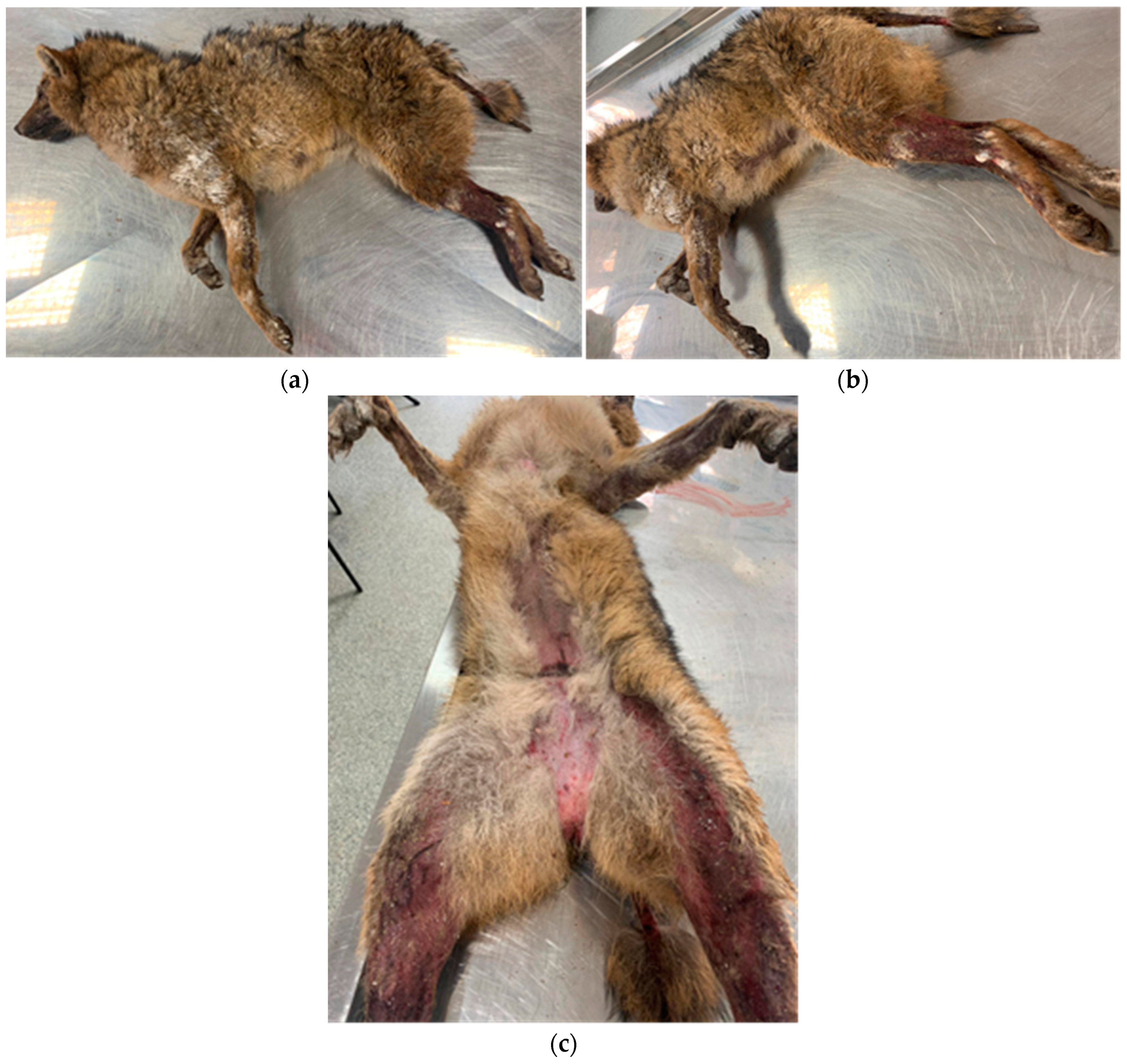
2.1.1. Clinical Examination
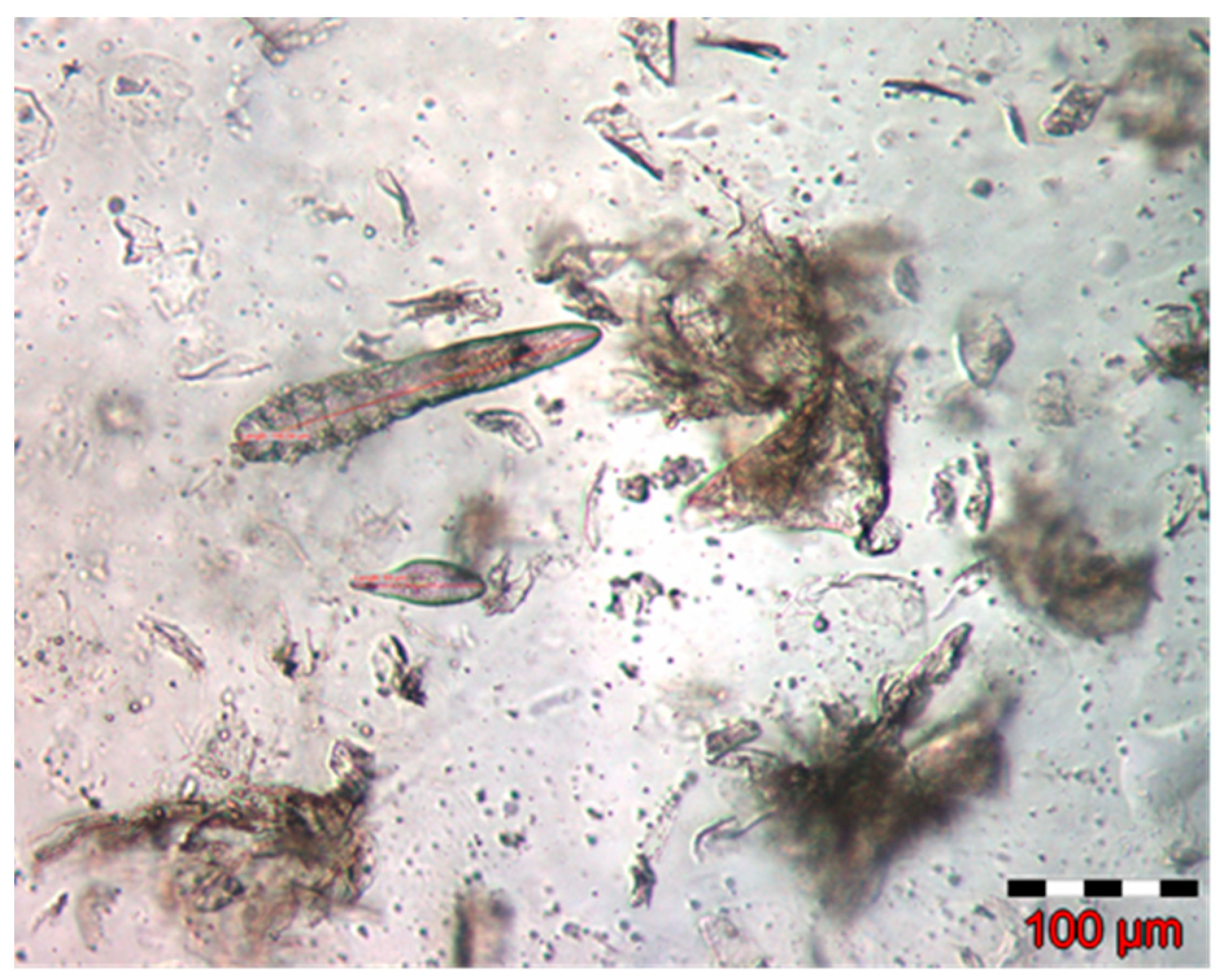
A corpse of a golden jackal female almost 2 years old, found in a silvo-steppe area in the vicinity of a small town in the south of Timiș County, was examined in the Parasitic Diseases clinic of FVM Timișoara. Unfortunately, the cause of death has not been established. The animal was severely emaciated and presented erythema and extensive severe alopecia accompanied by lichenification, seborrhea, and scaling. The lesions were located on different areas of the body: skin folds, feet, tail, and axillary and inguinal regions (Figure 1).

2.1.2. Diagnosis

After clinical examination, some laboratory tests such as skin scrapes, microscopic examination, trichogram (hair plucking), acetate tape test (impression), and fungal culture were performed in order to establish the diagnosis.
Deep skin scrape is commonly used to identify mites such as Demodex spp., Sarcoptes spp., and Notoedres spp. This collecting technique involves obtaining material from the edge of the lesions, which is mixed with a clarifying liquid and homogenized on a microscope slide, then covered with a coverslip and examined with the 10× microscope objective.
A trichogram has the same objectives as the deep scrape, to which is also added to the identification of the presence of dermatophytes. A small clump of hairs from the edge of the lesions is plucked after which it is placed on a microscope slide, followed by other steps identical to those of the skin scrape.
Acetate tape testing is convenient and reliable. A clear adhesive tape is pressed onto the surface of the skin to collect keratinocytes, any superficial microbes, and other ectoparasites such as Cheyletiella, a non-burrowing mite.
Fungal culture was performed by inoculating the hair and crust samples into the dermatophyte test medium (DTM) plate. The plate was incubated at 27 °C for 12 days.
Measurements were also performed in order to establish the specific sizes of the different developmental stages of mites (Table 1). They were made for 16 adults (11 males and 5 females), 5 nymphs, 5 larvae, and 8 eggs (Figure 2).

2.1.3. DNA Extraction and Molecular Analysis

The PCR reaction was carried out according to the technique described by Frank et al. 2013 [23] and was also used to identify D. cati in the same location [24]. The amplification was carried out by classical PCR and was based on the amplification of a sequence of the ~330 bp (fragment of the 16S rDNA) for Demodex spp., modified for the requirements of the mixture.
According to the protocol, both 5’ACTGTGCTAAGGTAGCGAAGTCA3’ forward primer and 5’TCAAAAGCCAACATCGAG3’ reverse primer [23] were used. They are considered highly conservative for several Demodex species, mainly D. gatoi, D. caprae, D. brevis, D. folliculorum, D. canis, and D. injai [23,25]. The reaction used a Master Mix MyTaqTM Red Mix (BIOLINE®®, Cincinnati, OH, USA) to achieve the results. The final volume of the PCR reaction was 25 μL, of which 12,5 μL MyTaqTM Red Mix (BIOLINE®®), 1 μL 1st primer, 1 μL 2nd primer (diluted to a concentration of 10 pmol/μL according to the protocol described by the manufacturer), and DNA extracted from the sample was analyzed, as well as ultrapure water. Amplification was accomplished with the My Cycler thermocycler (BioRad®®). This program included a stage of DNA denaturation at 95 °C for 90 sec, followed by 35 cycles of 55 °C for 30 s, 68 °C for 120 s, and 94 °C for 30 s, one of 55 °C for 30 s, and a final cycle 68 °C for 5 min. The analysis and control of amplicons was performed by horizontal electrophoresis in a submerse system of electrophoresis in 1.5% agarose gel, with the addition of flour dye MidoriGreen (Nippon Genetics®® Europe Gmbh, Düren, Germany) to a voltage of 120 V and 90 mA for 60 min. A 100 bp size Ladder DNA marker was used in the first well of the gel. The gel image with the migrated DNA fragments was captured using an UV photodocumentation system (UVP®®).
PCR products were sequenced at Macrogen Europe®® Company (Amsterdam, The Netherlands) and compared with those available in the GenBank database, using BLAST alignment (Figures S1 and S2).

3. Results

The laboratory tests revealed the presence of some cocci and rods, which were associated with Staphylococcus spp., respectively Pseudomonas aeruginosa and a few Malassezia spp. organisms. None of the usual ringworms grew on the DTM.
The scrapes proved positive for the presence of mites. Based on specific morphological characteristics (Table 1), the identified mites were classified as species of Demodex injai (Figure 2).
Thus, the total length of the adult mite was 238.56 µm and on segments, it was 25.53 µm for the gnathosoma, 63.05 µm for the podosoma, and 135.62 µm for the opisthosoma, respectively. The deutonymph measured 218.69 µm, the larval stage 137.89 µm, and the egg 65.11/25.64 µm.

4. Discussion

It is known that D. injai is the largest species of Demodex found in carnivores, especially in dogs. Basically, it is a prostigmatous mite with a vermiform appearance, which lives in the pilosebaceous follicles of the hosts and which, following intense proliferation, produces a chronic dermatological condition, sometimes generalized, rarely relapsing, called demodicosis. It usually results in folliculitis and/or furunculosis, accompanied by hyperseborrhea, erythema, hyperpigmentation, alopecia, comedones, and a characteristic odor of the affected areas [26,27,28].
In a study carried out in Poland between 2001 and 2008, D. injai was identified in two dog skin samples out of 39 examined and collected post-mortem [29]. The average mite length was 367 µm (309–411 µm) for the male and 339 µm (282–396 µm) for the female, three times higher that of D. cornei and almost double that of D. canis. Another study carried out more recently in 2020 by Chaudhary et al. [30], based on the morphometric analysis of 50 individuals of D. injai, revealed the following average dimensions of the parasite: gnathosoma—22.42 ± 0.60 µm, podosoma—74.94 ± 0.77 µm, opisthosoma—166.24 ± 2.55 µm, and total length—263.61 ± 2.83 µm. Samples were collected from 40 clinically manifested dogs in the Indian Veterinary Research Institute, Izatnagar, Bareilly, India. These sizes are smaller than those reported by Izdebska [29] in 2010 and close to those measured in our study.
Using the PCR technique, the result was identical, finding that the species involved was D. injai. Products of approximately 330 bp were obtained after PCR amplification. The rRNA sequences obtained from D. injai in the hind legs and axillary and inguinal areas were similar to those reported from morphologically alike parasites obtained from the skin. In the BLAST search, our sample was closely matched (96%) to the GenBank dog origin D. injai sequence (HE 817765) from Spain.
This case represents the first identification of D. injai in the golden jackal in Romania. Although initially the diagnosis was based on microscopic examination, confirmation and exact identification of the mite was achieved by PCR. However, it seems that the dimensions of the same parasite are variable depending on the body area from which it is collected [31]. This can, in fact, explain the differences in size recorded in the studies mentioned above [29,30]. D. injai was more frequently identified in terriers, especially in those with compromised immune status, in which seborrheic dermatitis of the dorsal area of the trunk was described [32,33].
In the case of the golden jackal, the specialized literature is very poor regarding demodicosis. In Israel, a case of simultaneous parasitism with two species of mites (Sarcoptes scabiei and an unnamed species of Demodex) was described, manifested as a chronic diffuse but severe dermatitis and folliculitis [17]. Another study carried out in the Caucasus area identifies the presence of D. canis in five out of 150 examined jackals, with a prevalence of 3.33% [2]. Controversial information about the presence of Demodex spp. was provided from the examination of five golden jackal specimens in Bangladesh, without specifying the mite species, prevalence, and intensity of parasitism [18].
The etiopathogenetic mechanisms of Demodex infection are not yet very well known, but it seems that a dysfunction of CD4+ T lymphocytes triggers the disease. This dysfunction may be due to parasitic co-infections, endocrine diseases (hypothyroidism, hyperadrenocorticism, or diabetes mellitus), neoplasms, or allergies that cause immunosuppression [34]. Thus, the synergy between immune factors and other intrinsic and extrinsic stressors is determinant in breaking the balance between the parasite and the host [35].

5. Conclusions

Analyzing data obtained from the specialized literature, the present study reports D. injai infection in the golden jackal for the first time in Europe. The increasing density of these populations highlights their potential role in the transmission of D. injai to congeners or to other species of carnivores with which it comes into contact, together with other zoonotic parasites. Additionally, they contribute to the maintenance of natural disease foci.

Supplementary Materials

The following supporting information can be downloaded at: https://www.mdpi.com/article/10.3390/pathogens12030412/s1, Figure S1: The sequences of the 16S rRNA gene obtained from sample 1 were identical to the Demodex injai sequence (GenBank nr. HE817765.1); Figure S2: Sequences with the highest similarity to the PCR product obtained from the amplification of samples.

Author Contributions

Conceptualization, S.M., F.M. and N.M.; methodology, A.-M.M. and M.I.; software, D.-C.P.; validation, S.M. and N.M.; investigation, A.-M.M., M.M.F.M. and N.M.; resources, A.-M.M., M.M.F.M. and D.-C.P.; data curation, N.M.; writing—original draft preparation, S.M., F.M. and N.M.; writing—review and editing, S.M., F.M. and N.M., visualization, A.-M.M., M.M.F.M. and V.I.; supervision, N.M.; project administration, S.M. and N.M. All authors have read and agreed to the published version of the manuscript.

Funding

This research received no external funding.

Institutional Review Board Statement

This study received approval (reference 164/21.12.2022) issued by the Bioethics Commission of the University of Life Sciences “King Michael I” from Timişoara.

Informed Consent Statement

Not applicable.

Data Availability Statement

Not applicable.

Conflicts of Interest

The authors declare no conflict of interest.

References

  1. Kemenszky, P.; Jánoska, F.; Nagy, G.; Csivincsik, Á. The golden jackal (Canis aureus) and the African swine fever pandemic: Its role is controversial but not negligible (a diet analysis study). Vet. Med. Sci. 2022, 8, 97–103. [Google Scholar] [CrossRef] [PubMed]
  2. Gherman, C.M.; Mihalca, A.D. A synoptic overview of golden jackal parasites reveals high diversity of species. Parasites Vectors 2017, 10, 419. [Google Scholar] [CrossRef] [Green Version]
  3. Koepfli, K.-P.; Pollinger, J.; Godinho, R.; Robinson, J.; Lea, A.; Hendricks, S.; Schweizer, R.M.; Thalmann, O.; Silva, P.; Fan, Z.; et al. Genome-wide evidence reveals that African and Eurasian golden jackals are distinct species. Curr. Biol. 2015, 25, 2158–2165. [Google Scholar] [CrossRef] [Green Version]
  4. Shamir, M.; Yakobson, B.; Baneth, G.; King, R.; Dar-Verker, S.; Markovics, A.; Aroch, I. Antibodies to selected canine pathogens and infestation with intestinal helminths in golden jackals (Canis aureus) in Israel. Vet. J. 2001, 162, 66–72. [Google Scholar] [CrossRef]
  5. Aguirre, A.A. Wild canids as sentinels of ecological health: A conservation medicine perspective. Parasites Vectors 2009, 2 (Suppl. S1), S7. [Google Scholar] [CrossRef] [Green Version]
  6. Papadopoulos, H.; Himonas, C.; Papazahariadou, M.; Antoniadou-Sotiriadou, K. Helminths of foxes and other wild carnivores from rural areas in Greece. J. Helminthol. 1997, 71, 227–231. [Google Scholar] [CrossRef]
  7. Georgieva, D.; Koinarski, V.T.; Ivanov, A.I.; Prelesov, P.N.; Kirkova, Z.T. Role of wild carnivores in the epizootology and epidemiology of trichinellosis. Bulg. J. Vet. Med. 2000, 3, 199–204. [Google Scholar]
  8. Blaga, R.; Gherman, C.; Seucom, D.; Cozma, V.; Boireau, P. First identification of Trichinella sp. in golden jackal (Canis aureus) in Romania. J. Wildl. Dis. 2008, 44, 457–459. [Google Scholar] [CrossRef] [PubMed] [Green Version]
  9. Kirkova, Z.; Raychev, E.; Georgieva, D. Studies on feeding habits and parasitological status of red fox, golden jackal, wild cat and stone marten in Sredna Gora, Bulgaria. J. Life Sci. 2011, 5, 264–270. [Google Scholar]
  10. Takács, A.; Szabó, L.; Juhász, L.; Takács, A.A.; Lanszki, J.; Takács, P.T.; Heltai, M. Data on the parasitological status of golden jackal (Canis aureus L., 1758) in Hungary. Acta Vet. Hung. 2014, 62, 33–41. [Google Scholar] [CrossRef] [PubMed] [Green Version]
  11. Ionică, A.M.; Matei, I.A.; D’Amico, G.; Daskalaki, A.A.; Juránková, J.; Ionescu, D.T.; Mihalca, A.D.; Modry, D.; Gherman, C.M. Role of golden jackals (Canis aureus) as natural reservoirs of Dirofilaria spp. in Romania. Parasites Vectors 2016, 9, 240. [Google Scholar] [CrossRef] [PubMed] [Green Version]
  12. Ilić, T.; Becskei, Z.; Petrović, T.; Polaček, V.; Ristić, B.; Milić, S.; Stepanović, P.; Radisavljević, K.; Dimitrijević, S. Endoparasitic fauna of red foxes (Vulpes vulpes) and golden jackals (Canis aureus) in Serbia. Acta Parasitol. 2016, 61, 389–396. [Google Scholar] [CrossRef]
  13. Dumitrache, M.O.; Gherman, C.M.; Cozma, V.; Mircean, V.; Györke, A.; Sándor, A.D.; Mihalca, A.D. Hard ticks (Ixodidae) in Romania: Surveillance, host associations, and possible risks for tick-borne diseases. Parasitol. Res. 2012, 110, 2067–2070. [Google Scholar] [CrossRef]
  14. Hornok, S.; Fuente, J.; Horváth, G.; Fernández de Mera, I.G.; Wijnveld, M.; Tánczos, B.; Farkas, B.; Jongejan, F. Molecular evidence of Ehrlichia canis and Rickettsia massiliae in ixodid ticks of carnivores from South Hungary. Acta Vet. Hung. 2013, 61, 42–50. [Google Scholar] [CrossRef] [PubMed] [Green Version]
  15. Duscher, G.G.; Kübber-Heiss, A.; Richter, B.; Suchentrunk, F. A golden jackal (Canis aureus) from Austria bearing Hepatozoon canis-import due to immigration into a non-endemic area? Ticks Tick Borne Dis. 2013, 4, 133–137. [Google Scholar] [CrossRef] [PubMed]
  16. D’Amico, G.; Dumitrache, M.O.; Matei, I.A.; Ionică, A.M.; Gherman, C.M.; Sándor, A.D.; Modry, D.; Mihalca, A.D. Ixodid ticks parasitizing wild carnivores in Romania. Exp. Appl. Acarol. 2017, 71, 139–149. [Google Scholar] [CrossRef] [PubMed]
  17. Berkovitz, A.; Waner, T.; King, R.; Perl, S. Concurrent parasitation with Sarcoptes and Demodex in a golden jackal. Isr. J. Vet. Med. 2009, 64, 10–11. [Google Scholar]
  18. Yousuf, M.A.; Bashu, J.; Pervin, M.; Islam, M.T.; Das, P.M.; Khan, M.A.H.N.A. Identifying diseases of golden jackals of Bangladesh Agricultural University campus, Mymensingh, Bangladesh. Bangl. J. Vet. Med. 2014, 12, 217–224. [Google Scholar] [CrossRef] [Green Version]
  19. Salvadori, C.; Formenti, N.; Trogu, T.; Lanfranchi, P.; Papini, R.A.; Poli, A. Demodicosis in chamois (Rupicapra rupicapra subsp. rupicapra) in the Italian Alps, 2013–2014. J. Wildl. Dis. 2016, 52, 433–435. [Google Scholar] [CrossRef] [Green Version]
  20. Ringwaldt, E.M.; Brook, B.W.; Carver, S.; Buettel, J.C. The patterns and causes of dermatitis in terrestrial and semi-aquatic mammalian wildlife. Animals 2021, 11, 1691. [Google Scholar] [CrossRef]
  21. Arlian, L.G.; Morgan, M.S. A review of Sarcoptes scabiei: Past, present and future. Parasites Vectors 2017, 10, 297. [Google Scholar] [CrossRef] [PubMed] [Green Version]
  22. Escobar, L.E.; Carver, S.; Cross, P.C.; Rossi, L.; Almberg, E.S.; Yabsley, M.J.; Niedringhaus, K.D.; Van Wick, P.; Dominguez-Villegas, E.; Gakuya, F.; et al. Sarcoptic mange: An emerging panzootic in wildlife. Transbound. Emerg. Dis. 2021, 69, 927–942. [Google Scholar] [CrossRef]
  23. Frank, L.A.; Kania, S.A.; Chung, K.; Brahmbhatt, R. A molecular technique for the detection and differentiation of Demodex mites on cats. Vet. Dermatol. 2013, 24, 367-e83. [Google Scholar] [CrossRef] [PubMed]
  24. Ilie, M.S.; Imre, M.; Giubega, S.; Luca, I.; Florea, T.; Morariu, S. Feline demodicosis case report-First molecular characterization of Demodex mites in Romania. Pathogens 2021, 10, 1474. [Google Scholar] [CrossRef] [PubMed]
  25. Bernstein, J.A.; Frank, L.A.; Kania, S.A. PCR amplification and DNA sequence identification of an unusual morphological form of Demodex cati in a cat. Vet. Dermatol. 2014, 25, 487-e80. [Google Scholar] [CrossRef]
  26. Desch, C.E.; Hillier, A. Demodex injai: A new species of hair follicle mite (Acari: Demodecidae) from domestig dog (Canidae). J Med. Entomol. 2003, 40, 146–149. [Google Scholar] [CrossRef]
  27. Ferrer, L.; Ravera, I.; Silbermayr, K. Immunology and pathogenesis of canine demodicosis. Vet. Dermatol. 2014, 25, 427-e65. [Google Scholar] [CrossRef]
  28. Silva Sgarbossa, R.S.A.; Vieira Sechi, G.; Duarte Pacheco, B.; Buba Lucina, S.; Paulo, M.R.; dos Santos Monti, F.; Rodrigues de Farias, M. The epidemiological and clinical aspects of Demodex injai demodicosis in dogs: A report of eight cases. Semin. Cienc. Agrar. 2017, 38, 3387–3394. [Google Scholar] [CrossRef] [Green Version]
  29. Izdebska, J.N. Demodex sp. (Acari, Demodecidae) and demodecosis in dogs: Characteristics, symptoms, occurrence. Bull. Vet. Inst. Pulawy 2010, 54, 335–338. [Google Scholar]
  30. Chaudhary, A.K.; Dimri, U.; Ajith, Y.; Madesh, E.; Yadav, S.; Kavitha, K.; Angmo, S. Comparative morphometric analysis for differentiation of three Demodex mite species causing canine demodicosis. Int. J. Curr. Microbiol. Appl. Sci. 2020, 9, 2151–2155. [Google Scholar] [CrossRef]
  31. Milosevic, M.A.; Frank, L.A.; Brahmbhatt, R.A.; Kania, K.A. PCR amplification and DNA sequencing of Demodex injai from otic secretions of a dog. Vet. Dermatol. 2013, 24, 286-e66. [Google Scholar] [CrossRef] [PubMed]
  32. Hillier, A.; Desch, C.E. Large-bodied Demodex mite infestation in 4 dogs. J. Am. Vet. Med. Assoc. 2002, 220, 623–627. [Google Scholar] [CrossRef]
  33. Ordeix, L.; Bardagi, M.; Scarampella, F.; Ferrer, L.; Fondati, A. Demodex injai infestation and dorsal greasy skin and hair in eight wirehaired fox terrier dogs. Vet. Dermatol. 2009, 20, 267–272. [Google Scholar] [CrossRef] [PubMed]
  34. Oliveira, C.D.; Larsson, C.E.; Camargo, M.M. Longitudinal assessment of T-lymphocyte subpopulations during generalized demodicosis in dogs and their relationship with remission. Vet. Dermatol. 2015, 26, 18–22. [Google Scholar] [CrossRef]
  35. Mederle, N.; Mederle, O.; Morariu, S.; Herman, V.; Negrescu, A.; Marin, A.M.; Suici, T.; Dărăbuș, G. Study regarding the role and medical and social implications of Demodex spp. parasitism in dogs and humans. Rev. Rom. Med. Vet. 2020, 30, 39–46. [Google Scholar]
Figure 1. Extensive alopecic areas on fore- and hind limbs, on the ventral abdomen and inguinal, including the tail, with erythema, hyperpigmentation, lichenification, and some follicular papules and pustules; (a,b)—lateral view of the entire body; (c)—enlarged view of the inguinal area and the medial part of the hind limbs.
Pathogens 12 00412 g001
Figure 2. Nymph and egg of Demodex injai.
Pathogens 12 00412 g002
Table 1. Demodex mites’ measurements according to their developmental stages.
Developmental StageTotal Length µm
(min.–max.)
Gnathosoma µm
(min.–max.)
Podosoma µm
(min.–max.)
Podosoma Width µm
(min.–max.)
Opisthosoma µm
(min.–max.)
Egg Width µm
(min.–max.)
Adult (n = 16)238.56
(203.86–249.19)
25.53
(18.71–27.95)
63.05
(56.31–77.20)
36.41
(33.17–40.19)
135.62
(109.37–157.10)
-
Nymph (n = 5)218.69
(196.23–234.69)
23.72
(17.66–26.33)
61.49
(61.60–63.89)
38.65
(33.04–44.49)
134.45
(113.64–150.68)
-
Larva (n = 5)137.89
(98.67–190.30)
--29.54
(23.35–36.78)
--
Egg (n = 8)65.11
(56.85–71.78)
----25.64
(20.25–32.08)
Disclaimer/Publisher’s Note: The statements, opinions and data contained in all publications are solely those of the individual author(s) and contributor(s) and not of MDPI and/or the editor(s). MDPI and/or the editor(s) disclaim responsibility for any injury to people or property resulting from any ideas, methods, instructions or products referred to in the content.

