
Bacterial Co-Infections and Antimicrobial Resistance in Patients Hospitalized with Suspected or Confirmed COVID-19 Pneumonia in Kazakhstan
 , and
, and
Abstract
:1. Introduction
2. Materials and Methods
2.1. Study Participants
- Mild Illness: Individuals who have any of the various signs and symptoms of coronavirus infection (e.g., fever, cough, sore throat, malaise, headache, muscle pain, nausea, vomiting, diarrhea, loss of taste and smell) but who do not have shortness of breath, dyspnea, or abnormal chest imaging.
- Moderate Illness: Individuals who show evidence of lower respiratory disease during clinical assessment or imaging and who have an oxygen saturation (SpO2) ≥ 94% on room air at sea level.
- Severe Illness: Individuals who have SpO2 < 94% on room air at sea level, a ratio of arterial partial pressure of oxygen to fraction of inspired oxygen (PaO2/FiO2) < 300 mm Hg, respiratory frequency > 30 breaths / min, or lung infiltrates > 50%.
- Critical Illness: Individuals who have respiratory failure, septic shock, and/or multiple organ dysfunction.
2.2. Laboratory Testing
2.3. Data Analysis
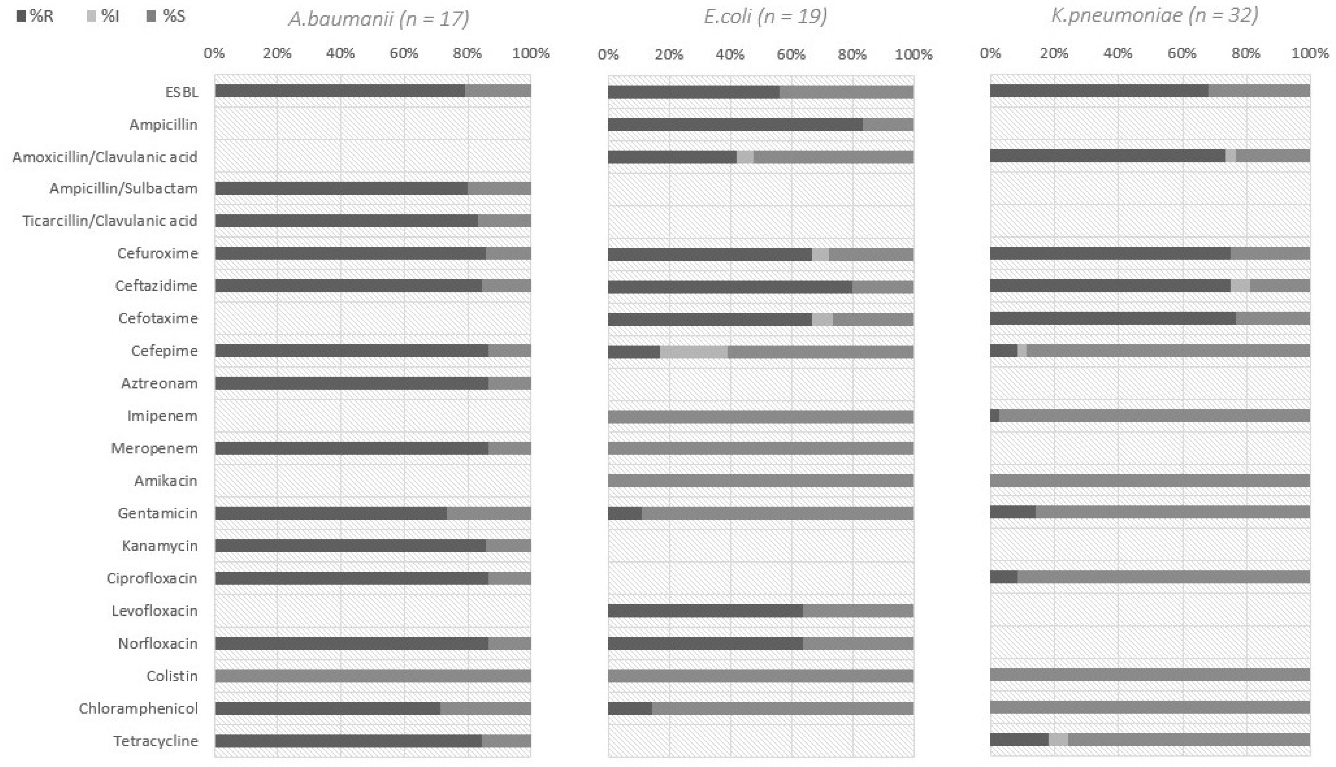
3. Results
4. Discussion
Author Contributions
Funding
Institutional Review Board Statement
Informed Consent Statement
Data Availability Statement
Acknowledgments
Conflicts of Interest
References
- Munster, V.J.; Koopmans, M.; van Doremalen, N.; van Riel, D.; de Wit, E. A Novel Coronavirus Emerging in China—Key Questions for Impact Assessment. N. Engl. J. Med. 2020, 382, 692–694. [Google Scholar] [CrossRef] [PubMed]
- Rebold, N.; Holger, D.; Alosaimy, S.; Morrisette, T.; Rybak, M. COVID-19: Before the Fall, an Evidence-Based Narrative Review of Treatment Options. Infect. Dis. Ther. 2021, 10, 93–113. [Google Scholar] [CrossRef] [PubMed]
- Elekhnawy, E.; Kamar, A.A.; Sonbol, F. Present and future treatment strategies for coronavirus disease 2019. Future J. Pharm. Sci. 2021, 7, 84. [Google Scholar] [CrossRef] [PubMed]
- Xu, X.; Chen, P.; Wang, J.; Feng, J.; Zhou, H.; Li, X.; Zhong, W.; Hao, P. Evolution of the novel coronavirus from the ongoing Wuhan outbreak and modeling of its spike protein for risk of human transmission. Sci. China Life Sci. 2020, 63, 457–460. [Google Scholar] [CrossRef] [Green Version]
- Huang, C.; Wang, Y.; Li, X.; Ren, L.; Zhao, J.; Hu, Y.; Zhang, L.; Fan, G.; Xu, J.; Gu, X.; et al. Clinical features of patients infected with 2019 novel coronavirus in Wuhan, China. Lancet 2020, 395, 497–506. [Google Scholar] [CrossRef] [Green Version]
- Lansbury, L.; Lim, B.; Baskaran, V.; Lim, W.S. Co-infections in people with COVID-19: A systematic review and meta-analysis. J. Infect. 2020, 81, 266–275. [Google Scholar] [CrossRef]
- Li, X.; Wang, L.; Yan, S.; Yang, F.; Xiang, L.; Zhu, J.; Shen, B.; Gong, Z. Clinical Characteristics of 25 Death Cases with COVID-19: A Retrospective Review of Medical Records in a Single Medical Center, Wuhan, China. Int. J. Infect. Dis. 2020, 94, 128–132. [Google Scholar] [CrossRef]
- Sulaiman, I.; Chung, M.; Angel, L.; Tsay, J.-C.J.; Wu, B.G.; Yeung, S.T.; Krolikowski, K.; Li, Y.; Duerr, R.; Schluger, R.; et al. Microbial Signatures in the Lower Airways of Mechanically Ventilated COVID-19 Patients Associated with Poor Clinical Outcome. Nat. Microbiol. 2021, 6, 1245–1258. [Google Scholar] [CrossRef]
- Dickson, R.P.; Schultz, M.J.; van der Poll, T.; Schouten, L.R.; Falkowski, N.R.; Luth, J.E.; Sjoding, M.W.; Brown, C.A.; Chanderraj, R.; Huffnagle, G.B. Lung Microbiota Predict Clinical Outcomes in Critically Ill Patients. Am. J. Respir. Crit. Care Med. 2020, 201, 555–563. [Google Scholar] [CrossRef]
- Langford, B.J.; So, M.; Raybardhan, S.; Leung, V.; Westwood, D.; MacFadden, D.R.; Soucy, J.-P.R.; Daneman, N. Bacterial Co-Infection and Secondary Infection in Patients with COVID-19: A Living Rapid Review and Meta-Analysis. Clin. Microbiol. Infect. 2020, 26, 1622–1629. [Google Scholar] [CrossRef]
- Murray, A.K. The Novel Coronavirus COVID-19 Outbreak: Global Implications for Antimicrobial Resistance. Front. Microbiol. 2020, 11, 1020. [Google Scholar] [CrossRef] [PubMed]
- Rawson, T.M.; Ming, D.; Ahmad, R.; Moore, L.S.P.; Holmes, A.H. Antimicrobial Use, Drug-Resistant Infections and COVID-19. Nat. Rev. Microbiol. 2020, 18, 409–410. [Google Scholar] [CrossRef] [PubMed]
- Schons, M.J.; Caliebe, A.; Spinner, C.D.; Classen, A.Y.; Pilgram, L.; Ruethrich, M.M.; Rupp, J.; Nunes de Miranda, S.M.; Römmele, C.; Vehreschild, J.; et al. All-Cause Mortality and Disease Progression in SARS-CoV-2-Infected Patients with or without Antibiotic Therapy: An Analysis of the LEOSS Cohort. Infection 2022, 50, 423–436. [Google Scholar] [CrossRef] [PubMed]
- World Health Organization. Strategic Preparedness and Response Plan for the New Coronavirus. 2020. Available online: https://www.who.int/publications-detail/covid-19-strategy-update-13-april-2020 (accessed on 15 April 2020).
- Hsu, J. How COVID-19 Is Accelerating the Threat of Antimicrobial Resistance. BMJ 2020, 369, m1983. [Google Scholar] [CrossRef] [PubMed]
- Zhussupova, G.; Skvirskaya, G.; Reshetnikov, V.; Dragojevic-Simic, V.; Rancic, N.; Utepova, D.; Jakovljevic, M. The Evaluation of Antibiotic Consumption at the Inpatient Level in Kazakhstan from 2011 to 2018. Antibiotics 2020, 9, 57. [Google Scholar] [CrossRef] [PubMed] [Green Version]
- Ministry of Healthcare of Kazakhstan. Guideline for Diagnosis and Treatment “Coronovirus Infection COVID-19 in Adults”; Protocol No. 124 of Republic of Kazakhstan; Ministry of Healthcare of Kazakhstan: Astana, Kazakhstan, 2020.
- COVID-19 Treatment Guidelines Panel. Coronavirus Disease 2019 (COVID-19) Treatment Guidelines. National Institutes of Health. Available online: https://www.covid19treatmentguidelines.nih.gov/ (accessed on 5 January 2022).
- World Health Organization. COVID-19 Clinical Management: Living Guidance. 2021. Available online: https://apps.who.int/iris/bitstream/handle/10665/349321/WHO-2019-nCoV-clinical-2021.2-eng.pdf (accessed on 15 April 2020).
- Miller, J.M.; Binnicker, M.J.; Campbell, S.; Carroll, K.C.; Chapin, K.C.; Gilligan, P.H.; Gonzalez, M.D.; Jerris, R.C.; Kehl, S.C.; Patel, R.; et al. A Guide to Utilization of the Microbiology Laboratory for Diagnosis of Infectious Diseases: 2018 Update by the Infectious Diseases Society of America and the American Society for Microbiology. Clin. Infect. Dis. 2018, 67, e1–e94. [Google Scholar] [CrossRef] [Green Version]
- Clinical and Laboratory Standards Institute (CLSI). Performance Standards for Antimicrobial Disk Susceptibility Tests, 11th ed.; Approved Standard. CLSI Document M02-A11; Clinical and Laboratory Standards Institute: Wayne, PA, USA, 2012; ISBN 1-56238-781-2. [Google Scholar]
- Charlson, E.S.; Bittinger, K.; Haas, A.R.; Fitzgerald, A.S.; Frank, I.; Yadav, A.; Bushman, F.D.; Collman, R.G. Topographical Continuity of Bacterial Populations in the Healthy Human Respiratory Tract. Am. J. Respir. Crit. Care Med. 2011, 184, 957–963. [Google Scholar] [CrossRef] [Green Version]
- He, Y.; Li, W.; Wang, Z.; Chen, H.; Tian, L.; Liu, D. Nosocomial infection among patients with COVID-19: A retrospective data analysis of 918 cases from a single center in Wuhan, China. Infect. Control Hosp. Epidemiol. 2020, 41, 982–983. [Google Scholar] [CrossRef] [Green Version]
- Mirzaei, R.; Goodarzi, P.; Asadi, M.; Soltani, A.; Aljanabi, H.A.A.; Jeda, A.S.; Dashtbin, S.; Jalalifar, S.; Mohammadzadeh, R.; Teimoori, A.; et al. Bacterial Co-infections with SARS-CoV-2. IUBMB Life 2020, 72, 2097–2111. [Google Scholar] [CrossRef]
- Chen, N.; Zhou, M.; Dong, X.; Qu, J.; Gong, F.; Han, Y.; Qiu, Y.; Wang, J.; Liu, Y.; Wei, Y.; et al. Epidemiological and Clinical Characteristics of 99 Cases of 2019 Novel Coronavirus Pneumonia in Wuhan, China: A Descriptive Study. Lancet 2020, 395, 507–513. [Google Scholar] [CrossRef] [Green Version]
- Rawson, T.M.; Moore, L.S.P.; Zhu, N.; Ranganathan, N.; Skolimowska, K.; Gilchrist, M.; Satta, G.; Cooke, G.; Holmes, A. Bacterial and Fungal Coinfection in Individuals with Coronavirus: A Rapid Review to Support COVID-19 Antimicrobial Prescribing. Clin. Infect. Dis. 2020, 71, 2459–2468. [Google Scholar] [CrossRef] [PubMed]
- Rice, L.B. Federal Funding for the Study of Antimicrobial Resistance in Nosocomial Pathogens: No ESKAPE. J. Infect. Dis. 2008, 197, 1079–1081. [Google Scholar] [CrossRef] [PubMed]
- Lim, Y.K.; Kweon, O.J.; Kim, H.R.; Kim, T.-H.; Lee, M.-K. Impact of Bacterial and Viral Coinfection in Community-Acquired Pneumonia in Adults. Diagn. Microbiol. Infect. Dis. 2019, 94, 50–54. [Google Scholar] [CrossRef] [PubMed]
- Guo, L.; Wei, D.; Zhang, X.; Wu, Y.; Li, Q.; Zhou, M.; Qu, J. Clinical Features Predicting Mortality Risk in Patients with Viral Pneumonia: The MuLBSTA Score. Front. Microbiol. 2019, 10, 2752. [Google Scholar] [CrossRef] [PubMed] [Green Version]
- Zhou, F.; Yu, T.; Du, R.; Fan, G.; Liu, Y.; Liu, Z.; Xiang, J.; Wang, Y.; Song, B.; Gu, X.; et al. Clinical Course and Risk Factors for Mortality of Adult Inpatients with COVID-19 in Wuhan, China: A Retrospective Cohort Study. Lancet 2020, 395, 1054–1062. [Google Scholar] [CrossRef]
- Morosini, M.I.; García-Castillo, M.; Coque, T.M.; Valverde, A.; Novais, A.; Loza, E.; Baquero, F.; Cantón, R. Antibiotic coresistance in extended-spectrum-beta-lactamase-producing Enterobacteriaceae and in vitro activity of tigecycline. Antimicrob. Agents Chemother. 2006, 50, 2695–2699. [Google Scholar] [CrossRef] [Green Version]
- Ministry of Health of the Republic of Kazakhstan. Сommunity-Acquired Pneumonia in Adults. Clinical Protocol of Diagnosis and Treatment No. 169. 2022. Available online: https://diseases.medelement.com/disease/внебoльничная-пневмoния-у-взрoслых-кр-рк-2019/17355 (accessed on 15 April 2020).
- Brolund, A. Overview of ESBL-producing Enterobacteriaceae from a Nordic perspective. Infect. Ecol. Epidemiol. 2014, 4, 24555. [Google Scholar] [CrossRef] [Green Version]
- Van, T.D.; Dinh, Q.-D.; Vu, P.D.; Nguyen, T.V.; Pham, C.V.; Dao, T.T.; Phung, C.D.; Hoang, H.T.T.; Tang, N.T.; Do, N.T.; et al. Antibiotic Susceptibility and Molecular Epidemiology of Acinetobacter Calcoaceticus-Baumannii Complex Strains Isolated from a Referral Hospital in Northern Vietnam. J. Glob. Antimicrob. Resist. 2014, 2, 318–321. [Google Scholar] [CrossRef] [Green Version]
- Thompson, D.S. Methicillin-Resistant Staphylococcus Aureus in a General Intensive Care Unit. J. R. Soc. Med. 2004, 97, 521–526. [Google Scholar] [CrossRef]
- Rello, J.; Torres, A.; Ricart, M.; Valles, J.; Gonzalez, J.; Artigas, A.; Rodriguez-Roisin, R. Ventilator-Associated Pneumonia by Staphylococcus Aureus. Comparison of Methicillin-Resistant and Methicillin-Sensitive Episodes. Am. J. Respir. Crit. Care Med. 1994, 150, 1545–1549. [Google Scholar] [CrossRef]
- Bhat, N.; Wright, J.G.; Broder, K.R.; Murray, E.L.; Greenberg, M.E.; Glover, M.J.; Likos, A.M.; Posey, D.L.; Klimov, A.; Lindstrom, S.E.; et al. Influenza-Associated Deaths among Children in the United States, 2003–2004. N. Engl. J. Med. 2005, 353, 2559–2567. [Google Scholar] [CrossRef] [PubMed]
- Martins-Filho, P.R.; Tavares, C.S.S.; Santos, V.S. Factors Associated with Mortality in Patients with COVID-19. A Quantitative Evidence Synthesis of Clinical and Laboratory Data. Eur. J. Intern. Med. 2020, 76, 97–99. [Google Scholar] [CrossRef] [PubMed]


| Total | SARS-CoV-2 RT-PCR Result | p-Value | ||
|---|---|---|---|---|
| (+) | (−) | |||
| n (%) or (SD) | n (%) or (SD) | n (%) (SD) | ||
| Total | 209 | 116 (56) | 93 (44) | 0.07 |
| City | 0.7 | |||
| Almaty | 64 (31) | 33 (28) | 31 (33) | 0.5 |
| Atyrau | 50 (24) | 25 (22) | 25 (27) | 0.4 |
| Karaganda | 95 (45) | 58 (50) | 37 (40) | 0.1 |
| Male | 114 (55) | 56 (48) | 58 (62) | 0.5 |
| Female | 95 (45) | 60 (52) | 35 (38) | 0.03 * |
| Age, median [range] years | 60 (14) | 61 (13) | 60 (14) | 0.4 |
| % lung damage, mean [SD] | 44% (21) | 43% (21) | 54% (26) | 0.1 |
| On antibiotics | 189 (90) | 112 (97) | 77 (83) | <0.01 * |
| Sputum sample results | ||||
| No growth | 62 (30) | − | − | − |
| Normal microbiota | 50 (24) | 28 (24) | 22 (24) | 0.7 |
| Pathogenic bacteria detected | 97 (71) | 50 (43) | 47 (51) | |
| Species of isolates (n = 106) | ||||
| All Enterobacterales | 70 (44) | 34 (29) | 36 (39) | 0.9 |
| Klebsiella pneumoniae | 32 (15) | 2 (2) | 1 (1) | 0.9 |
| Acinetobacter baumannii | 17 (11) | 7 (6) | 10 (11) | 0.4 |
| Escherichia coli | 19 (9) | 12 (10) | 7 (8) | 0.6 |
| Pseudomonas aeruginosa | 3 (2) | 2 (2) | 1 (1) | 0.9 |
| Stenotrophomonas maltophilia | 3 (2) | 2 (2) | 1 (1) | 0.6 |
| Staphylococcus aureus | 10 (6) | 6 (5) | 4 (4) | 0.9 |
| Streptococcus pneumoniae | 3 (1) | 2 (2) | 1 (1) | 0.9 |
| Candida spp. | 124 (15) | 73 (63) | 51 (55) | 0.2 |
| Species Cultured | N | Disease Severity Classification | ||||
|---|---|---|---|---|---|---|
| Mild or Moderate | Severe or Critical | |||||
| n | % | n | % | p-Value | ||
| Normal Microbiota [22] | 50 | 13 | 26% | 37 | 74% | 0.4 |
| A. baumannii | 17 | 0 | 0% | 17 | 100% | <0.01 * |
| P. aeruginosa | 3 | 2 | 66% | 1 | 33% | 0.5 |
| S. maltophilia | 3 | 0 | 0% | 3 | 100% | 0.3 b |
| K. pneumoniae | 32 | 2 | 6% | 30 | 94% | <0.01 * |
| E. coli | 19 | 5 | 26% | 14 | 74% | 0.9 |
| Other Enterobacteralesa | 19 | 3 | 16% | 16 | 84% | 0.3 |
| S. aureus | 10 | 4 | 40% | 6 | 60% | 0.9 |
| S. pneumoniae | 3 | 0 | 0% | 3 | 100% | 0.3b |
| Candida spp. | 124 | 39 | 23% | 95 | 77% | 0.9 |
Disclaimer/Publisher’s Note: The statements, opinions and data contained in all publications are solely those of the individual author(s) and contributor(s) and not of MDPI and/or the editor(s). MDPI and/or the editor(s) disclaim responsibility for any injury to people or property resulting from any ideas, methods, instructions or products referred to in the content. |
© 2023 by the authors. Licensee MDPI, Basel, Switzerland. This article is an open access article distributed under the terms and conditions of the Creative Commons Attribution (CC BY) license (https://creativecommons.org/licenses/by/4.0/).
Share and Cite
Lavrinenko, A.; Kolesnichenko, S.; Kadyrova, I.; Turmukhambetova, A.; Akhmaltdinova, L.; Klyuyev, D. Bacterial Co-Infections and Antimicrobial Resistance in Patients Hospitalized with Suspected or Confirmed COVID-19 Pneumonia in Kazakhstan. Pathogens 2023, 12, 370. https://doi.org/10.3390/pathogens12030370
Lavrinenko A, Kolesnichenko S, Kadyrova I, Turmukhambetova A, Akhmaltdinova L, Klyuyev D. Bacterial Co-Infections and Antimicrobial Resistance in Patients Hospitalized with Suspected or Confirmed COVID-19 Pneumonia in Kazakhstan. Pathogens. 2023; 12(3):370. https://doi.org/10.3390/pathogens12030370
Chicago/Turabian StyleLavrinenko, Alyona, Svetlana Kolesnichenko, Irina Kadyrova, Anar Turmukhambetova, Lyudmila Akhmaltdinova, and Dmitriy Klyuyev. 2023. "Bacterial Co-Infections and Antimicrobial Resistance in Patients Hospitalized with Suspected or Confirmed COVID-19 Pneumonia in Kazakhstan" Pathogens 12, no. 3: 370. https://doi.org/10.3390/pathogens12030370