Article

Optimization and Application of Real-Time qPCR Assays in Detection and Identification of Chlamydiales in Products of Domestic Ruminant Abortion

Tuberculosis and Brucellosis Research Programme, Department of Veterinary Tropical Diseases, Faculty of Veterinary Science, University of Pretoria, Pretoria 0002, South Africa
*
Author to whom correspondence should be addressed.
Pathogens 2023, 12(2), 290; https://doi.org/10.3390/pathogens12020290
Submission received: 23 December 2022 / Revised: 7 February 2023 / Accepted: 8 February 2023 / Published: 9 February 2023
(This article belongs to the Special Issue Bacterial, Fungal and Parasitic Zoonoses)

Abstract

:
Domestic ruminant abortions due to infectious agents represent an important cause of economic losses in the agricultural industry. This study aimed to optimise and apply qPCR assays for detection of Chlamydiales in domestic ruminant abortion cases. Primers and probes for detection of the order Chlamydiales, Chlamydia abortus, Chlamydia pecorum, Parachlamydia acanthamoeba and Waddlia chondrophila were taken from the literature to create one singleplex and two duplex assays and the assays were optimised. Placentitis and pneumonia are pathological lesions associated with Chlamydiales infection. In a previous study, twenty-five clinical cases had pathological lesions of placentitis or pneumonia. These cases were investigated further by application of the qPCR assays in this study. Chlamydiales were detected in 16 cases. C. abortus, P. acanthamoeba and W. chondrophila were detected in bovine; and C. pecorum and W. chondrophila in ovine and caprine cases. Chlamydiales were detected in three previously inconclusive cases. Identification was improved from genus to species level (C. pecorum). Four cases remained inconclusive. In conclusion, detection of Chlamydiales and differentiation to species level was improved. This study reports the first detection of P. acanthamoeba and W. chondrophila in abortion cases in South Africa, indicating a potentially significant role in abortions in this country.

1. Introduction

Infectious agents are the most common causes of domestic ruminant abortions [1] representing important economic losses in the agricultural industry [2]. Abortion is defined as the premature end of a pregnancy with the expulsion of a non-viable foetus [3]. Three families in the order Chlamydiales are of significance as agents of abortion, namely Chlamydiaceae, Waddliaceae and Parachlamydiaceae [4]. Chlamydiales are adapted to a very broad spectrum of hosts and are agents of a variety of diseases in animals and humans [4]. Several members of the order are of zoonotic significance. Chlamydia abortus (C. abortus), Waddlia chondrophila (W. chondrophila) and Parachlamydia acanthamoebae (P. acanthamoebae) have been detected in human cases of miscarriage and pneumonia [5,6,7].
Members of the order Chlamydiales are small, gram negative, obligate intracellular bacteria with a biphasic developmental cycle [4]. Faeces, urine, placenta and/or discharges from the nose, eyes or vulva of an infected animal may serve as sources of infection [8,9,10]. A primary infection is established in the tonsils following which the bacteria may disseminate via blood or lymph to the placenta [4].
The most important family is the Chlamydiaceae to which Chlamydia species belong [11]. Chlamydia abortus, the cause of ovine enzootic abortion (OEA), can also infect cattle and goats and is among the most common bacterial causes of ruminant abortion worldwide [4]. It can become endemic in herds [11] and results in late-term abortion or weak full-term calves, lambs or kids [10,12,13]. Studies suggest that Chlamydia pecorum (C. pecorum) is endemic in cattle, sheep and goats worldwide [9,14]. It is often carried in ruminant gastrointestinal tracts, but only causes sporadic abortions [9,14]. An Australian study by Clune et al. [15] indicated that it could be an important cause of abortion in that country. Development of detection methods and research into the significance of C. pecorum in domestic ruminants has increased in recent years in other countries [9]. However, little is reported on these bacteria in South Africa.
Other Chlamydiales of interest are Waddlia chondrophila (family Waddliaceae) [11] and Parachlamydia acanthamoebae (family Parachlamydiaceae). Both species were implicated in bovine abortions in several studies in the Northern Hemisphere [10,16,17,18]. P. acanthamoeba was detected in material form ovine abortion cases in a Tunisian study by Barkallah et al. [19]. No literature on P. acanthamoebae or W. chondrophila detection in the Southern Hemisphere was found. The reason may be that routine abortion investigations do not commonly include detection of these bacteria [4,10]. The role that P. acanthamoebae and W. chondrophila play in abortion worldwide needs to be investigated further [10,20].
Chlamydiales infections often lead to placentitis and bronchopneumonia in the foetus [10,21]. Chlamydia abortus, C. pecorum and W. chondrophila infections lead to a purulent and/or necrotising placentitis, often including vasculitis [4,13,16,21,22]. P. acanthamoeba infection leads to a purulent and/or necrotising placentitis, most commonly without vasculitis [22,23]. Both Parachlamydia species and W. chondrophila infection have been associated with pneumonia in the foetus [16,24].
Members of the Chlamydiales fall in the difficult-to-culture category [25]. Historically, isolation in cell culture or chicken embryos is the ‘gold standard’ [26]. Due to the low sensitivity, conventional cell-culture methods are seldom used and molecular-based methods are investigated for detection of these bacteria [27]. Cases of bovine, ovine and caprine abortion where the aetiology remains unknown, but the presence of an infectious agent is indicated by a purulent or necrotising placentitis, or septicaemia, are a problem encountered worldwide [2,20]. Foetuses that died 24 to 48 h before abortion may already be autolytic by the time they are aborted [12]. This presents an additional challenge as autolysis masks lesions and the agent of abortion may be outcompeted by other organisms [28]. These challenges have prompted the application of methods that are less affected by autolysis.
Immunohistochemical staining methods (IHC) use antibodies directed against chlamydial lipopolysaccharides or other surface antigens to demonstrate these bacteria in cells. However, these methods are often not specific for a bacterial species [10,14].
Molecular analysis offers more sensitive and specific methods in the form of conventional or quantitative real-time PCR (qPCR). These methods offer the opportunity to improve the diagnostic rate of traditional methods, such as smear examination and culture [29]. However, sensitivity and specificity differ between different PCR protocols. Real-time PCR methods provide increased sensitivity compared with conventional PCR, and qPCR methods provide quantification of target DNA [30]. A study in Switzerland reported an improvement in the overall diagnostic rate of 51.9% when qPCR was employed in abortion investigation in addition to conventional culture [31]. Duplexing is the simultaneous amplification and quantification of two target sequences in a single qPCR assay [20]. This approach increases sample throughput, reduces labour, minimises the impact of pipetting errors, and saves on reagent costs [20].
A screening PCR for the order Chlamydiales followed by species-specific PCRs is valuable for detection of Chlamydia and Chlamydia-like bacteria in products of abortion and swabs of the genital tract [17,32]. A Chlamydiales-specific real-time qPCR (pan-Chlamydiales qPCR) with locked nucleic acids targeting the conserved 16S rRNA gene was published by Lienard et al. [32]. This PCR was successfully applied to screen human and cattle samples for Chlamydiales [17,32,33]. In the study by Wheelhouse et al. [33], amplicons from PCR-positive cattle abortion samples were sequenced and revealed C. abortus, P. acanthamoebae and Neochlamydiae hartmannellae. In the Tunisian study by Barkallah et al. [17], further analysis of amplicons from PCR-positive cattle vaginal swab samples by PCR together with sequencing led to the identification of C. abortus, C. pecorum, P. acanthamoeba and W. chondrophila. As vaginal swab samples were used, the bacteria detected could not be correlated with placental or foetal lesions to determine their role in abortions.
Species-specific singleplex qPCR assays were developed to detect the ompA gene of C. abortus [34] and C. pecorum [35], a fragment of the 16S rRNA gene of W. chondrophila [27] and of P. acanthamoeba [36]. These qPCR assays were successfully used to detect DNA of C. abortus, C. pecorum, P. acanthamoeba and W. chondrophila in animal samples [17,22,34,35,37].
Different multiplex PCR assays for detection of Chlamydiales and other bacteria in products of abortion are reported in the literature [20,38,39,40]. In addition, multiple potential agents of abortion are often reported [17,20,31]. This raises the question of the role mixed infections play in abortion. In order to differentiate between infection and colonisation by C. abortus, a study by Gutierrez et al. [37] established a diagnostic cut-off point (DNA copies: ≥106) for the duplex qPCR they used.
The aim of this study was to optimise and apply singleplex screening and duplex species-specific qPCR assays for detection of Chlamydiales DNA in products of domestic ruminant abortions, where placentitis and/or pneumonia was previously reported as pathological lesion. The use of a diagnostic cut-off point to determine the clinical significance of C. abortus results was investigated.

2. Materials and Methods

In this descriptive study, synthetic controls as well as DNA from bacterial cultures and ruminant abortion cases were used to optimise three qPCR assays for the detection of Chlamydiales DNA in products of ruminant abortion cases. The qPCR assays were optimised by means of a two-phase test validation process. During the first phase, analytical test validation comprised the following steps: (a) testing synthetic-DNA-positive controls of the organisms of interest (sensitivity), (b) testing pure cultures of non-target organisms that are commonly found in products of abortion (specificity), (c) testing extracted DNA of foetal stomach content, lung and placenta spiked with synthetic DNA controls of the pathogens of interest to determine limit of detection. During the second phase, diagnostic test validation comprised: (a) parallel testing of field abortion samples where the causative pathogen was detected previously by histopathology, immunohistochemistry or PCR to demonstrate diagnostic sensitivity and (b) testing of foetal tissue/placenta of known uninfected animals to demonstrate diagnostic specificity.

2.1. Synthetic Controls

Synthetic oligonucleotides were used for determination of specificity, sensitivity and limit of detection of the qPCR assays. Synthetic oligonucleotides (gBlocks® Gene Fragments) were obtained from Integrated DNA Technologies, Coralville, IA, USA (www.idtdna.com, accessed on 20 November 2017) (IDT). The synthetic oligonucleotides were designed to contain three regions, the forward and reverse primers, and the probe of each target. The design was based on genomic reference sequences obtained from Genbank (http://www.ncbi.nlm.nih.gov/GenBank/index.html, accessed on 6 February 2022): Nr 036834.1 Chlamydia abortus strain Ov/B577 AY601755.1 Chlamydophila abortus clone 5 OmpA gene, KX388207.1 Chlamydia pecorum isolate M17/Ocular, Nr 074886.1 Waddlia chondrophila WSU86.1044 and Nr 026357.1 Parachlamydia acanthamoeba Strain B179. The synthetic controls were reconstituted according to the manufacturer’s instructions.

2.2. Microorganisms

Specificity of the PCR assays was tested using DNA extracted from different non-target bacteria often associated with products of abortion. Live intracellular control strains C. abortus (ATCC VR-165), P. acanthamoeba (ATCC VR-1476) and W. chondrophila (ATCC VR-1470) were procured from the American Type Culture Collection, Manassas, VA, USA. These bacteria were cultivated in five millilitres serum-casein-glucose-yeast extract medium (SCGYEM) at 30 °C for 6 days with Acanthamoeba castellani (ATCC 50739) as host according to ATCC instructions. Cultures were then harvested and centrifuged at 5000× g for 5 min. The supernatant was discarded and the pellet was resuspended in one millilitre of sterile PCR grade water.
Live bacterial control strains that grow on acellular media (Table 1) were streaked on 5% blood agar plates and incubated in 5% CO2 at 37 °C until growth was observed. Bacterial suspensions were made in 5% saline to a turbidity equal to a 0.5 McFarland standard. Fifty microlitres of Acholeplasma laidlawlii (NCTC 10116) suspension was inoculated in Mycoplasma broth (Oxoid CM0403 & SR0059, Thermoscientific, Gauteng, South Africa) and incubated in normal air at 37 °C until the broth was turbid. The bacterial suspensions and Acholeplasma laidlawlii broth culture were then diluted 1:100 in sterile PCR grade water.

2.3. Clinical Samples

All 25 clinical bovine, ovine and caprine abortion cases where necrotic placentitis and/or pneumonia were reported were selected from the 135 clinical abortion cases in the same study [41]. Fifty samples (17 placentas, 19 stomach contents, 14 lungs) from these 25 cases were analysed in this study.
DNA from 49 samples (14 placenta, 17 stomach content, 18 lung) from clinical ruminant abortion cases (13 bovine, 9 ovine, 10 caprine), which were available from a previous study [41], were used in specificity and limit of detection analyses. Only samples that had pathological lesions other than placentitis or pneumonia, or no lesions were selected.

2.4. DNA Extraction

DNA was extracted according to the manufacturer’s instructions from all culture suspensions and the Acholeplasma broth culture (500 μL) and clinical samples (25 mg) using the NucleoSpin® Tissue kit (Macherey-Nagel GMBH & Co., Düren, Germany).

2.5. Primers and Probes for Detection of Chlamydiales

Primers and probes were selected from published studies according to base pair size and annealing temperature [27,32,34,35,36]. The probes for Pan-Chlamydiales, W. chondrophila and P. acanthamoeba were slightly modified by replacing locked nucleic acids by a Minor Groove Binder (MGB) probe. Primers were synthesised by Integrated DNA Technologies, Coralville, IA, USA (www.idtdna.com, accessed on 20 November 2017) and probes by Thermo Fisher Scientific, Waltham, MA, USA (www.thermofischer.com, accessed on 13 November 2017). Sequences and targets for primers and probes are as in Table 2.
Table 2. Sequences, target genes, base pair sizes and concentration of primers and probes (For: Forward; Rev: Reverse; P: Probe; FAM: 6-carboxy-fluorescein; TAMRA: 6-carboxy-tetramethylrhodamin; VIC: 2′chloro-7phenyl-1,4-dichloro-6-carboxy-fluorescein; MGB: Minor Groove Binder).
ChlamydialesTarget GenePrimer and Probe Sequences (5′ to 3′)Base Pair SizesConcentration (μM) References
Singleplex qPCR assay
Pan-Chlamydiales 16S rRNAFor CCGCCAACACTGGGACT207 to 2150.1[32]
Rev GGAGTTAGCCGGTGCTTCTTTAC 0.1
P VIC-CTACGGGAGGCTGCAGTCGAGAATC-MGB 0.1
Multiplex qPCR assays
Assay 1
Chlamydia abortus
ompAFor GCAACTGACACTAAGTCGGCTACA820.9[34]
Rev ACAAGCATGTTCAATCGATAAGAGA 0.9
P FAM-TAAATACCACGAATGGCAAGTTGGTTTAGCG-TAMRA 0.2
Chlamydia pecorumompAFor CCATGTGATCCTTGCGCTACT760.9[35]
Rev TGTCGAAAACATAATCTCCGTAAAAT 0.9
P VIC-TGCGACGCGATTAGCTTACGCGTAG-TAMRA 0.2
Assay 2
Waddlia chondrophila
16SrRNAFor GGCCCTTGGGTCGTAAAGTTCT1010.5 (This study)[27]
Rev CGGAGTTAGCCGGTGCTTCT 0.5
P VIC-CATGGGAACAAGAGAAGGATG-MGB 0.2
Parachlamydia acanthamoebae16SrRNAFor CTCAACTCCAGAACAGCATTT1030.5 (This study)[36]
Rev CTCAGCGTCAGGAATAAGC 0.5
P FAM-TTCCACATGTAGCGGTGAAATGCGTAGATATG-MGB 0.2
The Pan-Chlamydiales qPCR was used as a singleplex assay. The C. abortus and C. pecorum qPCRs were combined in a duplex qPCR assay, while the P. acanthamoeba and W. chondrophila qPCRs were combined in a second duplex qPCR assay.

2.6. Quantitative Real-Time PCR (qPCR)

Amplification and detection of PCR products were performed in clear PCR strip tubes with caps (Applied Biosystems, Carlsbad, CA, USA) using a StepOnePlus® Real Time PCR system (Applied Biosystems, Carlsbad, CA, USA). The final volume of reaction mixtures was 20 μL. Pan-Chlamydiales qPCR primer and probe concentrations were as published by Lienard et al. [32]. The C. abortus and C. pecorum qPCR primer and probe concentrations were used as published [34,35]. In this study, P. acanthamoeba and W. chondrophila qPCR primer and probe concentrations were increased to 0.5 and 0.2 µM, respectively. The reaction mixture was completed by addition of 4 μL 5x Hot FirePol Probe Universal qPCR mix (Solis BioDyne, Tartu, Estonia), 10 μL ultrapure water and 2.5 μL DNA sample. Cycling conditions were: 95 °C for 10 min (initial activation), and 40 cycles of 95 °C for 15 s (denaturation) and 60 °C for 1 min (annealing/elongation).

2.7. Analytical Sensitivity

PCR amplification efficiency was established by standard curves where the amplification efficiency is calculated from the slope of the log-linear portion of the standard curve [42,43]. Reconstituted synthetic controls were diluted to 108 DNA copies/μL. Then 1:10 serial dilutions were prepared to 10 DNA copies/μL. Analytical sensitivity was determined in triplicate.

2.8. Analytical Specificity

Analytical specificity for each qPCR assay was evaluated using three Chlamydiales and nine non-Chlamydiales bacteria that are likely to occur as contaminants in products of abortion. The Acanthamoeba castellani ATCC control was included since it was used as a host for cultivating the live Chlamydiales controls.

2.9. Limit of Detection of the qPCR Assays

Samples of stomach content, placenta and lung from cases where no placentitis or pneumonia was reported were analysed with the qPCRs in this study. DNA from samples where there was no amplification of target DNA was spiked with 10, 5, 2 and 1 DNA copies (C. abortus/C. pecorum & P. acanthamoeba/W. chondrophila) or 1000, 500, 250 and 62 DNA copies (Pan-Chlamydiales) of the synthetic controls (Integrated DNA Technologies, Coralville, IA, USA). Six repetitions of the spiked samples were analysed to determine detection limits of the singleplex and duplex qPCR assays.

2.10. Diagnostic Sensitivity

Diagnostic sensitivity is the ability of a test to detect samples identified by a reference method as positive. It is calculated as follows: TP/(TP + FN), where TP = true positive and FN = false negative [43]. Samples of placenta, stomach content and lung from cases where placentitis and/or pneumonia was observed previously by histopathology were analysed by the qPCR assays in this study and the results were used to calculate diagnostic sensitivity.

2.11. Diagnostic Specificity

Diagnostic specificity is the ability of a test to identify samples that are found to be negative by a reference method. The formula is TN/(TN + FP), where TN = true negative and FP = false positive [43]. Samples of placenta, stomach content and lung from cases where no placentitis or pneumonia was observed previously by histopathology were analysed by the qPCR assays in this study.

2.12. Quality Assurance

A β-actin internal amplification control (Taqman® Gene Expression Assays, Applied Biosystems, Carlsbad, CA, USA) was included to assess the efficiency of the DNA extraction process and presence/absence of DNA polymerase inhibitors. Nuclease-free water was included as a negative control.

2.13. Data Analysis

Data analysis was performed with Applied Biosystems software (version 2.3) in the StepOnePlus® Real Time PCR system as well as Microsoft Excel 2016.

3. Results

3.1. Sensitivity, Specificity, Efficiency, and Limit of Detection of qPCR Assays for Detection of Chlamydiales

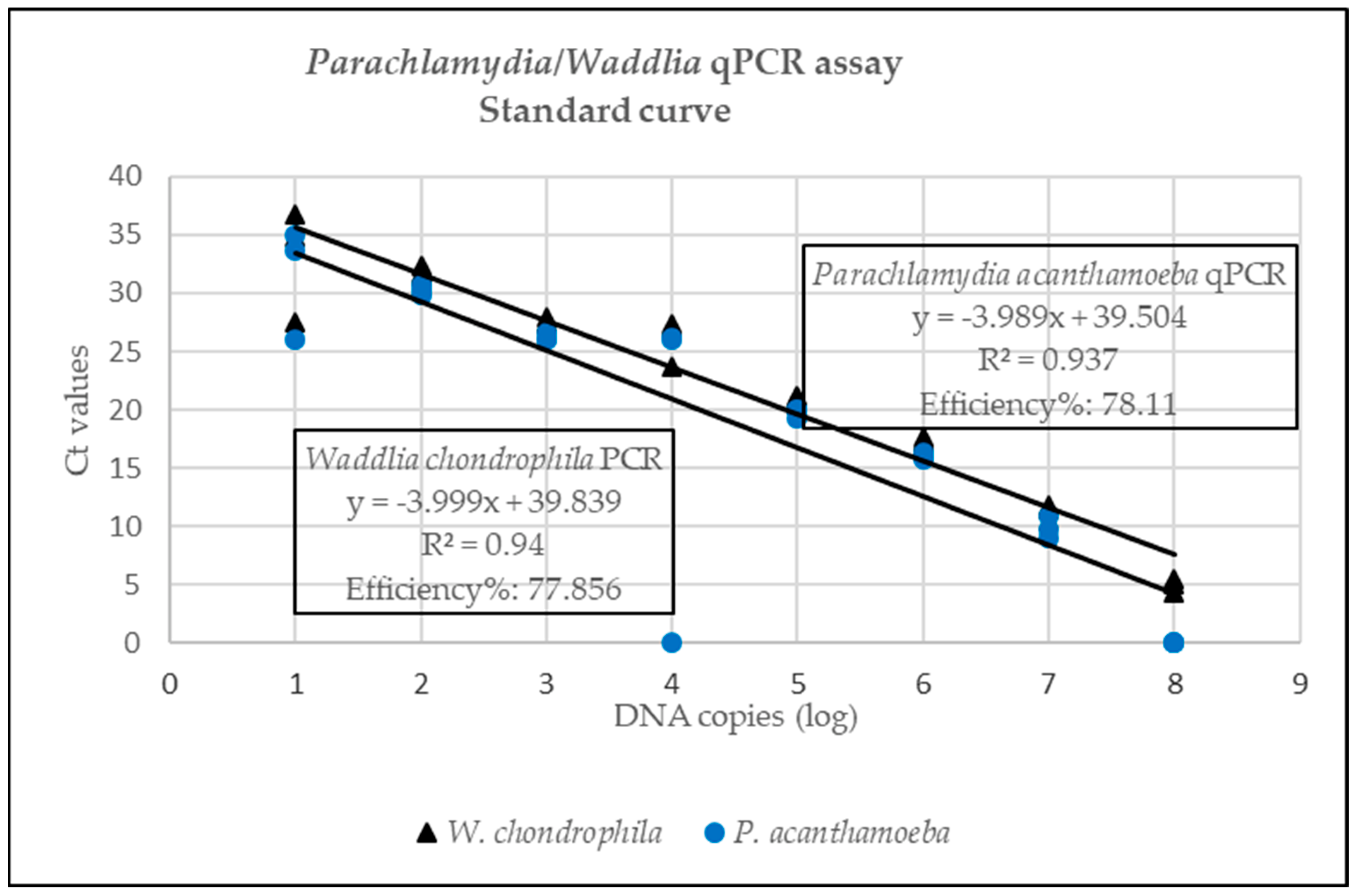
3.1.1. Analytical Sensitivity

Standard curves for the Pan-Chlamydiales, C. abortus/C. pecorum and P. acanthamoeba/W. chondrophila are represented in Figure 1, Figure 2 and Figure 3. The data for the singleplex Pan-Chlamydiales qPCR assay were not linear, which had a negative influence on correlation coefficient (R2) and efficiency calculations. An R2 of 0.9585 and an efficiency of 104.432% were calculated. The R2 and efficiency were lower than the C. abortus/C. pecorum duplex assay, but higher than the P. acanthamoeba/W. chondrophila duplex assay. The R2 values in the C. abortus/C. pecorum duplex assay were slightly better than the R2 of the singleplex qPCRs. Efficiency in the duplex assay was 99.5% (C. abortus) and 99% (C. pecorum). Multiplexing of P. acanthamoeba and W. chondrophila qPCR assays resulted in data that were not linear, also with a negative influence on R2 and efficiency calculations. Efficiency of the W. chondrophila qPCR decreased from 97.98% (singleplex) to 77.85% (duplex), and R2 decreased from 0.97 (singleplex) to 0.94 (duplex). The Ct value was 21 when a sample contained 105 DNA copies. The P. acanthamoeba qPCR increased slightly in efficiency from 74.51% (singleplex) to 78.11% (duplex), and R2 decreased from 0.97 (singleplex) to 0.93 (duplex). The Ct value was 19 when a sample contained 105 DNA copies.

3.1.2. Limit of Detection

Synthetic controls were added to extracted DNA of placenta, stomach content and lung samples. Spiked samples were analysed to determine the limit of detection of the qPCR assays. The limit of detection was defined as the lowest amount of target DNA that could be detected in 95% of replicates [42,43]. Pan-Chlamydiales qPCR limits of detection were <62.5 DNA copies in placenta, stomach content and <125 DNA copies in lung DNA. Limits of detection of the Chlamydia abortus/Chlamydia pecorum qPCR assay were five DNA copies in placenta and stomach content, and two DNA copies in lung DNA. Limits of detection for the P. acanthamoeba/W. chondrophila qPCR assay were <1 DNA copy (P. acanthamoeba) and 1 DNA copy (W. chondrophila) in placenta, <1 DNA copy in stomach content and <1 DNA copy in lung (Supplementary Tables S2–S12).

3.1.3. Analytical Specificity

None of the three qPCR assays detected any of the non-target organisms.

3.2. Clinical Samples

Fifty clinical samples with necrotic placentitis and/or pneumonia lesions from 25 abortion cases (17 placentas, 19 stomach contents and 14 lungs) were analysed. Animal species involved were bovine (n = 17), ovine (n = 6) and caprine (n = 2) (Table 3). In 16 cases, Chlamydiales were detected in at least one sample. In four of these cases, Chlamydiales were detected in two samples each. In two cases, Chlamydia had been detected previously by immunohistochemistry [36] and, in this study, Chlamydiales were again detected using qPCR. In eight cases, no Chlamydiales were detected in this study or the previous study by Jonker et al. [41]. A cut-off point (106 DNA copies) experimentally determined by Gutierrez et al. [37] was applied to C. abortus results to determine diagnostic significance of results.
Chlamydiales were detected in ten bovine, four ovine and two caprine cases and most often in placenta (n = 12). The number of DNA copies was highest in placenta. Where Chlamydiales DNA was detected in more than one sample in a case, DNA copies were lower in stomach content and lung than in placenta. C. abortus was detected in three bovine cases only. C. pecorum was only detected in ovine and caprine cases. P. acanthamoeba was detected in three bovine cases. W. chondrophila was detected in bovine, ovine and caprine cases. Chlamydia pecorum (n = 2) and W. chondrophila (n = 1) were detected in cases where pneumonia lesions had been reported previously.
One case that was previously positive for C. abortus by PCR was not positive in this study. In one bovine and one caprine case, Chlamydiales could not be identified further by the assays in this study.
Seven cases had pathological lesions (necrotic placentitis (n = 5); pneumonia (n = 2)) that pointed to bacterial infection, but previously no agent could be detected [36]. In this study, Chlamydiales were detected in samples from three of these cases. Chlamydia pecorum was detected in this study in one case where Chlamydia species were detected previously by IHC. In four cases, no agent of abortion could be detected in this study and the previous study by Jonker et al. [36].
β-actin was detected in all samples, indicating efficient extraction and absence of inhibition factors that could lead to false negative results.

3.2.1. Diagnostic Sensitivity

Diagnostic sensitivity was calculated using the data in Table 3. The results were as follows: Pan-Chlamydiales assay: 0.38 (38%); C. abortus/C. pecorum assay: 0.2 (20%); P. acanthamoeba/W. chondrophila assay: 0.22 (22%).

3.2.2. Diagnostic Specificity

Forty-nine samples of placenta, stomach content and lung from bovine (n = 19), ovine (n = 9) and caprine (n = 10) cases where no placentitis or pneumonia was observed, were analysed using the qPCR assays in this study (Supplementary Table S12). In 45 samples either no target DNA was detected or Ct values higher than 32 were returned. Chlamydiales were detected at Ct values lower than 32 in five samples. Results of diagnostic specificity calculations were as follows: Pan-Chlamydiales assay: 0.67 (67.3%); C. abortus/C. pecorum assay: C. abortus: 0.94 (94%) and C. pecorum: 1 (100%); P. acanthamoeba/W. chondrophila assay: P. acanthamoeba: 0.18 (18.4%) and W. chondrophila: 1 (100%).

4. Discussion

The aim of this study was to optimise and apply qPCR assays for detection of members of the order Chlamydiales and differentiation of C. abortus, C. pecorum, P. acanthamoeba and W. chondrophila in products of abortion, with necrotic placentitis and/or pneumonia lesions, from domestic ruminants in order to improve detection of intracellular bacteria that cannot be cultured on acellular media. Primers and probes for detection of DNA of the order Chlamydiales and of the species C. abortus, C. pecorum, P. acanthamoeba and W. chondrophila were selected from the literature. One singleplex and two duplex qPCR assays were optimised. These assays were applied to 50 samples from 25 clinical abortion cases, where necrotic placentitis and/or pneumonia were described histopathologically. An additional 49 samples where no pathological lesions were reported or where lesions other than placentitis and pneumonia were reported were also analysed using the three qPCR assays.
Slight modification of the Pan-Chlamydiales qPCR using a MGB probe instead of locked nucleic acids resulted in a correlation coefficient (R2) of 0.959 and an efficiency of 104.4%. Although efficiency lies between 90 and 105% [43], R2 was only 0.959, indicating that the serial dilution of the control was not accurately done [43]. The efficiency of this screening assay was much lower than the C. abortus/C. pecorum assay. This could be due to the use of a fragment of 16S RNA for the design of the primers and probe. Consequently, the Pan-Chlamydiales assay could miss positive cases that would be detected by the C. abortus/C. pecorum assay. Improvement of this assay should be attempted by amplifying a fragment from a different gene such as ompA. The C. abortus/C. pecorum assay had a high efficiency percentage at 99.5% (C. abortus) and 99% (C. pecorum) and R2 of 0.995 and 0.99 for C. abortus and C. pecorum, respectively. These results were slightly better than results when the assays were run in singleplex fashion. Efficiency of the W. chondrophila qPCR, which was 97.98% as a singleplex assay, decreased in the duplex assay, while efficiency of the P. acanthamoeba qPCR improved slightly (P. acanthamoeba: 78.11% and W. chondrophila: 77.86%). The use of an MGB probe improved the efficiency of this assay slightly; however, detection of targets was still impaired [42]. Improvement of the efficiency of the P. acanthamoeba/W. chondrophila qPCR assays must be attempted in future studies. Similar to the Pan-Chlamydiales assay, amplifying a fragment from a different gene such as ompA must be investigated to improve the assay. Improvement is essential if this assay is to be used for quantification studies in future since PCR efficiency plays an important role in quantification of target DNA [43].
Direct diagnosis of Chlamydiales consists of demonstration of microorganisms in tissues by methods such as the Modified Ziehl Neelsen staining method and IHC [26]. Chlamydiales were detected in 16 of the 25 clinical cases with histopathological lesions of necrotic placentitis or pneumonia. These results provide a preliminary indication of diagnostic sensitivity of the assays in this study, because Chlamydiales were detected in DNA from samples where histopathological lesions that are associated with infection by these bacteria were reported [4]. The calculated diagnostic sensitivity was quite low for all three assays, indicating either that the inclusivity of the PCR was low or that the limit of detection of the reference method was higher than the tested qPCR. Unfortunately, results of previous IHC or PCR analyses for Chlamydiales were only available in three cases. In two of these cases, Chlamydiales were also detected in this study. Further analysis of diagnostic sensitivity of the qPCR assays in this study by comparison with culture, IHC and other PCR methods is necessary. The most sensitive combination of methods in a diagnostic setting needs to be determined.
Analysis of samples from cases without lesions resulted in several Ct values higher than 32. Since there were no histopathological lesions, these results are more likely to be an indication of contamination. Diagnostic specificity results of the C. abortus/C. pecorum assay were highest, indicating good ability to identify samples found negative by the reference method. The results for the Pan-Chlamydiales and P. acanthamoeba/W. chondrophila assays were lower, indicating poorer exclusivity of the assays [43]. Another explanation is that the sensitivity of the reference method (histopathology) was low and the assays could detect more positive samples [43]. In addition to the reproductive tract, Chlamydiales can also colonise the intestinal tract [23]. Elementary bodies, the environmentally stable, infectious phase of the Chlamydiales developmental cycle [10], are shed into the environment together with products of abortion or faeces. Environmental contamination of placentae and foetuses can also lead to positive PCR results [14,23]. Therefore, the interpretation of PCR results by the laboratory diagnostician is of major importance. qPCR includes the option to quantify the amount of an agent present in a sample [14], which can be helpful in the discrimination between aetiologically significant pathogens and contaminants to prevent overestimation of the significance of a result.
A singleplex qPCR only detects one pathogen at a time. Several singleplex qPCR assays may be performed to detect different pathogens, but this can become quite costly and time consuming [44]. The minimum number of tests is often selected for economic reasons. For this reason, results of abortion investigations are often inconclusive [31]. A multiplex qPCR, on the other hand, can screen samples for several different pathogens at the same time, allowing the development of a more time and cost-effective analysis [44]. In this study, detection of potential agents of abortion improved from 19 to 21 out of 25 cases when multiplex qPCR was used and sample bias was removed by analysing all samples with necrotic placentitis and/or pneumonia lesions.
The assays in this study enabled the detection of C. pecorum in two ovine cases. In both cases pneumonia was reported, which is not the case in reports by other authors [13,15]. The role of C. pecorum in ovine abortion was investigated by an Australian study [15] and it was found to be significant in some herds. In South Africa, the significance of this Chlamydia species is currently unknown and its investigation is warranted.
Waddlia chondrophila was detected in five bovine, three ovine and one caprine case, and P. acanthamoeba in two bovine cases in this study. P. acanthamoeba and W. chondrophila were detected before in samples from bovine, ovine and caprine abortion cases in countries in the Northern Hemisphere [16,24,33,45]. To the best of the authors’ knowledge, this is the first report of detection of P. acanthamoeba and W. chondrophila in South Africa. The role of these members of Chlamydiales as agents of domestic ruminant abortion in South Africa is currently unknown.
In this study, like many others, several co-infections were detected. In an effort to differentiate between infection and colonisation or contamination, a diagnostic cut-off point (106 DNA copies) experimentally determined for C. abortus by Gutierrez et al. [37] was applied. In this study, this cut-off point excluded all cases where C. abortus was detected except for one bovine case. However, there may be fewer than 106 DNA copies of a causative agent in a sample due to sampling or processing. So, although a diagnostic cut-off point can be a useful tool it can also not be used in isolation to determine the significance of a qPCR result. A combination of analyses such as histopathology, IHC and qPCR as well as evaluation of all results by the laboratory diagnostician is once again essential to attach appropriate significance to detection of Chlamydiales. Since C. abortus is not commonly associated with bovine abortion it may not be selected from a list of singleplex PCR options. In this study, the C. abortus was detected, because the analysis was included in a multiplex assay targeting bacteria that cause necrotic placentitis or pneumonia and test selection was not possible. This illustrates the advantage of a multiplex assay that can detect several agents associated with a particular pathological lesion in one reaction.
Chlamydiales were detected in samples from two ovine cases, C. pecorum in one case and W. chondrophila in the other, where only pathological lesions pointing to an infectious cause were reported previously by Jonker et al. [41]. Lack of any other possible agents of abortion increases their significance in these cases. However, the efficiency of the P. acanthamoeba/W. chondrophila qPCR used in this study was considered too low to aid in the diagnosis, indicating the need for the design of new assays.
In one case, C. abortus was detected previously, but not in this study. The reason may be that a different sample from the same case was used and that there was not enough DNA in the sample. Chlamydiales detected in three bovine and one caprine case could not be identified further by the assays in this study. Internationally, more than one author has reported Chlamydiales they could not identify [31,46]. This serves as an indication that other Chlamydiales may be agents of abortion. Further investigation by PCR and sequencing is necessary to identify these agents and determine their role in abortions. The findings in this study indicate that further investigation of the role of Chlamydiales in ruminant abortion in South Africa is warranted.

5. Conclusions

Application of the three qPCR assays in this study improved detection of members of the order Chlamydiales in samples from cases with necrotic placentitis and/or pneumonia. In addition, differentiation of the Chlamydiales to species level was improved. Application of a diagnostic cut-off point was useful to identify clinically significant Ct values for C. abortus. Detection of C. pecorum, P. acanthamoebae and W. chondrophila in ruminant abortion cases in South Africa indicate that these bacteria potentially play a role in abortions.
It is recommended that, in future, these qPCR assays form part of a comprehensive diagnostic approach to ruminant abortions. These results serve as a reminder that ruminant abortion material should be handled with appropriate safety precautions since C. abortus, P. acanthamoebae and W. chondrophila are potentially zoonotic agents.

Supplementary Materials

The following supporting information can be downloaded at: https://www.mdpi.com/article/10.3390/pathogens12020290/s1, Table S1: Synthetic controls; Table S2–S11: Limit of detection data; Table S12: Results of analysis of samples without placentitis or pneumonia.

Author Contributions

Conceptualization, A.J.; methodology, A.J.; formal analysis, A.J.; investigation, A.J.; data curation, A.J.; writing—original draft preparation, A.J.; writing—review and editing, A.L.M.; supervision, A.L.M.; project administration, A.J.; funding acquisition, A.J. All authors have read and agreed to the published version of the manuscript.

Funding

This research was funded by AGRISeta (ref no. BC19UP57-18.1), Red Meat Research and Development South Africa (ref no 1395058846) and ITM-DGD FA4.

Institutional Review Board Statement

The animal study protocol was approved by the Faculty Ethics Committee and Animal Ethics Committee of University of Pretoria (V092-17: 2017, 2018, 2019, 2020 and REC 070-21: 2021, 2022).

Informed Consent Statement

Not applicable.

Data Availability Statement

Data are contained within the article.

Acknowledgments

The authors wish to thank Idexx Laboratories and the Pathology Section at the Faculty of Veterinary Science, University of Pretoria for submitting samples from abortion cases. F. Nkozi and A. Bosman are acknowledged for valuable inputs in the project.

Conflicts of Interest

The authors declare no conflict of interest. The funders had no role in the design of the study; in the collection, analyses, or interpretation of data; in the writing of the manuscript; or in the decision to publish the results.

References

  1. Sebastiani, C.; Curcio, L.; Ciullo, M.; Cruciani, D.; Crotti, S.; Pesca, C.; Torricelli, M.; Sebastianelli, M.; Felici, A.; Biagetti, M. A multi-screening Fast qPCR approach to the identification of abortive agents in ruminants. J. Microbiol. Meth. 2018, 148, 12–17. [Google Scholar] [CrossRef] [PubMed]
  2. Clothier, K.; Anderson, M. Evaluation of bovine abortion cases and tissue suitability for identification of infectious agents in California diagnostic laboratory cases from 2007 to 2012. Theriogenology 2016, 85, 933–938. [Google Scholar] [CrossRef] [PubMed]
  3. Baumgartner, W. Fetal Disease and abortion: Diagnosis and causes. In Bovine Reproduction, 1st ed.; Hopper, R.M., Ed.; John Wiley & Sons: Hoboken, NJ, USA, 2015; pp. 481–517. [Google Scholar] [CrossRef]
  4. Borel, N.; Polkinghorne, A.; Pospischil, A. A review on chlamydial diseases in animals: Still a challenge for pathologists? Vet. Pathol. 2018, 55, 1–17. [Google Scholar] [CrossRef] [PubMed]
  5. Ammerdorffer, A.; Stojanov, M.; Greub, G.; Baud, D. Chlamydia trachomatis and Chlamydia-like bacteria: New enemies of human pregnancies. Curr. Opin. Infect. Dis. 2017, 30, 289–296. [Google Scholar] [CrossRef]
  6. Pichon, N.; Guindre, L.; Laroucau, K.; Cantaloube, M.; Nallatamby, A.; Parreau, S. Chlamydia abortus in pregnant woman with acute respiratory distress syndrome. Emerg. Infect. Dis. 2020, 26, 628–629. [Google Scholar] [CrossRef]
  7. Greub, G.; Raoult, D. Parachlamydiaceae: Potential Emerging Pathogens. Emerg. Infect. Dis. 2002, 8, 626–630. [Google Scholar] [CrossRef]
  8. Essig, A.; Longbottom, D. Chlamydia abortus: New aspects of infectious abortions in sheep and potential risk for pregnant women. Curr. Clin. Microbiol. Rep. 2015, 2, 22–34. [Google Scholar] [CrossRef]
  9. Walker, E.; Lee, E.J.; Timms, P.; Polkinghorne, A. Chlamydia pecorum infections in sheep and cattle: A common and under recognised infectious disease with significant impact on animal health. Vet. J. 2015, 206, 252–260. [Google Scholar] [CrossRef]
  10. Borel, N.; Frey, C.F.; Gottstein, B.; Hilbe, M.; Pospischil, A.; Franzoso, F.D.; Waldvogel, A. Laboratory diagnosis of ruminant abortion in Europe. Vet. J. 2014, 200, 218–229. [Google Scholar] [CrossRef] [Green Version]
  11. Markey, B.; Leonard, F.; Archambault, M.; Cullinane, A.; Maguire, D. Clinical Veterinary Microbiology, 2nd ed.; Mosby Elsevier: Beijing, China, 2013. [Google Scholar]
  12. Givens, M.D.; Marley, M.S.D. Infectious causes of embryonic and fetal mortality. Theriogenology 2008, 70, 270–285. [Google Scholar] [CrossRef]
  13. Gianitti, F.; Anderson, M.; Miller, M.; Rowe, J.; Sverlow, K.; Vasquez, M.; Cantón, G. Chlamydia pecorum: Fatal and placental lesions in sporadic caprine abortion. J. Vet. Diagn. Investig. 2015, 28, 184–189. [Google Scholar] [CrossRef] [PubMed]
  14. Sachse, K.; Vretou, E.; Livingstone, M.; Borel, N.; Pospischil, A.; Longbottom, D. Recent developments in the laboratory diagnosis of chlamydial infections. Vet. Microbiol. 2009, 135, 2–21. [Google Scholar] [CrossRef]
  15. Clune, T.; Besier, S.; Hair, S.; Hancock, S.; Lockwood, A.; Thompson, A.; Jelocnik, M.; Jacobson, C. Chlamydia pecorum detection in aborted and stillborn lambs from Western Australia. BMC Vet. Res. 2021, 52, 84. [Google Scholar] [CrossRef] [PubMed]
  16. Henning, K.; Schares, G.; Granzow, H.; Polster, U.; Hartmann, M.; Hotzel, H.; Sachse, K.; Peters, M.; Rauser, M. Neospora caninum and Waddlia chondrophila strain 2032/99 in a septic stillborn calf. Vet. Microbiol. 2002, 85, 285–292. [Google Scholar] [CrossRef] [PubMed]
  17. Barkallah, M.; Gharbi, Y.; Ben Hassena, A.; Ben Silma, A.; Mallek, Z.; Gautier, M.; Greub, G.; Gdoura, R.; Fendri, I. Survey of infectious etiologies of bovine abortion during mid-to late gestation in dairy herds. PLoS ONE 2014, 9, e91549. [Google Scholar] [CrossRef]
  18. Ruhl, S.; Casson, N.; Kaiser, C.; Thoma, R.; Pospischil, A.; Greub, G.; Borel, N. Evidence for Parachlamydia in bovine abortion. Vet. Microbiol. 2009, 135, 169–174. [Google Scholar] [CrossRef] [PubMed]
  19. Barkallah, M.; Jribi, H.; Ben Silma, A.; Gharbi, Y.; Mallek, Z.; Gautier, M.; Fendri, I.; Gdoura, R. Molecular prevalence of Chlamydia and Chlamydia-like bacteria in Tunisian domestic ruminant farms and their influencing risk factors. Transbound. Emerg. Dis. 2018, 65, e329–e338. [Google Scholar] [CrossRef] [PubMed]
  20. Barkallah, M.; Gharbi, Y.; Ben Silma, A.; Elleuch, F.; Mallek, Z.; Ben Saad, R.; Gautier, M.; Gdoura, R.; Fendri, I. Simultaneous detection of Waddlia chondrophila and Listeria monocytogenes in aborted ruminant samples by real-time quantitative PCR. J. Microbiol. Methods 2016, 125, 64–69. [Google Scholar] [CrossRef]
  21. Hazlett, M.J.; McDowall, R.; DeLay, J.; Stalker, M.; McEwen, B.; van Dreumel, T.; Spinato, M.; Binnington, B.; Slavic, D.; Carman, S.; et al. A prospective study of sheep and goat abortion using real-time polymerase chain reaction and cut point estimation shows Coxiella burnetii and Chlamydophila abortus infection concurrently with other major pathogens. J. Vet. Diagn. Investig. 2013, 25, 359–368. [Google Scholar] [CrossRef] [Green Version]
  22. Blumer, S.; Greub, G.; Waldvogel, A.; Hässig, M.; Thoma, R.; Tschuor, A.; Pospischil, A.; Borel, N. Waddlia, Parachlamydia and Chlamydiaceae in bovine abortion. Vet. Microbiol. 2011, 152, 385–393. [Google Scholar] [CrossRef]
  23. Borel, N.; Ruhl, S.; Casson, N.; Kaiser, C.; Pospischil, A.; Greub, G. Parachlamydia spp. and related Chlamydia-like organisms and bovine abortion. In Emerg. Infect. Dis.; 2007; 13, p. 12. Available online: http://www.cdc.gov/EID/content/13/12/1904.htm (accessed on 6 February 2022). [CrossRef]
  24. Ruhl, S.; Goy, G.; Casson, N.; Thoma, R.; Pospischil, A.; Greub, G.; Borel, N. Parachlamydia acanthamoeba infection and abortion in small ruminants. Emerg. Infect. Dis. 2008, 14, 1966–1968. [Google Scholar] [CrossRef] [PubMed]
  25. Vidal, S.; Kegler, K.; Greub, G.; Aeby, S.; Borel, N.; Dagleish, M.P.; Posthaus, H.; Perreten, V.; Rodriguez-Campos, S. Neglected zoonotic agents in cattle abortion: Tackling the difficult to grow bacteria. BMC Vet. Res. 2017, 13, 373. [Google Scholar] [CrossRef] [PubMed]
  26. Rodolakis, A.; Laroucau, K. Chlamydiaceae and chlamydial infections in sheep or goats. Vet. Microbiol. 2015, 181, 107–118. [Google Scholar] [CrossRef] [PubMed]
  27. Goy, G.; Croxatto, A.; Posfay-Barbe, K.M.; Gervaix, A.; Greub, G. Development of a real-time PCR for the specific detection of Waddlia chondrophila in clinical samples. Eur. J. Clin. Microbiol. Infect. Dis. 2009, 28, 1483–1486. [Google Scholar] [CrossRef]
  28. Kirkbride, C. Examination of Bovine and Ovine fetuses. Vet. Clin. N. Am. Food Anim. 1986, 2, 61–83. [Google Scholar] [CrossRef] [PubMed]
  29. Jiang, L.; Ren, H.; Zhou, H.; Quin, T.; Chen, Y. Simultaneous detection of nine key bacterial respiratory pathogens using Luminex xTAG® Technology. Int. J. Env. Res. Public Health 2017, 14, 223. [Google Scholar] [CrossRef]
  30. Iraola, G.; Pérez, R.; Betancor, L.; Marandino, A.; Morsella, C.; Méndez, A.; Paolicchi, F.; Piccirillo, A.; Tomás, G.; Velilla, A.; et al. A novel real-time PCR assay for quantitative detection of Campylobacter fetus based on ribosomal sequences. BMC Vet. Res. 2016, 12, 286. [Google Scholar] [CrossRef]
  31. Schnydrig, P.; Vidal, S.; Brodard, I.; Frey, C.F.; Posthaus, H.; Perreten, V.; Rodrigues-Campos, S. Bacterial, fungal, parasitological, and pathological analyses of abortions in small ruminants from 2012–2016. SAT/ASMV 2017, 12, 647–656. [Google Scholar] [CrossRef]
  32. Lienard, J.; Croxatto, A.; Aeby, S.; Jaton, K.; Posfay-Barbe, K.; Gervaix, A.; Greub, G. Development of a new Chlamydiales-specific real-time PCR and its application to respiratory clinical samples. J. Clin. Microbiol. 2011, 49, 2637–2642. [Google Scholar] [CrossRef]
  33. Wheelhouse, N.; Longbottom, D.; O’Donovan, J. Chlamydia in cases of bovine abortion in Ireland. Vet. Rec. 2014, 174, 560–561. [Google Scholar] [CrossRef] [PubMed]
  34. Pantchev, A.; Sting, R.; Bauerfeind, R.; Tyczka, J.; Sachse, K. New real-time PCR tests for species-specific detection of Chlamydophila psittaci and Chlamydophila abortus from tissue samples. Vet. J. 2009, 181, 145–150. [Google Scholar] [CrossRef] [PubMed]
  35. Pantchev, A.; Sting, R.; Bauerfeind, R.; Tyczka, J.; Sachse, K. Detection of all Chlamydophila and Chlamydia spp. of veterinary interest using species-specific real-time PCR assays. Comp. Immunol. Microbiol. Infect. Dis. 2010, 33, 473–484. [Google Scholar] [CrossRef] [PubMed]
  36. Casson, N.; Posfay-Barbe, K.M.; Gervaix, A.; Greub, G. New diagnostic real-time PCR for specific detection of Parachlamydia acanthamoebae DNA in clinical samples. J. Clin. Microbiol. 2008, 46, 1491–1493. [Google Scholar] [CrossRef] [PubMed]
  37. Gutierrez, J.; O’Donovan, J.; Proctor, A.; Brady, C.; Marques, P.X.; Worral, S.; Nally, J.E.; McElroy, M.; Basset, H.; Fagan, J.; et al. Application of quantitative real-time polymerase chain reaction for the diagnosis of toxoplasmosis and enzootic abortion of ewes. J. Vet. Diagn. Inv. 2012, 24, 846–854. [Google Scholar] [CrossRef] [PubMed]
  38. Berri, M.; Rekiki, A.; Sidi Boumedine, K.; Rodolakis, A. Simultaneous differential detection of Chlamydophila abortus, Chlamydophila pecorum and Coxiella burnetii from aborted ruminant’s clinical samples using multiplex PCR. BMC Microbiol. 2009, 9, 130. Available online: http://www.biomedcentral.com/1471-2180/9/130 (accessed on 6 February 2022). [CrossRef]
  39. Azeem, S.; Ghorbani-Nezami, S.; Allam, A.; Cooper, V.L.; Burrough, E.; Schwartz, K.; Plummer, P.; Arruda, P.; Gauger, P.; Kinyon, J.M.; et al. A multiplex real-time PCR panel for detection of major bacterial and viral agents causing abortion in small ruminants. In Proceedings of the American Association of Veterinary Laboratory Diagnosticians Annual Conference, Providence, RI, USA, 22–28 October 2015. [Google Scholar]
  40. Tramutra, C.; Lacerenza, D.; Zoppi, S.; Goria, M.; Dondo, A.; Ferroglio, E.; Nebbia, P.; Rosati, S. Development of a set of multiplex standard polymerase chain reaction assays for the identification of infectious agents from aborted bovine clinical samples. J. Vet. Diagn. Investig. 2011, 23, 657–664. [Google Scholar] [CrossRef]
  41. Jonker, A.; Thompson, P.N.; Michel, A.L. Approaches to increase recovery of bacterial and fungal abortion agents in domestic ruminants, Onderstepoort. J. Vet. Res. 2022, 90, a2010. [Google Scholar] [CrossRef]
  42. Bustin, S.A.; Benes, V.; Garson, J.A.; Hellemans, J.; Hugget, J.; Kubista, M.; Mueller, R.; Nolan, T.; Pfaffl, M.W.; Shipley, G.L.; et al. The MIQE Guidelines: Minimum information for publication of quantitative Real-Time PCR experiments. Clin. Chem. 2009, 55, 611–622. [Google Scholar] [CrossRef] [Green Version]
  43. Kralik, P.; Ricchi, M. A basic guide to real time PCR in microbial diagnostics: Definitions, parameters, and everything. Front. Microbiol. 2017, 8, 108. [Google Scholar] [CrossRef]
  44. Selim, A.; Elhaigh, M.M.; Gaede, W. Development of multiplex real-time PCR assay for the detection of Brucella spp., Leptospira spp. and Campylobacter foetus. Vet. Ital. 2014, 50, 269–275. [Google Scholar] [CrossRef] [PubMed]
  45. Deuchande, R.; Gidlow, J.; Caldow, G.; Baily, J.; Longbottom, D.; Wheelhouse, N.; Borel, N.; Greub, G. Parachlamydia involvement in bovine abortions in a beef herd in Scotland. Vet. Rec. 2010, 166, 598–599. [Google Scholar] [CrossRef] [PubMed]
  46. Merdja, S.-E.; Khaled, H.; Aaziz, R.; Vorimore, F.; Bertin, C.; Dahmani, A.; Bouyoucef, A.; Laroucau, K. Detection and genotyping of Chlamydia species responsible for reproductive disorders in Algerian small ruminants. Trop. Anim. Health 2015, 47, 437–443. [Google Scholar] [CrossRef] [PubMed]
Figure 1. Standard curve for the Pan-Chlamydiales assay where Ct values (y-axis) are plotted against the log of the concentration of DNA copies (x-axis).
Pathogens 12 00290 g001
Figure 2. Standard curve for the Chlamydia abortus/Chlamydia pecorum assay where Ct values (y-axis) are plotted against the log of the concentration of DNA copies (x-axis).
Pathogens 12 00290 g002
Figure 3. Standard curve for the Parachlamydia acanthamoeba/Waddlia acanthamoeba duplex qPCR where Ct values (y-axis) are plotted against the log of the concentration of DNA copies (x-axis).
Pathogens 12 00290 g003
Table 1. Bacteria and amoeba species used as negative controls to test specificity.
BacteriaSource or Strain
Acholeplasma laidlawliiNCTC 10116
Brucella abortusClinical sample
Chlamydia abortusATCC VR-165
Escherichia coliATCC 25922
Ochrobactrum anthropiATCC 49687
Parachlamydia acanthamoebaATCC VR-1476
Pasteurella multocidaATCC 12945
Salmonella TyphimuriumATCC 13311
Staphylococcus aureusATCC 25923
Streptococcus agalactiaeATCC 27956
Trueperella pyogenesClinical sample
Waddlia chondrophilaATCC VR-1470
Amoeba
Acanthamoeba castellaniATCC 50739
Table 3. Chlamydiales DNA detected in bovine, ovine and caprine cases in this study together with the samples where they were detected, necropsy lesions, bacteria detected previously and cycle thresholds. (ND = not detected, Ct = cycle threshold, PanChl = Pan-Chlamydiales, Chlab = Chlamydia abortus, Chlpec = Chlamydia pecorum, Pacanth = Parachlamydia acanthamoeba, Wchon = Waddlia chondrophila).
Case No.PathologyBacteria/Fungi
(Previous Study [41])
SamplesDNA Detected
(Mean Ct Values)
Bovine
1Necrotic placentitis
Purulent bronchopneumonia
Bacillus licheniformis
Rhizopus sp.
Stomach contentND
LungND
2Necrotic placentitis and vasculitisMycoplasma sp.
Aspergillus fumigatus
PlacentaPanChl (36.05)
Stomach contentPanChl (33.38)
Chlab (32.33)
Chlpec (36.90)
Wchon (31.85)
LungND
3Necrotic placentitisNDStomach contentND
4Necrotic placentitisSalmonella sp.PlacentaPanChl (32.90)
Wchon (26.02)
5Necrotic placentitis with vasculitisCoxiella burnetiiStomach contentPanChl (32.25)
Chlab (32.18)
Chlpec (35.86)
6Necrotic placentitisStreptococcus dysgalactiae
Acholeplasma sp.
PlacentaPanChl (28.63)
Stomach contentWchon (34.36)
LungND
7Purulent placentitisNDPlacentaND
8Necrotic placentitis Lichtheimia corymbiferaPlacentaPanChl (31.21)
Wchon (23.97)
Stomach contentND
LungND
9Meningitis bronchointerstitial pneumoniaBrucella abortus biovar 1Stomach contentND
10Meningoencephalitis
Necrotic placentitis
Hepatic necrosis
NDStomach contentND
11Necrotic placentitis with vasculitisNDPlacentaND
NDLungND
12Necrotic placentitisCampylobacter fetus
Mannheimia varigena
Streptococcus pluranimalium
Mycoplasma sp.
PlacentaPanChl (30.02)
Pacanth (36.88)
Wchon (32.47)
Stomach contentND
LungND
13Necrotic placentitis
Purulent pneumonia
Chlamydia sp.
Salmonella sp.
PlacentaPanChl (31.63)
Stomach contentPanChl (33.94)
LungND
14Necrotic placentitisNDPlacentaPanChl (5.03)
Chlab (5.65)
Stomach contentND
15Viral placentitisPasteurella multocidaPlacentaChlab (30.79)
16Interstitial pneumoniaBrucella abortus biovar 1Stomach contentND
17Necropurulent placentitisTrueperella pyogenesPlacentaPanChl (34.68)
Pacanth (33.48)
18Necrotic placentitis
Pneumonia
Chlamydia sp.PlacentaPanChl (23.13)
Chlpec (18.96)
Stomach contentPanChl (31.69)
Chlpec (29.83)
19Bacterial & fungal placentitis PleuropneumoniaChlamydia abortusStomach contentND
LungND
20BronchopneumoniaNDStomach contentPanChl (27.42)
Chlpec (22.19)
Wchon (35.17)
LungPanChl (25.74)
Chlpec (23.42)
Wchon (35.39)
21Bacterial placentitisArcobacter sp.PlacentaND
LungND
22Necrotic placentitisSalmonella Budapest
Mycoplasma species
PlacentaPanChl (35.03)
Wchon (33.35)
Stomach contentND
23Fibrinopurulent bacterial pneumoniaNDPlacentaPanChl (29.49)
Chlpec (34.48)
Wchon (32.52)
LungND
24Necrotic placentitisCoxiella burnetiid
Salmonella typhimurium
PlacentaND
Stomach content Foetus AND
Lung Foetus APanChl (32.41)
Chlpec (35.84)
Wchon (31.57)
Stomach content Foetus BND
Lung Foetus BND
25Necrotizing placentitis
Subcutaneous oedema
Hydrocephalus
E. coli
Lichtheimia corymbifera
PlacentaPanChl (10.27)
Stomach contentND
LungND
Disclaimer/Publisher’s Note: The statements, opinions and data contained in all publications are solely those of the individual author(s) and contributor(s) and not of MDPI and/or the editor(s). MDPI and/or the editor(s) disclaim responsibility for any injury to people or property resulting from any ideas, methods, instructions or products referred to in the content.

