Article

Pathological Features and Genomic Characterization of an Actinobacillus equuli subsp. equuli Bearing Unique Virulence-Associated Genes from an Adult Horse with Pleuropneumonia

1
Department of Infectious Diseases and Public Health, Jockey Club College of Veterinary Medicine, City University of Hong Kong, Hong Kong SAR, China
2
Louisiana Animal Disease Diagnostic Laboratory, School of Veterinary Medicine, Louisiana State University, Baton Rouge, LA 70803, USA
3
Department of Pathobiological Sciences, School of Veterinary Medicine, Louisiana State University, Baton Rouge, LA 70803, USA
4
Veterinary Teaching Hospital and Department of Veterinary Clinical Sciences, School of Veterinary Medicine, Louisiana State University, Baton Rouge, LA 70803, USA
*
Author to whom correspondence should be addressed.
Pathogens 2023, 12(2), 224; https://doi.org/10.3390/pathogens12020224
Submission received: 14 December 2022 / Revised: 27 January 2023 / Accepted: 27 January 2023 / Published: 31 January 2023
(This article belongs to the Section Bacterial Pathogens)

Abstract

:
Actinobacillus equuli subsp. equuli is the etiological agent of sleepy foal disease, an acute form of fatal septicemia in newborn foals. A. equuli is commonly found in the mucous membranes of healthy horses’ respiratory and alimentary tracts and rarely causes disease in adult horses. In this study, we report a case of a 22-year-old American Paint gelding presenting clinical signs associated with an atypical pattern of pleuropneumonia subjected to necropsy. The gross and histopathological examinations revealed a unilateral fibrinosuppurative and hemorrhagic pleuropneumonia with an infrequent parenchymal distribution and heavy isolation of A. equuli. The whole genome sequence analysis indicated that the isolate shared 95.9% homology with the only other complete genome of A. equuli subsp. equuli available in GenBank. Seven virulence-associated genes specific to the isolate were identified and categorized as iron acquisition proteins, lipopolysaccharides (LPS), and capsule polysaccharides. Moreover, four genes (glf, wbaP, glycosyltransferase family 2 protein, and apxIB) shared higher amino acid similarity with the invasive Actinobacillus spp. than the reference A. equuli subsp. equuli genome. Availability of the whole genome sequence will allow a better characterization of virulence determinants of A. equuli subsp. equuli, which remain largely elusive.

1. Introduction

Actinobacillus species (spp.) are gram-negative, anaerobic, and pleomorphic bacteria in the family Pasteurellaceae [1]. Actinobacillus spp. reside on the mucosal membranes of healthy animals’ respiratory and genitourinary tracts [2]. Clinical manifestations caused by these organisms vary, including respiratory infections, septicemia, metritis, mastitis, arthritis, endocarditis, meningitis, or stillbirth [3,4,5,6,7,8,9]. In horses, A. equuli subspecies (subsp.) equuli or A. equuli subsp. haemolyticus are commonly found in the normal oral flora of healthy horses and the alimentary and genital tracts, whereas A. ligneieresii and A. pleuropneumoniae are less frequently found [10,11,12]. Interestingly, A. equuli subsp. equuli differs from A. equuli subsp. haemolyticus by manifestation of different disease patterns and epidemiology. A. equuli subsp. equuli is primarily associated with “sleepy foal disease”, an acute form of fatal septicemia in newborn foals that can develop into a chronic state with kidney, joint, and lung lesions [3,13,14]. Although A. equuli rarely causes disease in adult horses, a few acute and chronic cases of peritonitis have been previously reported [4,15,16]. In addition, it has been recently reported that A. equuli acts as a primary pathogen in sows and piglets, given favorable conditions [13,14]. Actinobacillus equuli subsp. equuli is non-hemolytic and CAMP-negative, which are the main features that differentiate from A. equuli subsp. haemolyticus [13]. Actinobacillus equuli subsp. equuli does not contain repeats-in-toxins (RTX) activity referred to as the A. equuli toxin (Aqx) or any other related RTX genes, which confers the hemolytic phenotype associated with pulmonary hemorrhage and endothelial damage in an adult horse [15]. Although several virulence factors associated with pathotypes of certain Actinobacillus spp. have been identified, such as autotransporters (ATs), outer membrane proteins (OMPs), capsule polysaccharide (CPS), lipopolysaccharide (LPS), iron acquisition mechanisms, RTX, and urease, the virulence factors associated with A. equuli subsp. equuli are largely unknown [2,15,16,17]. We hypothesized that a comprehensive genome analysis of A. equuli subsp. equuli isolated from a clinical case can improve our understanding of the potential invasion determinants and the virulence factors associated with the pathogenicity and disease phenotype.
Here, we describe a clinical case of A. equuli subsp. equuli associated with an atypical pattern of pleuropneumonia in an adult horse, in which this infection is usually rare or sporadic. The gross and histopathological examinations revealed lesions in the left lung consistent with fibrinosuppurative and hemorrhagic pleuropneumonia associated with heavy isolation of A. equuli. To further understand the basis of the disease phenotype, full-length genome sequence of the isolate was obtained and characterized. Availability of the complete genome sequence will allow a better characterization of virulence determinants of A. equuli subsp. equuli, which remain largely elusive.

2. Materials and Methods

2.1. Postmortem Examination and Sample Collection

A postmortem examination of a 22-year-old American Paint horse with atypical patterns of pleuropneumonia was performed at the Louisiana Animal Disease Diagnostic Laboratory (LADDL). Specimens from the left lung were aseptically collected and submitted for additional ancillary testing (bacteriological culture). Samples from all organs were fixed in 10% neutral buffered formalin for 24–48 h for histopathologic examination.

2.2. Histopathology

Formalin-fixed tissues (lung, liver, adrenal glands, spleen, kidneys, heart, encephalon [cerebrum, cerebellum, brainstem], pituitary gland, mesenteric lymph node, pancreas, and gastrointestinal tract) were embedded in paraffin. Subsequently, formalin-fixed paraffin-embedded (FFPE)-tissue sections (4 μm) were stained with hematoxylin and eosin (H&E) according to a standard laboratory procedure before histological evaluation.

2.3. Bacteriology, MALDI-TOF Mass Spectrometry, and Antibiotic Sensitivity

Samples from the lungs were subjected to routine bacteriological testing by inoculating commercial blood agar, chocolate agar, and MacConkey agar plates (Remel, San Diego, CA, USA). Inoculated blood and chocolate agar plates were incubated at 37 °C with 5% CO2 for the first 24 h and without CO2 for the next 48 h. Inoculated MacConkey agar plates were incubated at 37 °C for up to 72 h. Bacterial colonies were subsequently screened using a MALDI Biotyper® Compass (Bruker, Billerica, MA, USA) [(automated identification system based on matrix-assisted laser desorption/ionization time of flight (MALDI-TOF)] for identification using the direct transfer method. Identification was further clarified with phenotypic presentation combined with Christie–Atkins–Munch-Peterson (CAMP) test results using pure cultures following standard procedures. Antibiotic sensitivities were performed using the broth microdilution method (Sensititre™ EQUIN1F Vet AST Plate) to determine minimum inhibitory concentrations (MICs).

2.4. DNA Extraction, Whole Genome Sequencing, and Assembly

Genomic DNA was extracted from pure bacterial colonies using Qiagen’s DNeasy Blood and Tissue Kit (Qiagen, Germantown, MD, USA) on a QIAcube automated workstation (Qiagen). DNA concentrations were determined using the Qubit dsDNA HS Assay Kit on a Qubit flex fluorometer (ThermoFisher Scientific, Waltham, MA, USA). DNA libraries were constructed with the Nextera XT DNA Library Preparation Kit (Illumina®, San Diego, CA, USA) using 0.2 ng/µL of genomic DNA. Resultant libraries were sequenced on an Illumina MiSeq desktop sequencer using v2 sequencing chemistry with 2 × 250 bp pair-end reads per the manufacturer’s protocol. The quality control of raw sequences was performed by FastQC v0.11.9, and the de novo assembly was done using SPAdes v3.14.1. The final contigs of the isolated strain (designated strain 4524) were subjected to BLASTn to identify the reference genome, which shows the highest identity with the contigs. The genome of the 4524 strain was reordered using progressive Mauve with the A. equuli subsp. equuli ATCC 19392 genome as the reference genome [18]. The FASTA file of the 4524 genomes was used for genome annotation and further analysis.

2.5. A. equuli Subsp. equuli Genome Annotation and Analysis

The whole genome sequence of the 4524 strain was annotated using Bakta v.1.3.0, Prokka v.1.12, and NCBI Prokaryotic Genome Annotation Pipeline [19,20]. The proteome of the 4524 strain was subjected to BLASTp against the Cluster of Orthologous Groups (COGs) database at E-value < 1e−5 to identify the COGs [21]. The CRISPI web interface with parameters set to the default values was used to find CRISPR sequences in the genomes [22]. The CGView Server [23] (http://stothard.afns.ualberta.ca/cgview_server/ (accessed on 12 May 2022) was used to generate the circular genome map of the 4524 strain with A. equuli subsp. equuli ATCC 19392 as the reference genome. The GenBank files of A. equuli subsp. equuli ATCC 19392 and 4524 strains were submitted to EDGAR for comparative genomic analyses [24]. The EDGAR private project was developed for generating Venn diagrams and verifying shared genes and core genomes between the two strains. Virulence-associated genes described in previous studies were identified through the genome of the 4524 strain and aligned to the homologous genes in the ATCC 19392 genome.

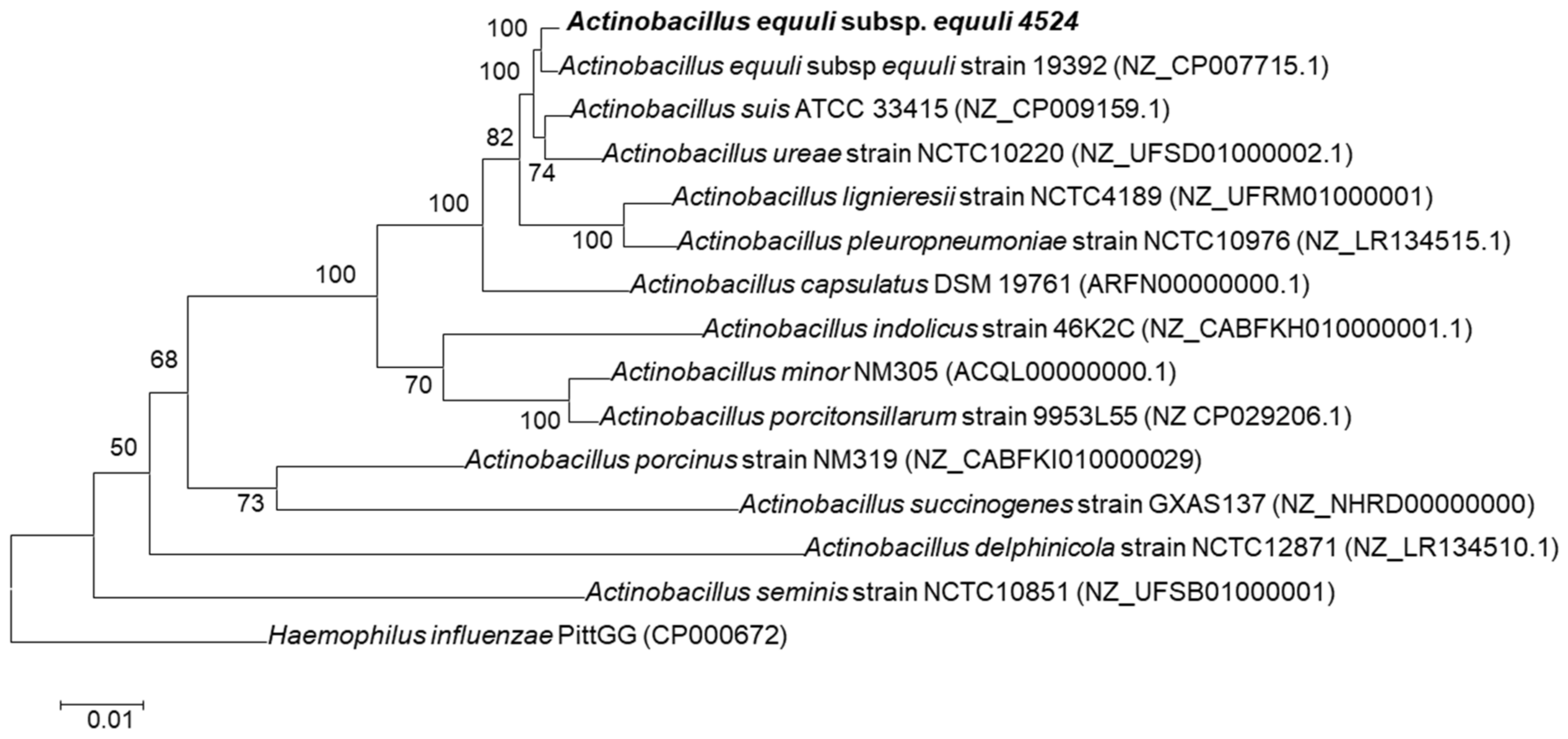
2.6. Phylogenetic Analysis

Thirteen Actinobacillus bacteria belonging to various species were selected, and their respective nucleotide sequences were retrieved from the GenBank database. The selected strains and their characteristics are presented in Supplementary Table S1. Whole-genome alignments were performed using REALPHY (http://realphy.unibas.ch (accessed on 20 June 2022)) [25]. The phylogenetic tree was constructed using the maximum likelihood method, and the evolutionary distances were computed using the general time-reversible model along with gamma-distributed rates (G = 0.6) and invariant site (I) by MEGA v.7 [26,27,28]. Branch validity was evaluated by the bootstrap test with 1000 replications. The average nucleotide identity (ANI) values of selected Actinobacillus spp. strains were calculated using the server EzBioCloud (http://www.ezbiocloud.net/tools/ani (accessed on 1 July 2022)) [29]. According to the algorithm developed by Goris et al. [30], a 95∼96% cut-off value was used for the species boundary [31]. The web-based DSMZ service (http://ggdc.dsmz.de (accessed on 3 July 2022)) [32] with 70% species and sub-species cut-off was used to estimate the in silico genome-to-genome distance values for the 4524 isolate and selected strains.

3. Results

3.1. Case Presentation

In March 2020, a 22-year-old American Paint gelding presented to the Louisiana State University Veterinary Teaching Hospital due to acute onset of illness with tachypnea, bilateral purulent to hemorrhagic nasal discharge, and cough with bloody sputum. The gelding was kept at a boarding facility and used as a lesson horse. The illness presented suddenly, and he was brought immediately to the Veterinary Teaching Hospital for evaluation and treatment due to the rapid onset and progression of clinical signs. The horse had no previous illness and was not receiving treatment for any other known underlying condition. Due to a possible infectious disease concern, the gelding was immediately admitted to the isolation facility. On presentation, major findings included a marked leukocytosis (12.1 × 10³/μL [reference range: 5.0–11.0 × 10³/μL) with left shift (5.8% bands; 0.7 × 10³/μL [reference range: 0.0–0.1 × 10³/μL]), toxic neutrophils, and monocytosis (12%; 1.5 × 10³/μL [reference range: 0.0–0.8 × 10³/μL]). Ultrasonographically, pleural effusion and pulmonary consolidation of the left lung were noticed. A thoracocentesis revealed neutrophilic exudation with short, intracytoplasmic bacterial rods, indicating marked septic neutrophilic inflammation. The direct smears of pleural fluid contained moderate numbers of nucleated cells and erythrocytes. The nucleated cells comprised approximately 70% of non-degenerate neutrophils, 30% of macrophages, and rarely small lymphocytes. In addition, a few neutrophils containing phagocytosed short bacilliform bacteria were observed. The macrophages were variably vacuolated, with some cells having more rounded borders and increased nuclear-to-cytoplasmic ratios. Due to the overall poor prognosis, humane euthanasia was elected by the owner, and the horse was submitted for a thorough postmortem examination.

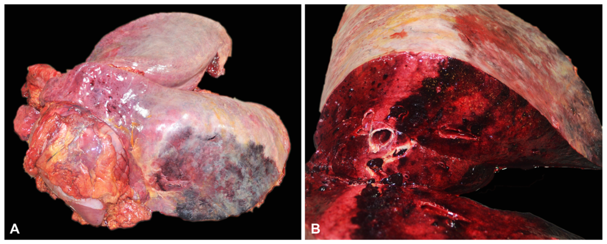
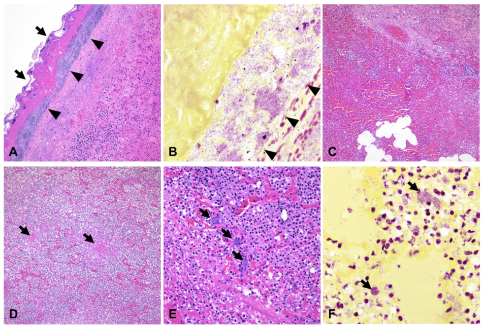
3.2. Gross and Histopathological Findings

Macroscopically, an extensive area (approximately 50 cm in diameter) of the pulmonary parenchyma at the level of the caudoventral aspect of the left lung was dark red and firm, with the lining pleura overlaid by a thick layer of fibrin (Figure 1). The cranial part of the left lung also had multiple dark red pin-point foci. There was no evidence of exudate within nasal passages, paranasal sinuses, guttural pouches, laryngeal, tracheal, or bronchial lumina. The abdominal cavity contained approximately 0.5–1 L of yellow-tinged fluid. No other significant gross abnormalities were found.
Histologically, the affected parenchyma was characterized by liquefactive necrosis and fibrinosuppurative inflammation with loss of alveolar septa and/or alveoli diffusely filled with necrotic debris abundant of degenerate neutrophils, fibrin exudation, edema, and hemorrhage. Bronchioles were frequently filled with large necrotic cell debris and degenerate neutrophils, and numerous, variably sized blood vessels were occluded by fibrin thrombi. The interlobular septa were distended by edema, fibrin exudation, and hemorrhage. The pleura was significantly expanded by abundant fibrin exudation and degenerate neutrophils and coated by a thick layer of polymerized fibrin and myriad colonies of short, gram-negative bacterial rods (Figure 2). Additional findings included fibrinous capsulitis and tubulointerstitial nephritis, and mild neutrophilic hepatitis. Overall, the gross and histological changes in the left lung were consistent with fibrinosuppurative and hemorrhagic pleuropneumonia associated with heavy isolation of A. equuli, in agreement with the clinical history and differential diagnosis considered at the time of euthanasia.

3.3. Bacteriology, MALDI-TOF Mass Spectrometry, and Antibiotic Sensitivity

Initially, MALDI-TOF identified the organism as Actinobacillus suis with a score of 2.47. However, the MALDI patterns and the 16S rRNA gene sequences of A. arthritidis, A. capsulatus, A, equuli, A. lignieresii, A. pleuropneumoniae, and A. suis are highly similar; therefore, they cannot be distinguished from each other. Since A. suis did not fit with the case, we used the growth patterns and CAMP test to speciate further. The isolate was non-hemolytic on blood agar with no growth on MacConkey agar. In addition, the CAMP test was negative. Using this additional information, the isolate was identified as A. equuli. Finally, the isolate was submitted for WGS for further confirmation of the identification. Broth microdilution minimum inhibitory concentration (MIC) results can be seen in Table 1.

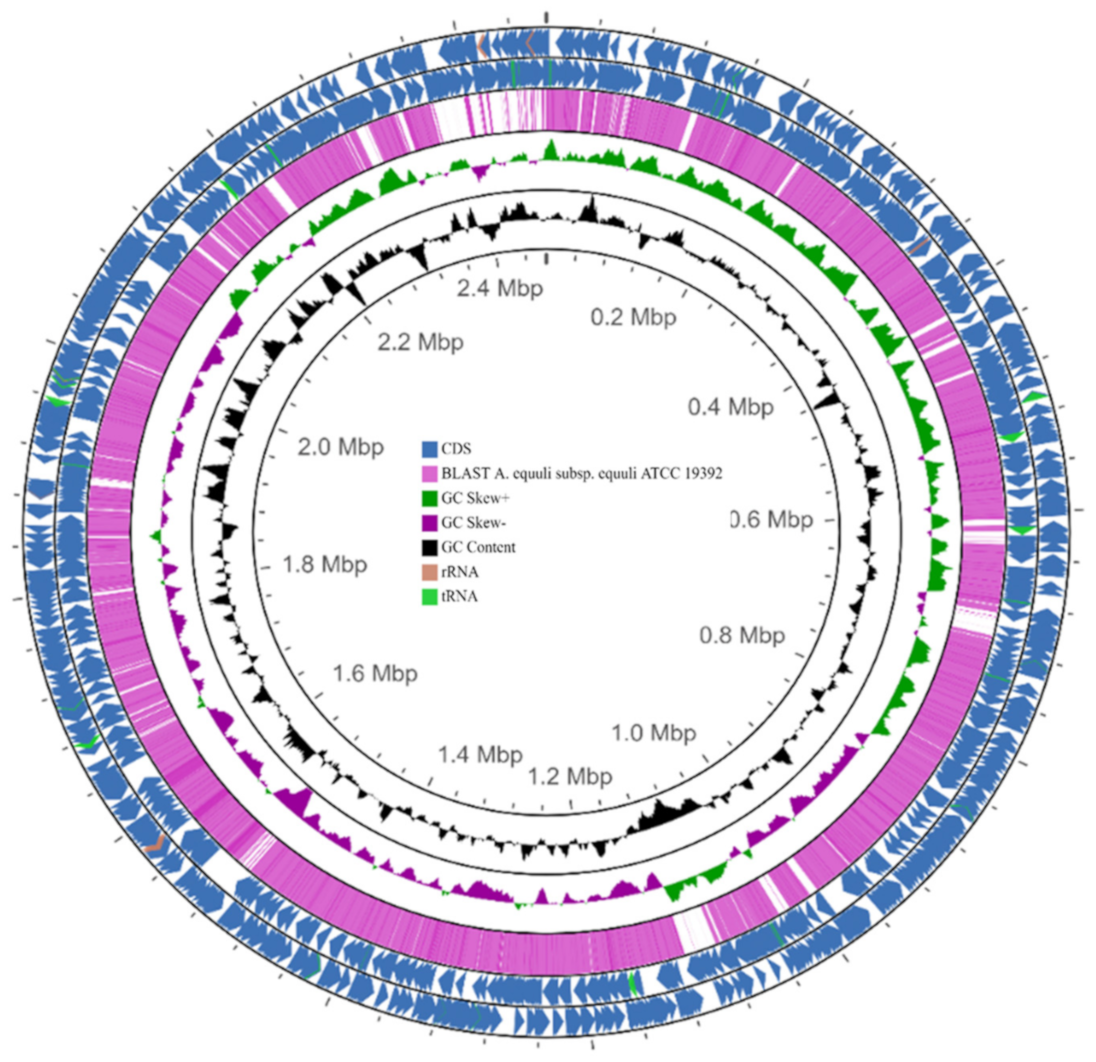
3.4. Genome Properties of the A. equuli subsp. equuli 4524 Isolate

Whole-genome sequencing was performed to characterize the genetic properties of the A. equuli, 4524 strain, isolated from this clinical case. The assembled genome contained 118 contigs, with N50 of 213,479 bp and approximately 43× sequence coverage. The full-length genome was obtained with a length of 2,464,726 bp, a G + C content of 40.2%, and 2362 predicted genes consisting of 2277 protein-coding genes, and 85 for RNA (61 tRNA, 1 tmRNA, 8 rRNA, and 15 ncRNA genes) (Table 2). Approximately 60% of the predicted genes were assigned to 26 functional COG categories (Table 3).
The phylogenetic analysis using the whole genome sequences of Actinobacillus spp. identified the 4524 isolate as A. equuli subsp. equuli owing to clustering together with A. equuli subsp. equuli ATCC 19392 strain (Figure 3).
The genome of the isolate displayed the highest average nucleotide identity (ANI) value (95.9%) and the lowest genome-to-genome distance calculation (GGDC) values (0.0418) to the genome of A. equuli subsp. equuli ATCC 19392 (Table 4). Notably, the highest DNA-DNA hybridization (DDH) value (66.1) was measured between the whole genomes of the 4524 and ATCC 19392 strains, with ≥70% probability indicating that these two strains belong to the same species (Table 3). DDH value ≤ 70% is interpreted as two distinct species, while DDH value ≥ 70% is inferred as two tested organisms belonging to the same species.
Further, the BLASTn comparison of the genome sequences between 4524 and ATCC 19392 strains using the CGView Comparison Tool (CCT) showed they are very similar, suggesting the isolate belongs to A. equuli subsp. equuli (Figure 4).

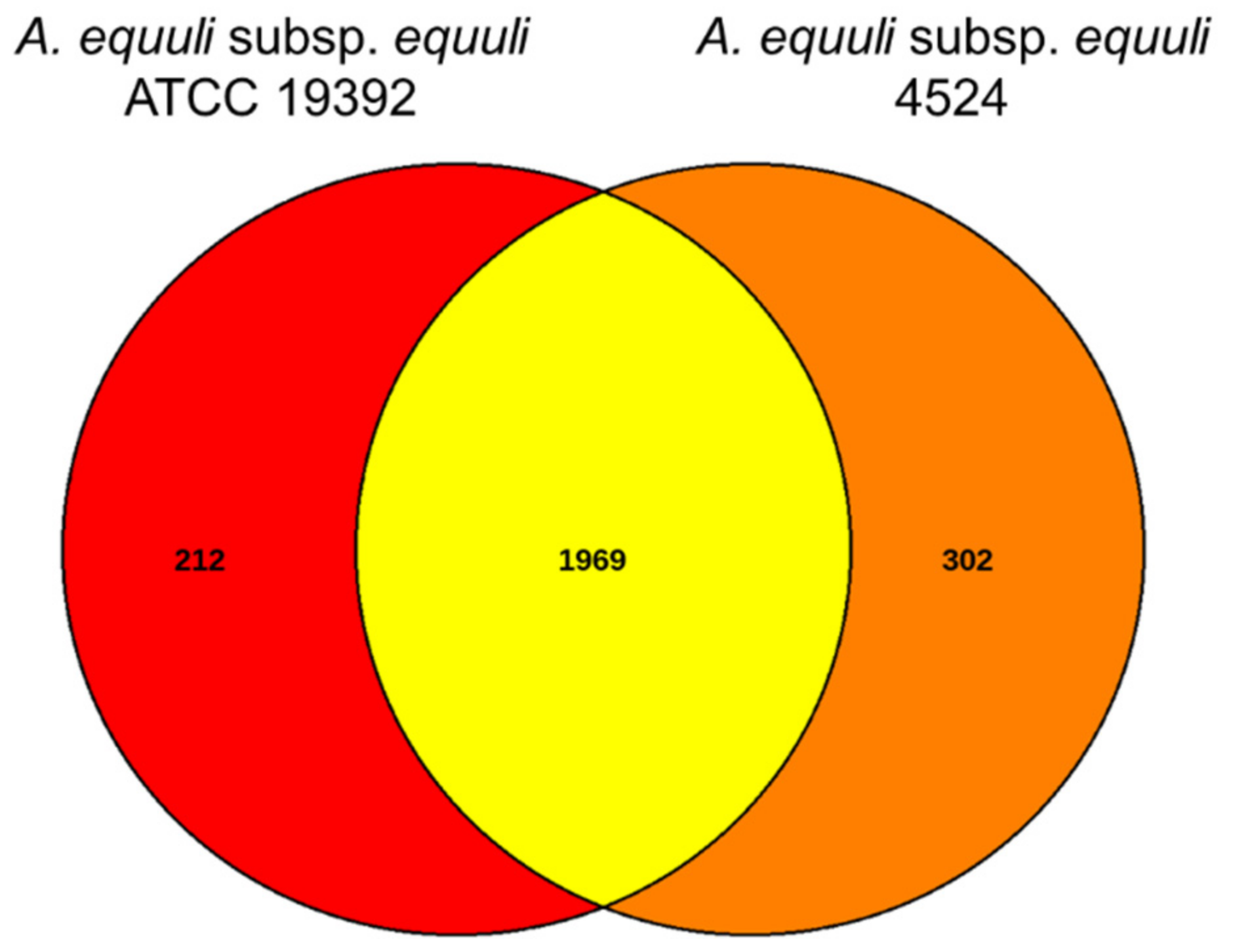
3.5. Comparative Genome Analysis of A. equuli Subsp. equuli 4524 with ATCC 19392 Strain

The genome of A. equuli subsp. equuli 4524 was compared with ATCC 19392 strain using EDGAR pipeline. The EDGAR pipeline found 2483 CDS in the genomes of 4524 and ATCC 19392 strains, of which the two strains shared 1969 CDS (79.3%). Venn diagram of the two strains showed that there are 302 genes unique to isolate 4524 while 212 genes were specific to the ATCC 19392 strain (Figure 5).
In addition, 136 putative virulence genes previously identified in other Actinobacillus spp. were detected in the genome of both strains, of which 122 were present in both strains. In comparison, seven virulence-associated genes were only detected in the ATCC 19392 genome, and seven genes were only identified in the genome of 4524 isolate (Table 5, Table S2).
Of 129 virulence-associated genes identified in the genome of 4524 strain, 72 genes encoded RTX toxins, lipopolysaccharides (LPS), capsular polysaccharide (CPS), outer membrane protein (OMP), salicylic acid, and biofilm (pgaA, pgaB, pgaC). In addition, several putative iron acquisition systems, including those for hemoglobin (hgbA), transferrin (TonB and TonB-exbBD), and siderophore (fhuBCD), were detected in the genome. In addition, different types of adhesins, including filamentous hemagglutinin (fhaC), tight adherence protein (tadA-F), fimbria-like protein (flpB, flpC), and autotransporters that may be involved in adhesion of the bacteria to the target cells were also found (Table S2). Among the strain-specific genes of A. equuli subsp. equuli 4524 strain, three were categorized as iron acquisition proteins (TonB-dependent receptor plug domain protein, FhuD, and FhuB), two LPS (glycosyltransferase family 2 protein and rfaF), one CPS (putative glycosyltransferase YkoT), and one miscellaneous (pgaC) (Table S2). The shared genes between A. equuli subsp. equuli 4524 and ATCC 19392 strains showed > 90% nucleotide similarity, except for three genes categorized as LPS (glf, wbaP, and glycosyltransferase family 2 protein) and the apxIB gene encoding toxin exporter. The three LPS genes, including two glf genes and WbaP, encoded protein sequences of 94% similarity with homologous proteins present in A. pleuropneumoniae and A. suis, respectively (Table S2). Another LPS gene encoding glycosyltransferase family 2 protein was 99.3% identical to the homologous protein in the genome of A. ureae (Table S2). The LPS gene transfers activated sugar residue from a donor substrate to a proper acceptor [33]. Interestingly, the apxIB gene synthesizing RTX toxin exporter in the 4524 strain showed only 65% similarity at both nucleotide and amino acid levels compared to that of ATCC 19392 strain. In contrast, it was 99.7% and 99.9% similar at the nucleotide and amino acid levels to the aqxB protein gene of A. equuli subsp. haemolyticus strain. (Table S2).

4. Discussion

Actinobacillus equuli subsp. equuli has traditionally been associated with neonatal septicemia. In contrast, infections in adult horses are less common and often associated with concurrent bacterial or viral infections as well as other predisposing factors such as drug-associated damage or stressful events that compromise the mucosal integrity [7,12,15,16]. In this study, we described a case of a 22-year-old American Paint gelding that suffered respiratory distress with tachypnea, bilateral purulent to hemorrhagic nasal discharge, and cough with bloody sputum. In agreement with the clinical history and differential diagnosis considered at the time of euthanasia, the gross and histopathological changes observed in the left lung were consistent with fibrinosuppurative and hemorrhagic pleuropneumonia associated with heavy isolation of A. equuli. Further whole genome sequencing and phylogenetic analyses of the isolate identified A. equuli subsp. equuli as the causative agent of fibrinosuppurative and hemorrhagic pleuropneumonia. Actinobacillus equuli is classified into two subspecies: A. equuli subsp. equuli and A. equuli subsp. haemolyticus. Actinobacillus equuli subsp. equuli usually is innocuous in the oral cavity and alimentary tract of adult horses, whereas A. equuli subsp. haemolyticus resides preferentially in the respiratory tract and is frequently isolated from the normal oral cavity and tracheal wash fluids [3,13,14]. In contrast to this notion, a retrospective study of equine actinobacillosis indicated that A. equuli subsp. equuli was isolated more frequently than hemolytic Actinobacillus spp. (presumably A. equuli subsp. haemolyticus) from the respiratory tract of foals and adult horses with respiratory disease manifestations, suggesting that A. equuli subsp. equuli can likely be primary pathogens under favorable conditions [12].
To enhance our knowledge about the potential invasion determinants and the virulence factors associated with A. equuli subsp. equuli, comprehensive genome analysis of the 4524 isolate was performed and compared with the reference A. equuli subsp. equuli ATCC 19392 strain available in GenBank. Infection of invasive species of Actinobacillus is a multifactorial process governed by various virulence factors, such as RTX toxins, LPS, CPS, OMPs, and biofilm, among others, to establish a productive infection in the host [2,20,21]. The RTX toxins are prevalent in Actinobacillus spp. and play an important role in their pathogenicity [34]. Actinobacillus equuli subsp. haemolyticus and A. equuli subsp. equuli differ by the presence or absence of an RTX, referred to as the A. equuli toxin (Aqx) encoded by the aqx gene, responsible for the hemolytic phenotype [34]. RTX toxins also contribute to the host specificity of the two bacterial species, A. suis and A. equuli subsp. haemolyticus containing the apx and the aqx genes, respectively. The hemolytic species A. suis had the apxI and apxII genes but not aqx, which was only found in strains of equine origin, suggesting that different hemolysins are associated with the hemolytic phenotype in these species [35,36]. In addition, the ApxI and ApxII were found to be more cytotoxic to swine lymphocytes. In contrast, the Aqx was more toxic to equine lymphocytes, indicating the host cell-specific activity of RTX [35]. Interestingly, apxIB gene encoding toxin exporter, but not aqx, was detected in the genome of the 4524 isolate, suggesting that this isolate could encode the protein for toxin secretion. Furthermore, the sequence of the toxin protein was highly similar to the aqxB gene of its hemolytic counterpart, A. equuli subsp. haemolyticus, the causative agent of fatal pulmonary hemorrhage in the adult horse [15]. Accordingly, we can speculate that the RTX toxin gene detected in A. equuli subsp. equuli isolate 4524 might have evolved to increase the host-specific cytotoxicity or transferred from its counterpart through the horizontal gene transfer mechanisms, resulting in increased virulence.
Endotoxin, associated with all gram-negative bacteria, damages endothelium, causing vascultilis and thrombosis [37]. Interestingly, several strain-specific putative surface polysaccharides that contribute to bacterial adherence, such as LPS (glycosyltransferase family 2 protein and rfaF) and CPS (glycosyltransferase YkoT) were also detected in the genome of A. equuli subsp. equuli 4524 isolate. Notably, the nucleotide and amino acid sequences of wbaP and glf genes of the 4524 isolate showed higher similarity to those of A. suis NCTC12996 and ATCC 33415 strains and A. pleuropneumoniae 3906 strain, respectively, compared to that of the reference A. equuli subsp. equuli ATCC 19392 strain. This observation suggested that the 4524 isolate may express the adherence molecules required for the firm adherence of the bacteria to the host cells, similar to those of invasive species, such as A. suis and A. pleuropneumoniae [38,39], resulting in enhancement of the pathogenicity of the 4524 strain; however, further investigation is needed.
Lastly, multiplication of the pathogenic microorganism within host tissues is essential for establishing infection. It partially depends on the ability of the pathogen to scavenge essential nutrients. Iron is essential for bacterial growth and regulating the expression of many virulence factors. The genome of the A. equuli subsp. equuli 4524 strain contained different high-affinity iron-acquisition systems to uptake iron from siderophores, transferrin, and hemoglobin proteins associated with TonB-dependent energy transduction. The tonB genes encode the proteins needed for the active transport of iron through the outer membrane [40]. The genome content of 4524 strain indicated the potential of the bacteria in transporting iron from transferrin, hemoglobin (hgbA), or ferric siderophore (fhuBCD) across the outer membrane depending on the exbB–exbD–TonB system [40,41]. The identified operon fhuBCD in the 4524 isolate might be involved in the translocation of ferric hydroxamate from the outer to the inner membrane (fhuD) and in the internalization of ferric hydroxamate (fhuBC) [41,42]. Taken together, A. equuli subsp. equuli 4524 isolate is genetically well-equipped to overcome iron shortages during infection. The results of genome mining suggested that infection by A. equuli subsp. equuli 4524 strain is a multifactorial process, and the putative virulence-associated genes may play a role in pathogenesis and disease phenotype in an adult horse.

5. Conclusions

Here, we report a case of an adult equine actinobacillosis associated with fibrinosuppurative and hemorrhagic pleuropneumonia caused by A. equuli. The phylogenetic analyses identified the isolate as A. equuli subsp. equuli owing to clustering together with A. equuli subsp. equuli ATCC 19392 reference strain. Further genome comparative analysis revealed the presence of seven unique virulence-associated genes in the A. equuli subsp. equuli 4524 isolate, but not in the reference strain, related to the pathogenesis and invasiveness in certain invasive species of Actinobacillus genus. Thus, we speculate that these unique features together with the putative virulence-associated genes (glycosyltransferase family 2 protein, rfaF, glycosyltransferase YkoT, glf, wbaP, and apxIB) related to the pathogenesis and invasiveness in certain invasive species of Actinobacillus genus, may be closely associated with the virulence phenotype observed in our clinical case. However further studies will be needed to compare 4524 and non-laboratory-adapted A. equuli subsp. equuli strains, other than the type strain, as well as A. equuli subsp. haemolyticus strains, to reveal more information on the functions of virulence-associated genes related to the pathogenesis of A. equuli subsp. equuli.

Supplementary Materials

The following supporting information can be downloaded at: https://www.mdpi.com/article/10.3390/pathogens12020224/s1, Table S1: Actinobacillus species used in this study and their characteristics; Table S2: Shared and strain-specific virulence-associated genes detected in A. equuli subsp. equuli 4524 and ATCC 19392 strains.

Author Contributions

Conceptualization, M.C., F.D.P. and Y.-Y.G.; methodology, M.K., M.C., L.P., M.S.M., J.W. and R.B.; software, M.K. and F.L.; formal analysis, M.K., F.L. and Y.-Y.G.; investigation, L.P., M.S.M., J.W. and R.B.; data curation, M.C. and Y.-Y.G.; writing—original draft preparation, M.K., M.C. and Y.-Y.G.; writing—review and editing, M.K., M.C., Á.K., U.B.R.B. and Y.-Y.G.; supervision, M.C. and F.D.P.; funding acquisition, M.C. and Y.-Y.G. All authors have read and agreed to the published version of the manuscript.

Funding

Yun Young Go is supported by start-up (9610462) and intramural (APRC-9610530) funds from the City University of Hong Kong, Hong Kong SAR, China. Mariano Carossino was also supported by start-up funds (PG009641) from the School of Veterinary Medicine, LSU, Louisiana, USA. In addition, support was also obtained from F.D.A. Veterinary Laboratory Investigation and Response Network (Vet-LIRN; grant number AWD-47109-1/U18FD006442) awarded to Udeni Balasuriya and the Louisiana Animal Disease Diagnostic Laboratory (PG009095).

Data Availability Statement

The complete genome sequence of A. equuli subsp. equuli 4524 isolate was deposited in NCBI GenBank under the accession number JAPHVQ000000000.

Acknowledgments

We would like to kindly acknowledge the members of the Histology section at the Louisiana Animal Disease Diagnostic Laboratory (LADDL), School of Veterinary Medicine, Louisiana State University, for their assistance.

Conflicts of Interest

The authors declare no conflict of interest. The funders had no role in the design of the study; in the collection, analyses, or interpretation of data; in the writing of the manuscript; or in the decision to publish the results.

References

  1. MacInnes, J.I. Actinobacillus; Wiley-Blackwell: Ames, IA, USA, 2010. [Google Scholar]
  2. Bujold, A.R.; Shure, A.E.; Liu, R.; Kropinski, A.M.; MacInnes, J.I. Investigation of putative invasion determinants of Actinobacillus species using comparative genomics. Genomics 2019, 111, 59–66. [Google Scholar] [CrossRef] [PubMed]
  3. Kuhnert, P.; Berthoud, H.; Christensen, H.; Bisgaard, M.; Frey, J. Phylogenetic relationship of equine Actinobacillus species and distribution of RTX toxin genes among clusters. Vet. Res. 2003, 34, 353–359. [Google Scholar] [CrossRef] [PubMed] [Green Version]
  4. Matthews, S.; Dart, A.J.; Dowling, B.A.; Hodgson, J.L.; Hodgson, D.R. Peritonitis associated with Actinobacillus equuli in horses: 51 cases. Aust. Vet. J. 2001, 79, 536–539. [Google Scholar] [CrossRef] [PubMed]
  5. Aalbaek, B.; Ostergaard, S.; Buhl, R.; Jensen, H.E.; Christensen, H.; Bisgaard, M. Actinobacillus equuli subsp. equuli associated with equine valvular endocarditis. APMIS 2007, 115, 1437–1442. [Google Scholar] [CrossRef]
  6. Castagnetti, C.; Rossi, M.; Parmeggiani, F.; Zanoni, R.G.; Pirrone, A.; Mariella, J. Facial cellulitis due to Actinobacillus equuli infection in a neonatal foal. Vet. Rec. 2008, 162, 347–349. [Google Scholar] [CrossRef]
  7. Donahue, J.M.; Sells, S.F.; Bolin, D.C. Classification of Actinobacillus spp isolates from horses involved in mare reproductive loss syndrome. Am. J. Vet. Res. 2006, 67, 1426–1432. [Google Scholar] [CrossRef]
  8. Stewart, A.J.; Hinchcliff, K.W.; Saville, W.J.; Jose-Cunilleras, E.; Hardy, J.; Kohn, C.W.; Reed, S.M.; Kowalski, J.J. Actinobacillus sp. bacteremia in foals: Clinical signs and prognosis. J. Vet. Intern. Med. 2002, 16, 464–471. [Google Scholar] [CrossRef]
  9. Ward, C.L.; Wood, J.L.; Houghton, S.B.; Mumford, J.A.; Chanter, N. Actinobacillus and Pasteurella species isolated from horses with lower airway disease. Vet. Rec. 1998, 143, 277–279. [Google Scholar] [CrossRef]
  10. Edwards, P.R. Studies on Shigella equirulis (Bact. viscosum equi). Bulletin- Kentucky, Agricultural Experiment Station, 1931; 289–330.11. [Google Scholar]
  11. Sternberg, S.; Brandstrom, B. Biochemical fingerprinting and ribotyping of isolates of Actinobacillus equuli from healthy and diseased horses. Vet. Microbiol. 1999, 66, 53–65. [Google Scholar] [CrossRef]
  12. Layman, Q.D.; Rezabek, G.B.; Ramachandran, A.; Love, B.C.; Confer, A.W. A retrospective study of equine actinobacillosis cases: 1999–2011. J. Vet. Diagn. Investig. 2014, 26, 365–375. [Google Scholar] [CrossRef]
  13. Benavente, C.E.; Fuentealba, I.C. Actinobacillus suis and Actinobacillus equuli, emergent pathogens of septic embolic nephritis, a new challenge for the swine industry. Arch. Med. Vet. 2012, 44, 99–107. [Google Scholar] [CrossRef] [Green Version]
  14. Thompson, A.B.; Postey, R.C.; Snider, T.; Pasma, T. Actinobacillus equuli as a primary pathogen in breeding sows and piglets. Can. Vet. J. 2010, 51, 1223–1225. [Google Scholar]
  15. Pusterla, N.; Jones, M.E.; Mohr, F.C.; Higgins, J.K.; Mapes, S.; Jang, S.S.; Samitz, E.M.; Byrne, B.A. Fatal pulmonary hemorrhage associated with RTX toxin producing Actinobacillus equuli subspecies haemolyticus infection in an adult horse. J. Vet. Diagn. Investig. 2008, 20, 118–121. [Google Scholar] [CrossRef] [Green Version]
  16. Bosse, J.T.; Janson, H.; Sheehan, B.J.; Beddek, A.J.; Rycroft, A.N.; Kroll, J.S.; Langford, P.R. Actinobacillus pleuropneumoniae: Pathobiology and pathogenesis of infection. Microbes Infect. 2002, 4, 225–235. [Google Scholar] [CrossRef]
  17. Bujold, A.R.; MacInnes, J.I. Identification of putative adhesins of Actinobacillus suis and their homologues in other members of the family Pasteurellaceae. BMC Res. Notes 2015, 8, 675. [Google Scholar] [CrossRef] [Green Version]
  18. Darling, A.C.; Mau, B.; Blattner, F.R.; Perna, N.T. Mauve: Multiple alignment of conserved genomic sequence with rearrangements. Genom. Res. 2004, 14, 1394–1403. [Google Scholar] [CrossRef] [Green Version]
  19. Seemann, T. Prokka: Rapid prokaryotic genome annotation. Bioinformatics 2014, 30, 2068–2069. [Google Scholar] [CrossRef] [Green Version]
  20. Schwengers, O.; Jelonek, L.; Dieckmann, M.A.; Beyvers, S.; Blom, J.; Goesmann, A. Bakta: Rapid and standardized annotation of bacterial genomes via alignment-free sequence identification. Microb. Genom. 2021, 7, 000685. [Google Scholar] [CrossRef]
  21. Tatusov, R.L.; Fedorova, N.D.; Jackson, J.D.; Jacobs, A.R.; Kiryutin, B.; Koonin, E.V.; Krylov, D.M.; Mazumder, R.; Mekhedov, S.L.; Nikolskaya, A.N.; et al. The COG database: An updated version includes eukaryotes. BMC Bioinform. 2003, 4, 41. [Google Scholar] [CrossRef] [Green Version]
  22. Grissa, I.; Vergnaud, G.; Pourcel, C. CRISPRFinder: A web tool to identify clustered regularly interspaced short palindromic repeats. Nucleic Acids Resm. 2007, 35, W52–W57. [Google Scholar] [CrossRef] [Green Version]
  23. Grant, J.R.; Stothard, P. The CGView Server: A comparative genomics tool for circular genomes. Nucleic Acids Res. 2008, 36, W181–W184. [Google Scholar] [CrossRef] [PubMed]
  24. Blom, J.; Albaum, S.P.; Doppmeier, D.; Puhler, A.; Vorholter, F.J.; Zakrzewski, M.; Goesmann, A. EDGAR: A software framework for the comparative analysis of prokaryotic genomes. BMC Bioinform. 2009, 10, 154. [Google Scholar] [CrossRef] [PubMed] [Green Version]
  25. Bertels, F.; Silander, O.K.; Pachkov, M.; Rainey, P.B.; van Nimwegen, E. Automated reconstruction of whole-genome phylogenies from short-sequence reads. Mol. Biol. Evol. 2014, 31, 1077–1088. [Google Scholar] [CrossRef] [PubMed]
  26. Kumar, S.; Stecher, G.; Tamura, K. MEGA7: Molecular Evolutionary Genetics Analysis Version 7.0 for Bigger Datasets. Mol. Biol. Evol. 2016, 33, 1870–1874. [Google Scholar] [CrossRef] [Green Version]
  27. Felsenstein, J. Evolutionary trees from DNA sequences: A maximum likelihood approach. J. Mol. Evol. 1981, 17, 368–376. [Google Scholar] [CrossRef]
  28. Nei, M.; Kumar, S. Molecular Evolution and Phylogenetics; Oxford University Press: New York, NY, USA, 2000. [Google Scholar]
  29. Yoon, S.H.; Ha, S.M.; Kwon, S.; Lim, J.; Kim, Y.; Seo, H.; Chun, J. Introducing EzBioCloud: A taxonomically united database of 16S rRNA gene sequences and whole-genome assemblies. Int. J. Syst. Evol. Microbiol. 2017, 67, 1613–1617. [Google Scholar] [CrossRef]
  30. Goris, J.; Konstantinidis, K.T.; Klappenbach, J.A.; Coenye, T.; Vandamme, P.; Tiedje, J.M. DNA-DNA hybridization values and their relationship to whole-genome sequence similarities. Int. J. Syst. Evol. Microbiol. 2007, 57, 81–91. [Google Scholar] [CrossRef] [Green Version]
  31. Richter, M.; Rossello-Mora, R. Shifting the genomic gold standard for the prokaryotic species definition. Proc. Natl. Acad. Sci. USA 2009, 106, 19126–19131. [Google Scholar] [CrossRef] [Green Version]
  32. Meier-Kolthoff, J.P.; Auch, A.F.; Klenk, H.P.; Goker, M. Genome sequence-based species delimitation with confidence intervals and improved distance functions. BMC Bioinform. 2013, 14, 60. [Google Scholar] [CrossRef] [Green Version]
  33. Schmid, J.; Heider, D.; Wendel, N.J.; Sperl, N.; Sieber, V. Bacterial Glycosyltransferases: Challenges and Opportunities of a Highly Diverse Enzyme Class Toward Tailoring Natural Products. Front. Microbiol. 2016, 7, 182. [Google Scholar] [CrossRef] [Green Version]
  34. Berthoud, H.; Frey, J.; Kuhnert, P. Characterization of Aqx and its operon: The hemolytic RTX determinant of Actinobacillus equuli. Vet. Microbiol. 2002, 87, 159–174. [Google Scholar] [CrossRef]
  35. Kuhnert, P.; Berthoud, H.; Straub, R.; Frey, J. Host cell specific activity of RTX toxins from haemolytic Actinobacillus equuli and Actinobacillus suis. Vet. Microbiol. 2003, 92, 161–167. [Google Scholar] [CrossRef]
  36. Frey, J. The role of RTX toxins in host specificity of animal pathogenic Pasteurellaceae. Vet. Microbiol. 2011, 153, 51–58. [Google Scholar] [CrossRef]
  37. Bannerman, D.D.; Goldblum, S.E. Mechanisms of bacterial lipopolysaccharide-induced endothelial apoptosis. Am. J. Physiol. Lung Cell Mol. Physiol. 2003, 284, L899–L914. [Google Scholar] [CrossRef] [Green Version]
  38. Paradis, S.E.; Dubreuil, D.; Rioux, S.; Gottschalk, M.; Jacques, M. High-molecular-mass lipopolysaccharides are involved in Actinobacillus pleuropneumoniae adherence to porcine respiratory tract cells. Infect. Immun. 1994, 62, 3311–3319. [Google Scholar] [CrossRef] [Green Version]
  39. Jacques, M.; Paradis, S.E. Adhesin-receptor interactions in Pasteurellaceae. FEMS Microbiol. Rev. 1998, 22, 45–59. [Google Scholar] [CrossRef]
  40. Beddek, A.J.; Sheehan, B.J.; Bosse, J.T.; Rycroft, A.N.; Kroll, J.S.; Langford, P.R. Two TonB systems in Actinobacillus pleuropneumoniae: Their roles in iron acquisition and virulence. Infect. Immun. 2004, 72, 701–708. [Google Scholar] [CrossRef] [Green Version]
  41. Postle, K. TonB protein and energy transduction between membranes. J. Bioenerg. Biomembr. 1993, 25, 591–601. [Google Scholar] [CrossRef]
  42. Jacques, M. Surface polysaccharides and iron-uptake systems of Actinobacillus pleuropneumoniae. Can. J. Vet. Res. 2004, 68, 81–85. [Google Scholar]
Figure 1. Gross findings in the lungs of the Actinobacillus equuli-infected horse. (A) The left lung is unilaterally affected by an extensive, well-demarcated dark red to black area of hemorrhage and necrosis coated by fibrinous exudate (hemorrhagic and fibrinous pleuropneumonia). (B) Left lung, cut surface. The cut surface shows an extensive and relatively well-delimited area of necrosis and hemorrhage.
Pathogens 12 00224 g001
Figure 2. Microscopic features of Actinobacillus equuli-associated pleuropneumonia. (A) The pleura is extensively coated by a thick layer of polymerized fibrin (arrows) and myriad coccobacillary bacteria underlying the fibrinous exudate and extending into the subpleural interstitium (arrowheads). The underlying pulmonary parenchyma is affected by hemorrhage with loss of alveolar septa and replacement by numerous degenerate leukocytes. (B) The bacteria underlying the fibrinous exudate are gram-negative (arrowheads). (C,D) The pulmonary parenchyma is affected by extensive hemorrhage, fibrinous exudation, and necrosis and is infiltrated by numerous degenerate leukocytes. Within areas of less pronounced necrosis, alveolar spaces are filled with degenerate leukocytes (D), and the pulmonary vasculature is multifocally occluded by fibrin thrombi ((D), arrows). (E,F) Within affected areas of the parenchyma, there are multifocal extracellular and intracellular colonies of gram-negative (F) coccobacilli (arrows). (A,C,D,E); H&E, 200×, 100×, 100×, and 400×, respectively. (B,F); Gram stain, 1000×.
Pathogens 12 00224 g002
Figure 3. Maximum likelihood phylogenetic tree of the complete genome sequences of isolate 4524 and other Actinobacillus spp. based on REALPY. Numbers at nodes represent the percentages of occurrence of nodes in 1000 bootstrap trials. The Haemophilus influenzae PittGG (CP000672) was used as an outgroup.
Pathogens 12 00224 g003
Figure 4. Circular map of the genome of A. equuli subsp. equuli 4524 strain generated using the CGView Server. From the outside to the center: coding sequences (CDSs) in the positive strand, CDSs in the reverse strand, BLASTn vs. A. equuli subsp. equuli ATCC 19392 (NZ_CP007715.1), GC skew, and GC content.
Pathogens 12 00224 g004
Figure 5. Venn diagram of the complete genomes of A. equuli subsp. equuili 4524 and ATCC 19392 strains.
Pathogens 12 00224 g005
Table 1. Broth microdilution MIC results.
AntimicrobicResultInterpretation
Amikacin=16Susceptible
Ampicillin≤0.25No Interpretation
Azithromycin≤0.25No Interpretation
Cefazolin≤4No Interpretation
Ceftazidime≤1Susceptible
Ceftiofur≤0.25No Interpretation
Chloramphenicol≤4Susceptible
Clarithromycin≤1No Interpretation
Doxycycline≤2Susceptible
Enrofloxacin≤0.25No Interpretation
Erythromycin=0.5No Interpretation
Gentamicin=8Intermediate
Imipenem≤1Susceptible
Oxacillin≤0.25No Interpretation
Penicillin=0.25No Interpretation
Rifampicin=4No Interpretation
Tetracycline≤2No Interpretation
Ticarcillin≤8No Interpretation
Ticarcillin-Clavulanate≤8Susceptible
Trimethoprim-sulfamethoxazole≤0.5Susceptible
Table 2. Genome statistics of A. equuli subsp. equuli 4524 isolate.
AttributeValue
Genome size (bp)2,464,726
Total genes2362
Protein coding genes2277
RNA genes85
tRNA genes61
tmRNA genes1
rRNA genes8
ncRNA genes15
Genes assigned to COGs1419
CRISPR repeats2
Table 3. The number of genes associated with general Cluster of Orthologous Group (COG) functional categories.
CodeValueDescription
J203Translation, ribosomal structure, and biogenesis
A1RNA processing and modification
K83Transcription
L89Replication, recombination, and repair
B0Chromatin structure and dynamics
D33Cell cycle control, cell division, chromosome partitioning
Y0Nuclear structure
V30Defense mechanisms
T36Signal transduction mechanisms
M148Cell wall/membrane/envelope biogenesis
N6Cell motility
Z0Cytoskeleton
W1Extracellular structures
U28Intracellular trafficking, secretion, and vesicular transport
O88Posttranslational modification, protein turnover, chaperones
C104Energy production and conversion
G127Carbohydrate transport and metabolism
E168Amino acid transport and metabolism
F68Nucleotide transport and metabolism
H100Coenzyme transport and metabolism
I42Lipid transport and metabolism
P108Inorganic ion transport and metabolism
Q8Secondary metabolites biosynthesis, transport, and catabolism
R69General function prediction only
X4Mobilome: prophages, transposons
S78Function unknown
Table 4. Average nucleotide identity (ANI) and Genome-to-Genome Distance Calculation (GGDC), DNA-DNA hybridization (DDH) values between A. equuli 4524 and selected Actinobacillus spp.
SpeciesStrainANI (%)GGDCDDHProb. DDH ≥ 70%
A. capsulatus DSM 1976192.70.07394916.6
A. delphinicola NCTC1287170.50.1742250.01
A. equuli subsp. equuliATCC 1939295.90.041866.170
A. indolicus 46K2C74.50.212620.70
A. lignieresii NCTC418986.80.134531.50.19
A. minor NM30576.40.193622.60
A. pleuropneumoniae NCTC1097686.50.133531.70.2
A. porcinus NM319730.178524.40.01
A. porcitonsillarum 9953L5576.30.19322.70
A. seminis NCTC1085171.80.185723.50
A. succinogenes GXAS13771.80.184723.70
A. suis ATCC3341593.20.07175019
A. ureae NCTC1022092.70.072849.517.8
Table 5. Shared and strain-specific virulence genes in A. equuli subsp. equuli 4524 and ATCC 19392 genomes.
Virulence FactorNumber of Shared Genes in 4524 and ATCC 19392Number of Genes Specific to ATCC 19392Number of Genes Specific to 4524
Iron acquisition4013
LPS4132
CPS1811
Miscellaneous1521
OMP400
Salicylic acid200
RTX toxin100
Autotransporter100
Total12277
Disclaimer/Publisher’s Note: The statements, opinions and data contained in all publications are solely those of the individual author(s) and contributor(s) and not of MDPI and/or the editor(s). MDPI and/or the editor(s) disclaim responsibility for any injury to people or property resulting from any ideas, methods, instructions or products referred to in the content.

