Article

Morphological and Molecular Identification of Dactylogyrus gobiocypris (Monogenea: Dactylogyridae) on Gills of a Model Fish, Gobiocypris rarus (Cypriniformes: Gobionidae)

1
State Key Laboratory of Freshwater Ecology and Biotechnology and Key Laboratory of Aquaculture Disease Control, Ministry of Agriculture, Institute of Hydrobiology, Chinese Academy of Sciences, Wuhan 430072, China
2
College of Science, Tibet University, Lhasa 850000, China
3
University of Chinese Academy of Sciences, Beijing 100049, China
*
Author to whom correspondence should be addressed.
Pathogens 2023, 12(2), 206; https://doi.org/10.3390/pathogens12020206
Submission received: 8 January 2023 / Revised: 21 January 2023 / Accepted: 22 January 2023 / Published: 28 January 2023

Abstract

:
The rare minnow Gobiocypris rarus is an ideal model organism for toxicological research. Dactylogyrus species are usually found on the gills of this rare minnow in laboratory farming systems. Dactylogyrid infection may change the sensibility of fish to toxicants and affect toxicological evaluations. In the present study, dactylogyrid infection was investigated, and species of Dactylogyrus collected from rare minnows were determined. Based on the observed ‘D. wunderi’ type anchors, with a shorter outer root and elongated inner root, and accessory piece consisting of two parts, the dactylogyrids were identified as D. gobiocypris. A partial 18S-ITS1 rDNA sequence was firstly sequenced, and the highest sequence identity (86.7%) was to D. cryptomeres. Phylogenetic analysis revealed that D. gobiocypris formed a clade with D. squameus, D. finitimus, and D. cryptomeres, all of which have been recorded in the family Gobionidae. Histopathology analysis indicated that a heavy burden of D. gobiocypris caused necrosis of gill filaments. Inflammatory responses, such as tumefaction and hyperaemia, were also observed on gills with severe dactylogyrid infection. Supplementary morphological characteristics and 18S-ITS1 rDNA sequence provided basic data for identification of this parasite species.

1. Introduction

The rare minnow Gobiocypris rarus Ye et Fu, 1983 (Gobionidae) is a small gobionid fish endemic to China, mainly distributed at the edge of the west and northwest area of the Sichuan Basin [1,2]. This rare minnow possesses particular biological characteristics, such as high sensitivity to chemicals, small body size, short life cycle, and being easy to rear in laboratory, which make it an excellent model organism for ecotoxicology studies [3,4,5]. Since 1995, rare minnows have been widely used in acute and subchronic toxicity experiments on heavy metals [6,7], organics [8,9,10], and endocrine-disrupting chemicals [11,12,13].
The monogenean family Dactylogyridae Bychowsky, 1933 is one of the most species-rich groups of helminths, with more than 1000 species recognised worldwide [14]. Forty-one species of Dactylogyrus have been recorded from fishes in the family Gobionidae in China [15], and twenty-six species of Dactylogyrus are found on fishes in the family Gobionidae in Europe [16]. Species of Dactylogyrus were found on gills of rare minnows in a laboratory in China, and D. gobiocypris Yao, 1995 was described based on sclerotized parts of the anchor and copulatory complex [17]. No studies have reported D. gobiocypris since.
Dactylogyrids can infect the gills of cypriniform fishes [18], causing serious hyperplasia of the gill filament epithelium, copious mucus, and eventually affecting respiratory function [19,20]. Fish heavily infected with dactylogyrids are also susceptible to bacterial infections [21,22,23].
The present study provides supplementary morphological characteristics, novel sequences of the 18S ribosomal RNA subunit and the first internal transcribed spacer region of rDNA (ITS1), and histopathological analysis of D. gobiocypris parasitizing G. rarus specimens reared in the laboratory in the Institute of Hydrobiology, Chinese Academy of Sciences, Wuhan City, Hubei province, China.

2. Materials and Methods

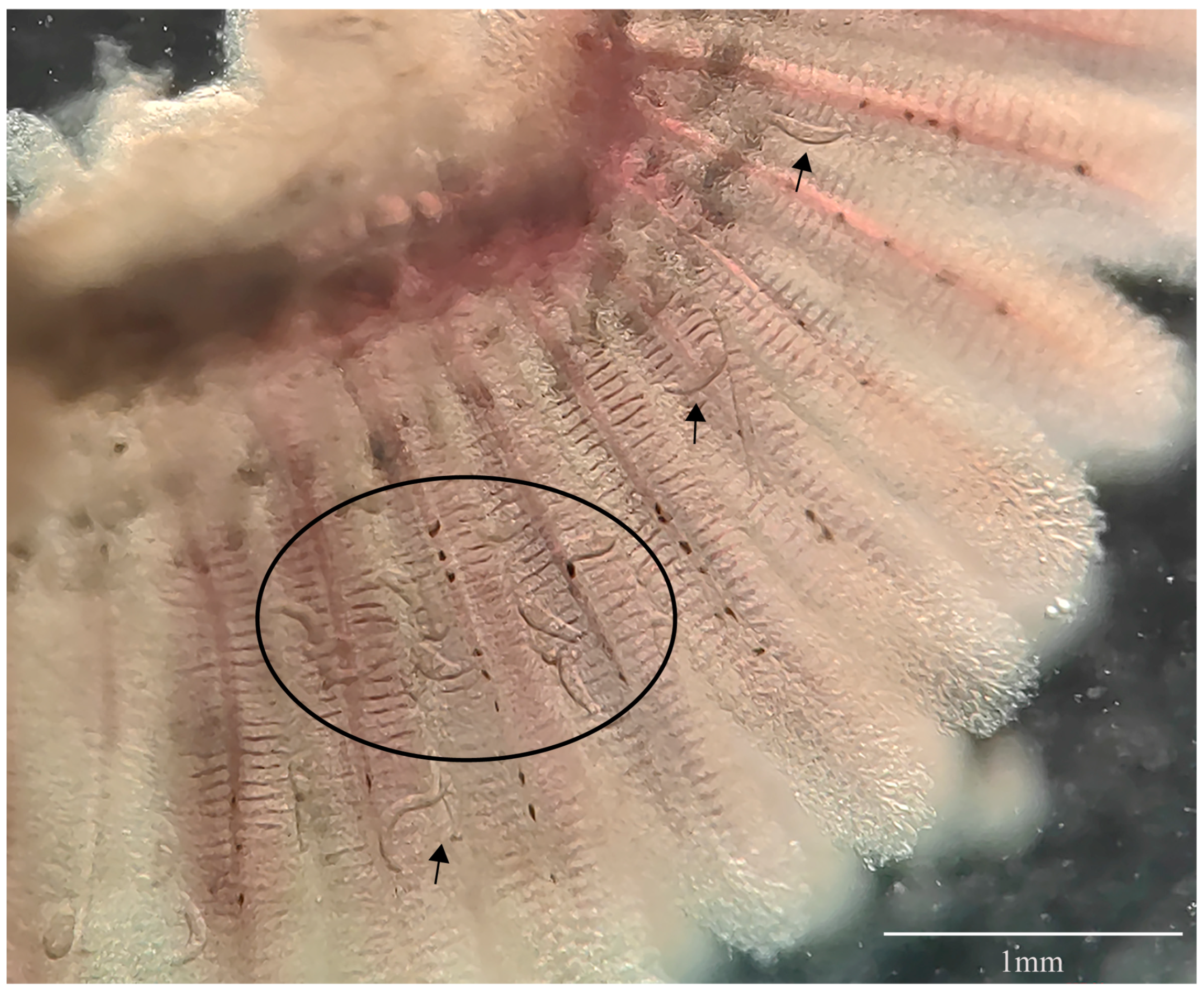
2.1. Parasite Collection

Samples of rare minnow were obtained from the laboratory in the Institute of Hydrobiology, Chinese Academy of Sciences in April 2022. Thirty fish (with total body length of 4.2 ± 0.9 cm) were randomly selected and anesthetized with 0.02% MS-222 (tricaine methanesulfonate) (Sigma, St Louis, MO, USA). Specimens of species of Dactylogyrus were then examined and collected using micro surgical needles under a stereoscopic microscope. Worms were rinsed several times with distilled water for further analyses.

2.2. Morphological Identification

A random subsample of dactylogyrids were mounted on a microscope slide and fixed in ammonium picrate glycerine (GAP) as whole mount following the procedure described by Ergens [24] and Malmberg [25] for morphological identification. GAP and Canada balsam were performed according to Ergens [24]. Additional specimens, with opisthaptors excised using a scalpel, were then individually subjected to proteolytic digestion according to the method described by Paladini et al. [26] and Tu et al. [22]. The tissue-free opisthaptoral sclerotized parts were mounted in GAP, and the excised body of each specimen was preserved in 95% ethanol for subsequent molecular analysis. Specimens were photographed using an optical microscope (Axioplan 2 imaging and Axiophot 2, Zeiss, Oberkochen, Germany). Measurements were taken according to Šimková et al. [27], and are given in micrometers (μm) unless otherwise stated. Identification of individual specimens was performed by comparing the morphology and measurements of anchors and the copulatory complex to the literature [17]. Five full worms, embedded in GAP and mounted on Canada balsam, were deposited as voucher specimens (accession nos. CJW-DG 202201-05) in the Museum of the Institute of Hydrobiology, Chinese Academy of Sciences, Wuhan City, Hubei province, China.

2.3. DNA extraction, Amplification and Sequencing

Genomic DNA was extracted from the excised bodies of 12 specimens using a Tissue Cell Genome Kit (TaKaRa, Beijing, China) according to the manufacturer’s instructions. The region of rDNA spanning the 3′ end of the 18S ribosomal RNA subunit, the entire ITS1 gene, and the 5′ end of the 5.8S ribosomal RNA subunit were targeted using primers S1 (5′-ATTCCGATAACGAACGAGACT-3′) and H7 (5′-GCTGCGTTCTTCATCGATACTCG-3′) [28,29]. PCR amplification was conducted using LA Taq polymerase (TaKaRa, Beijing, China) with the following profile: 5 min at 95 °C, 35 cycles of 1 min at 94 °C, 1 min at 55 °C, 1 min 30 s at 72 °C, and a final extension of 10 min at 72 °C. After purification, PCR products were cloned into the pGEM-T vector (Promega, Madison, USA), sequenced with the primers described above, produced by Sangon Biotech (Shanghai, China), and assembled manually with DNAStar’s SeqMan software (DNAStar, Madison, WI, USA).

2.4. Molecular Analyses

The obtained sequences of partial 18S rDNA, ITS1, and the flanking sequence of 5.8S rDNA were compared using BLAST in GenBank to assess similarity with other Dactylogyrus species. From the 12 specimens, 12 sequences of 18S-ITS1 rDNA were obtained to evaluate the intraspecific variation using BLAST. Sequences (Table 1) used for phylogenetic analyses were chosen from Dactylogyrus species from closely related hosts. Thaparocleidus vistulensis (Siwak, 1932), in the family Ancylodiscoididae, was used as the outgroup. Sequences were imported into PhyloSuite [30] and aligned with available 18S-ITS1 rDNA sequences of other Dactylogyrus spp. in GenBank using MAFFT 7.149 [31]. Ambiguously aligned fragments were trimmed using Gblocks [32] with the following parameter settings: minimum number of sequences for a conserved/flank position (6/6), maximum number of contiguous non-conserved positions (8), minimum length of a block (10), allowed gap positions (with half). Phylogenetic analyses based on the 18S-ITS1 sequences were performed using maximum likelihood (ML) and inference (BI) methods. TNe+G4 and K2P+G4 were chosen as the best-fit partition model for nucleotide evolution for ML and BI analyses, respectively using ModelFinder [33]. ML phylogenies were inferred using IQ-TREE [34], for 1000 standard bootstraps, as well as the Shimodaira–Hasegawa-like approximate likelihood-ratio test. BI phylogenies were inferred using MrBayes 3.2.6 [35], with two parallel runs (2,000,000 generations) in which the initial 25% of sampled data were discarded as burn-in.

2.5. Histopathology Analysis

The first gill arch of each fish was collected and fixed in 10% neutral buffered formalin (Yeasen, Shanghai, China). The fixing solution was diluted to 4% after 4 to 24 h, washed for 24 h and dehydrated in graded ethanol. Gills were embedded in paraffin (Yeasen, Shanghai, China), sliced into 5 μm-thick sections, and stained with the Hematoxylin and Eosin Staining Kit (Yeasen, Shanghai, China) according to Molnár [36]. The slides were mounted on Canada balsam and examined under an optical microscope (Axioplan 2 imaging and Axiophot 2, Zeiss, Oberkochen, Germany).
Table 1. Species included in the phylogenetic analysis.
Parasite SpeciesHost SpeciesLocalityGenBank IDRefs
Dactylogyrus cryptomeresGobio gobioMorava River basin, Czech RepublicAJ564123[28]
Dactylogyrus finitimusRomanogobio albipinatusMorava River basin, Czech RepublicAJ564133[28]
Dactylogyrus squameusPseudorasbora parvaMorava River basin, Czech RepublicAJ564156[28]
Dactylogyrus gobiocyprisGobiocypris rarusWuhan City, ChinaOP441417Present study
Dactylogyrus achmerowiCyprinus carpioMorava River basin, Czech RepublicAJ564108[28]
Dactylogyrus extensusCyprinus carpioMorava River basin, Czech RepublicAJ564129[28]
Dactylogyrus vastatorCarassius auratusLiangzi Lake, ChinaKC876016[37]
Dactylogyrus intermediusCarassius auratusLiangzi Lake, ChinaKC876017[37]
Dactylogyrus lamellatusCtenopharyngodon idellaMorava River basin, Czech RepublicAJ564141[28]
Outgroup
Thaparocleidus vistulensisSilurus glanisMorava River basin, Czech RepublicAJ490165[38]

3. Results

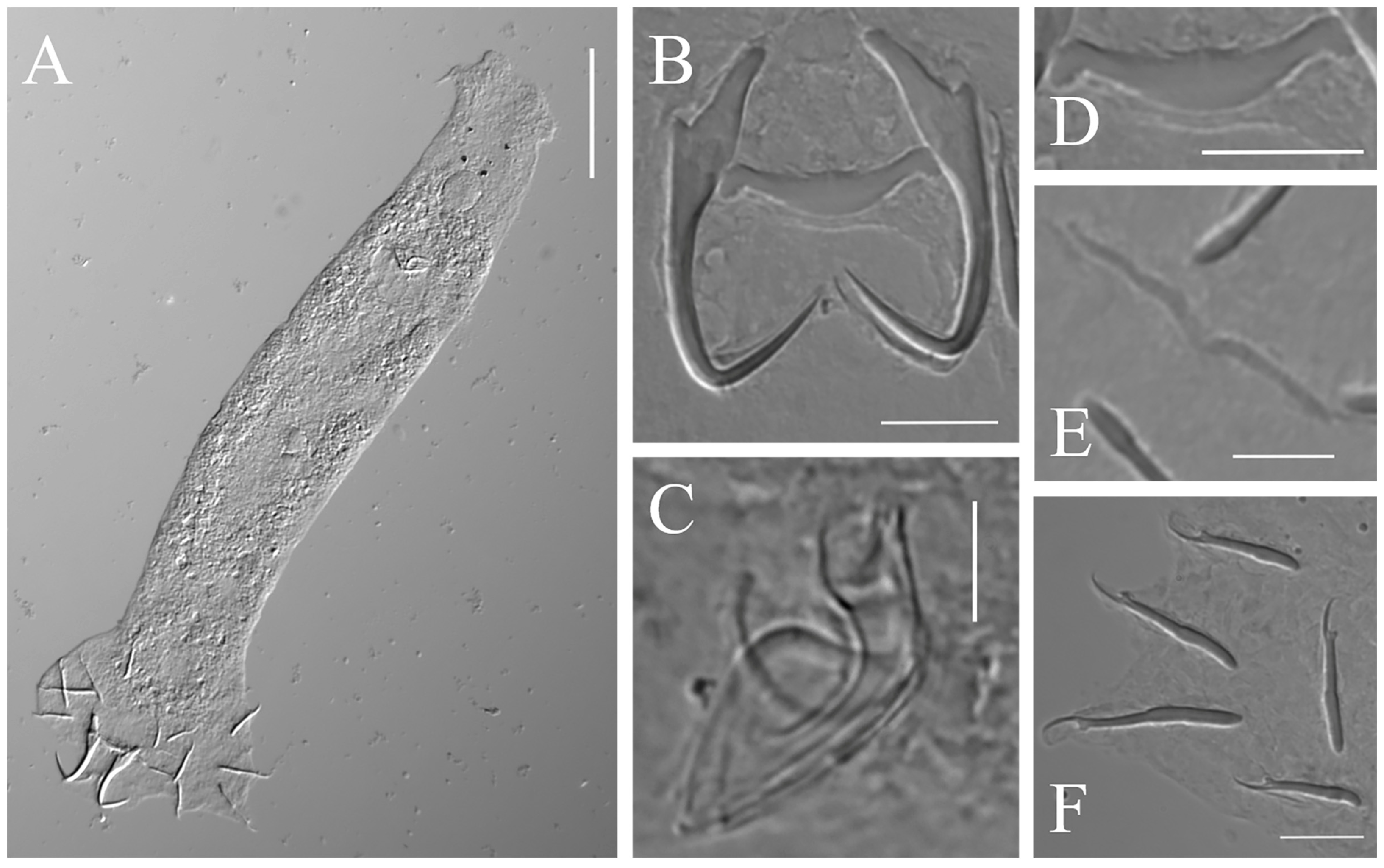
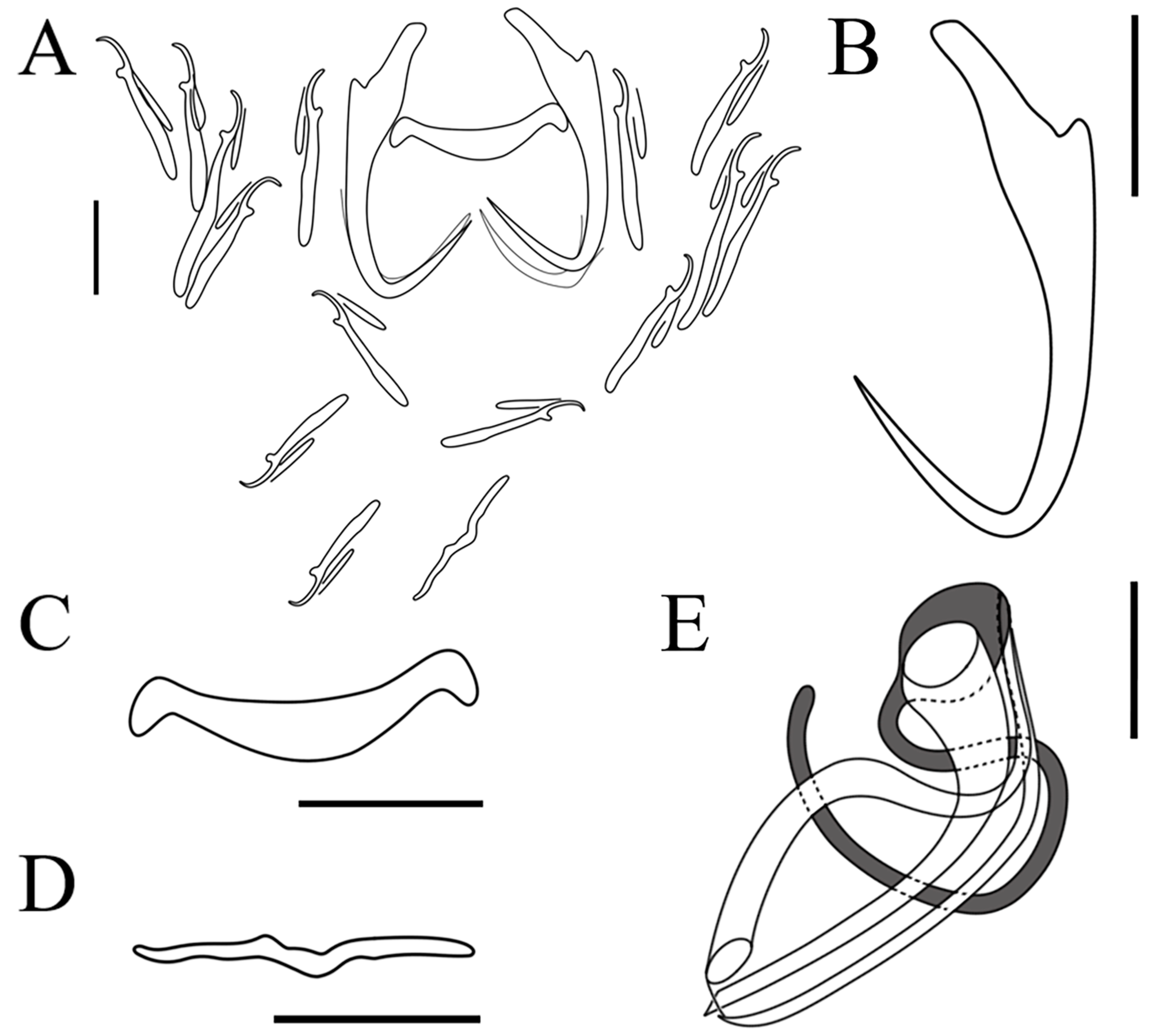
3.1. Taxonomic Summary

Dactylogyrus gobiocypris Yao, 1995

Host: Gobiocypris rarus Ye et Fu, 1983 (Cypriniformes: Gobionidae).
Site of infection: gill filaments (Figure 1).
Locality: specimens collected from cultured rare minnow in the laboratory in the Institute of Hydrobiology, Chinese Academy of Sciences (30°54′74.1″ N, 114°35′’04.3″ E), Wuhan City, Hubei province, China.
Prevalence and mean abundance: 96.7% and 60.8 ± 84.5 (3–408), respectively.
Deposition of specimens: deposited in the Museum of the Institute of Hydrobiology (accession nos. CJW-DG 202201–05), Chinese Academy of Sciences, Wuhan City, Hubei province, China.
DNA reference sequences: a sequence (1042 bp) spanning the region of the 3′ end of the 18S ribosomal RNA subunit and ITS1 to the 5′ end of the 5.8S ribosomal RNA subunit was deposited in GenBank (OP441417).
Description: Based on 56 specimens fixed in GAP. Body length, 182 (118–248; n = 32); width, 45 (28–74; n = 32). Eye spots: two pairs. Pharynx diameter, 11 (7–17; n = 33). Copulatory complex: composed of penis and accessory piece, posterior to pharynx. Penis: tubular and well sclerotized; length, 12 (10–14; n = 30). Accessory piece: composed of two parts, one horseshoe-shaped and the other semicapsular; both intersect at the proximal part of the penis; length, 16 (13–17; n = 30). Vaginal armament: absent. Anchor: total length, 26 (24–30; n = 55); base length, 21 (18–24; n = 55); point length, 12 (10–13; n = 55); anchor inner root elongate length, 7 (6–9; n = 55); outer root length, 1 (1–2; n = 55). Ventral bar: rod-shaped, ends slightly enlarged, middle portion slightly convex posteriorly; total length, 4 (2–6; n = 56); median length, 2 (2–5; n = 56); width, 18 (15–21; n = 56). Dorsal bar: V-shaped, slightly extended; total length, 2 (2–4; n = 52); median length, 1 (1–2; n = 52); width, 16 (13–21; n = 52). Marginal hooks: seven pairs; total length, 17 (14–23; n = 55); shaft length, 12 (9–18; n = 55); sickle length, 5 (4–6; n = 55); filament loop length, 8 (7–9; n = 55) (Table 2) (Figure 2 and Figure 3).

3.2. Morphological Characterization

Dactylogyrus gobiocypris Yao, 1995 was the only Dactylogyrus species described on the gills of this rare minnow in China [17]. More detailed morphometric measurements are provided herein, since new data on the morphology and phylogeny of D. gobiocypris were obtained in the present study. The measurements and shape of the sclerotized parts of the anchors of the specimens collected in the present study were almost identical to the original descriptions of D. gobiocypris by Yao [17]. However, the ventral bar was flatter and straighter, and the copulatory complex was shorter, than that of the D. gobiocypris described by Yao [17] (penis length 10–14 μm vs. 15–20 μm, and accessory piece 13–17 μm vs. 18–21, respectively). The morphometrical parameters of the sclerotized parts of dactylogyrids sometimes vary over seasons, temperature, and fixation and measurement procedure [29,40,41]. We used a substantial sample size in the present study, while the original descriptions by Yao were based on seven specimens. Thus, these discrepancies are judged to demonstrate intraspecific variation.
According to the studied morphological characteristics, D. gobiocypris most closely resembles D. trullaeformis in the shape of the anchors, with ‘D. wunderi’ type anchors, having a shorter outer root and elongated inner root. However, D. gobiocypris differs from D. trullaeformis [39] in: (1) the longer length (15–21 μm vs. 14–16 μm, respectively) and shape of the middle portion of ventral bar (slightly convex posteriorly), which is flatter and straighter in D. trullaeformis; (2) the accessory piece of D. gobiocypris consists of two parts, one of which is horseshoe-shape and the other semicapsular, while the accessory piece of D. trullaeformis consists of only one part and is shaped as a heterogeneous groove (Figure 4).

3.3. Molecular Analyses

Sequences (18S-ITS1) collected from the 12 specimens were identical; the length was 1042 bp. The BLAST search showed that Dactylogyrus gobiocypris displayed the highest sequence identity, 86.7%, to D. cryptomeres, which was collected from Gobio gobio (Linnaeus, 1758) (Cypriniformes: Gobionidae). The sequence of D. gobiocypris was then submitted in GenBank for the first time.
Phylogenetic analyses, based on the BI and ML criteria of the 18S rDNA-ITS1 sequence, showed identical topology and only minor differences in statistical support values for some nodes (Figure 5). Dactylogyrus gobiocypris formed a clade with D. squameus, D. finitimus, and D. cryptomeres, all of which parasitize on the family Gobionidae. D. lamellatus, parasitic on Ctenopharyngodon Idella (Valenciennes, 1844) (Cypriniformes: Xenocyprididae), then formed a clade with those Dactylogyrus species above.

3.4. Histopathology Analysis

The histopathological responses of the host to D. gobiocypris were investigated by serially sectioning the gills of naturally infected fishes. The gill lamellae of uninfected G. rarus were structurally intact, with consistent thickness at the base and end, uniform morphology, and visible gaps between gill lamellae (Figure 6 A,C). Histological examination showed that the infected gills were damaged, to some extent, by necrosis. Additionally, the infection caused hyperplasia of the respiratory epithelium between gill lamellae, with a tendency for adjacent gill filaments to fuse (Figure 6 B). Gill lamellae were affected by the anchors of D. gobiocypris, with a breakdown of cell integrity. Cell proliferation was also observed on the base of gill lamellae, which resulted in adhesion of adjacent gill lamellae (Figure 6D).

4. Discussion

Species of the genus Dactylogyrus are a group of monogenean gill parasites that are highly specific to freshwater fishes of the family Cyprinidae [28]. Basing on the measurements and shape of sclerotized parts of opisthaptor and copulatory complex, the dactylogyrids collected from gills of a rare minnow were identified as D. gobiocypris. To date, D. gobiocypris represents the only monogenean species reported infecting this rare minnow in China [17]. The present study provides additional measurements of sclerotized structures of the opisthaptor of this species, along with its molecular characterization and histopathological responses.
In general, the taxonomy of dactylogyrid monogeneans depends on accurate descriptions of the size and shape of the sclerotized parts of the opisthaptor and reproductive organs [29]. The measurements and morphology of the sclerotized parts of the specimens collected in the present study were almost identical to those of D. gobiocypris provided by Yao [17]. Of the other species infecting closely related hosts in the Gobionidae family, D. gobiocypris most closely resembles D. trullaeformis in the shape of the anchors. However, there are distinct differences in the structure of the copulatory complex between the two species. The accessory piece of D. gobiocypris consists of two parts, one of which is horseshoe-shaped and the other semicapsular, whereas in D. trullaeformis it consists of only one part and is shaped as a heterogeneous groove. The 18S ribosomal RNA subunit and the internal transcribed spacer region (ITS1) are common molecular markers for identification of Dactylogyrus species [29,42]. The results of the BLAST search suggested the sequence of D. gobiocypris displayed the highest overall identity (86.7%) to D. cryptomeres, collected from Gobio gobio. The sequence of D. gobiocypris was obtained and submitted in GenBank for the first time.
Phylogenetic trees (BI / ML) of Dactylogyrus species, constructed based on partial 18S-ITS1 rDNA sequences, are divided into two clades: (1) one clade includes dactylogyrids from Cyprinus carpio (Linnaeus, 1758) and Carassius auratus Linnaeus, 1758, both representatives of Cyprinidae; (2) the other clade includes parasite species of C. idella (Xenocyprididae) and Gobionidae. Dactylogyrus gobiocypris exhibited a relatively close phylogenetic relationship with D. squameus, D. finitimus, and D. cryptomeres, all of which parasitize fishes of the Gobionidae family. The molecular phylogeny shows a consistent pattern of relationships among Dactylogyrus species. This suggests that there is a high degree of host specificity among the Dactylogyrus species that parasitize Gobionidae fishes, which has been displayed in previous molecular phylogenetic studies [28,42,43].
Dactylogyrus gobiocypris was found on all individuals of G. rarus investigated, with a high abundance which reached 60.8±84.5 parasites per fish. Prevalence and mean abundance of Dactylogyrus infection in cultured rare minnow under laboratory conditions is higher than Dactylogyrus spp. in wild and farmed goldfish Carassius auratus [42,44]. The IHB rare minnow is a closed laboratory animal colony, the offspring of 50 wild G. rarus specimens collected in Hanyuan County of Sichuan Province in 2006 and bred using methods that prevent inbreeding [45]. The higher prevalence and mean abundance of Dactylogyrus gobiocypris infection may be related to declining genetic diversity and regular supplementation of the number of susceptible hosts. Dactylogyrus gobiocypris can be achieved by in vivo culture under laboratory conditions, and the host is singly infected with D. gobiocypris. The rare minnow (Gobiocypris rarus)–D. gobiocypris artificial infection system can be used as a new host–parasite laboratory model, which will provide support for further investigation.
Observation of histopathological sections of gills of G. rarus infected with D. gobiocypris indicated that D. gobiocypris infection could lead to damage of gill lamellae, causing serious hyperplasia and fusion of the gill filament epithelium. These lesions may reduce the area of gas exchange, affect the respiratory function of gills, and even cause potential secondary infections leading to serious disease with adverse consequences [20,21,46]. In the present study, G. rarus infected with a high abundance of D. gobiocypris did not have obvious typical clinical symptoms or high mortalities. This lack of symptoms is perhaps caused by decreased parasite virulence or increased host tolerance with a long coevolutionary history.
Parasitic infection may be capable of modifying the resistance of the host to other stressors [46,47,48]. The susceptibility to toxicants of G. rarus may be affected under the stress of high abundance of D. gobiocypris, thus interfering with the outcome of toxicological evaluation [49]. Fish hosts infected with parasites have been proven to be more sensitive to toxicants than uninfected conspecifics [50,51,52,53]. Most research to date on tolerance to chemicals and environmental pollutants appears to have overlooked the effects of parasites. Therefore, parasite infection in model organisms should be considered during aquatic toxicity testing and chemical safety assessment.

Author Contributions

Investigation, J.C.; data curation, J.C.; writing—original draft preparation, J.C.; writing—review and editing, H.Z., M.L., J.W., G.W. and W.L.; funding acquisition, G.W. and W.L. All authors have read and agreed to the published version of the manuscript.

Funding

This research was funded by the National Natural Science Foundation of China, grant number 32230109 and the earmarked fund for CARS, grant number CARS-45.

Institutional Review Board Statement

The animal study protocol was approved by the Animal Ethics Committee of the Institute of Hydrobiology, Chinese Academy of Sciences (project identification code: IHB/LL/2020025; date of approval: 27 July 2022).

Informed Consent Statement

Not applicable.

Data Availability Statement

Not applicable.

Acknowledgments

We are very grateful to the anonymous reviewers for their helpful suggestions.

Conflicts of Interest

The authors declare no conflict of interest.

References

  1. Ye, M.; Fu, T. Description of a new genus and species of Danioninae from China (Cypriniformes: Cyprinidae). Acta Zootaxonomica Sin. 1983, 8, 434–437. [Google Scholar]
  2. He, Y.; Wang, J.; Blanchet, S.; Lek, S. Genetic structure of an endangered endemic fish (Gobiocypris rarus) in the upper Yangtze River. Biochem. Syst. Ecol. 2012, 43, 214–225. [Google Scholar] [CrossRef]
  3. Zhou, Y.; Cheng, S.; Hu, W.; Sun, M. A new toxicity test organism-Gobiocypris rarus. Zool. Res. 1995, 16, 59. [Google Scholar]
  4. Zhong, X.; Xu, Y.; Liang, Y.; Liao, T.; Wang, J. The Chinese rare minnow (Gobiocypris rarus) as an in vivo model for endocrine disruption in freshwater teleosts: A full life-cycle test with diethylstilbestrol. Aquat. Toxicol. 2005, 71, 85–95. [Google Scholar] [CrossRef] [PubMed]
  5. Wang, J.; Cao, W. Gobiocypris rarus as a Chinese native model organism: History and current situation. Asian J. Ecotoxicol. 2017, 12, 20–33. [Google Scholar] [CrossRef]
  6. Li, Z.; Chen, L.; Wu, Y.; Li, P.; Li, Y.; Ni, Z. Effects of mercury on oxidative stress and gene expression of potential biomarkers in larvae of the Chinese rare minnow Gobiocypris rarus. Arch. Environ. Contam. Toxicol. 2014, 67, 245–251. [Google Scholar] [CrossRef] [PubMed]
  7. Zhu, B.; Liu, L.; Li, D.; Ling, F.; Wang, G. Developmental toxicity in rare minnow (Gobiocypris rarus) embryos exposed to Cu, Zn and Cd. Ecotoxicol. Environ. Saf. 2014, 104, 269–277. [Google Scholar] [CrossRef]
  8. Wu, W.; Li, W.; Xu, Y.; Wang, J. Long-term toxic impact of 2, 3, 7, 8-tetrachlorodibenzo-p-dioxin on the reproduction, sexual differentiation, and development of different life stages of Gobiocypris rarus and Daphnia magna. Ecotoxicol. Environ. Saf. 2001, 48, 293–300. [Google Scholar] [CrossRef]
  9. Zhu, B.; Liu, T.; Hu, X.; Wang, G. Developmental toxicity of 3, 4-dichloroaniline on rare minnow (Gobiocypris rarus) embryos and larvae. Chemosphere 2013, 90, 1132–1139. [Google Scholar] [CrossRef]
  10. Yan, S.; Wang, J.; Zheng, Z.; Ji, F.; Yan, L.; Yang, L.; Zha, J. Environmentally relevant concentrations of benzophenones triggered DNA damage and apoptosis in male Chinese rare minnows (Gobiocypris rarus). Environ. Int. 2022, 164, 107260. [Google Scholar] [CrossRef]
  11. Zha, J.; Sun, L.; Spear, P.A.; Wang, Z. Comparison of ethinylestradiol and nonylphenol effects on reproduction of Chinese rare minnows (Gobiocypris rarus). Ecotoxicol. Environ. Saf. 2008, 71, 390–399. [Google Scholar] [CrossRef] [PubMed]
  12. Wei, Y.; Liu, Y.; Wang, J.; Tao, Y.; Dai, J. Toxicogenomic analysis of the hepatic effects of perfluorooctanoic acid on rare minnows (Gobiocypris rarus). Toxicol. Appl. Pharmacol. 2008, 226, 285–297. [Google Scholar] [CrossRef] [PubMed]
  13. Zhang, Y.; Wu, L.; Zhang, G.; Guan, Y.; Wang, Z. Effect of low-dose malathion on the gonadal development of adult rare minnow Gobiocypris rarus. Ecotoxicol. Environ. Saf. 2016, 125, 135–140. [Google Scholar] [CrossRef]
  14. Gibson, D.I.; Timofeeva, T.A.; Gerasev, P.I. A catalogue of the nominal species of the monogenean genus Dactylogyrus Diesing, 1850 and their host genera. Syst. Parasitol. 1996, 35, 3–48. [Google Scholar] [CrossRef]
  15. Wu, B.; Lang, S.; Wang, J. Fauna sinica. Platyhelminthes. Monogenea; Science Press: Beijing, China, 2000; pp. 222–268. [Google Scholar]
  16. Pugachev, O.; Gerasev, P.; Gussev, A.; Ergens, R.; Khotenowsky, I. Guide to Monogenoidea of Freshwater Fish of Palaearctic and Amur Regions; Ledizione-LediPublishing: Milan, Italy, 2009; pp. 398–525. [Google Scholar]
  17. Yao, W. A new species of Dactylogyrus parasitic from gills of Gobiocypris rarus. Trans. Res. Fish Dis. 1995, 2, 119–120. [Google Scholar]
  18. Wierzbicka, J. Monogenoidea of gills of certain Cyprinidae fish species. Acta Parasitol. Pol. 1974, 22, 149–163. [Google Scholar]
  19. Jalali, B.; Barzegar, M. Dactylogyrids (Dactylogyridae: Monogenea) on common carp (Cyprinus carpio L.) in freshwaters of Iran and description of the pathogenicity of D. sahuensis. J. Agric. Sci. Technol. 2005, 7, 9–16. [Google Scholar]
  20. Rahanandeh, M.; Sharifpour, I.; Jalali, B.; Kazemi, R.; Fatideh, B.; Sabet, S. Survey on Dactylogyrosis in Caspian frisian roach (Rutilus frisii kutum) caused by Dactylogyrus frisii. Glob. Vet. 2010, 4, 515–518. [Google Scholar]
  21. Reed, P.; Francis-Floyd, R.; Klinger, R.; Petty, D. Monogenean parasites of fish. In Publication Series of Fisheries and Aquatic Sciences Department, Institute of Food and Agricultural Sciences, University of Florida; University of Florida: Gainesville, FL, USA, 2009; pp. 1–10. [Google Scholar]
  22. Tu, X.; Ling, F.; Huang, A.; Wang, G. The first report of Dactylogyrus formosus Kulwiec, 1927 (Monogenea: Dactylogyridae) from goldfish (Carassius auratus) in central China. Parasitol. Res. 2015, 114, 2689–2696. [Google Scholar] [CrossRef]
  23. Dove, A.D.; Ernst, I. Concurrent invaders-four exotic species of Monogenea now established on exotic freshwater fishes in Australia. Int. J. Parasitol. 1998, 28, 1755–1764. [Google Scholar] [CrossRef]
  24. Ergens, R. The suitability of ammonium picrate-glycerin in preparing slides of lower Monogenoidea. Folia Parasitol. 1969, 16, 320. [Google Scholar]
  25. Malmberg, G. The excretory systems and the marginal hooks as a basis for the systematics of Gyrodactylus (Trematoda, Monogenea). Ark. För Zool. 1970, 23, 1–235. [Google Scholar]
  26. Paladini, G.; Gustinelli, A.; Fioravanti, M.L.; Hansen, H.; Shinn, A.P. The first report of Gyrodactylus salaris Malmberg, 1957 (Platyhelminthes, Monogenea) on Italian cultured stocks of rainbow trout (Oncorhynchus mykiss Walbaum). Vet. Parasitol. 2009, 165, 290–297. [Google Scholar] [CrossRef] [PubMed] [Green Version]
  27. Šimková, A.; Pečínková, M.; Řehulková, E.; Vyskočilová, M.; Ondračková, M. Dactylogyrus species parasitizing European Barbus species: Morphometric and molecular variability. Parasitology 2007, 134, 1751–1765. [Google Scholar] [CrossRef] [PubMed]
  28. Šimková, A.; Morand, S.; Jobet, E.; Gelnar, M.; Verneau, O. Molecular phylogeny of congeneric monogenean parasites (Dactylogyrus): A case of intrahost speciation. Evolution 2004, 58, 1001–1018. [Google Scholar] [CrossRef]
  29. Sharma, P.; Agarwal, N.; Kumar, S. Ribosomal DNA and morphological analysis of Dactylogyrus species from freshwater fishes of India. J. Parasit. Dis. 2011, 35, 210–214. [Google Scholar] [CrossRef] [Green Version]
  30. Zhang, D.; Gao, F.; Jakovlić, I.; Zou, H.; Zhang, J.; Li, W.; Wang, G. PhyloSuite: An integrated and scalable desktop platform for streamlined molecular sequence data management and evolutionary phylogenetics studies. Mol. Ecol. Resour. 2020, 20, 348–355. [Google Scholar] [CrossRef]
  31. Nakamura, T.; Yamada, K.D.; Tomii, K.; Katoh, K. Parallelization of MAFFT for large-scale multiple sequence alignments. Bioinformatics 2018, 34, 2490–2492. [Google Scholar] [CrossRef]
  32. Talavera, G.; Castresana, J. Improvement of phylogenies after removing divergent and ambiguously aligned blocks from protein sequence alignments. Syst. Biol. 2007, 56, 564–577. [Google Scholar] [CrossRef] [Green Version]
  33. Kalyaanamoorthy, S.; Minh, B.Q.; Wong, T.K.F.; von Haeseler, A.; Jermiin, L.S. ModelFinder: Fast model selection for accurate phylogenetic estimates. Nat. Methods 2017, 14, 587–589. [Google Scholar] [CrossRef] [Green Version]
  34. Minh, B.Q.; Schmidt, H.A.; Chernomor, O.; Schrempf, D.; Woodhams, M.D.; von Haeseler, A.; Lanfear, R. IQ-TREE 2: New models and efficient methods for phylogenetic inference in the genomic era. Mol. Biol. Evol. 2020, 37, 1530–1534. [Google Scholar] [CrossRef] [PubMed] [Green Version]
  35. Ronquist, F.; Teslenko, M.; van der Mark, P.; Ayres, D.L.; Darling, A.; Höhna, S.; Larget, B.; Liu, L.; Suchard, M.A.; Huelsenbeck, J.P. MrBayes 3.2: Efficient Bayesian phylogenetic inference and model choice across a large model space. Syst. Biol. 2012, 61, 539–542. [Google Scholar] [CrossRef] [PubMed] [Green Version]
  36. Molnár, K. Studies on gill parasitosis of the grasscarp (Ctenopharyngodon idella) caused by Dactylogyrus lamellatus Achmerov, 1952. Acta Vet. Acad. Sci. Hung. 1972, 22, 9–24. [Google Scholar] [PubMed]
  37. Cheng, J.; Pan, Y.; Ma, X.; Wang, G.; Li, W. Morphological and molecular identification of 7 species of Dactylogyrus on gills of the goldfish (Carassius auratus). Acta Hydrobiol. Sin. 2023, 47, 345–354. [Google Scholar]
  38. Šimková, A.; Plaisance, L.; Matějusová, I.; Morand, S.; Verneau, O. Phylogenetic relationships of the Dactylogyridae Bychowsky, 1933 (Monogenea: Dactylogyridea): The need for the systematic revision of the Ancyrocephalinae Bychowsky, 1937. Syst. Parasitol. 2003, 54, 1–11. [Google Scholar] [CrossRef]
  39. Gusev, A. Monogenetic trematodes of fish from the Amur River system. Tr. Zool. Inst. Akad. Nauk. SSSR 1955, 19, 171–398. [Google Scholar]
  40. Zolovs, M.; Ozuna, A.; Kirjušina, M. Seasonal variation of attachment apparatus and copulatory organ morphometric variables of Dactylogyrus crucifer Wagener, 1857 (Monogenea: Dactylogyridae) on the gills of roach (Rutilus rutilus L.) in Latvian water bodies. Acta Biol. Univ. Daugavp. 2012, 12, 191–198. [Google Scholar]
  41. Vignon, M.; Sasal, P. The use of geometric morphometrics in understanding shape variability of sclerotized haptoral structures of monogeneans (Platyhelminthes) with insights into biogeographic variability. Parasitol. Int. 2010, 59, 183–191. [Google Scholar] [CrossRef]
  42. Ling, F.; Tu, X.; Huang, A.; Wang, G. Morphometric and molecular characterization of Dactylogyrus vastator and D. intermedius in goldfish (Carassius auratus). Parasitol. Res. 2016, 115, 1755–1765. [Google Scholar] [CrossRef]
  43. Simkova, A.; Morand, S. Co-evolutionary patterns in congeneric monogeneans: A review of Dactylogyrus species and their cyprinid hosts. J. Fish Biol. 2008, 73, 2210–2227. [Google Scholar] [CrossRef]
  44. Li, W.X.; Zou, H.; Wu, S.G.; Xiong, F.; Li, M.; Ma, X.R.; Marcogliese, D.J.; Locke, S.A.; Wang, G.T. Composition and diversity of communities of Dactylogyrus spp. in wild and farmed goldfish Carassius auratus. J. Parasitol. 2018, 104, 353–358. [Google Scholar] [CrossRef]
  45. Li, H.; Wang, C.; Wang, J. Genetic structure of IHB rare minnow (Gobiocypris rarus). J. Hydroecol. 2018, 39, 83–90. [Google Scholar]
  46. Zhang, C.; Li, D.; Chi, C.; Ling, F.; Wang, G. Dactylogyrus intermedius parasitism enhances Flavobacterium columnare invasion and alters immune-related gene expression in Carassius auratus. Dis. Aquat. Org. 2015, 116, 11–21. [Google Scholar] [CrossRef] [PubMed]
  47. Horoszewicz, L. The influence of parasites, handling of fish and the methods of investigations on the evaluation of their tolerance and thermal resistance. Rocz Nauk. Roln 1972, 94, 35–53. [Google Scholar]
  48. Sindermann, C.J. Pollution-associated diseases and abnormalities of fish and shellfish: A review. Fish. Bull. 1979, 76, 717–749. [Google Scholar]
  49. Su, L.; Xu, C.; Cai, L.; Qiu, N.; Hou, M.; Wang, J. Susceptibility and immune responses after challenge with Flavobacterium columnare and Pseudomonas fluorescens in conventional and specific pathogen-free rare minnow (Gobiocypris rarus). Fish Shellfish. Immunol. 2020, 98, 875–886. [Google Scholar] [CrossRef]
  50. Perevozchenko, I.; Davydov, C. DDT and its metabolites in some cestodes in fishes. Gidrobiol. Zhurnal 1974, 10, 86–90. [Google Scholar]
  51. Boyce, N.P.; Yamada, S.B. Effects of a parasite, Eubothrium salvelini (Cestoda: Pseudophyllidea), on the resistance of juvenile sockeye salmon, Oncorhynchus nerka, to Zinc. J. Fish. Board Can. 1977, 34, 706–709. [Google Scholar] [CrossRef]
  52. Pascoe, D.; Cram, P. The effect of parasitism on the toxicity of cadmium to the three-spined stickleback, Gasterosteus aculeatus L. J. Fish Biol. 1977, 10, 467–472. [Google Scholar] [CrossRef]
  53. Moles, A. Sensitivity of parasitized coho salmon fry to crude oil, toluene, and naphthalene. Trans. Am. Fish. Soc. 1980, 109, 293–297. [Google Scholar] [CrossRef]
Figure 1. Dactylogyrus gobiocypris infection on gills of Gobiocypris rarus. Scale-bar: 1 mm.
Pathogens 12 00206 g001
Figure 2. Light micrographs of Dactylogyrus gobiocypris: (A) whole parasite in ventral view; (B) opisthaptoral central hook complex; (C) copulatory complex (dorsal view); (D) ventral bar; (E) dorsal bar; (F) marginal hooks. Scale-bars: (A) 50 μm; (B,D,F) 10 μm; (C,E) 5 μm.
Pathogens 12 00206 g002
Figure 3. Line drawings of Dactylogyrus gobiocypris: (A) opisthaptor; (B) anchor; (C) ventral bar; (D) dorsal bar; (E) copulatory complex (dorsal view). Scale-bars: (AD) 10 μm; (E) 5 μm.
Pathogens 12 00206 g003
Figure 4. Comparisons of the opisthaptoral and copulatory sclerotized parts among Dactylogyrus gobiocypris in the present study, D. gobiocypris Yao, 1995 [17] and D. trullaeformis Gussev, 1955 [39]: (A) anchors; (B) ventral bars; (C) copulatory complex; (D) dorsal bars. Scale-bars: (A,B,D) 10 μm; (C) 5 μm.
Pathogens 12 00206 g004
Figure 5. Phylogenetic analysis of Dactylogyrus gobiocypris estimated by Bayesian Inference, using 18S-ITS1 rDNA sequences of related species of Dactylogyrus. Thaparocleidus vistulensis was used as outgroup. Newly generated sequence is in bold. The higher taxa names to the right are for hosts. Posterior probabilities (BI) and bootstrap values (ML) are given below the nodes (posterior probabilities < 0.50 and bootstrap values < 50 are not shown).
Pathogens 12 00206 g005
Figure 6. Comparison of histopathological sections of gills of Gobiocypris rarus, uninfected (A,C) and infected (B,D) with Dactylogyrus gobiocypris. The black arrow shows epithelial hyperplasia and the proliferating cells between adjacent gill lamellae. The black triangle indicates the end cells of the gill lamellae are damaged. The five-pointed star points to anchors of D. gobiocypris in gill tissues. Scale-bars: (AD) 50 μm.
Pathogens 12 00206 g006
Table 2. Morphometric parameters of Dactylogyrus gobiocypris in this study, D. gobiocypris Yao, 1995 [17] and D. trullaeformis Gussev, 1955 [39]. N, the number of D. gobiocypris specimens measured.
Source of DataNDactylogyrus gobiocyprisDactylogyrus trullaeformis
Gobiocypris rarusG. rarusGnathopogon strigatus
Squalidus chankaensis
this StudyYao, 1995 [17] (n = 7)Gussev, 1955 [39]
Body
Total length32182.0 ± 34.6 (117.7–248.4)102.5–113.0150–300
Total width3244.8 ± 9.6 (27.8–74.1)27.5–28.130–50
Pharynx diameter3311.2 ± 2.2 (7.2–16.8)34.516–19
Anchor
Total length5526.4 ± 1.6 (23.8–30.1)26.5–30.027–30
Base length5521.1 ± 1.5 (18.2–24.3) 21–24
Outer root length551.0 ± 0.2 (0.6–1.9)1.51–2
Inner root length557.2 ± 0.7 (5.9–9.0)7.5–10.06–8
Point length5511.5 ± 0.8 (9.5–13.4)12.5–13.011–13
Ventral bar
Total length563.6 ± 0.7 (2.4–5.7)
Medium length562.3 ± 0.6 (1.5–4.6)1.8–3.41
Width5617.9 ± 1.4 (14.8–21.0)17.5–20.014–16
Dorsal bar
Total length522.4 ± 0.4 (1.5–3.5)
Medium length521.0 ± 0.2 (0.5–1.5)1.02
Width5215.8 ± 1.6 (13.0–20.6)11.0–15.010–19
Marginal hook
Total length5517.1 ± 1.5 (14.3–22.8)16.5–25.015–23
Sickle length555.0 ± 0.4 (4.0–6.3)
Shaft length5512.1 ± 1.5 (9.3–17.7)
Filament loop length557.7 ± 0.5 (6.9–9.0)
Copulatory complex
Penis length3011.9 ± 1.2 (10.3–14.4)15.0–20.016–21
Accessory piece3015.5 ± 1.1 (13.4–17.1)17.5–21.317
Disclaimer/Publisher’s Note: The statements, opinions and data contained in all publications are solely those of the individual author(s) and contributor(s) and not of MDPI and/or the editor(s). MDPI and/or the editor(s) disclaim responsibility for any injury to people or property resulting from any ideas, methods, instructions or products referred to in the content.

