Communication

Pathogenic Biofilm Removal Potential of Wild-Type Lacticaseibacillus rhamnosus Strains

by
Gregoria Mitropoulou
,
Vasiliki Kompoura
,
Grigorios Nelios
and
Yiannis Kourkoutas
*
Laboratory of Applied Microbiology and Biotechnology, Department of Molecular Biology and Genetics, Democritus University of Thrace, 68100 Alexandroupolis, Greece
*
Author to whom correspondence should be addressed.
Pathogens 2023, 12(12), 1449; https://doi.org/10.3390/pathogens12121449
Submission received: 30 October 2023 / Revised: 6 December 2023 / Accepted: 11 December 2023 / Published: 14 December 2023

Abstract

:
The emergence of antimicrobial resistance remains one of the greatest public health concerns. Biofilm formation has been postulated as a mechanism of microbial pathogens to resist antimicrobial agents. Lactic Acid Bacteria (LAB) and their metabolites have been proposed to combat bacterial biofilms due to their antimicrobial activity. In this vein, the aim of the present study was to investigate the biofilm removal potential of cell-free supernatants (CFSs) of five wild-type Lacticaseibacillus rhamnosus strains, isolated from Greek natural products, in comparison to the commercially available L. rhamnosus GG strain, against biofilms formed by common foodborne pathogens (Salmonella Enteritidis, Salmonella Typhimurium, Escherichia coli, Listeria monocytogenes, and Staphylococcus aureus). The biofilm removal activity of LAB was assessed on a two-day-old mature biofilm using a microtiter plate-based procedure. Both non-neutralized and neutralized CFSs removed biofilms in a concentration-dependent manner. The biofilm removal activity of the non-neutralized CFSs was significantly higher compared to the neutralized CFSs, as expected, with ranges of 60–89% and 30–80%, respectively. The biofilm removal efficiency of L. rhamnosus OLXAL-3 was significantly higher among the wild-type L. rhamnosus strains tested (20–100% v/v). In conclusion, our results suggest the great potential of the application of wild-type L. rhamnosus strain’ CFSs as effective natural agents against pathogenic bacterial biofilms.

Graphical Abstract

1. Introduction

The rising antimicrobial resistance of pathogens poses a worldwide risk to human health, and thus, efficient antimicrobial alternatives are urgently required [1]. Bacterial biofilms, formed by the polymeric metabolites secreted by microbes, are one of the main resistance mechanisms that bacteria utilize to survive against various stresses, including antibiotics, disinfectants, and host defenses. The use of Lactic Acid Bacteria (LAB) to combat bacterial biofilms is a rapidly growing trend. In addition, it has been documented that LAB produce several bioactive molecules, such as organic acids, alcohols, carbon dioxide, diacetyl, hydrogen peroxide, and bacteriocins [2,3], many of which exert powerful antimicrobial activity [2,4]. Generally, most of these compounds are secreted during cultivation in a broth medium following the proliferation of bacterial cells, which is known as a supernatant. Hence, it has been observed that the LAB culture supernatant acts efficiently against bacterial biofilms [5,6,7,8]. Thus, the antimicrobial agents produced by probiotic cell metabolism have recently been proposed as potential candidates for controlling bacterial biofilm formation against foodborne pathogens [9,10,11]. Interestingly, a previous study depicted that probiotics themselves and their dried supernatant could inhibit the formation of dental plaque caused by Streptococcus mutans [12].
Bacterial biofilm formation is a consequence of the accumulation and non-reversible attachment of bacterial cells on a biological or non-biological surface, as well as of a body of extracellular polymers (Extracellular Polymeric Substance—ESP), or glycocalyx, that is secreted by the same microorganisms [13,14].
In the food industry, microbial biofilms have been found in dairy, fish, and poultry products, as well as in the production lines, e.g., of manufacturing plants that produce ready-to-eat meals [15]. Members of the species Salmonella have been reported to form biofilms on a variety of surfaces and equipment, while Bacillus cereus, Escherichia coli, Shigella spp., and Staphylococcus aureus have been isolated during the production process of dairy products [16]. Furthermore, biofilm formation by E. coli was observed and isolated from surfaces during the production of cattle-derived meat products [15]. Frequently observed species related to biofilm formation are staphylococci, Enterobacteriaceae, and the foodborne pathogen Listeria monocytogenes. Therefore, addressing and controlling the formation of microbial biofilms from foodborne pathogens is a challenge for the food industry, where the need to produce safe products is of paramount importance.
Hence, the aim of the present study was to assess the biofilm removal potential of cell-free supernatant (CFS) of five LAB strains belonging to the Lacticaseibacillus rhamnosus species, isolated from traditional Greek foods, against biofilms formed by common foodborne pathogens.

2. Materials and Methods

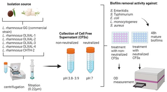
2.1. Microbial Strains

Five wild-type lactic acid bacteria, the technological and functional properties of which have been described in a previous study [17], isolated from Greek natural products, were used, as shown in Table 1. All strains were grown in de Man, Rogosa, and Sharpe broth (MRS) (10 g/L of Bacteriological peptone, 2 g/L of Dipotassium phosphate, 0.05 g/L of Manganese sulfate, 5 g/L of Sodium acetate, 4 g/L of Yeast extract, 20 g/L of Dextrose, 0.2 g/L of Magnesium sulfate, 8 g/L of Beef extract, 1 g/L of Tween80, 2 g/L of Ammonium citrate) (Condalab, Madrid, Spain) at 37 °C for 24 h. The commercial Lacticaseibacillus rhamnosus GG served as the reference strain, since its activity against pathogenic biofilm formation is reported in the literature [18].
Salmonella enterica subsp. enterica ser. Enteritidis FMCC B56 PT4, Salmonella enterica subsp. enterica ser. Τyphimurium DSMZ 554, Escherichia coli ATCC 25922, Listeria monocytogenes NCTC 10527 serotype 4b, and Staphylococcus aureus ATCC 25923 were grown in Brain–Heart Infusion (BHI) broth (2 g/L of Glucose, 10 g/L of Enzymatic digest of animal tissues, 5 g/L of Beef heart infusion, 5 g/L of Sodium chloride, 12.5 g/L of Calf brain infusion, 2.5 g/L of Di-sodium hydrogen phosphate) (Condalab, Madrid, Spain) at 37 °C for 24 h.

2.2. Preparation of LAB CFS (Cell-Free Supernatants)

An overnight bacterial culture of each isolated wild-type LAB strain (109 cfu/mL) was centrifuged at 8500× g, 4 °C for 15 min, and the supernatant (pH 3.8–3.9) was collected. In order to study the neutralized CFSs (pH 7) against bacterial biofilm removal, part of the collected CFSs was neutralized using 5 M of NaOH solution using a pH meter (WTW Ph 330i, WTW, Weilheim, Germany). All CFSs were then sterilized through filtration (0.22 μm, Merck, Darmstadt, Germany) (cell-free supernatant—CFS).

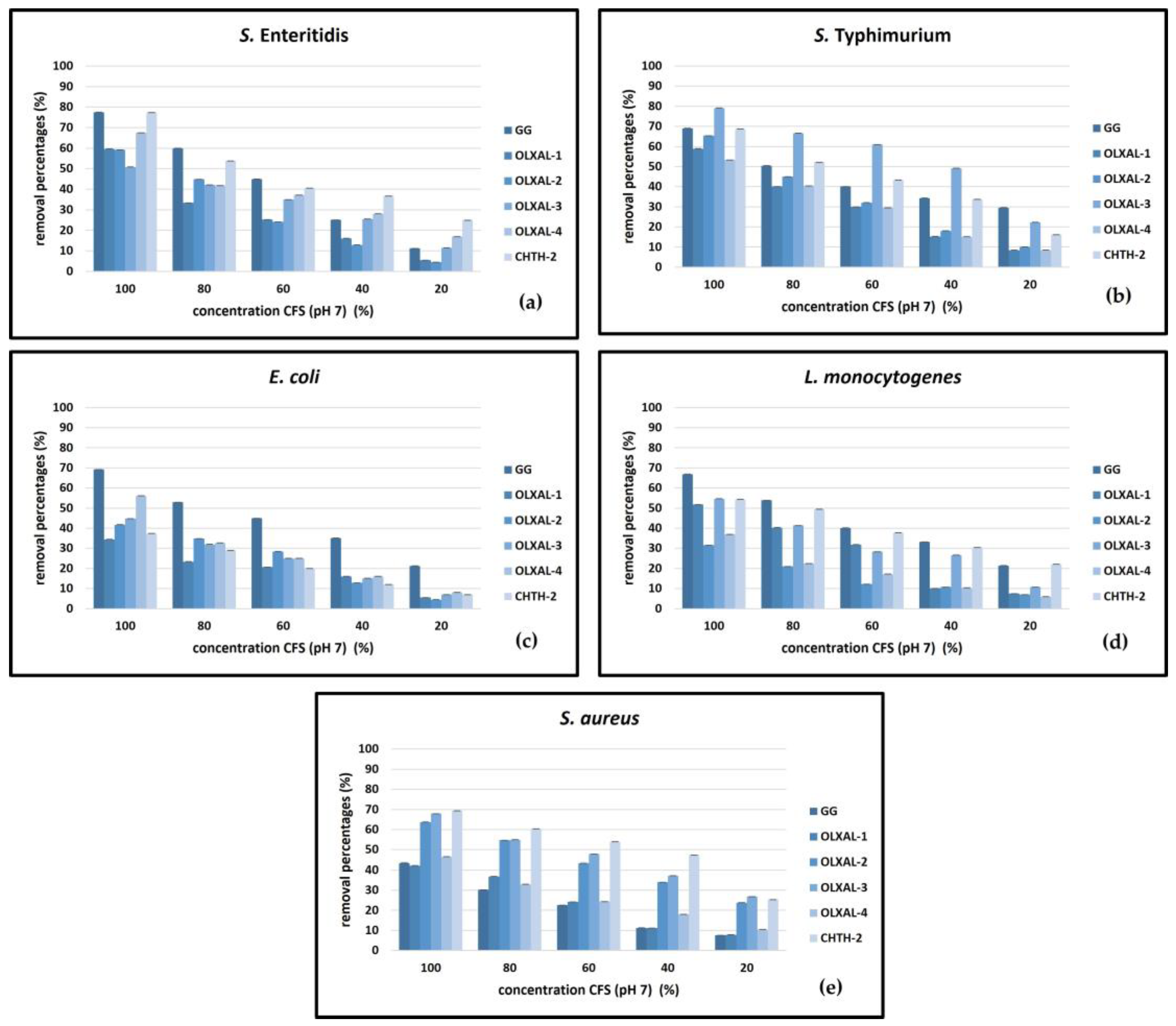
2.3. Biofilm Removal Activity

Biofilm removal activity was determined using 24-well polystyrene microtiter plates, according to Koohestani et al. [19], with some modifications. In each well, 1800 μL of Brain–Heart Infusion broth and 200 μL of bacterial suspension were poured after two ten-fold dilutions to obtain a final concentration of 106 logcfu/mL. For the establishment of the bacterial biofilms, the microplates were incubated at 37 °C for 48 h. After incubation, the free (planktonic) cells were discarded and the wells were washed with ¼ Ringer’s (2.25 g/L of NaCl, 0.105 g/L of KCl, 0.12 g/L of CaCl2H12O6, and 0.05 g/L of NaHCO3) sterilized solution to remove any weakly attached cells. An optical density (OD) measurement of approximately 1.4 after 48 h suggested a relatively dense biofilm formation, as OD values above 1 generally indicate high cell or biofilm densities (Table S1) [20,21].
In the next step, CFSs (neutralized and non-neutralized) of all LAB strains were gently added to the wells at concentrations of 100%, 80%, 60%, 40%, or 20% and left undisturbed for 1 h at ambient temperature (approximately at 24 °C). Then, the CFSs were vigorously decanted and the wells were washed again with ¼ Ringer’s sterilized solution. The remaining attached bacterial biofilms were stabilized with the addition of 2000 μL of methanol solution and allowed to dry for 5 min. Cell staining was performed using 1% w/v crystal violet (Sigma-Aldrich, St. Louis, MO, USA) for 30 min, followed by a final wash with distilled H2O, in order to remove any excess staining solution. The remaining crystal violet at the bottom of the wells was diluted in ethanol/acetone solution (Chem-Lab NV, Zedelgem, Belgium) at a ratio of 80/20 for 15 min. The solutions were then transferred into clear bottom 96-well microplates to determine the optical absorbance at 540 nm. Wells containing only BHI broth and bacterial suspension without CFSs were used as the negative and positive controls, respectively.
The estimation of the reduction percentage of biofilms exposed to the different CFSs was calculated according to the following equation:
Reduction   percentage = C B ( T B ) ( C B ) × 100 ,
where C represents the OD values of positive control wells, B represents the OD values of negative control wells, and T represents the OD values of CFS-treated wells at 540 nm.

2.4. Statistical Analysis

All experiments were performed at least in quadruple, and the results were analyzed for statistical significance with analysis of variance (ANOVA). Duncan’s test was used to determine significant differences (coefficients, ANOVA tables, and significance (p < 0.05) were computed using Statistica for Windows, v.12.5 (StatSoft, Tulsa, OK, USA)).

3. Results and Discussion

This study investigated the biofilm removal potential of cell-free supernatants (CFSs) obtained from five wild-type Lacticaseibacillus rhamnosus strains along with the commercially available L. rhamnosus GG against biofilms formed by common foodborne pathogens. The efficacies of both non-neutralized and neutralized CFSs were assessed, revealing notable strain-specific activities.
A comparison of CFS effectiveness across pathogens revealed that non-neutralized CFSs exhibited significant biofilm removal activity against S. Enteritidis, S. Typhimurium, E. coli, L. monocytogenes, and S. aureus (60–89% removal) (Figure 1 and Figure 2). The neutralization of CFSs led to a reduction in efficacy, but still maintained significant removal activity (32–80% removal) compared to the biofilm removal percentages of non-neutralized CFSs (p < 0.05).
Among the CFSs of the strains tested, the highest (p < 0.05) removal activity was observed with non-neutralized L. rhamnosus OLXAL-3 and OLXAL-4 CFSs against S. Enteritidis (88–89%), S. aureus (84–89%), E. coli (84–87%), and L. monocytogenes (81–84%). Non-neutralized CFS of OLXAL-3 exhibited strong biofilm removal activity against S. Typhimurium (73–88% removal). On the contrary, non-neutralized CFS of OLXAL-4 was not as effective against biofilms of the same pathogen (51–68% removal). Similarly, substantial removal activity of L. rhamnosus OLXAL-2 and OLXAL-3 CFSs against S. aureus biofilms (87–89% removal) was recorded. Non-neutralized L. rhamnosus CHTH-2 CFSs exhibited notable removal efficacy against all pathogens (70–81% removal), but lower (p < 0.05) efficacy compared to non-neutralized OLXAL-3 and OLXAL-4 CFSs. However, moderate biofilm removal percentages were observed for non-neutralized L. rhamnosus OLXAL-1 CFSs, with ranges of 44–69%, 41–69%, 57–75%, 51–76%, and 37–65% against S. Enteritidis, E. coli, S. Typhimurium, L. monocytogenes, and S. aureus biofilms, respectively. Effective biofilm removal activity against specific pathogens was observed with non-neutralized L. rhamnosus GG CFSs (used as the reference), as removal percentages of 46–88%, 55–82%, 57%–86%, 68–86%, and 62–74% against E. coli, L. monocytogenes, S. Enteritidis, S. Typhimurium, and S. aureus biofilms, respectively, were recorded. However, these values were significantly lower (p < 0.05) than OLXAL-3 non-neutralized CFS activity.
Importantly, noteworthy removal activity against both S. Enteritidis and S. Typhimurium (35–51% and 37–67%, respectively) with neutralized L. rhamnosus OLXAL-3 CFSs was also noted. Similarly, both neutralized L. rhamnosus CHTH-2 and OLXAL-2 CFSs demonstrated noteworthy activity against S. aureus biofilms (69% and 64% removal, respectively), while neutralized L. rhamnosus GG CFSs were effective against E. coli and L. monocytogenes biofilms (67–69% removal). Of note, after neutralization, the efficacy of OLXAL-4 CFSs on biofilm removal against all pathogens tested was in the range of 37–67%. In contrast, reduced biofilm removal activity against all pathogens tested (34–59% removal) was observed with neutralized L. rhamnosus OLXAL-1 CFSs.
Different strains exhibited distinct biofilm removal effectiveness against various pathogens, indicating inherent strain-specific bioactivity variations. CFS neutralization led to reduced activity; yet, certain strains retained notable removal activity, suggesting resilience to pH alterations. Although generally high biofilm removal percentages were recorded with L. rhamnosus GG CFSs used as the reference, certain wild-type strains exceeded their efficiency against specific pathogens, suggesting a strain-specific effectiveness, that varied according to the bacterial pathogen.
The above findings are in accordance with previous studies that demonstrated the antimicrobial and biofilm removal activity of LAB CFSs against foodborne pathogens in a concentration-dependent manner [19,22,23]. Notably, it has been recently reported by Nelios et al. [17] that non-neutralized L. rhamnosus CFSs exhibited the growth inhibition of foodborne spoilage and pathogenic microorganisms up to 90%, but their activity was significantly reduced after neutralization. Discrepancies in efficacy between the strains might be attributed to variations in metabolite profiles, including the production of organic acids and bacteriocins that might contribute to biofilm removal, suggesting the need for further investigation into the specific mechanisms of action.
The results of the present study are in agreement with previous research by Divyashree et al. [24], in which the effect of non-neutralized CFSs of Lactobacillus casei MYSRD 108 and Lactobacillus plantarum MYSRD 71 on Salmonella paratyphi bacterial biofilms at a 15% v/v CFS concentration was studied, reporting a removal activity over 75% with the non-neutralized Lactobacillus casei MYSRD 108 CFS and 81% with the non-neutralized Lactobacillus plantarum MYSRD 71 CFS against S. paratyphi biofilms. As expected, neutralized CFSs had significantly lower activity. The greater biofilm removal activity of the non-neutralized CFSs of the two strains was due to the presence of organic acids, according to Divyashree et al. [24]. In another study [23], the non-neutralized CFS of the probiotic strain Weissella confusa WM36 resulted in the removal of 95.68% of Salmonella typhi biofilm at 20% v/v, while the non-neutralized CFS of Weissella viridescens WM33 removed 66.46% of Salmonella Typhimurium biofilms at 15% v/v. Similarly, Tazehabadi et al. [25] studied the biofilm removal activities of Bacillus subtilis KATMIRA 1933 and Bacillus amyloliquefaciens B-1895 CFS against Salmonella enterica subsp. enterica serovar Hadar, Salmonella enterica subsp. enterica serovar Enteritidis phage type 4, and Salmonella enterica subsp. enterica serovar Thompson biofilms. The CFS of Bacillus subtilis KATMIRA 1933 removed 51.1, 48.3, and 56.9% of the biofilms formed by the Salmonella species studied, while the corresponding removal percentages observed with Bacillus amyloliquefaciens B-1895 CFS were 30.4, 28.6, and 35.5%, respectively. According to Tazehabadi et al. [24], the activity against biofilms was not associated with low pH and organic acid production, but with the production of subtilisin peptides, as the pH of CFS was approximately 5.85 for both strains.
Abdelhamid et al. [26] studied the activity of non-neutralized CFSs of six probiotic bacteria of the genera Bifidobacterium and Lacticaseibacillus against biofilms of multi-resistant E. coli WW1 and IC2 strains. The CFSs of B. longum and L. plantarum removed 57.94 and 64.57% of E. coli IC2 and E. coli WW1 biofilms, respectively. Furthermore, the skim milk CFSs fermented with L. helveticus or L. rhamnosus, separately, removed 31.52 and 17.68% of E. coli IC2 biofilms, respectively, while the corresponding removal percentages recorded with B. longum or L. helveticus CFS were 70.81 and 69.49%. In a similar study published by Apiwatsiri et al. [27], neutralized and non-neutralized CFSs of Lactobacillus plantarum 22F, 25F, and Pediococcus acidilactici 72N strains showed significant removal activity against bacterial biofilm strains of E. coli resistant to the antibiotic colistin.
According to a study by Shao et al. [28], non-neutralized CFSs of three Leuconostoc mesenteroides strains effectively removed L. monocytogenes biofilms. Leuconostoc species produce bacteriocins, such as mesentericin Y105, which is formed by Leuconostoc mesenteroides spp. mesenteroides; leucocin A-UAL 187, which is produced by Leuconostoc gelidum; carnosin 44A, which is a metabolic product of Leuconostoc carnosum; and leuconocin S, which is produced by Leuconostoc paramesenteroides [29]. Bacteriocins of Leuconostoc species possess significantly high removal activity against Listeria biofilms. Moreover, Moradi et al. [30] studied the effect of L. acidophilus LA5 and L. casei 431 CFS on L. monocytogenes biofilms. A greater biofilm removal activity was noted for L. acidophilus CFS, and it was found that L. acidophilus CFS contains 1.8% lauric acid, which is considered a possible surfactant; yet, the mechanism of action is not fully understood. According to Moradi et al. [30], and in agreement with our results, the action of LAB CFSs in the removal of bacterial biofilms was strain-specific and due to the presence of compounds, such as exopolysaccharides, organic acids, and surfactants.
In a recent study, Koohestani et al. [19] explored the removal activity of L. acidophilus LA5 and L. casei 431 CFSs against S. aureus bacterial biofilms. In accordance with our findings, their results highlighted the significant removal activity of both CFSs tested in a concentration-dependent manner. Of note, the subsequent neutralization of both Lactobacillus CFSs resulted in reduced activity, which is in agreement with our results, as well as previous reports [31].
This study demonstrated the significant potential of LAB-derived CFSs in mitigating biofilms formed by various foodborne pathogens. Understanding their mechanisms of action and optimizing their applications could lead to valuable interventions in food safety protocols. Future research could explore additional microbial metabolites and their synergistic effects for enhanced biofilm control strategies.

4. Conclusions

A concentration-dependent removal activity (60–89%) of S. Enteritidis, S. Typhimurium, E. coli, L. monocytogenes, and S. aureus biofilms was observed with the CFSs of the wild-type L. rhamnosus strains studied. However, their effectiveness was reduced after neutralization, but still remained significant (32–79% removal activity). Thus, more research is still required to identify the presence of other microbial metabolites, such as bacteriocins, hydrogen peroxide, diacetyl, etc., as well as the expression of specific biofilm genes, and clarify their effect and mechanisms of action in biofilm removal.
Since the food industry is susceptible to the risks associated with biofilm formation and biofilms not only have an impact on food manufacturing operations, but also pose a wide public health risk through the contamination of food products, a better understanding of the mechanisms of their formation in the food chain is essential for developing prevention and control measures.

Supplementary Materials

The following supporting information can be downloaded at: https://www.mdpi.com/article/10.3390/pathogens12121449/s1. Table S1. OD values (540 nm) of positive controls before treatment with Lacticaseibacillus rhamnosus strains CFSs.

Author Contributions

Conceptualization, Y.K.; methodology, G.M.; validation, G.M., V.K., G.N. and Y.K.; formal analysis, G.M. and V.K.; investigation, G.M., V.K. and G.N.; resources, Y.K.; data curation, G.M., V.K., G.N. and Y.K.; writing—original draft preparation, G.M., V.K. and G.N.; writing—review and editing, Y.K.; visualization, G.M., V.K., G.N. and Y.K.; supervision, Y.K.; project administration, Y.K.; funding acquisition, Y.K. All authors have read and agreed to the published version of the manuscript.

Funding

The research work was supported by the Hellenic Foundation for Research and Innovation (H.F.R.I.) under the “First Call for H.F.R.I. Research Projects to support Faculty members and Researchers and the procurement of high-cost research equipment grant” (Project number: HFRI-FM17-2496, acronym: iFUNcultures).

Institutional Review Board Statement

Not applicable.

Informed Consent Statement

Not applicable.

Data Availability Statement

The data presented in this study are available upon request from the corresponding author. The data are not publicly available due to the restrictions of the funding authorities.

Acknowledgments

Salmonella enterica subsp. enterica ser. Enteritidis FMCC B56 PT4 was kindly provided by Nychas G.J.E., the Agricultural University of Athens, Athens, Greece.

Conflicts of Interest

The authors declare no conflict of interest.

References

  1. De Oliveira, D.M.P.; Forde, B.M.; Kidd, T.J.; Harris, P.N.A.; Schembri, M.A.; Beatson, S.A.; Paterson, D.L.; Walker, M.J. Antimicrobial Resistance in ESKAPE Pathogens. Clin. Microbiol. Rev. 2020, 33, 10-1128. [Google Scholar] [CrossRef] [PubMed]
  2. Šušković, J.; Kos, B.; Beganović, J.; Pavunc, A.; Habjanič, K.; Matošić, S. Antimicrobial Activity—The Most Important Property of Probiotic and Starter Lactic Acid Bacteria. Food Technol. Biotechnol. 2010, 3, 296–307. [Google Scholar]
  3. Júnior, W.; Ferrari, Í.; Viana de Souza, J.; Silva, C.; Costa, M.; Dias, F. Characterization and Evaluation of Lactic Acid Bacteria Isolated from Goat Milk. Food Control 2015, 53, 96–103. [Google Scholar] [CrossRef]
  4. Tejero-Sariñena, S.; Barlow, J.; Costabile, A.; Gibson, G.R.; Rowland, I. In Vitro Evaluation of the Antimicrobial Activity of a Range of Probiotics against Pathogens: Evidence for the Effects of Organic Acids. Anaerobe 2012, 18, 530–538. [Google Scholar] [CrossRef]
  5. Ait Ouali, F.; Al Kassaa, I.; Cudennec, B.; Abdallah, M.; Bendali, F.; Sadoun, D.; Chihib, N.-E.; Drider, D. Identification of Lactobacilli with Inhibitory Effect on Biofilm Formation by Pathogenic Bacteria on Stainless Steel Surfaces. Int. J. Food Microbiol. 2014, 191, 116–124. [Google Scholar] [CrossRef]
  6. Aminnezhad, S.; Kasra-Kermanshahi, R. Antibiofilm Activity of Cell-Free Supernatant from Lactobacillus Casei in Pseudomonas Aeruginosa. Feyz 2014, 81, 30–37. [Google Scholar]
  7. Khiralla, G.M.; Mohamed, E.A.H.; Farag, A.G.; Elhariry, H. Antibiofilm Effect of Lactobacillus pentosus and Lactobacillus plantarum Cell-Free Supernatants against Some Bacterial Pathogens. J. Biotech. Res. 2015, 6, 86–95. [Google Scholar]
  8. Bulgasem, B.Y.; Lani, M.N.; Hassan, Z.; Wan Yusoff, W.M.; Fnaish, S.G. Antifungal Activity of Lactic Acid Bacteria Strains Isolated from Natural Honey against Pathogenic Candida Species. Mycobiology 2016, 44, 302–309. [Google Scholar] [CrossRef]
  9. Jain, M.; Stitt, G.; Son, L.; Enioutina, E.Y. Probiotics and Their Bioproducts: A Promising Approach for Targeting Methicillin-Resistant Staphylococcus aureus and Vancomycin-Resistant Enterococcus. Microorganisms 2023, 11, 2393. [Google Scholar] [CrossRef]
  10. Xin, W.-G.; Li, X.-D.; Zhou, H.-Y.; Li, X.; Liu, W.-X.; Lin, L.-B.; Wang, F. Isolation, Antibacterial Characterization, and ATF-Based Preparation of Viable Cells of Lacticaseibacillus paracasei XLK 401 and Its Potential Application in Milk Preservation. J. Dairy Sci. 2023, in press. [Google Scholar] [CrossRef]
  11. Zhang, H.; Zhang, Z.; Li, J.; Qin, G. New Strategies for Biocontrol of Bacterial Toxins and Virulence: Focusing on Quorum-Sensing Interference and Biofilm Inhibition. Toxins 2023, 15, 570. [Google Scholar] [CrossRef] [PubMed]
  12. Khanafari, A.; Porgham, S.H. Investigation of Probiotic Chocolate Effect on Streptococcus Mutans Growth Inhibition. Jundishapur J. Microbiol. 2012, 5, 590–597. [Google Scholar] [CrossRef]
  13. Donlan, R.M. Biofilms: Microbial Life on Surfaces. Emerg. Infect. Dis. 2002, 8, 881–890. [Google Scholar] [CrossRef]
  14. Barzegari, A.; Kheyrolahzadeh, K.; Hosseiniyan Khatibi, S.M.; Sharifi, S.; Memar, M.Y.; Zununi Vahed, S. The Battle of Probiotics and Their Derivatives Against Biofilms. Infect. Drug Resist. 2020, 13, 659–672. [Google Scholar] [CrossRef]
  15. Srey, S.; Jahid, I.K.; Ha, S.-D. Biofilm Formation in Food Industries: A Food Safety Concern. Food Control 2013, 31, 572–585. [Google Scholar] [CrossRef]
  16. Shi, X.; Zhu, X. Biofilm Formation and Food Safety in Food Industries. Trends Food Sci. Technol. 2009, 20, 407–413. [Google Scholar] [CrossRef]
  17. Nelios, G.; Santarmaki, V.; Pavlatou, C.; Dimitrellou, D.; Kourkoutas, Y. New Wild-Type Lacticaseibacillus rhamnosus Strains as Candidates to Manage Type 1 Diabetes. Microorganisms 2022, 10, 272. [Google Scholar] [CrossRef]
  18. Lebeer, S.; De Keersmaecker, S.C.J.; Verhoeven, T.L.A.; Fadda, A.A.; Marchal, K.; Vanderleyden, J. Functional Analysis of luxS in the Probiotic Strain Lactobacillus rhamnosus GG Reveals a Central Metabolic Role Important for Growth and Biofilm Formation. J. Bacteriol. 2007, 189, 860–871. [Google Scholar] [CrossRef]
  19. Kohestani, M.; Moradi, M.; Tajik, H.; Badali, A. Effects of Cell-Free Supernatant of Lactobacillus acidophilus LA5 and Lactobacillus casei 431 against Planktonic Form and Biofilm of Staphylococcus aureus. Vet. Res. Forum 2018, 9, 301. [Google Scholar] [CrossRef]
  20. Fernández-Calderón, M.C.; Hernández-González, L.; Gómez-Navia, C.; Blanco-Blanco, M.T.; Sánchez-Silos, R.; Lucio, L.; Pérez-Giraldo, C. Antifungal and Anti-Biofilm Activity of a New Spanish Extract of Propolis against Candida Glabrata. BMC Complement. Med. Ther. 2021, 21, 147. [Google Scholar] [CrossRef]
  21. Galán-Ladero, M.A.; Blanco-Blanco, M.T.; Hurtado, C.; Pérez-Giraldo, C.; Blanco, M.T.; Gómez-García, A.C. Determination of Biofilm Production by Candida Tropicalis Isolated from Hospitalized Patients and Its Relation to Cellular Surface Hydrophobicity, Plastic Adherence and Filamentation Ability. Yeast 2013, 30, 331–339. [Google Scholar] [CrossRef] [PubMed]
  22. Ben Slama, R.; Kouidhi, B.; Zmantar, T.; Chaieb, K.; Bakhrouf, A. Anti-listerial and Anti-biofilm Activities of Potential Probiotic Lactobacillus Strains Isolated from Tunisian Traditional Fermented Food. J. Food Saf. 2013, 33, 8–16. [Google Scholar] [CrossRef]
  23. Pelyuntha, W.; Chaiyasut, C.; Kantachote, D.; Sirilun, S. Cell-Free Supernatants from Cultures of Lactic Acid Bacteria Isolated from Fermented Grape as Biocontrol against Salmonella Typhi and Salmonella Typhimurium Virulence via Autoinducer-2 and Biofilm Interference. PeerJ 2019, 7, e7555. [Google Scholar] [CrossRef] [PubMed]
  24. Divyashree, S.; Anjali, P.G.; Somashekaraiah, R.; Sreenivasa, M.Y. Probiotic Properties of Lactobacillus casei-MYSRD 108 and Lactobacillus plantarum-MYSRD 71 with Potential Antimicrobial Activity against Salmonella paratyphi. Biotechnol. Rep. 2021, 32, e00672. [Google Scholar] [CrossRef]
  25. Tazehabadi, M.H.; Algburi, A.; Popov, I.V.; Ermakov, A.M.; Chistyakov, V.A.; Prazdnova, E.V.; Weeks, R.; Chikindas, M.L. Probiotic Bacilli Inhibit Salmonella Biofilm Formation Without Killing Planktonic Cells. Front. Microbiol. 2021, 12, 615328. [Google Scholar] [CrossRef] [PubMed]
  26. Abdelhamid, A.G.; Esaam, A.; Hazaa, M.M. Cell Free Preparations of Probiotics Exerted Antibacterial and Antibiofilm Activities against Multidrug Resistant E. coli. Saudi Pharm. J. 2018, 26, 603–607. [Google Scholar] [CrossRef] [PubMed]
  27. Apiwatsiri, P.; Pupa, P.; Yindee, J.; Niyomtham, W.; Sirichokchatchawan, W.; Lugsomya, K.; Shah, A.A.; Prapasarakul, N. Anticonjugation and Antibiofilm Evaluation of Probiotic Strains Lactobacillus plantarum 22F, 25F, and Pediococcus acidilactici 72N Against Escherichia Coli Harboring Mcr-1 Gene. Front. Vet. Sci. 2021, 8, 614439. [Google Scholar] [CrossRef]
  28. Shao, X.; Fang, K.; Medina, D.; Wan, J.; Lee, J.; Hong, S.H. The probiotic, Leuconostoc mesenteroides, inhibits Listeria monocytogenes biofilm formation. J. Food Saf. 2020, 40, e12750. [Google Scholar] [CrossRef]
  29. Stiles, M.E. Bacteriocins Produced by Leuconostoc Species. J. Dairy Sci. 1994, 77, 2718–2724. [Google Scholar] [CrossRef]
  30. Moradi, M.; Mardani, K.; Tajik, H. Characterization and Application of Postbiotics of Lactobacillus Spp. on Listeria Monocytogenes in Vitro and in Food Models. LWT 2019, 111, 457–464. [Google Scholar] [CrossRef]
  31. Mahasneh, A.M.; Hamdan, S.; Mahasneh, S.A. Probiotic Properties of Lactobacillus Species Isolated from Local Traditional Fermented Products. JJBS 2015, 8, 81–87. [Google Scholar] [CrossRef]
Figure 1. Biofilm removal activity of non-neutralized CFSs of five wild-type L. rhamnosus strains in comparison to L. rhamnosus GG against (a) S. Enteritidis, (b) S. Typhimurium, (c) E. coli, (d) L. monocytogenes, and (e) S. aureus.
Pathogens 12 01449 g001
Figure 2. Biofilm removal activity of neutralized (pH, 7) CFSs of five wild-type L. rhamnosus strains in comparison to L. rhamnosus GG against (a) S. Enteritidis, (b) S. Typhimurium, (c) E. coli, (d) L. monocytogenes and (e) S. aureus.
Pathogens 12 01449 g002
Table 1. Wild-type Lactic Acid Bacteria (LAB) used in the present study.
Isolate CodeBacterial SpeciesSource of Isolation
GG (ATCC 53103)Lacticaseibacillus rhamnosusHuman intestines
OLXAL-1Lacticaseibacillus rhamnosusOlive (fruit)
OLXAL-2Lacticaseibacillus rhamnosusOlive (fruit)
OLXAL-3Lacticaseibacillus rhamnosusOlive (fruit)
OLXAL-4Lacticaseibacillus rhamnosusOlive (fruit)
CHTH-2Lacticaseibacillus rhamnosusFeta-type cheese
Disclaimer/Publisher’s Note: The statements, opinions and data contained in all publications are solely those of the individual author(s) and contributor(s) and not of MDPI and/or the editor(s). MDPI and/or the editor(s) disclaim responsibility for any injury to people or property resulting from any ideas, methods, instructions or products referred to in the content.

