Article

Campylobacter jejuni Response When Inoculated in Bovine In Vitro Fecal Microbial Consortia Incubations in the Presence of Metabolic Inhibitors

1
Department of Animal Science, University of Wyoming, Laramie, WY 82071, USA
2
United States Department of Agriculture, Agricultural Research Service, Southern Plains Agricultural Research Center, College Station, TX 77845, USA
3
Agricultural Sciences, Blinn College, Bryan, TX 77833, USA
4
Ruminant Nutrition, Ruminant Microbiology, and Preharvest Food Safety, Department of Animal and Dairy Science, University of Georgia, Athens, GA 30602, USA
5
Meat Science and Animal Biologics Discovery Program, Department of Animal and Dairy Sciences, University of Wisconsin, Madison, WI 53706, USA
*
Author to whom correspondence should be addressed.
Pathogens 2023, 12(12), 1391; https://doi.org/10.3390/pathogens12121391
Submission received: 16 October 2023 / Revised: 11 November 2023 / Accepted: 23 November 2023 / Published: 26 November 2023
(This article belongs to the Collection Campylobacter Infections Collection)

Abstract

:
Infection with the foodborne pathogen Campylobacter is the leading bacterial cause of human foodborne illness in the United States. The objectives of this experiment were to test the hypothesis that mixed microbial populations from the bovine rumen may be better at excluding Campylobacter than populations from freshly voided feces and to explore potential reasons as to why the rumen may be a less favorable environment for Campylobacter than feces. In an initial experiment, C. jejuni cultures inoculated without or with freshly collected bovine rumen fluid, bovine feces or their combination were cultured micro-aerobically for 48 h. Results revealed that C. jejuni grew at similar growth rates during the first 6 h of incubation regardless of whether inoculated with the rumen or fecal contents, with rates ranging from 0.178 to 0.222 h−1. However, C. jejuni counts (log10 colony-forming units/mL) at the end of the 48 h incubation were lowest in cultures inoculated with rumen fluid (5.73 log10 CFUs/mL), intermediate in cultures inoculated with feces or both feces and rumen fluid (7.16 and 6.36 log10 CFUs/mL) and highest in pure culture controls that had not been inoculated with the rumen or fecal contents (8.32 log10 CFUs/mL). In follow-up experiments intended to examine the potential effects of hydrogen and hydrogen-consuming methanogens on C. jejuni, freshly collected bovine feces, suspended in anaerobic buffer, were incubated anaerobically under either a 100% carbon dioxide or 50:50 carbon dioxide/hydrogen gas mix. While C. jejuni viability decreased <1 log10 CFUs/mL during incubation of the fecal suspensions, this did not differ whether under low or high hydrogen accumulations or whether the suspensions were treated without or with the mechanistically distinct methanogen inhibitors, 5 mM nitrate, 0.05 mM 2-bromosulfonate or 0.001 mM monensin. These results suggest that little if any competition between C. jejuni and hydrogen-consuming methanogens exists in the bovine intestine based on fecal incubations.

1. Introduction

Campylobacter has been isolated from most food-producing animals with a particularly high prevalence in swine [1,2] and poultry [3,4,5,6] and ranging from low levels to greater than 89% prevalence in ruminants [7]. Campylobacter spp. can colonize the gastrointestinal tracts of food-producing animals as well as wild and feral animals [2,5]. Campylobacter jejuni is the predominant species associated with poultry and cattle [7]. Campylobacter coli is recognized as the primary species in swine [7]; however, considerable numbers of pigs can be colonized with C. jejuni [8,9].
Campylobacter originating from food animals continues to be a major public health concern and a leading cause of human gastrointestinal diseases both in the United States as well as worldwide [6,10,11]. In addition, in some human cases, Campylobacter has been associated with post-infection involving immune-mediated neuropathies known as Guillian Barré Syndrome [6]. Campylobacter is also recognized as a reservoir of genes that encode for resistance to antibiotics that are important for the clinical treatment of human disease [12]. With the emergence of molecular methodologies such as whole-genome sequencing and more advanced applications of polymerase chain assays for rapid detection, there have been an increasing number of species and subspecies of Campylobacter identified in a wide range of sources [13]. However, C. jejuni remains the cause of most of the human illness cases, followed by C. coli [11].
Campylobacter associated with ruminants represents a potential public health concern along with poultry and swine [2,5]. Dairy sources have been reported to be one of the highest causes of campylobacteriosis in humans, and C. jejuni isolates bearing a close genetic relationship with human strains have been detected in dairy cattle fecal material as well as environmental samples and exhibit increased cases in rural communities with high densities of dairy cattle [14,15]. Further concerns have been raised when some Campylobacter spp. including C. jejuni isolates from dairy farms exhibit antibiotic resistance and/or carry the lipooligosaccharide classes potentially responsible for triggering Guillian Barré Syndrome [16,17]. Beef cattle farms, feedlots, and cow–calf operations have also been proven to be sources of Campylobacter that display antibiotic resistance [18,19,20,21]. This may also be a public health concern as antibiotic-resistant Campylobacter spp. have been isolated from beef products such as beef livers [22,23,24]. In cattle, Campylobacter is more likely to colonize the lower gastrointestinal tract and has exhibited only limited ability to survive in rumen in vitro incubations [25,26]. This would suggest that fecal sources may be more important as a vector for Campylobacter in cattle than the rumen and upper gastrointestinal tract populations. Thus, the objectives of this experiment were to test the hypothesis that mixed microbial populations from the bovine rumen may be better at excluding Campylobacter than populations from freshly voided feces and to explore potential reasons as to why the rumen may be a less favorable environment for Campylobacter than feces.

2. Materials and Methods

2.1. Microbial Sources

A poultry field isolate of Campylobacter jejuni was used in these studies [27]. Campylobacter jejuni used as an inoculum was grown in 18 × 150 mm crimp-top culture tubes containing nonantibiotic supplemented Bolton broth (Oxoid Ltd., Basingstoke, UK) as described by Anderson et al. [28] that was flushed with an microaerobic gas phase (10% CO2, 5% O2, 85% N2) and incubated 24 h at 42 °C to yield approximately 108 colony-forming units (CFUs)/mL as the final concentration. Collections of bovine ruminal and fecal micro-organisms were conducted on the morning of each experiment (10:00 AM) from a ruminally cannulated Jersey cow that was not lactating and grazing on coastal bermudagrass pasture. Rumen contents withdrawn from the cannula were strained through a nylon paint strainer into an insulated container until full and capped immediately to minimize oxygen exposure. Feces collected via rectal palpation were placed into a Nasco Whirl-Pak® (Madison, WI, USA) and immediately closed. Transport of rumen fluid and feces to the laboratory occurred within 30 min of collection. Measurements of pH of undiluted rumen fluid and freshly diluted fecal suspensions (8% w/v diluted in water) using a pH meter ranged between 6.34 to 6.53. Campylobacter status of the freshly collected rumen and fecal samples, was determined by plating 1 mL or 1 g portions of each freshly collected sample combined with 1 mL 0.1 M phosphate buffer (pH 6.5) on Campy Cefex agar as described previously [29]. The presence or absence of Campylobacter colonies was confirmed after 48 h microaerobic incubation. Husbandry procedures for animal care were approved by the Southern Plains Agricultural Research Center’s Animal Care and Use Committee.

2.2. Comparison of C. jejuni in Fecal versus Rumen Mixed Microbial Populations

A Campylobacter jejuni culture grown for 24 h was added (0.35 mL) to a 350 mL batch of freshly prepared Bolton broth amended to contain glucose, cellobiose and xylose (Sigma-Aldrich, St. Louis, MO, USA), each at 0.2% w/v. The carbohydrates were added to the Bolton broth to serve as substrates for rumen micro-organisms. Ten-milliliter volumes of the C. jejuni-inoculated, sugar-amended Bolton broth were then distributed under a continuous flow of microaerobic gas mix (10 % CO2, 5 % O2, 85 % N2) to presterilized 18 × 150 mm crimp-top culture tubes. Triplicate sets of the culture tubes were subsequently inoculated individually (0.2 mL) or jointly (0.1 mL each) with suspensions of the freshly collected gastrointestinal tract populations, previously serially 10-fold-diluted in anaerobic dilution solution [30] to 1:10,000, to compare effects of the bovine rumen, bovine fecal micro-organisms or their combination. The freshly collected bovine rumen and fecal samples were each diluted to deplete endogenous substrate and to dilute to extinction the potential effect of wildtype Campylobacter that may have been present in the samples. Once all additions to the culture tubes were added, the tubes were closed with rubber stoppers, crimped, and incubated at 39 °C for 48 h under the 10% CO2, 5% O2, 85% N2 headspace gas phase. During incubation, samples (1 mL) were collected from each tube at 0, 6, 24 and 48 h for colorimetric measurement of ammonia [31] and enumeration of Campylobacter via plating of serial 10-fold dilutions (in 0.1 M phosphate buffer, pH 6.5) to Campy Cefex agar. Colonies exhibiting typical Campylobacter morphology on the Campy Cefex agar were counted after 48 h of incubation at 42 °C. Representative colonies picked at random during mixed culture studies were confirmed as Campylobacter based on the amplification and detection of the ceuE gene [32]. Additionally, the number of total anaerobes was quantified in the original diluted fecal and ruminal fluid samples used as treatments as well as in samples collected from each culture tube at the end of the 48 h incubation period via plating of serial 10-fold dilutions to anaerobic Brucella Blood Agar (Anaerobe Systems, Morgan Hill, CA, USA) as performed earlier [28]. Dilution and plating of the fluid samples for enumeration of anaerobes and subsequent incubation (39 °C for 48 h) of inoculated Brucella agar were carried out in a Bactron IV Anaerobic Environmental Chamber (Sheldon Manufacturing Inc., Cornelius, OR, USA) under an 85% N2, 15% CO2 and 5% H2 atmosphere.

2.3. Impact of Different Gas Atmospheres and Anti-Methanogenic Treatments on C. jejuni Survivability and Select Incubation Characteristics in Mixed Rumen and Fecal Populations

For experimental incubations with the anti-methanogenic treatments 2-bromosulfonate and nitrate, 80 g of freshly collected feces were suspended in 500 mL anaerobic dilution solution supplemented with 35 g BactoTM casamino acids (Becton, Dickinson and Co., Sparks, MD, USA) to achieve a 14% w/v fecal suspension containing 7% casamino acids. The suspension was mixed vigorously via 15 min of high-speed stirring on a stir plate while continuously under 100% carbon dioxide and then separated into two equal volume batches, with one batch being inoculated with 0.3 mL of a 1:10 dilution of C. jejuni grown overnight but not the other batch. After mixing, 10 mL volumes of each fecal suspension were transferred to 18 × 150 mm glass tubes preloaded without or with 0.2 mL of stock concentrations of 24 mM 2-bromosulfonate, 250 mM nitrate (each in water) or both to achieve initial concentrations at the start of incubation of 0.001, 0.05 or their respective combinations. Depending on the experimental design, all additions were made to the tubes while under a continuous flow of either 100% carbon dioxide or a mixture of 50:50 hydrogen/carbon dioxide with each tube subsequently being closed by a rubber stopper and immediately sealed after introducing all additions. Experimental incubations testing the effects of 0.05 mM 2-bromosulfonate, 0.001 mM sodium monensin (both from Sigma-Aldrich) or their combination were conducted similarly except with a fecal suspension inoculated with 10% w/v of freshly collected feces. Monensin, being poorly soluble in water, was added at 0.1 mL from a 0.1 mM stock solution prepared in ethanol. For consistency, 0.1 mL ethanol (Sigma-Aldrich) was added to untreated controls and 2-bromosulfonate-treated incubations. The fecal suspension contained 7% casamino acids as above, but for these incubations only carbon dioxide was used (100% was the only gas phase tested). For all incubations, tubes were maintained upright with no agitation at 39 °C for 24 h. Samples of 1.5 mL fluid were removed from each tube with separate 1 cc syringes at 0, 6 and 24 h for colony enumeration of Campylobacter on Campy Cefex agar via serial 10-fold dilution of 1 mL aliquots from each sample. Portions of the remaining fluid samples from 0 and 24 h collections were used for colorimetric determination of nitrate concentrations [33]. At the end of the 24 h incubation, 1 mL headspace was collected from each tube and analyzed via gas chromatography on a Gow-Mac gas chromatograph as described previously [34]. Gas volumes in each tube were measured via volume displacement using a 20 mL gas-tight glass syringe and gas concentrations were calculated using the ideal gas law.

2.4. Statistical Analysis

Statistical analyses of Campylobacter counts as log10 transformations were conducted at each sampling time for impact of microbial populations or their mixtures using a general analysis of variance. Ammonia concentrations, pH and log10 transformations of total culturable anaerobes measured at the end of the 48 h incubations were likewise analyzed for effects on microbial populations or their mixtures. In the second experiment, comparisons of the main effects of the gas phase, anti-methanogen treatment, log10 concentrations of C. jejuni or their potential interactions were likewise analyzed via general analysis of variance. Total gas generated, hydrogen and methane final concentrations and the net change in Campylobacter jejuni populations were determined in C. jejuni inoculated cultures. Main effect comparisons of C. jejuni inoculation, treatment and their potential interaction were determined in cultures that had been either inoculated or not inoculated with C. jejuni for total gas produced, final hydrogen, and methane concentrations. To avoid confounding effects of the supplied gas phases, separate comparisons were conducted on incubations initiated under 100% carbon dioxide or the 50:50 hydrogen/carbon dioxide mix. Results from the third experiment testing the effects of 2-bromosulfonate, monensin or their combination were analyzed similarly except, due to the presence of wildtype Campylobacter, the comparisons of the main effects of C. jejuni inoculation and relevant interactions were omitted. When detected, physiologically significant interactions between the gas phase and anti-methanogenic treatments or Campylobacter and anti-methanogenic treatments were noted. When the main effects or interactions were significant, means were separated based on an LSD multiple comparison of means. All analyses were conducted using Statistix version10 Analytical Software (Tallahassee, FL, USA).

3. Results

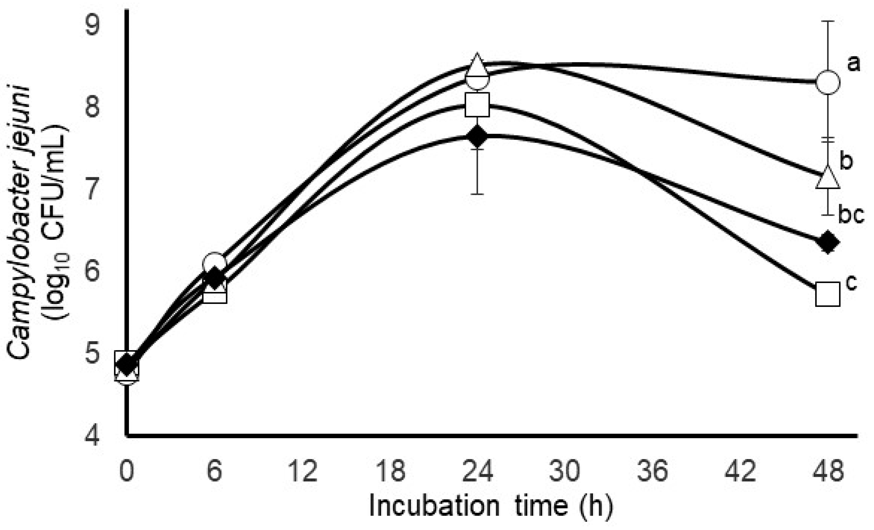
3.1. Comparison of C. jejuni Survival in Mixed Rumen and Bovine Fecal In Vitro Microbial Cultures

No wildtype background Campylobacter were detected in bovine or fecal samples prior to use in these initial set of experiments. The growth curves for C. jejuni in eitherpure culture or experimentally inoculated into the bovine rumen or fecal micro-organisms mixed populations are presented in Figure 1. In support of our hypothesis that rumen micro-organisms may be more antagonistic to C. jejuni than fecal micro-organisms, we observed that the C. jejuni concentrations measured at the end of the 48 h incubation period were more than 2.5 log10 CFUs/mL lower (p < 0.05) in the mixed culture with rumen micro-organisms than in the pure culture and 1.4 log10 units lower in the mixed culture with fecal micro-organisms (Figure 1). The antagonistic effect of the rumen micro-organisms against C. jejuni appeared to be restricted to the later incubation period; however, C. jejuni counts did not differ between the pure or mixed cultures (p > 0.05) when measured at 0, 6 or 24 h of incubation (Figure 1). Similarly, the mean specific growth rates of C. jejuni determined over the first 6 h of incubation did not differ (p > 0.05) between the mixed or pure cultures, further indicating a near-unimpeded growth of C. jejuni during the early incubation periods (Table 1). The concentrations of rumen and fecal anaerobes, initially at 4.0 and 3.0 log10 CFUs/mL at the start of the incubations, did not differ (p > 0.05) between the mixed cultures after 48 h incubation (Table 1). The pH of the mixed population incubations of rumen or fecal micro-organisms or their combinations cultured with C. jejuni, initially at pH 6.50, were lower (p < 0.05) after 48 h incubation than the pure cultures of C. jejuni incubated likewise (Table 1). Based on the 3 to 4 log-fold increase in total anaerobes and the resulting decrease in pH during the incubations, likely due to the greater fermentative activity of the rumen and fecal anaerobes, it is reasonable to suspect that given sufficient time, the anaerobes were able to actively compete against the inoculated C. jejuni. However, net accumulations of ammonia did not differ (p > 0.05) between the pure C. jejuni cultures or any of the mixed microbial populations cultured with C. jejuni, thus revealing no obvious disruption in ammonia production or consumption.

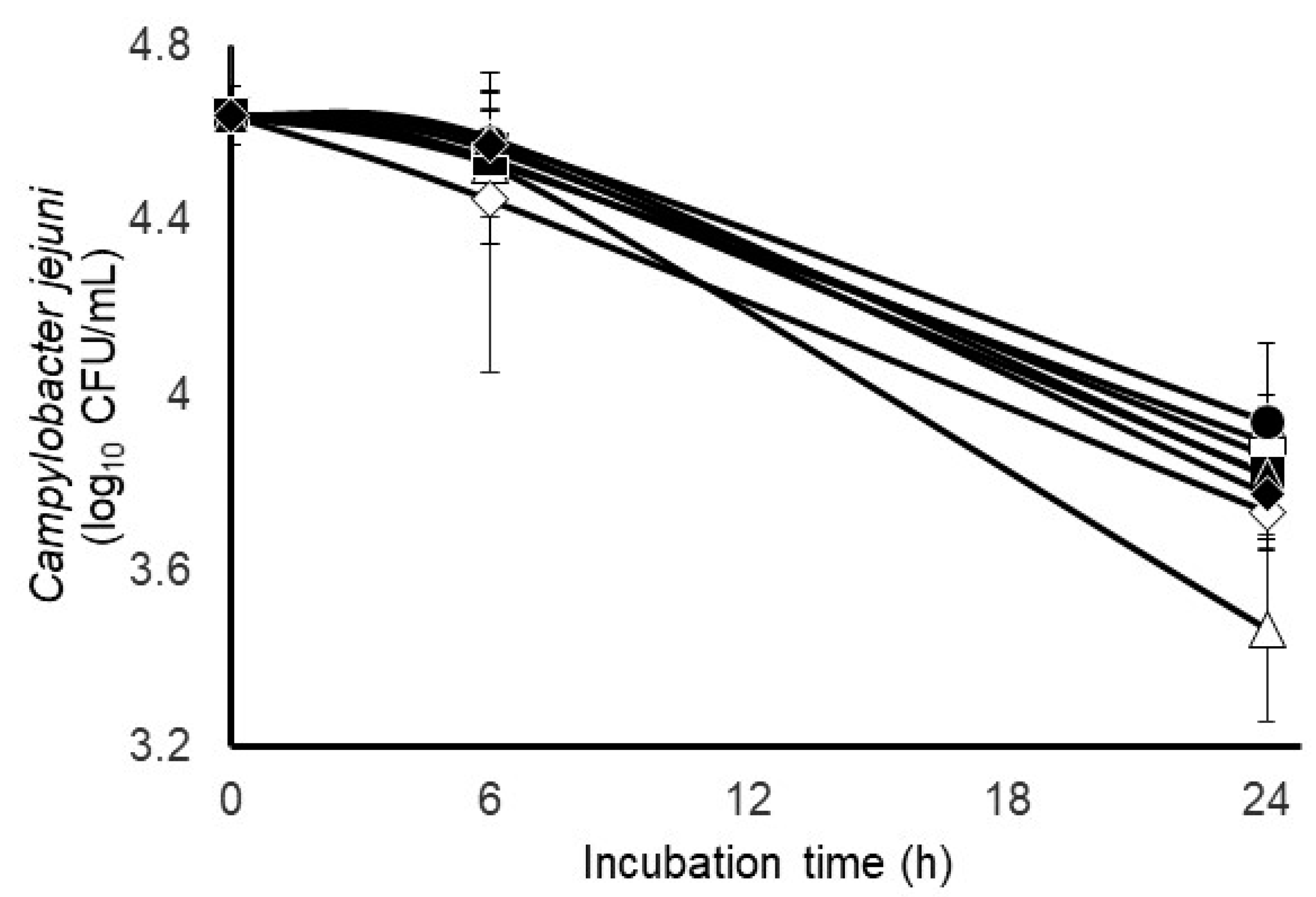
3.2. Impact of Different Gas Atmospheres and Anti-Methanogenic Treatments on C. jejuni Survivability and Select Incubation Characteristics in Mixed Fecal Populations

The quantitative results from the bacteriological culture of the bovine feces collected for this particular experiment revealed the presence of 1.1 × 10⁵ CFUs of wildtype Campylobacter/g of feces; consequently, only mixed fecal populations that had been inoculated with C. jejuni were analyzed and no attempt was made to differentiate the inoculated C. jejuni strain from the wildtype. The survival characteristics of C. jejuni during incubation with mixed populations of bovine fecal microbial populations under two initially different gas phases (carbon dioxide or 50:50 hydrogen/carbon dioxide) and without or with two anti-methanogenic treatments that would be considered mechanistically distinct, as well as their combination, are provided in Figure 2. No main effect of the gas phase was observed on the net change in Campylobacter concentrations after 24 h incubation of the mixed fecal populations. A main effect of gas phase was observed on total gas production, hydrogen and methane accumulation but not metabolized nitrate (Table 2). The amount of total gas volume and methane produced was slight in the fecal incubations. A main effect of anti-methanogen treatment was not observed (p > 0.05) on methane production, but a tendency for a treatment effect was observed on the net change in Campylobacter concentrations, with survivability appearing to be lowest after 24 h (but not 6 h) incubation of the mixed fecal populations treated with 5 mM sodium nitrate (Table 2). Less total gas was also produced by the fecal incubations treated individually with 5 mM nitrate or combined with 0.05 mM 2-bromosulfonate. The main effects of initial gas phase, 100% carbon dioxide or a 50:50 carbon dioxide/hydrogen mix, along with mechanistically different anti-methanogenic compounds bromosulfonate and monensin, administered alone or together to mixed fecal micro-organisms are presented in Table 3. Gas phase or anti-methanogenic compound did not statistically impact (p > 0.05) C. jejuni populations after 6 h, or total gas production, hydrogen. methane, and ammonia accumulation after 24 h.

4. Discussion

Campylobacter spp. have been isolated from a wide range of animals, farm, and urban environments as well as meat-processing plants [5]. Campylobacter are especially known for their high prevalence in cattle, swine, and poultry, leading to concerns over food safety when these animals are slaughtered, with the majority of disease outbreaks occurring from raw or undercooked meat products [5]. Poultry products have been considered one of the primary sources of campylobacteriosis, which is consistent with the ability of Campylobacter to readily colonize gastrointestinal tract populations and interact with the indigenous microbiota [5,35]. However, cattle-based meat products can also serve as a source of Campylobacter and can be detected in ruminants under a range of management conditions [2,36]. However, the ecology and sites of colonization in the gastrointestinal tract are not well known.
Although no indigenous Campylobacter were detected in rumen contents, inoculated C. jejuni could survive in rumen contents (Figure 1). However, based on the results presented in Figure 1, it appears that rumen micro-organisms are more antagonistic to C. jejuni than fecal micro-organisms, as C. jejuni levels at the end of the 48 h incubation period were 1.4 log10 CFUs/mL less in the presence of rumen micro-organisms than when incubated with fecal micro-organisms. This reduction was evident only at the end of the fermentation as no differences in mixed cultures of Campylobacter versus pure cultures were detected at earlier timepoints. Since the total anaerobic microbial populations did not change over time, this decrease may be due to a combination of specific nutrient limitations being reached for Campylobacter after 24 h in mixed cultures, but with sufficient nutrients left to sustain the general microbial rumen and fecal communities and/or the buildup of fermentative products that are antagonistic to Campylobacter. Decreases in pH at 48 h (Table 1) suggest that an accumulation of fermentation products over time that are antagonistic to Campylobacter is possible as organic acids have been identified as one of the mechanisms associated with anti-Campylobacter probiotic cultures [37].
It is less clear whether nutrient limitation was a factor as detectable increases in ammonia did not differ (Table 1) between the pure C. jejuni cultures or any of the mixed microbial populations cultured with C. jejuni. Presumably, amino acid fermentation would lead to increases in ammonia for pure cultures of C. jejuni since Mueller–Hinton agar contains 300 g dehydrated beef extract and 17.5 g casein hydrolysate per liter. However, it is conceivable that either these sources of amino acid were not optimal for C. jejuni to use these pathways or other substrates served as primary sources of carbon and energy. This could also be reflective of C. jejuni’s limited metabolic capabilities. Rath et al. [38] examined the metabolomic profiles of intestinal contents of pigs inoculated with either C. jejuni or C. coli and reported that C. coli could use a wide range of substrates including short-chain fatty acids, fucose, as well as serine and asparagine, while C. jejuni could only use serine. It would be of interest in future mixed fecal culture incubation studies to compare ammonia production from various Campylobacter spp., particularly C. coli versus C. jejuni.
In the current study, in vitro rumen and fecal incubations were used in the presence of methanogen inhibitors and nitrate to determine if there is a relationship between C. jejuni and the methanogenic population as a function of hydrogen. The survival characteristics of C. jejuni were not detectably altered (Figure 2) during incubation with mixed populations of bovine fecal micro-organisms under two initially different gas phases (carbon dioxide or 50:50 hydrogen/carbon dioxide) and without or with two mechanistically different anti-methanogenic treatments, or their combination. Unlike that observed with mixed rumen populations [26], a main effect of the gas phase was not observed on the net change in Campylobacter concentrations after 24 h incubation of the mixed fecal populations. However, a main effect of the gas phase was observed on total gas production, hydrogen and methane accumulation in the fecal incubations (Table 2). Compared to hydrogen accumulation, the relative levels of total gas and methane were modest, but this could have been associated with minimal methanogen activity occurring in fecal contents as opposed to what would typically be seen in rumen incubations. This is consistent with the lack of a detectable impact of anti-methanogen compounds on methane production. However, less total gas was also produced by the fecal incubations treated individually with 5 mM nitrate or combined with 0.05 mM 2-bromosulfonate, while only the combined compounds reduced hydrogen significantly. The anti-methanogenic compounds did apparently decrease Campylobacter’s survivability, with the lowest numerical levels in the fecal incubations treated with 5 mM sodium nitrate (Table 2).
It is not clear whether Campylobacter can become established in the mature rumen, but there is evidence that it can survive under certain circumstances such as in the presence of protozoa [35]. Campylobacter have also been shown to exhibit a trend to increase in the rumens of beef cattle undergoing preslaughter fasting [39]. The factors that influence the presence of Campylobacter in the rumen are unclear but 16S rDNA microbiome taxonomic data from previous poultry in vitro cecal incubation studies detected a potential inverse relationship between methanogens and Campylobacter [40]. In addition, metagenomics profiling of poultry cecal microbiota indicated that Campylobacter possessed uptake hydrogenases and was potentially one of the micro-organisms that could use hydrogen during cecal fermentation [41].
Clearly, Campylobacter represent a serious threat to public health, but, aside from marginally effective hygienic and biosecurity measures, there are few practical interventions for controlling the colonization of food-producing animals with these pathogens [42,43]. From a public health perspective, new strategies are needed to reduce the incidence and concentration of these pathogens both on the farm and during processing. Campylobacter may not be competitive in a rumen environment, but the fact that C. jejuni does survive better in fecal mixed cultures suggests that the lower gastrointestinal tract ecology of Campylobacter colonization in both swine and ruminants may be a primary concern for transmission. In cattle, it has been established in several studies that Campylobacter are more likely to colonize the lower gastrointestinal tract [25]. In swine, Rath et al. [44] demonstrated with weaned pigs either infected with either C. coli and/or C. jejuni that both Campylobacter spp. could colonize the jejunum as well as the cecum. This would suggest that control measures implemented for limiting Campylobacter in swine and ruminants may need to target delivery to the lower gastrointestinal tract of these food animals.
In conclusion, it appears that C. jejuni can survive better in bovine fecal contents compared to rumen contents. While the environmental conditions of the rumen are likely more hostile to Campylobacter, other factors may also contribute to this difference. Certainly, the availability of substrates and potentially more competitive indigenous microbiota could contribute to the hostile nature of the rumen environment. Both the composition of fecal material and the presence of less competitive micro-organisms including hydrogen utilizers may be more favorable to Campylobacter’s survival. The presence of fecal metabolites such as amino acids may represent a greater availability of preferred substrates for Campylobacter metabolism versus rumen metabolite profiles. In addition, the fecal microbial population may be a more supportive microbial consortium that can potentially serve as cross feeders with Campylobacter by generating end-product metabolites that Campylobacter can use as substrates. However, the current study can only suggest potential factors. To determine which of these factors are contributors to Campylobacter survival in bovine fecal material will require 16S rDNA microbiome analyses to determine the microbial populations present and determine if specific relationships exist among certain fecal micro-organisms and Campylobacter levels. Metabolomic analyses would help elucidate which metabolites are present that could serve as potential substrates for Campylobacter metabolism. Combining these approaches in future studies offers an opportunity to better understand the ecology of Campylobacter under these conditions and potentially lead to more targeted mitigation strategies for this pathogen.

Author Contributions

R.C.A. designed the study and developed original manuscript draft; D.K.D. conducted statistical analyses; T.L.P., T.L.C., R.B.H., N.A.K. and T.R.C. contributed to conducting the study; S.C.R. wrote and edited the final draft. All authors have read and agreed to the published version of the manuscript.

Funding

This research received no external funding.

Institutional Review Board Statement

The animal study protocol was approved by the Southern Plains Agricultural Research Center’s Institutional Animal Care and Use Committee (Protocols # 20071 and 2010005).

Informed Consent Statement

Not applicable.

Data Availability Statement

Data is privately held but can be made available upon reasonable request.

Conflicts of Interest

The authors declare no conflict of interest.

References

  1. Pearce, R.A.; Wallace, F.M.; Call, J.E.; Dudley, R.L.; Oser, A.; Yoder, L.; Sheridan, J.J.; Luchansky, J.B. Prevalence of Campylobacter within a swine slaughter and processing facility. J. Food Prot. 2003, 66, 1550–1556. [Google Scholar] [CrossRef] [PubMed]
  2. Moore, J.E.; Corcoran, D.; Dooley, J.S.G.; Fanning, S.; Lucey, B.; Matsuda, M.; McDowell, D.A.; Mégraud, F.; Millar, B.C.; O’Mahony, R.; et al.  Campylobacter. Vet. Res. 2005, 36, 351–382. [Google Scholar] [CrossRef] [PubMed]
  3. Corry, J.E.L.; Atabay, H.I. Poultry as a source of Campylobacter and related organisms. J. Appl. Microbiol. 2001, 90, 96S–114S. [Google Scholar] [CrossRef]
  4. Sahin, O.; Morishita, T.Y.; Zhang, Q. Campylobacter colonization in poultry: Sources of infection and modes of transmission. Anim. Health Res. Rev. 2002, 3, 95–105. [Google Scholar] [CrossRef]
  5. Horrocks, S.M.; Anderson, R.C.; Nisbet, D.J.; Ricke, S.C. Incidence and ecology of Campylobacter in animals. Anaerobe 2009, 15, 18–25. [Google Scholar] [CrossRef]
  6. Umaraw, P.; Prajapati, A.; Verma, A.K.; Pathak, V.; Singh, V.P. Control of Campylobacter in poultry industry from farm to poultry processing unit: A review. Critical Revs. Food Sci. Nutr. 2017, 57, 659–665. [Google Scholar] [CrossRef]
  7. Stanley, K.; Jones, K. Cattle and sheep farms as reservoirs of Campylobacter. J. Appl. Microbiol. 2003, 94, 104S–113S. [Google Scholar] [CrossRef]
  8. Harvey, R.B.; Young, C.R.; Ziprin, R.L.; Hume, M.E.; Genovese, K.J.; Anderson, R.C.; Droleskey, R.E.; Stanker, L.H.; Nisbet, D.J. Prevalence of Campylobacter spp. isolated from the intestinal tract of pigs raised in an integrated swine production system. J. Am. Vet. Med. A 1999, 215, 1601–1604. [Google Scholar]
  9. Jensen, A.N.; Dalsgaard, A.; Baggesen, D.L.; Nielsen, E.M. The occurrence and characterization of Campylobacter jejuni and C. coli in organic pigs and their outdoor environment. Vet. Microbiol. 2006, 116, 96–105. [Google Scholar] [CrossRef]
  10. Natsos, G.; Koutoulis, K.C.; Sossidou, E.; Chemaly, M.; Mouttotou, N.K. Campylobacter spp. infection in humans and poultry. J. Hell. Vet. Med. Soc. 2016, 67, 65–82. [Google Scholar] [CrossRef]
  11. Chlebicz, A.; Śliżewska, K. Campylobacteriosis, salmonellosis, yersiniosis, and listeriosis as zoonotic foodborne diseases: A review. Int. J. Environ. Res. Public Health 2018, 15, 863. [Google Scholar] [CrossRef]
  12. Yang, Y.; Feye, K.M.; Shi, Z.; Pavlidis, H.O.; Kogut, M.; Ashworth, J.A.; Ricke, S.C. A historical review on antibiotic resistance of foodborne Campylobacter. Front. Microbiol. 2019, 10, 1509. [Google Scholar] [CrossRef]
  13. Ricke, S.C.; Feye, K.M.; Chaney, W.E.; Shi, Z.; Pavlidis, H.; Yang, Y. Developments in rapid detection methods for the detection of foodborne Campylobacter in the United States. Front. Microbiol. 2019, 9, 3280. [Google Scholar] [CrossRef] [PubMed]
  14. Grout, L.; Marshall, J.; Hales, S.; Baker, M.G. Dairy cattle density and temporal patterns of human campylobacteriosis and cryptosporidiosis in New Zealand. EcoHealth 2022, 19, 273–289. [Google Scholar] [CrossRef]
  15. An, J.-U.; Ho, H.; Kim, J.; Kim, W.H.; Kim, J.; Lee, S.; Mun, S.-H.; Guk, J.-H.; Hong, S.; Cho, S. Dairy cattle, a potential reservoir of human campylobacteriosis: Epidemiological and molecular characterization of Campylobacter jejuni from cattle farms. Front. Microbiol. 2018, 9, 3136. [Google Scholar] [CrossRef]
  16. Aksomaitiene, J.; Ramonaite, S.; Tamuleviciene, E.; Novoslavskij, A.; Alter, T.; Malakauskas, M. Overlap of antibiotic resistant Campylobacter jejuni MLST genotypes isolated from humans, broiler products, dairy cattle and wild birds in Lithuania. Front. Microbiol. 2019, 10, 1377. [Google Scholar] [CrossRef] [PubMed]
  17. St Charles, J.L.; Brooks, P.T.; Bell, J.A.; Ahmed, H.; Van Allen, M.; Manning, S.D.; Mansfield, L.S. Zoonotic transmission of Campylobacter jejuni to caretakers from sick pen calves carrying a mixed population of strains with and without Guillain Barré Syndrome-associated lipooligosaccharide loci. Front. Microbiol. 2022, 13, 800269. [Google Scholar] [CrossRef]
  18. Bae, W.; Hancock, D.D.; Call, D.R.; Park, Y.H.; Berge, A.C.B.; Finger, R.M.; Sischo, W.M.; Besser, T.E. Dissemination of antimicrobial resistant strains of Campylobacter coli and Campylobacter jejuni among cattle in Washington State and California. Vet. Microbiol. 2007, 122, 306–315. [Google Scholar] [CrossRef]
  19. Bae, W.; Kaya, K.N.; Hancock, D.D.; Call, D.R.; Park, Y.H.; Besser, T.E. Prevalence and antimicrobial resistance of thermophilic Campylobacter spp. from cattle farms in Washington state. Appl. Environ. Microbiol. 2005, 71, 169–174. [Google Scholar] [CrossRef]
  20. Karama, M.K.; Kambuyi, B.T.; Cenci-Goga, M.; Malahlela, A.; Jonker, C.; He, J.; Ombui, T.; Tshuma, T.; Etter, E.; Kalake, A. Occurrence and antimicrobial resistance profiles of Campylobacter jejuni, Campylobacter coli, and Campylobacter upsaliensis in beef cattle on cow–calf operations in South Africa. Foodborne Path. Dis. 2020, 17, 440–446. [Google Scholar] [CrossRef]
  21. Plishka, M.; Sargeant, J.M.; Greer, A.L.; Hookey, S.; Winder, C. The prevalence of Campylobacter in live cattle, turkey, chicken, and swine in the United States and Canada: A systematic review and meta-analysis. Foodborne Path. Dis. 2021, 18, 230–242. [Google Scholar] [CrossRef] [PubMed]
  22. Premarathne, J.M.K.J.K.; Anuar, A.S.; Thung, T.Y.; Satharasinghe, D.A.; Jambari, N.N.; Abdul-Mutalib, N.A.; Huat, J.T.Y.; Basri, D.F.; Rukayadi, Y.; Nakaguchi, Y.; et al. Prevalence and antibiotic resistance against tetracycline in Campylobacter jejuni and C. coli in cattle and beef meat from Selangor, Malaysia. Front. Microbiol. 2017, 8, 2254. [Google Scholar] [CrossRef] [PubMed]
  23. Karki, A.B.; Marasini, D.; Oakey, C.K.; Mar, K.; Fakhr, M.K. Campylobacter coli from retail liver and meat products is more aerotolerant than Campylobacter jejuni. Front. Microbiol. 2018, 9, 2951. [Google Scholar] [CrossRef] [PubMed]
  24. Ghatak, S.; He, Y.; Reed, S.; Irwin, P. Comparative genomic analysis of a multidrug-resistant Campylobacter jejuni strain YH002 isolated from retail beef liver. Foodborne Path. Dis. 2020, 17, 576–584. [Google Scholar] [CrossRef]
  25. Facciolà, A.; Riso, R.; Avventuroso, E.; Visalli, G.; Delia, S.A.; Lagana, P. Campylobacter: From microbiology to prevention. J. Prev. Med. Hyg. 2017, 58, E79–E92. [Google Scholar]
  26. Dittoe, D.K.; Anderson, R.C.; Krueger, N.A.; Harvey, R.B.; Poole, T.L.; Crippen, T.L.; Callaway, T.R.; Ricke, S.C. Survival of Campylobacter jejuni during in vitro culture with mixed bovine ruminal microorganisms in the presence of methanogen inhibitors. J. Environ. Sci. Health B 2023, 58, 711–718. [Google Scholar] [CrossRef]
  27. Byrd, J.A.; Hargis, B.M.; Caldwell, D.J.; Herron, K.L.; McReynolds, J.L.; Brewer, R.L.; Anderson, R.C.; Bischoff, K.M.; Callaway, T.R.; Kubena, L.F. Effect of lactic acid administration in the drinking water during preslaughter feed withdrawal on Salmonella and Campylobacter contamination of broilers. Poult. Sci. 2001, 80, 278–283. [Google Scholar] [CrossRef]
  28. Anderson, R.C.; Krueger, N.A.; Byrd, J.A.; Harvey, R.B.; Callaway, T.R.; Edrington, T.S.; Nisbet, D.J. Effects of thymol and diphenyliodonium chloride against Campylobacter spp. during pure and mixed culture in vitro. J. Appl. Microbiol. 2009, 107, 1258–1268. [Google Scholar] [CrossRef]
  29. Stern, N.J.; Wojton, B.; Kwiater, K. A differential selective medium, and dry-ice generated atmosphere for recovery of Campylobacter jejuni. J. Food Prot. 1992, 55, 514–517. [Google Scholar] [CrossRef]
  30. Bryant, M.P.; Burkey, L.A. Cultural methods and some characteristics of some of the more numerous groups of bacteria in the bovine rumen. J. Dairy Sci. 1953, 36, 205–217. [Google Scholar] [CrossRef]
  31. Chaney, A.L.; Marbach, E.P. Modified reagents for determination of urea and ammonia. Clin. Chem. 1962, 8, 130–132. [Google Scholar] [CrossRef] [PubMed]
  32. Horrocks, S.M.; Jung, Y.S.; Huwe, J.K.; Harvey, R.B.; Ricke, S.C.; Carstens, G.E.; Callaway, T.R.; Anderson, R.C.; Ramlachan, N.; Nisbet, D.J. Effects of short-chain nitrocompounds against Campylobacter jejuni and Campylobacter coli in vitro. J. Food Sci. 2007, 72, M50–M55. [Google Scholar] [CrossRef] [PubMed]
  33. Cataldo, D.A.; Haroon, M.; Schrader, L.E.; Youngs, V.L. Rapid colorimetric determination of nitrate in plant tissue by nitration of salicylic acid. Commun. Soil Sci. Plant Anal. 1975, 6, 71–80. [Google Scholar] [CrossRef]
  34. Anderson, R.C.; Ripley, L.H.; Bowman, J.G.P.; Callaway, T.R.; Genovese, K.J.; Beier, R.C.; Harvey, R.B.; Nisbet, D.J. Ruminal fermentation of anti-methanogenic nitrate- and nitro-containing forages in vitro. Front. Vet. Sci 2016, 3, 62. [Google Scholar] [CrossRef] [PubMed]
  35. Indikova, I.; Humphrey, T.J.; Hilbert, F. Survival with a helping hand: Campylobacter and microbiota. Front. Microbiol. 2015, 6, 1266. [Google Scholar] [CrossRef] [PubMed]
  36. Krueger, N.A.; Anderson, R.C.; Krueger, W.K.; Horne, W.J.; Callaway, T.R.; Edrington, T.S.; Carstens, G.E.; Harvey, R.B.; Nisbet, D.J. Prevalence and concentration of Campylobacter in rumen contents and feces in pasture and feedlot fed cattle. Foodborne Path. Dis. 2008, 5, 571–577. [Google Scholar] [CrossRef]
  37. Deng, W.; Dittoe, D.K.; Pavilidis, H.O.; Chaney, W.E.; Yang, Y.; Ricke, S.C. Current perspectives and potential of probiotics to limit foodborne Campylobacter in poultry. Front. Microbiol. 2020, 11, 583429. [Google Scholar] [CrossRef]
  38. Rath, A.; Rautenschlein, S.; Rzeznitzeck, J.; Lalk, M.; Methling, K.; Rychlik, I.; Peh, E.; Kittler, S.; Waldmann, K.-H.; von Altrock, A. Investigation on the colonisation of Campylobacter strains in the pig intestine depending on available metabolites. Comp. Immunol. Microbiol. Infect. Dis. 2022, 88, 101865. [Google Scholar] [CrossRef]
  39. Welch, C.B.; Lourenco, J.M.; Seidel, D.S.; Krause, T.R.; Rothrock, M.J.; Pringle, T.D.; Callaway, T.R. The impact of pre-slaughter fasting on the ruminal microbial population of commercial Angus steers. Microorganisms 2021, 9, 2625. [Google Scholar] [CrossRef]
  40. Feye, K.M.; Rubinelli, P.M.; Chaney, W.E.; Pavlidis, H.O.; Kogut, M.H.; Ricke, S.C. The preliminary development of an in vitro poultry cecal culture model to evaluate the effects of Original XPCTM for the reduction of Campylobacter jejuni and its potential effects on the microbiota. Front. Microbiol. 2020, 10, 3062. [Google Scholar] [CrossRef]
  41. Sergeant, M.J.; Constantinidou, C.; Cogan, T.A.; Bedford, M.R.; Penn, C.W.; Pallen, M.J. Extensive microbial and functional diversity within the chicken cecal microbiome. PLoS ONE 2014, 9, e91941. [Google Scholar] [CrossRef] [PubMed]
  42. Epps, S.V.R.; Harvey, R.B.; Hume, M.E.; Phillips, T.D.; Anderson, R.C.; Nisbet, D.J. Foodborne Campylobacter: Infections, metabolism, pathogenesis and reservoirs. Int. J. Environ. Res. Public Health. 2013, 10, 6292–6304. [Google Scholar] [CrossRef] [PubMed]
  43. Pattison, M. Practical intervention strategies for Campylobacter. J. Appl. Microbiol. 2001, 90, 121S–125S. [Google Scholar] [CrossRef] [PubMed]
  44. Rath, A.; Rautenschlein, S.; Rzeznitzeck, J.; Breves, G.; Hewicker-Trautwein, M.; Waldmann, K.-H.; von Altrock, A. Impact of Campylobacter spp. on the integrity of the porcine gut. Animals 2021, 11, 2742. [Google Scholar] [CrossRef]
Figure 1. Comparison of growth/survival characteristics of Campylobacter jejuni grown in pure culture (circles) or with mixed populations of bovine rumen micro-organisms (squares), bovine fecal micro-organisms (triangles) or their combination (closed diamonds). Values at each timepoint are least-squares means ± standard deviations from cultures incubated in triplicate. Means with different letter affiliations differ at p < 0.05 based on an LSD multiple comparison of means.
Pathogens 12 01391 g001
Figure 2. Comparison of survival characteristics of experimentally inoculated Campylobacter jejuni during culture with mixed populations of bovine fecal micro-organisms initiated with 100% carbon dioxide (open symbols) or a 50:50 hydrogen/carbon dioxide gas phase and treated without (circles) or with 0.05 mM 2-bromosulfonate (squares), 5 mM sodium nitrate (triangles) or their combination (diamonds). Values at each timepoint are least-squares means ± standard deviation from cultures incubated in triplicate.
Pathogens 12 01391 g002
Table 1. Comparisons of numbers of total culturable anaerobes, pH and ammonia concentrations after 48 h incubation of Campylobacter jejuni with mixed populations of bovine rumen or fecal micro-organisms or their combinations.
Campylobacter jejuni Mean Specific Growth Rate (h−1)Total Culturable Anaerobes
(log10 CFUs/mL)
pHAmmonia
(µmol/mL)
Treatmentsduring initial 6 h incubationAfter 48 h incubation
Campylobacter jejuni only0.222NA 6.42 a1.25
2X Rumen fluid0.1457.136.18 b1.90
2X Feces0.1817.176.16 b1.75
1X Rumen fluid/1X feces0.1787.156.17 b1.83
Treatment effectp = 0.1860 p = 0.7122 p = 0.0474p = 0.1983
Standard error of the mean0.0220.0370.0620.211
NA; not applicable. a, b Means within columns with unlike superscripts differ at p < 0.05 based on an LSD multiple comparison of means.
Table 2. Main effects of initial gas phase, 100% carbon dioxide or a 50:50 carbon dioxide/hydrogen mix, and mechanistically different anti-methanogenic compounds, administered alone or together, on microbial and fermentation parameters during incubation of mixed fecal micro-organisms under a carbon dioxide alone or mixed with hydrogen and inoculated with Campylobacter jejuni to achieve 4.6 ± 0.1 log10 colony-forming units (CFUs)/mL incubation fluid.
Change in Campylobacter jejuni
(log10 CFUs/mL)
Gas
Produced (mL)
Hydrogen Accumulation (µmol/mL)Methane Accumulation (µmol/mL)Nitrate Metabolized (µmol/mL)
ParameterAfter 6 hAfter 24 hAfter 24 hAfter 24 hAfter 24 hAfter 24 h
Initial gas phase
100% carbon dioxide−0.12−0.903.81 a0.52 b0.51 a2.06
50:50 Hydrogen/carbon dioxide−0.07−0.802.11 b32.07 a0.04 b1.99
p value0.46170.21230.0296<0.00010.01390.9233
SEM0.0500.0550.5170.8540.1230.523
Anti-methanogen treatment
No treatment−0.08−0.723.75 x17.260.13ND
0.05 mM 2-Bromosulfonate−0.09−0.804.87 x18.290.21ND
5 mM sodium nitrate−0.08−0.991.50 y15.420.321.81
Combined −0.13−0.881.72 y14.200.432.24
p value0.96280.06130.00110.97730.74650.5678
SEM0.0750.0700.5797.130.2040.514
Comparisons of amounts of nitrate metabolized were made between cultures treated with nitrate alone or in combination with 2-bromosulfonate. ND; not done. a, b Means within columns with different superscripts differ at p < 0.05 based on an LSD multiple comparison of means. x, y Means within columns with different superscripts differ at p < 0.05 based on an LSD multiple comparison of means.
Table 3. Main effects of mechanistically different anti-methanogenic compounds, administered alone or together, on microbial and fermentation parameters during incubation of mixed fecal micro-organisms under a 100% carbon dioxide atmosphere and inoculated with Campylobacter jejuni to achieve 4.6 ± 0.1 log10 colony-forming units (CFUs)/mL incubation fluid.
Change in Campylobacter jejuni
(log10 CFUs/mL)
Gas
Produced (mL)
Hydrogen Accumulation (µmol/mL)Methane Accumulation (µmol/mL)Ammonia Accumulation (µmol/mL)
ParameterAfter 6 hAfter 24 hAfter 24 hAfter 24 hAfter 24 hAfter 24 h
Anti-methanogen treatment
No Treatment−0.16NA18.670.25Undectable0.96
0.05 mM 2-Bromosulfonate−0.08NA17.670.15Undectable0.04
0.001 mM Monensin−0.02NA19.000.54Undectable0.11
Combined −0.14NA18.000.40Undectable0.19
p value0.6080-0.11890.2086-0.0856
SEM0.080-0.3730.130-0.242
NA, not available due to unavoidable interruption of facilities services.
Disclaimer/Publisher’s Note: The statements, opinions and data contained in all publications are solely those of the individual author(s) and contributor(s) and not of MDPI and/or the editor(s). MDPI and/or the editor(s) disclaim responsibility for any injury to people or property resulting from any ideas, methods, instructions or products referred to in the content.

