Article

Molecular Characterisation of M. kansasii Isolates by Whole-Genome Sequencing

by
Priya Rajendran
,
Chandrasekaran Padmapriyadarsini
,
Naveenkumar Nagarajan
,
Roja Samyuktha
,
Vadivu Govindaraju
,
Radhika Golla
,
Shanmugavel Ashokkumar
and
Sivakumar Shanmugam
*
ICMR—National Institute for Research in Tuberculosis, Chennai 600031, India
*
Author to whom correspondence should be addressed.
Pathogens 2023, 12(10), 1249; https://doi.org/10.3390/pathogens12101249
Submission received: 27 July 2023 / Revised: 7 September 2023 / Accepted: 14 September 2023 / Published: 17 October 2023
(This article belongs to the Special Issue Recent Advances in Nontuberculous Mycobacteria (NTM))

Abstract

:
M. kansasii is the most common non-tuberculous mycobacteria, known to be causing pulmonary and extrapulmonary diseases in humans. Based on molecular methods, M. kansasii has been previously classified into seven different subtypes. Now, based on whole-genome sequence analysis, a new species designation was proposed, in which M. kansasii species was designated subtype 1 and is of pathogenic significance in both immunocompetent and immunocompromised patients. The aim of the study is to examine the distribution of subtypes, based on whole-genome sequence analysis, and identify the genetic determinants of drug resistance for the isolates. Whole-genome sequencing was performed using 12 isolates for which phenotypic DST results were available. A phylogenetic tree was constructed by alignment of each of the 12 isolates and the additional strains, as well as the M. kansasii reference strain, using the MAFFT algorithm. Based on this analysis, all 12 isolates were classified as subtype I. Drug-resistant mutations were identified by analysing the isolates with known drug-resistant loci of MTB and NTM. Although we had mutations in the drug-resistant genes, the significance of those mutations could not be explored due to the minimal availability of data available to compare. Further large-scale studies targeting the phenotypic and genotypic drug-resistance pattern, along with whole-genome analysis, will facilitate a better understanding of the resistance mechanisms involved in M. kansasii.

1. Introduction

Non-tuberculous mycobacteria (NTM) are environmental organisms and are known to cause opportunistic infection in humans. They comprise more than 180 species and are soon to be revised as more species are added every year [1]. Despite the fact that many of the NTM species are pathogenic, only a few have been demonstrated to have significant associations with human diseases. M. kansasii is one such species and is usually graded as the second or third most common NTM isolated from patients with lung diseases [2,3]. In addition to pulmonary diseases, M. kansasii is also known to be involved in infection of extrapulmonary sites, including the skin and lymph nodes, and as a disseminated disease. Previously, M. kansasii isolates were classified into seven different subtypes based on PCR and restriction fragment length polymorphisms (RFLPs) of the gene hsp65 [4]. Now, based on whole-genome sequence analysis, it has been demonstrated that each subtype, except subtype 7, parallels new species-level lineages of the M. kansasii complex [5]. Among the new species designated, the M. kansasii species comprises subtype 1 and is of pathogenic significance in both immunocompetent and immunocompromised patients [6], while M. persicum, formerly known as subtype 2, is isolated mainly from HIV-infected patients [5,7]. Other four species, namely M. pseudokansasii (subtype 3), M. ostraviense (subtype 4), M. innocens (subtype 5), and M. attenuatum (subtype 6) are known to be colonisers with no evidence of causing disease in humans [4,5]. Hence, when M. kansasii is identified during NTM diagnosis, insisting further subtype identification will help in understanding its pathogenic significance and appropriate patient management.
According to the ATS/ERS/ESCMID/IDSA guidelines in 2020, treatment of patients with rifampicin (RIF) who are susceptible to M. kansasii pulmonary disease includes a regimen of RIF, ethambutol (EMB), and either isoniazid (INH) or macrolide (either clarithromycin (CLR) or azithromycin) (conditional recommendation). On the contrary, patients with RIF-resistant M. kansasii or intolerance to one of the first-line drugs, fluoroquinolone (e.g., moxifloxacin) is suggested as part of a second-line regimen. [8]. As per CLSI guidelines, drug-susceptibility testing (DST) for M. kansasii is usually carried out for RIF and CLR. RIF resistance (minimum inhibitory concentration (MIC) > 2 µg/mL) is uncommon but can be observed in isolates from patients with prolonged exposure to rifamycin and unsuccessful treatment of a regimen containing rifamycin [9]. Resistance to CLR is determined by an MIC of 32 µg/mL or higher [9]. Susceptibility testing to other drugs (amikacin, ciprofloxacin, doxycycline, linezolid, minocycline, moxifloxacin, rifabutin, and trimethoprim-sulfamethoxazole) is conducted only when RIF resistance is identified [10]. In a study conducted by us previously, we discovered that out of the 18 strains of M. kansasii subjected to DST using the Sensititre method (broth microdilution), doxycycline showed the highest resistance pattern with 13 strains, followed by RIF and trimethoprim/sulfamethoxazole with 7 strains. Out of the 22 strains tested using the proportion sensitivity testing (PST) method, 20 and 10 demonstrated resistance to INH and EMB, respectively. However, there was a poor correlation between the resistance pattern of the drugs tested and the clinical outcome of the patients from whom these strains were isolated [11]. In this study, our objective was to undertake whole-genome sequence analysis of these isolates in order to examine potential genetic determinants of drug resistance and study the distribution of subtypes through phylogenetic analysis.

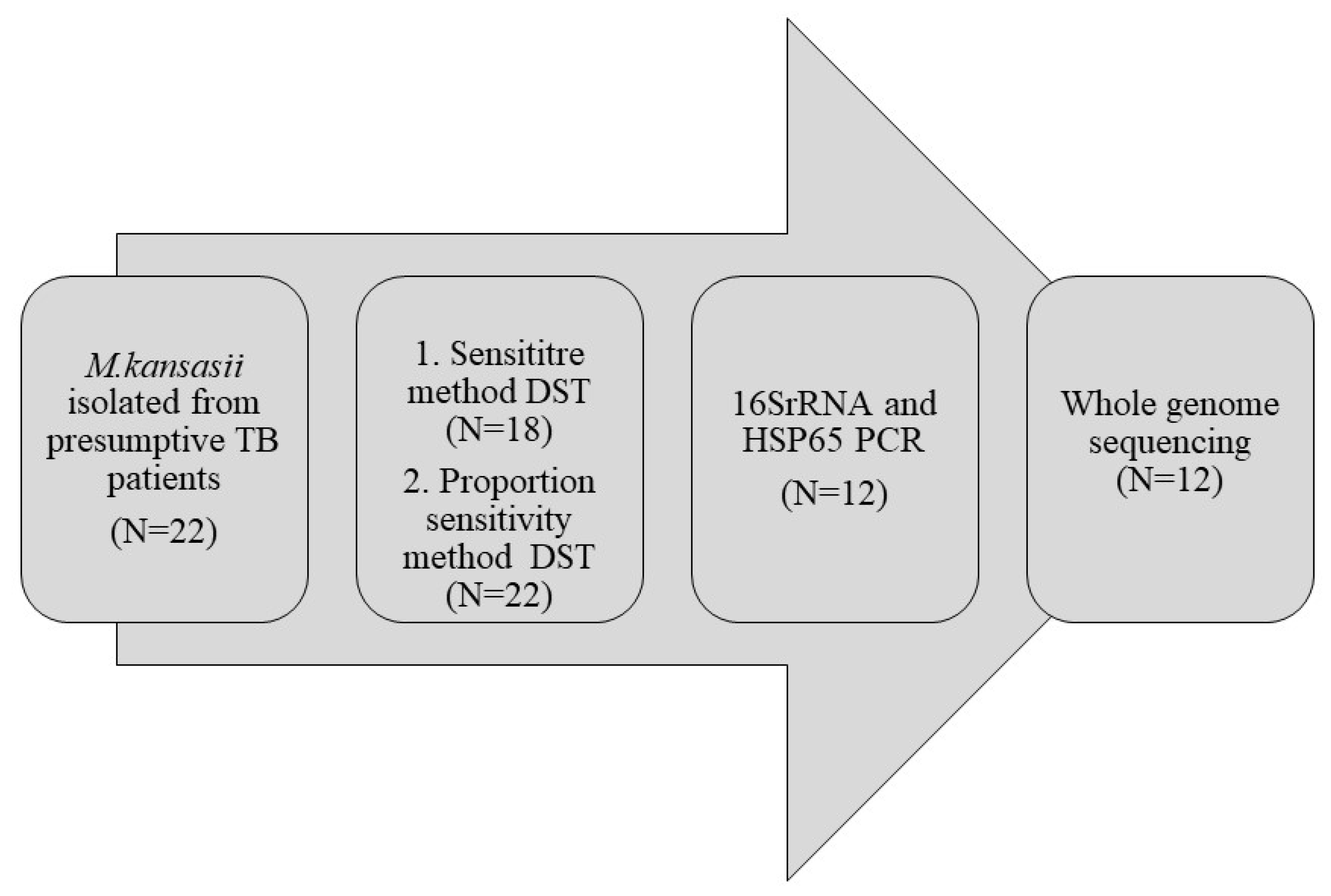
2. Materials and Methods

2.1. Preliminary Genotypic Identification Tests

A total of 12 M. kansasii isolates out of 22 reported earlier were available for this study and were subcultured onto LJ medium for further characterisation. Following conventional species identification methods and biochemical tests, molecular tests targeting the hsp65 and 16SrRNA gene was carried out. Briefly, the genomic DNA was extracted from LJ medium colonies using the CTAB (cetyltrimethylammonium bromide) extraction method [12]. The extracted DNA was subjected to 16SrRNA PCR using the primer sequences 5′-ATGCACCACCTGCACACAGG and 5′-GGTGGTTTGTCGCGTTGTTC and hsp65 PCR using the primer sequences Tbll (5′-ACCAACGATGGTGTGTCCAT) and Tb12 (5′-CTTGTCGAACCG CATACCCT). The reaction mixture and cycling conditions were adapted from the protocol published elsewhere [13,14]. The isolates confirmed by the molecular method were included for whole-genome sequence analysis (Figure 1).

2.2. Genome Sequencing

Genomic DNA extracted from clinical isolates using the CTAB was further purified using the Genomic DNA Clean and Concentrator kit (Zymo Research, Irvine, CA, USA). Purified DNA was assessed for quality and quantity using the Nano DropTM and QubitTM dsDNA assay kit method (ThermoFisher Scientific, Waltham, MA, USA). Sequencing libraries were prepared using the NEBNext Ultra DNA Library preparation kit. In brief, fragmented DNA was subjected to a series of enzymatic steps to repair the ends and tailing with dA-tail, followed by ligation of adapter sequences. Adapter ligated fragments were cleaned up using SPRI beads, and the clean fragments were indexed using limited cycle PCR to generate final libraries for paired-end sequencing on a HiSeq X 10 sequencer (Illumina, San Diego, Ca, USA). Paired-end libraries were prepared from 1 ng of high-quality genomic DNA with the Nextera XT DNA sample preparation kit according to the manufacturer’s instructions (Illumina Inc., San Diego, USA). The libraries were sequenced on a HiSeq 2500 or a NextSeq 500 instrument (Illumina, San Diego, CA, USA) at a read length of 2 × 150 bp.

2.3. Dataset

The dataset of the 12 isolates containing the phenotypic DST results and quality of sequence and alignment is shown in Table 1. The reference sequence of M. kansasii subtype was obtained from NC_022663.1. All 12 isolates were mapped to the reference sequence from the start and endpoint of 1 to 6,432,277.

2.4. Variant Calling

Open-source programmes, namely Genome Analysis Tool Kit (GATK) [15], BWA [16], SAMtools [17] and PICRD-tools, were employed for variant calling. Raw FASTQ sequences were mapped to the reference genome (Mycobacterium kansasii ATCC 12478) using the BWA-MEM algorithm and SAMtools. The mapped reads were sorted, duplicated, and saved in BAM format. GATK was used for the realignment of reads around insertions or deletions. SAMtools mpileup utility was used to call variants with four reads mapped in the forward and reverse direction at an allele frequency of 75% and with a minimum of four calls with a phred score of at least 20.

2.5. Phylogenetic Analysis

Consensus genomes for the 12 isolates were generated by a computational tool (iVar) [18] using default settings. Full-length sequences of 16SrRNA, along with genes such as rpoB, hsp65, tuf and the intergenic spacer region (ITS) of the M. Kansasii reference strain, were downloaded from NCBI and were used as a reference for alignments with the respective sequences of 12 isolates (Table 1) and the additional strains reported in other studies (Table 2). Sequences from the same loci of the consensus genomes were identified using the blastn algorithm. The gene sequences of the reference strains, 12 study strains, and the additional strains reported from other studies were concatenated and aligned using the MAFFT algorithm [19]. These alignments were directly given as the input for computing a maximum-likelihood phylogenetic tree using RAxML-NG version 1.1 [20]. RaxML uses the GTR-GAMMA model of nucleotide substitution, bootstrap, and a randomseed value to construct a phylogenetic tree. A best scoring tree with maximum likelihood is identified with bootstrap search. The branch support was provided by 1000 bootstrap replicates. The revised subtype classification data were obtained from previous studies and compared with our sequences for subtyping [4,21].

2.6. Identification of Mutations in Drug-Resistant Locus

Since a mutation catalogue exclusively for NTM was not available, intragenic drug-resistant locus of MTB from the WHO mutation catalogue [22,23] and drug-resistant locus available for NTM from the literature were obtained [24]. These known loci were analysed in the 12 consensus genomes to identify drug-resistant mutations. Drug-resistant gene sequences from the consensus genomes were translated using an online server [25], and the non-synonymous mutations were identified by alignment with the M. Kansasii reference sequence and the details of the same are provided in the Supplementary Materials.

3. Results

3.1. Identification by Molecular Methods

The product of the 16SrRNA and hsp65 gene with the size of 470 bp and 439 bp was amplified from all the DNA of culture isolates. The hsp65 PCR products were further subjected to RFLP for species identification of M. kansasii (Figure 2).

3.2. Subtype Classification

The phylogenetic tree was constructed using concatenated 16srRNA, rpoB, hsp65, ITS, and tuf genes. Based on this analysis, all 12 isolates were classified as subtype I when compared to the reference sequence and sequences obtained from previous studies based on this analysis (Figure 3, Table 2).

3.3. Association of Mutation with Drug Resistance

The drug-resistant locus of MTB and M. kansasii were aligned with same locus of 12 isolates and some mutations were identified as follows (Table 3).

3.3.1. RIF and INH Resistance

The genes rpoB and katG associated with RIF and INH resistance, respectively, in MTB were analysed for mutations in the M. kansasii isolates. There were no mutations found in comparison with both MTB and M. kansasii reference genomes.

3.3.2. EMB Resistance

Sequence analysis of the EMB-resistance-associated loci (embB and embCA) revealed mutations S272N, S565G, Q853R, and A1007T in the embB gene of 10 isolates when compared to the MTB genome. Out of these isolates, five of them were resistant to EMB, identified previously using the PST method of DST. When the sequences were compared to the M. kansasii reference sequence, one isolate had 5 non-synonymous mutations in the emb gene (Table 3), and this isolate was resistant to EMB according to the phenotypic method. However, the association of these mutations with drug resistance could not be explored since these mutations were not reported elsewhere.
When another resistance-determining region of EMB, aftB, was compared with the MTB reference sequence, three isolates had 9 mutations. Out of these three isolates, one was EMB resistant according to the phenotypic DST. When compared with the M. kansasii reference sequence, one isolate had 18 mutations, and this isolate was sensitive according to the phenotypic DST (Table 3).

3.3.3. Clarithromycin Resistance

Three isolates that were resistant to CLR according to the phenotypic DST did not have any significant mutations when compared with M. kansasii and the MTB reference genome. However, an isolate that was susceptible to CLR had a mutation in the rrl gene at a resistance-determining region (A2089G) when compared to the M. kansasii reference genome.

3.3.4. Quinolone Resistance

Two isolates that were previously reported to be resistant to ciprofloxacin and one isolate to moxifloxacin according to phenotypic DST had no mutations in the quinolone resistance-determining region (QRDR) of the gyrA and gyrB loci when compared with both MTB and the M. kansasii reference genome.

3.3.5. Amikacin Resistance

The sequences of two isolates that were resistant to Amikacin according to phenotypic DST when compared with the MTB sequences at eis genes, no mutations were found. However, there were three non-synonymous mutations (V301I, E348D, D352G) in two other isolates that were sensitive according to phenotypic DST. When compared with the M. kansasii reference sequence, two non-synonymous mutations were found (M293T, V297I) for these isolates.

4. Discussion

Of the NTM diseases reported worldwide, next to the M. avium complex and M. abscessus, M. kansasii is known to be the predominant organism isolated from pulmonary infections in a different geographical area [26,27]. While the prevalence of NTM in India ranged from 0.7% to 34% [28], distribution of M. kansasii among them ranged from 1.5% to 11.8% [29]. Although distribution of M. kansasii isolates have been documented throughout the country, further subtype analysis was not carried out in many studies. In our earlier studies, we reported the isolation of M. kansasii (46.8%) from presumptive pulmonary TB patients and its drug-resistance pattern via phenotypic DST. In this study, we carried out molecular analysis of these isolates [11,30] by hsp65 PCR and 16srRNA PCR and further phylogenetic analysis of the whole-genome sequences.
Out of the 18 M. kansasii isolates obtained, 12 were subjected to whole-genome sequencing, and all the isolates were grouped as subtype I (M. kansasii as per recent classification) [4]. The finding concurs with the fact that M. kansasii is associated with human disease in both immunocompetent and immunocompromised hosts, and in our study, all the patients were immunocompetent. Studies from other parts of the world like China, Europe, the United States, and Japan have also documented the role of subtype I in human infections [21,31], irrespective of the site of infection. Another study from China documented minimal distribution of subtypes (II, III and IV), along with the majority of subtype I associated with pulmonary disease [32].
As per CLSI guidelines, drug-susceptibility testing (DST) for M. kansasii is usually carried out for RIF and CLR. We reported a total of seven RIF-resistant isolates earlier by phenotypic DST in which the MIC was 2 µg/mL for three and 8 µg/mL for four isolates. In the rpoB gene of these isolates when compared to MTB and the M. kansasii reference genome, no mutations were found. Klein et al., have reported that mutations in the rifampin-resistant isolates (n = 5) appeared to be associated with high-level resistance (256 mg/mL). This could be a reason for the RIF-resistant isolates not having any mutations conferred in the rpoB gene. Moreover, other factors like role of the efflux pump and the persistent nature of bacilli due to prolonged exposure to the drug also need to be explored. Further genotypic studies are also needed to confirm the correlation between the mutations and high level of RIF resistance to demonstrate its clinical significance.
We could not find any mutations associated with INH resistance for all the isolates (n = 12) that were resistant to this drug by phenotypic DST. While the INH resistance in MTB is attributed to the role of mutations in the inhA and katG gene, studies documenting the role of these genes leading to INH resistance among NTM is absent [24,33].
Since embB and embCA genes are associated with EMB resistance in MTB, we looked for mutations in these genes with comparison to MTB and the M. kansasii reference genome for all 12 isolates. A total of ten isolates (five resistant and five susceptible by phenotypic DST) revealed four mutations when compared with the MTB genome, and one isolate (EMB resistant by phenotypic DST) had five mutations in comparison to the M. kansasii reference genome. This interesting concurrence between phenotypic and genotypic results for some isolates implicates the need for further genetic studies exploring the association of these mutations with drug resistance. Another study by Bakula et al., reported M306I, G406P, and M423I amino acid substitutions, which were associated with EMB resistance in MTB but not yet reported in NTM [24]. Moreover, as these mutations were found in both EMB-resistant and EMB-susceptible isolates, their specificity for EMB resistance in M. kansasii was not warranted.
M. kansasii isolates that were found to be resistant to CLR previously by phenotypic DST did not have mutations in the rrl gene. In contrast, an isolate susceptible to CLR was found to have a single mutation (A2089G) in the drug-resistant locus. Such a type of single but different mutation (A2266C) in the rrl gene has been reported in a study by Bakula et al., but concurring with phenotypic resistance [24]. However, a study from China reported a mix of concordance patterns where 9 CLR-resistant isolates had mutations at position 2058 and 2059 and discordance patterns where 16 CLR-resistant isolates were not found to have mutations in the rrl gene [32].
The mechanism of drug resistance in NTM could be either acquired in that it is mediated by drugs or by being inherently mediated by the efflux pump or porin channels present in the cell envelope. These porin channels present in the cell envelope may help in the low permeability of the cell wall to external stress due to drugs, resulting in a resistant phenotype [34]. Such mechanisms involved in NTM drug resistance need to be explored through whole-genome sequence analysis studies with a larger set of samples.
A major limitation of this study is the low number of isolates that were subjected to sequencing for the determination of drug-resistance genes. Another limitation is although we could identify few mutations associated with drug resistance, the phenotypic or genotypic drug-resistance patterns could not be correlated with treatment outcome. Without this correlation, it is difficult to mention if the mutation seen in sequence analysis has any relevance.

5. Conclusions

To our knowledge, this study is the first report from India on whole-genome sequence analysis of M. kansasii on its subtype distribution and drug-resistance association. Although we had many mutations in the drug-resistant genes, with the given minimal number of such studies published elsewhere, we could not compare these mutations (except for few) to explore its significance. Further studies involving both drug-susceptibility testing and whole-genome sequence analysis of M. kansasii isolates inclusive of structural inferences are needed to understand the molecular mechanisms involved in drug resistance and to further demonstrate its clinical significance.

Supplementary Materials

The following supporting information can be downloaded at: https://www.mdpi.com/article/10.3390/pathogens12101249/s1, Supplementary file: Alignment of isolates with M. kansasii reference sequence showing mutation in drug resistant loci.

Author Contributions

P.R.: Idea and conceptualization, methodology, data acquisition, Writing— draft preparation. C.P.: Writing—review and editing. N.N.: Data acquisition, Data analysis. R.S.: Data analysis and interpretation. V.G.: Methodology. R.G.: Methodology. S.A.: Methodology. S.S.: Methodology, Data acquisition and analysis, Writing—review, and editing. All authors have read and agreed to the published version of the manuscript.

Funding

Funded by ICMR extramural grant.

Institutional Review Board Statement

EC/IEC approval was obtained, informed consent taken, and the study followed ICMR National Ethical Guidelines and other applicable guidelines and regulations. Date of approval -03/11/2014. IEC approval number- IEC-2014020. The study was approved by Institutional (ICMR–National Institute for Research in Tuberculosis) ethics committee (IEC number 2014020).

Informed Consent Statement

Informed consent was obtained from all subjects involved in the study.

Data Availability Statement

Not applicable.

Conflicts of Interest

The authors declare no conflict of interest.

References

  1. Tortoli, E. Microbiological features and clinical relevance of new species of the genus Mycobacterium. Clin. Microbiol. Rev. 2014, 27, 727–752. [Google Scholar] [CrossRef]
  2. Field, S.K.; Cowie, R.L. Lung disease due to the more common nontuberculous mycobacteria. Chest 2006, 129, 1653–1672. [Google Scholar] [CrossRef] [PubMed]
  3. Prevots, D.R.; Marras, T.K. Epidemiology of human pulmonary infection with nontuberculous mycobacteria: A review. Clin. Chest Med. 2015, 36, 13–34. [Google Scholar] [CrossRef] [PubMed]
  4. Jagielski, T.; Borowka, P.; Bakula, Z.; Lach, J.; Marciniak, B.; Brzostek, A.; Dziadek, J.; Dziurzynski, M.; Pennings, L.; van Ingen, J.; et al. Genomic Insights Into the Mycobacterium kansasii Complex: An Update. Front. Microbiol. 2019, 10, 2918. [Google Scholar] [CrossRef] [PubMed]
  5. Tagini, F.; Aeby, S.; Bertelli, C.; Droz, S.; Casanova, C.; Prod’hom, G.; Jaton, K.; Greub, G. Phylogenomics reveal that Mycobacterium kansasii subtypes are species-level lineages. Description of Mycobacterium pseudokansasii sp. nov., Mycobacterium innocens sp. nov. and Mycobacterium attenuatum sp. nov. Int. J. Syst. Evol. Microbiol. 2019, 69, 1696–1704. [Google Scholar] [CrossRef]
  6. Taillard, C.; Greub, G.; Weber, R.; Pfyffer, G.E.; Bodmer, T.; Zimmerli, S.; Frei, R.; Bassetti, S.; Rohner, P.; Piffaretti, J.C.; et al. Clinical implications of Mycobacterium kansasii species heterogeneity: Swiss National Survey. J. Clin. Microbiol. 2003, 41, 1240–1244. [Google Scholar] [CrossRef]
  7. Shahraki, A.H.; Trovato, A.; Mirsaeidi, M.; Borroni, E.; Heidarieh, P.; Hashemzadeh, M.; Shahbazi, N.; Cirillo, D.M.; Tortoli, E. Mycobacterium persicum sp. nov., a novel species closely related to Mycobacterium kansasii and Mycobacterium gastri. Int. J. Syst. Evol. Microbiol. 2017, 67, 1766–1770. [Google Scholar] [CrossRef]
  8. Daley, C.L.; Iaccarino, J.M.; Lange, C.; Cambau, E.; Wallace, R.J., Jr.; Andrejak, C.; Böttger, E.C.; Brozek, J.; Griffith, D.E.; Guglielmetti, L.; et al. Treatment of Nontuberculous Mycobacterial Pulmonary Disease: An Official ATS/ERS/ESCMID/IDSA Clinical Practice Guideline. Clin. Infect. Dis. 2020, 71, e1–e36. [Google Scholar] [CrossRef]
  9. CLSI. Performance Standards for Susceptibility Testing of Mycobacteia, Nocardia spp., and Other Aerobic Actinonmyces; Clinical and Laboratory Standards Institute: Wayne, PA, USA, 2018. [Google Scholar]
  10. M24-A2; Susceptibility Testing of Mycobacteria, Nocardiae, and Other Aerobic Actinomycetes—2nd Edition. Clinical and Laboratory Standards Institute: Wayne, PA, USA, 2011.
  11. Rajendran, P.; Padmapriyadarsini, C.; Vijayaraghavan, V.; Manoharan, T.; Lokanathan, L.M.; Kadhar, P.B.; Jayabal, L.; Sivaramakrishnan, G. Drug susceptibility profiling of pulmonary Mycobacterium kansasii and its correlation with treatment outcome. Ann. Thorac. Med. 2021, 16, 323–328. [Google Scholar]
  12. De Almeida, I.N.; da Silva Carvalho, W.; Rossetti, M.L.; Costa, E.R.D.; De Miranda, S.S. Evaluation of six different DNA extraction methods for detection of Mycobacterium tuberculosis by means of PCR-IS6110: Preliminary study. BMC Res. Notes 2013, 6, 561. [Google Scholar] [CrossRef]
  13. Mendum, T.A.; Chilima, B.Z.; Hirsch, P.R. The PCR amplification of non-tuberculous mycobacterial 16S rRNA sequences from soil. FEMS Microbiol. Lett. 2000, 185, 189–192. [Google Scholar] [CrossRef] [PubMed]
  14. Telenti, A.; Marchesi, F.; Balz, M.; Bally, F.; Böttger, E.C.; Bodmer, T. Rapid identification of mycobacteria to the species level by polymerase chain reaction and restriction enzyme analysis. J. Clin. Microbiol. 1993, 31, 175–178. [Google Scholar] [CrossRef] [PubMed]
  15. Van der Auwera, G.A.; Carneiro, M.O.; Hartl, C.; Poplin, R.; Del Angel, G.; Levy-Moonshine, A.; Jordan, T.; Shakir, K.; Roazen, D.; Thibault, J.; et al. From FastQ data to high confidence variant calls: The Genome Analysis Toolkit best practices pipeline. Curr. Protoc. Bioinform. 2013, 43, 11.10.1–11.10.33. [Google Scholar] [CrossRef]
  16. Li, H.; Durbin, R. Fast and accurate short read alignment with Burrows-Wheeler transform. Bioinformatics 2009, 25, 1754–1760. [Google Scholar] [CrossRef] [PubMed]
  17. Li, H.; Handsaker, B.; Wysoker, A.; Fennell, T.; Ruan, J.; Homer, N.; Marth, G.; Abecasis, G.; Durbin, R.; 1000 Genome Project Data Processing Subgroup. The Sequence Alignment/Map format and SAMtools. Bioinformatics 2009, 25, 2078–2079. [Google Scholar] [CrossRef]
  18. Moshiri, N. ViralConsensus: A fast and memory-efficient tool for calling viral consensus genome sequences directly from read alignment data. Bioinformatics 2023, 39, btad317. [Google Scholar] [CrossRef]
  19. Katoh, K.; Misawa, K.; Kuma, K.I.; Miyata, T. MAFFT: A novel method for rapid multiple sequence alignment based on fast Fourier transform. Nucleic Acids Res. 2002, 30, 3059–3066. [Google Scholar] [CrossRef]
  20. Stamatakis, A. RAxML version 8: A tool for phylogenetic analysis and post-analysis of large phylogenies. Bioinformatics 2014, 30, 1312–1313. [Google Scholar] [CrossRef]
  21. Guo, Y.; Cao, Y.; Liu, H.; Yang, J.; Wang, W.; Wang, B.; Li, M.; Yu, F. Clinical and Microbiological Characteristics of Mycobacterium kansasii Pulmonary Infections in China. Microbiol. Spectr. 2022, 10, e0147521. [Google Scholar] [CrossRef]
  22. World Health Organization. Catalogue of Mutations in Mycobacterium Tuberculosis Complex and Their Association with Drug Resistance; World Health Organization: Geneva, Switzerland, 2021. [Google Scholar]
  23. Walker, T.M.; Miotto, P.; Köser, C.U.; Fowler, P.W.; Knaggs, J.; Iqbal, Z.; Hunt, M.; Chindelevitch, L.; Farhat, M.R.; Cirillo, D.M.; et al. The 2021 WHO catalogue of Mycobacterium tuberculosis complex mutations associated with drug resistance: A genotypic analysis. Lancet Microbe 2022, 3, e265–e273. [Google Scholar] [CrossRef]
  24. Bakuła, Z.; Modrzejewska, M.; Pennings, L.; Proboszcz, M.; Safianowska, A.; Bielecki, J.; van Ingen, J.; Jagielski, T. Drug Susceptibility Profiling and Genetic Determinants of Drug Resistance in Mycobacterium kansasii. Antimicrob. Agents Chemother. 2018, 62, e01788-17. [Google Scholar] [CrossRef]
  25. Bikandi, J.; Millán, R.S.; Rementeria, A.; Garaizar, J. In silico analysis of complete bacterial genomes: PCR, AFLP-PCR and endonuclease restriction. Bioinformatics 2004, 20, 798–799. [Google Scholar] [CrossRef] [PubMed]
  26. Brown-Elliott, B.A.; Wallace, R.J., Jr. Infections caused by nontuberculous mycobacteria other than Mycobacterium avium complex. In Mandell, Douglas, and Bennett’s Principles and Practice of Infectious Diseases, 8th ed.; Bennett, J.E., Dolin, R., Blaser, M.J., Eds.; Elsevier: Philadelphia, PA, USA, 2015. [Google Scholar]
  27. Rivero-Lezcano, O.M.; Gonzalez-Cortes, C.; Mirsaeidi, M. The unexplained increase of nontuberculous mycobacteriosis. Int. J. Mycobacteriol. 2019, 8, 1–6. [Google Scholar] [CrossRef] [PubMed]
  28. Kalpana, T.; Mugunthan, M.; Joseph, N.M.; Ellappan, K. A Comprehensive Review and Update on Epidemiology, Symptomatology and Management of Nontuberculous Mycobacteria (NTM). J. Pure Appl. Microbiol. 2022, 16, 814–824. [Google Scholar] [CrossRef]
  29. Sharma, S.K.; Upadhyay, V. Epidemiology, diagnosis & treatment of non-tuberculous mycobacterial diseases. Indian J. Med. Res. 2020, 152, 185–226. [Google Scholar]
  30. Gomathy, N.S.; Padmapriyadarsini, C.; Silambuchelvi, K.; Nabila, A.; Tamizhselvan, M.; Banurekha, V.V.; Lavanya, J.; Chandrasekar, C. Profile of patients with pulmonary non-tuberculous mycobacterial disease mimicking pulmonary tuberculosis. Indian J. Tuberc. 2019, 66, 461–467. [Google Scholar] [CrossRef] [PubMed]
  31. Zhang, Y.; Mann, L.B.; Wilson, R.W.; Brown-Elliott, B.A.; Vincent, V.; Iinuma, Y.; Wallace, R.J., Jr. Molecular analysis of Mycobacterium kansasii isolates from the United States. J. Clin. Microbiol. 2004, 42, 119–125. [Google Scholar] [CrossRef]
  32. Li, Y.; Pang, Y.; Tong, X.; Zheng, H.; Zhao, Y.; Wang, C. Mycobacterium kansasii Subtype I Is Associated with Clarithromycin Resistance in China. Front. Microbiol. 2016, 7, 2097. [Google Scholar] [CrossRef]
  33. Jagielski, T.; Bakuła, Z.; Roeske, K.; Kamiński, M.; Napiórkowska, A.; Augustynowicz-Kopeć, E.; Zwolska, Z.; Bielecki, J. Detection of mutations associated with isoniazid resistance in multidrug-resistant Mycobacterium tuberculosis clinical isolates. J. Antimicrob. Chemother. 2014, 69, 2369–2375. [Google Scholar] [CrossRef]
  34. Nasiri, M.J.; Haeili, M.; Ghazi, M.; Goudarzi, H.; Pormohammad, A.; Imani Fooladi, A.A.; Feizabadi, M.M. New Insights in to the Intrinsic and Acquired Drug Resistance Mechanisms in Mycobacteria. Front. Microbiol. 2017, 8, 681. [Google Scholar] [CrossRef]
Figure 1. Workflow of experiments carried out for M. kansasii isolates.
Pathogens 12 01249 g001
Figure 2. HSP65 PCR (a) and 16srRNA PCR (b) for the 12 isolates that were subjected to sequencing. Lane M: Marker.
Pathogens 12 01249 g002
Figure 3. Phylogenetic tree based on 16srRNA, rpoB, hsp65, ITS, and tuf gene sequences constructed using neighbour-joining method (* former classification).
Pathogens 12 01249 g003
Table 1. Mapping and coverage of NTM isolates with M. kansasii reference sequence.
S. No.Lab No.Resistance to Drugs by Phenotypic DSTGene Mutations ComparisonTreatment OutcomeAlignment Coverage
MTB
Genome
M. kansasii
Genome
No. of ReadsCoverageMean Depth
1NT 08 RIF/SXT/DOXembBNo mutationDied5,067,05599.650238.1
2NT 09EMBembBNo mutationCured4,350,93199.722638.2
3NT 12 CLR/DOXembB, aftBaftBCured4,199,38698.822637.9
4NT 13RIF/SXT/DOX/AMK/LZD/CIP/EMBembB, aftBNo mutationRelapse4,701,85698.819838.1
5NT 16No resistanceembB, eiseisCured4,740,11699.677137.9
6NT 22EMBembBembBCured4,626,35399.301838
7NT 27 DOX/EMBembBNo mutationDied4,862,09999.946838
8NT 28 SXT/DOX/
EMB
embB, eisNo mutationCured4,557,19399.689538.1
9NT 35No resistanceaftBNo mutationCured3,844,92099.319938.3
10NT 43 CLR/RFB/RIF/SXT/AMK/
DOX
embBNo mutationCured4,094,94399.635338.1
11NT 33No resistanceembBNo mutationCured4,372,57899.671438.1
12NT 47EMBNo mutationrrlCured1,856,34599.557840.9
Table 2. Accession details of M. kansasii genomes used in phylogenetic analysis (* former classification).
S. No.StrainFormer kansasii SubtypeGenBank No.
1ATCC 12478(M. kansasii) INC_022663.1_I *
2MK7(M. kansasii) IGCA_900565995.1
3Kuro-I(M. kansasii) IGCA_014701265.1
412MK(M. persicum) IINZ_MWQA01000001.1_II *
51010001469(M. persicum) IILWCM00000000.1
63MK(M. persicum) IIMWKX01.1
7MK142(M. pseudokansasii) IIINZ_UPHU01000001.1_III *
8732(M. pseudokansasii) IIIJANZ01.1
9174_15_11(M. pseudokansasii) IIINKRD01.152
10FDAARGOS_1613(M. ostraviense) IVNZ_CP089224.1_IV *
11241/15(M. ostraviense) IVGCA_002705925.1
121010001458(M. ostraviense) IVGCA_001632895.1
13MK21(M. innocens) VNZ_UPHQ01000197.1_V *
1449_11(M. innocens) VNKRC01.1
151010001454(M. innocens) VLWCH01.1
16MK41(M. attenuatum) VINZ_UPHT01000123.1_VI *
17MK191(M. attenuatum) VIUPHS01.1
18MK136(M. attenuatum) VIUPHP01.1
Table 3. List of mutations identified in the study isolates in correspondence to MTB and M. kansasii reference genome.
Target DrugDrug-Resistant LocusMutation in Comparison to MTB Reference SequenceMutation in Comparison to M. kansasii Reference Sequence
Amikacineis/MKAN_RS04925V301IM293T
E348DV297I
D352G
EthambutolembBS272NL78M
S565GG130A
Q853RA159G
A1007TA259T
Y737N
aftBS159AV100A
I202VV107A
S238GM127V
R401HA133V
M491LM192L
V511AF257L
A516QM331V
A524GV339L
Q561RI354V
K399R
V393L
L394S
G412V
E435D
T515S
I541L
K603R
S657P
Disclaimer/Publisher’s Note: The statements, opinions and data contained in all publications are solely those of the individual author(s) and contributor(s) and not of MDPI and/or the editor(s). MDPI and/or the editor(s) disclaim responsibility for any injury to people or property resulting from any ideas, methods, instructions or products referred to in the content.

