Article

Exploring the Biofilm Formation Capacity in S. pseudintermedius and Coagulase-Negative Staphylococci Species

by
Vanessa Silva
1,2,3,4,
Elisete Correia
5,
José Eduardo Pereira
6,7,
Camino González-Machado
8,9,
Rosa Capita
8,9,
Carlos Alonso-Calleja
8,9,
Gilberto Igrejas
2,3,4,† and
Patrícia Poeta
1,6,7,*,†
1
Microbiology and Antibiotic Resistance Team (MicroART), Department of Veterinary Sciences, University of Trás-os-Montes and Alto Douro (UTAD), 5000-801 Vila Real, Portugal
2
Department of Genetics and Biotechnology, University of Trás-os-Montes and Alto Douro (UTAD), 5000-801 Vila Real, Portugal
3
Functional Genomics and Proteomics Unit, University of Trás-os-Montes and Alto Douro (UTAD), 5000-801 Vila Real, Portugal
4
LAQV-REQUIMTE, Department of Chemistry, NOVA School of Science and Technology, Universidade Nova de Lisboa, 2829-516 Caparica, Portugal
5
Center for Computational and Stochastic Mathematics (CEMAT), Department of Mathematics, University of Trás-os-Montes and Alto Douro (UTAD), 5000-801 Vila Real, Portugal
6
CECAV—Veterinary and Animal Research Centre, University of Trás-os-Montes and Alto Douro (UTAD), 5000-801 Vila Real, Portugal
7
Associate Laboratory for Animal and Veterinary Science (AL4AnimalS), University of Trás-os-Montes and Alto Douro (UTAD), 5000-801 Vila Real, Portugal
8
Department of Food Hygiene and Technology, Veterinary Faculty, University of León, E-24071 León, Spain
9
Institute of Food Science and Technology, University of León, E-24071 León, Spain
*
Author to whom correspondence should be addressed.
These authors contributed equally to this work.
Pathogens 2022, 11(6), 689; https://doi.org/10.3390/pathogens11060689
Submission received: 14 April 2022 / Revised: 8 June 2022 / Accepted: 10 June 2022 / Published: 16 June 2022

Abstract

:
The ability of biofilm formation seems to play an important role in the virulence of staphylococci. However, studies reporting biofilm formation of coagulase-negative staphylococci isolated from animals are still very scarce. Thus, we aimed to evaluate the biofilm-forming capacity of CoNS and S. pseudintermedius isolated from several animal species and to investigate the effect of conventional antimicrobials on biofilm reduction. A total of 35 S. pseudintermedius and 192 CoNS were included. Biofilm formation was accessed by the microtiter plate assay and the biofilms were stained by crystal violet. Association between biofilm formation and staphylococci species and antimicrobial resistance was also performed. Biofilm susceptibility testing was performed with tetracycline and amikacin at the minimum inhibitory concentration (MIC) and 10 × MIC. The metabolic activity of the biofilm cells after antimicrobial treatment was accessed by the XTT assay. All isolates formed biofilm, with S. urealyticus producing the most biofilm biomass and S. pseudintermedius producing the least biomass. There was a positive association between biofilm formation and multidrug resistance as well as resistance to individual antimicrobials. Neither tetracycline nor amikacin were able to eradicate the biofilm, not even at the highest concentration used. This study provides new insights into biofilm formation and the effects of antimicrobials on CoNS species.

1. Introduction

Staphylococci are divided into two main categories, coagulase-negative staphylococci (CoNS) and coagulase-positive staphylococci (CoPS), based on their ability to induce clotting of mammalian serum [1]. Staphylococcus lugdunensis has become known as an “intermediate species” since, although belonging to the CoNS group, it shares several features with CoPS S. aureus displaying clinical features of both groups [2]. While S. aureus is a classical mammal pathogen and it can cause life-threatening diseases, CoNS were considered to be nearly nonpathogenic [3]. In fact, before the 1970s, CoNS were recognized as contaminants in clinical samples [4]. CoNS are commensal organisms that colonize the skin and mucous membranes of humans and animals; however, in recent years, it has been found that CoNS are also opportunistic pathogens in veterinary and human medicines, which have been established as common causes of many infections [2,3,5]. As stated above, CoNS colonize humans and animals, and although there are more than 50 known species of CoNS, some of them colonize both humans and animals [5,6]. Risk factors for commensal CoNS to cause infection in the host include a compromised immune system, breaking of the natural skin barrier, or the presence of indwelling medical devices [5]. CoNS can colonize specific body parts and cause specific infections; for example, S. haemolyticus is involved in native valve endocarditis and S. saprophyticus is responsible for up to 10% of urinary tract infections in women [7]. In addition, numerous studies have reported an increase in antimicrobial resistance in CoNS, which limits the therapeutic options [8,9,10]. Regardless of the host, CoNS might be generally prone to increased antimicrobial resistance carriage since CoNS isolated from various animal species also carried resistance to several classes of antimicrobials [11,12,13,14]. In addition, CoNS have the ability to form biofilms on indwelling medical devices, making these infections extremely difficult to treat [15]. Although it was previously thought that CoNS lack the enormous repertoire of virulence factors common in S. aureus strains, after molecular and genomic investigations, it appears that CoNS also have a considerable number of genes encoding for biofilm production, adhesion factors hemolysins, and exoenzymes [16].
Biofilms are structured aggregates of bacterial cells that form communities surrounded by an extracellular matrix (ECM) [17]. Staphylococci bacterial cells can attach to biotic and abiotic surfaces through the production of cell wall-anchored proteins of which the microbial surface components recognizing adhesive matrix molecules (MSCRAMMs) are the main family [17,18]. In CoNS biofilms, the bacterial intercellular adhesion is promoted by polysaccharide intercellular adhesin (PIA) [19]. PIA production is dependent upon four genes, icaADBC, encoded by the ica operon [17]. Biofilm formation impairs the action of the antimicrobials, disinfectants, and the host immune system; therefore, there is an enhanced resistance. Antimicrobial resistance in biofilm is mainly due to the action of the ECM, which prevents the diffusion of drugs and by the physiological changes in bacteria caused by the differences in the environmental conditions of the biofilm [20]. Biofilm cells can be between 10 and 1000 times more resistant than planktonic cells [21]. Furthermore, in biofilm cells, the mechanisms of resistance are altered and bacteria showing susceptibility to a given compound become quiescent, increasing their tolerance to that compound [22]. Therefore, the aim of this study was to investigate the biofilm formation of CoNS strains isolated from animals (pets, livestock and wild animals). This study also aimed to study the effect of antibiotics commonly used in veterinary medicine on the CoNS biofilm mass reduction and to investigate a possible correlation between staphylococci species, antimicrobial resistance, and biofilm production.

2. Material and Methods

2.1. Study Design and Bacterial Isolates

Part of this work was a retrospective study that included 226 non-aureus staphylococci, namely, 192 CoNS and 35 S. pseudintermedius isolates. The CoNS species included S. lentus (n = 68), S. sciuri (n = 75), S. urealyticus (n = 22), S. vitulinus (n = 6), S. xylosus (n = 6), S. haemolyticus (n = 5), S. epidermidis (n = 3), S. cohnii (n = 2), S. succinus, S. saprophyticus, S. hominis, and S. chromogenes. The isolates were recovered from animals’ infections and from healthy animals between 2018 and 2021: canine pyoderma (31 S. pseudintermedius), healthy poultry (36 S. lentus, 21 S. urealyticus, 15 S. sciuri, and 3 S. haemolyticus), wild owls (20 S. sciuri, 11 S. lentus, 2 S. vitulinus, 2 S. epidermidis, 2 S. haemolyticus, 2 S. xylosus, 1 S. saprophyticus, and 1 S. succinus), Miranda donkeys (17 S. sciuri, 2 S. lentus, 1 S. xylosus, and 1 S. vitulinus), camels (13 S. lentus, 12 S. sciuri, 3 S. xylosus, 1 S. epidermidis, 1 S. chromogenes, and 1 S. hominis), healthy dogs (5 S. lentus, 4 S. pseudintermedius, 2 S. sciuri, 2 S. cohnii, and 1 S. vitulinus), and wild hares (9 S. sciuri, 2 S. vitulinus, 1 S. lentus and 1 S. urealyticus) [11,23,24,25,26]. All isolates have been previously characterized regarding their antimicrobial resistance and will be used to perform statistical analysis to study the correlations between biofilm formation (evaluated in this study) and antimicrobial resistance of the isolates (Supplementary Table S1) [11,23,24,25,26,27,28,29,30]. S. aureus ATCC® 25,923 (quality control strain) was used as a positive control due to its great biofilm-forming capacity. The isolates were cryopreserved at −20 °C in skim milk.

2.2. Biofilm Formation Assay

The biofilm formation was investigated by the microtiter assay as previously described, with some modifications [31]. Briefly, two colonies of fresh cultures of staphylococci isolates were transferred to tubes containing 3 mL of Tryptic Soy Broth (TSB, Oxoid, Basingstoke, UK) and incubated at 37 °C for 16 ± 1 h with continuous shaking at 120 rpm (ES-80 Shaker-incubator, Grant Instruments, Cambridge, UK). Then, the standardized staphylococci suspension at 106 cfu/mL was prepared and 200 µL added into each well in the 96-well plate. S. aureus ATCC® 25,923 was included in all plates as a positive control. Uninoculated TSB was included as the negative control. The plates were incubated at 37 ºC for 24 h under static conditions. All experiments had seven technical replicates and were performed in triplicate.

Biofilm Biomass Quantification

Biofilm mass was quantified using the Crystal Violet (CV) Staining method as previously described by Peeters et al. (2008), with some modifications [32]. After incubation, the plate wells were washed twice with 200 µL of distilled water to remove non-attached bacterial cells and plates were then allowed to dry at room temperature for 2 h. Then, the biofilm cells were fixed with 100 µL of methanol (VWR International) and incubated for 15 min at room temperature. Methanol was removed, and the plates were allowed to dry in a laminar flow cabinet for 10 min. Then, the attached biofilm cells were stained using 100 µL of CV at 1% (v/v) and were added to each well for 10 min at room temperature. The excess dye was removed by rinsing the plates with distilled water and crystal violet-bound cells were solubilized with 33% (v/v) acetic acid. The absorbance was measured at 570 nm using a microplate reader BioTek ELx808U (BioTek, Winooski, VT, USA). To standardize the results, the biofilm formation of each isolate was normalized according to the results obtained from the positive-control strain S. aureus ATCC® 25923.

2.3. Confocal Laser Scanning Microscopy (CLSM)

One isolate of each species was selected for confocal laser scanning microscopy (CLSM) analysis for the visualization of biofilm aggregate structures. The isolates were selected according to their capacity of biofilm formation and antimicrobial resistance. In total, 13 strains representative of the bacterial collection were used.
Biofilm formation was carried out by the microtiter assay. After one hour of adhesion at 37 °C, the wells were washed with 150 mM of NaCl in order to eliminate any non-adherent cells and refilled with 250 µL of TSB. The plates were then incubated for 24 h at 37 °C. After incubation, the wells were washed with 150 mM of NaCl.
For staining with fluorescent dye, 1.00 µL of SYTO 9 (stock 20 mM in DMSO, Thermo Fisher Scientific, Madrid, Spain) was mixed with 1000 µL of NaCl 150 mM, and 250 µL of this solution was added to each well. The plate was then incubated in the dark at 37 °C for 20 min to enable fluorescent labelling of the bacteria.
CLSM image acquisition was performed using a Zeiss LSM 800 Airyscan confocal laser scanning microscope with ZEN 2.3 software (Carl Zeiss, Jena, Germany). Channel mode visualization was done using the 63× (0.8 NA) objective with oil immersion. The microscopic parameters used for the SYTO9-stained cells have been previously reported [33]. Stacks of horizontal plane images (512 × 512 pixels corresponding to 126.8 × 126.8 μm), with a z-step of 0.25 μm, were obtained for each well from three different randomly chosen areas. Three independent experiments were performed for each strain on different days. Original Zeiss files (CZI format) were imported into the IMARIS 9.1 software package (Bitplane, Zurich, Switzerland) for modelling in three dimensions for image analysis. Biovolume represented the amount of biofilm (μm3) in the observation field of 16,078.2 μm2. Surface coverage (%) reflected the efficiency of substratum colonization by the populations of bacteria. Roughness was an indicator of biofilm heterogeneity since it provided a measure of biofilm thickness. Biofilms of uniform thickness were represented with the value of zero. The maximum thickness (μm) of biofilms was determined directly from the confocal stack images. This experiment was performed in triplicate in three different days.

2.4. Effect of Antibiotics on 24 h-Old Biofilms

A total of 21 isolates were selected to investigate the efficacy of conventional antibiotics in reducing biofilm mass. Two isolates of each species were selected according to their biofilm-forming capacity: those that produced the most and the least biofilm of each species, except for S. saprophyticus, S. succinus, S. hominis, S. chromogenes, and S. cohnii, since only one isolate of each was isolated from animals. Two antibiotics used in veterinary medicine were chosen for this assay: tetracycline and amikacin. The minimal inhibitory concentrations (MICs) were determined by a standard broth microdilution method in sterile 96-well microplates according to the EUCAST guidelines and as described by Silva et al. [11]. Biofilm formation was performed as described in Section 2.2. After obtaining the 24 h biofilms, the medium was aspired and replaced with 200 µL of TSB supplemented with amikacin or tetracycline (to a final concentration at MIC, 5 × MIC, and 10 × MIC) and incubated at 37 °C for 24 h under static conditions. Positive controls were included in all pates by adding TBS without antimicrobials. After incubation with antimicrobials, biofilm mass was quantified using the CV staining method, as described in Section 2.2. All experiments had four technical replicates. Analysis was performed on two independent occasions and four technical replicates for each antimicrobial.

Effect of Antibiotics on Metabolic Activity

The effect of antimicrobials on the metabolic activity of biofilms was determined by the XTT colorimetric method. After the incubation period with antimicrobial agents, biofilms were washed twice with 200 µL of 0.9% (w/v) NaCl solution. A cell proliferation assay kit (XTT Kit, AppliChem Panreac, Barcelona, Spain) was used and the reagents were prepared according to the instructions of the manufacturer. Briefly, the reaction solution was prepared by adding 0.1 mL of PMS (N-methyl dibenzopyrazine methyl sulfate) to 5 mL of XTT reagent. Then, 50 μL of the reaction solution was added to each well and the plates were incubated for 5 h and the absorbance was measured with a microplate reader (BioTek ELx808U, Winooski, VT, USA) at a wavelength of 490 nm.

2.5. Statistical Analysis

Descriptive statistics of the data are presented as the mean (M) and standard deviation (SD) when appropriate. Skewness and kurtosis coefficients were computed for univariate normality analysis purposes. To determine the association between resistance and multi-resistance phenotypes and resistance to a particular antimicrobial with biofilm formation, a one-way analysis of variance (ANOVA) and Tukey’s pos-hoc and independent samples t-tests were performed. All statistical analysis was performed using SPSS (IBM SPSS Statistics 26). Statistically significant effects were assumed for p < 0.05.

3. Results

3.1. Biofilm Formation

A microtiter plate assay was used to measure the biofilm production of 225 non-aureus staphylococci, including S. pseudintermedius and CoNS, isolated from different animal species. The results were normalized against S. aureus ATCC 25,923 (biofilm-producer) so that the comparison of the results could be more consistent. Considered that some staphylococci species had only one or a few associated isolates and in order to have a balanced experimental design in which there is a similar number of isolates in all groups, isolates belonging to the most prevalent species were selected for the statistical analysis. All isolates had the capacity to form biofilm. Figure 1a shows the biofilm formation of each isolate grouped by the most prevalent staphylococcal species. S. urelyticus isolates significantly produced more biofilm biomass, with a percentage mean of biofilm formation of 174.7 ± 22.78, than S. sciuri (116.1 ± 26.38), S. lentus (112.3 ± 22.64) and S. pseudintermedius (106.6 ± 14.90) (p < 0.001). Among the least prevalent species (Figure 1b), there was no significant differences in biofilm formation (p = 0.096), but S. xylosus isolates produced the most biofilm. In this study, some CoNS species had only one isolate. The mean percentage of biofilm formation for those isolates was 103.89 ± 3.86, 101.27 ± 5.76, 108.88 ± 7.24 and 138.11 ± 5.09 for S. chromogenes, S. hominis, S. saprophyticus and S. succinus, respectively. Among the different animal species included in this study, staphylococci isolated from poultry produced more biofilm biomass than strains isolated from other animals, these differences being significant between isolates from poultry and isolates from hare and dog.

3.2. Antimicrobial Resistance and Biofilm Formation
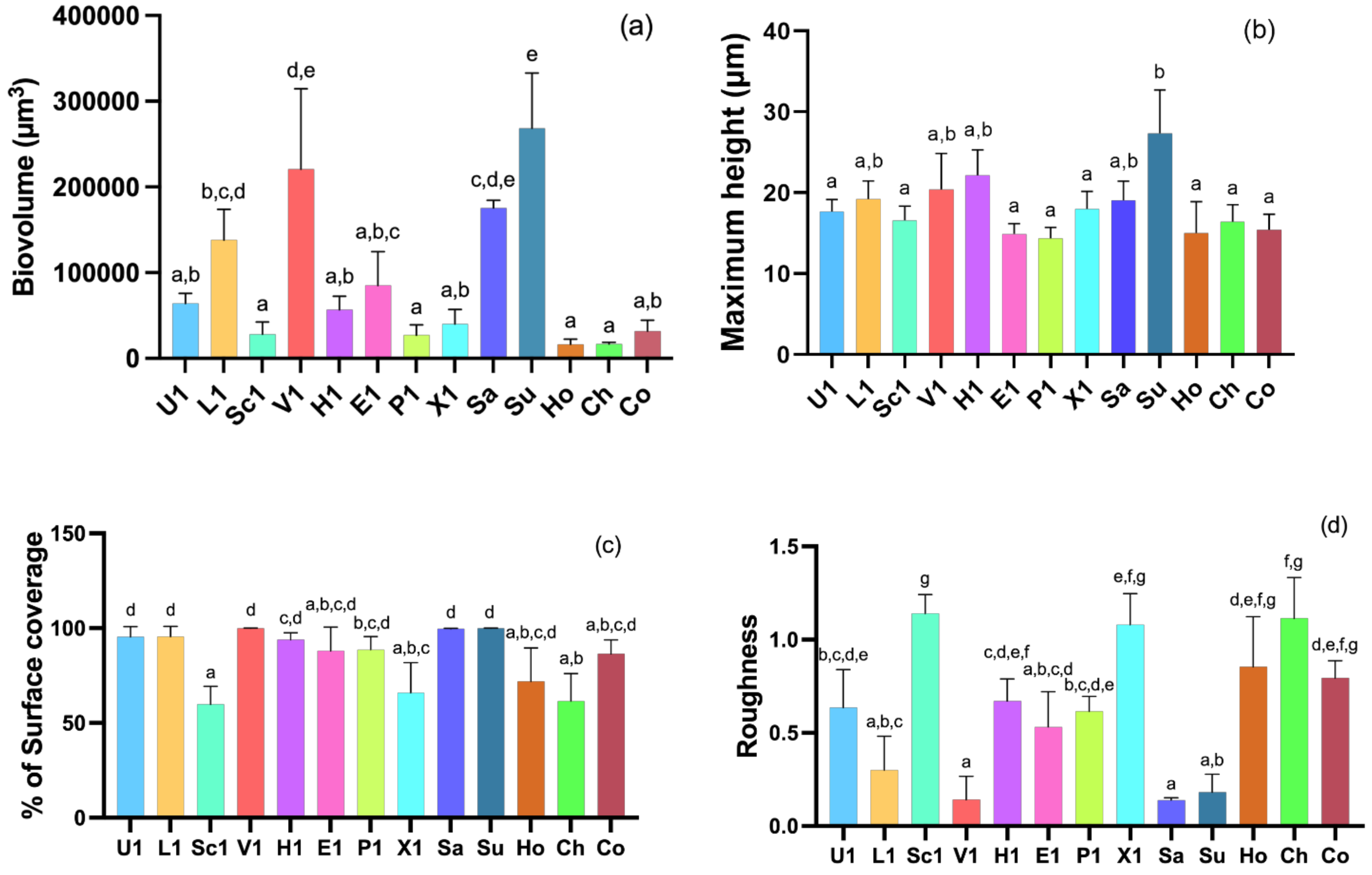
Staphylococci isolates were divided into two categories: multidrug resistant (MDR) and non-multidrug resistant (non-MDR). The relationship between the biofilm-forming capacity and MDR phenotype was investigated by statistical analysis using Student’s t-tests. As shown in Figure 2, the MDR strains significantly produced more biofilm than non-MDR isolates (p < 0.05). To determine whether biofilm formation is related to resistance to any particular antimicrobial, the biofilm formation was evaluated in isolates with different resistance profiles to 11 antimicrobials (Table 1). The results revealed that isolates resistant to cefoxitin, which is an indicator of methicillin resistance, tetracycline, and fusidic acid produced significantly more biofilm than susceptible isolates (p < 0.001). Isolates resistant to erythromycin, clindamycin, and chloramphenicol also formed stronger biofilms than susceptible isolates (p < 0.05). In contrast, staphylococci isolates resistant to ciprofloxacin and trimethoprim-sulfamethoxazole produced weaker biofilms when compared to resistant isolates (p < 0.05 and p < 0.001, respectively).

3.3. CLSM Analysis

Three-dimensional images of biofilms formed by the 13 different staphylococcal species are shown in Figure 3. In accordance with the results obtained din the microtiter assay, all isolates produced biofilms. Most isolates produced compact biofilm structures that covered the entire available surface, except for S. sciuri, S. pseudintermedius, S. xylosus, and S. chromogenes, which produced rough biofilms with irregular coverage and confluent growth areas.
The structural parameters, including biovolume, maximum height, percentage of covered surface, and roughness, were obtained from the battery of images acquired by CLSM, which allowed the quantification of biofilm biomass (Figure 4). The results obtained revealed a marked variability in the structure of the biofilms among the CoNS and S. pseudintermedius isolates. The biovolume of the biofilms ranges from 16,818.63 ± 2034.19 to 268,342.66 ± 64,584.58 µm3 in the observation field of 16,078.2 μm2. Accordingly, with the three-dimensional images of biofilms, S. chromogenes produced the least biofilm biomass while S. succinus produced the most biomass (p < 0.001), followed by S. vitulinus (220,867.43 ± 93,748.89 µm). Other isolates, such as S. hominis, S. pseudintermedius, and S. sciuri, also produced low amounts of biofilm biomass when compared to other isolates. The greatest biofilm thickness (27.33 ± 5.38 µm) was also observed in S. succinus biofilm while the smallest biofilm thickness was detected in S. pseudintermedius (14.33 ± 1.38 µm) and S. epidermidis (14.92 ± 1.23 µm) (p < 0.001). However, regarding the roughness, S. succinus biofilm had the lowest scores (0.18 ± 0.10) while S. sciuri obtained the highest roughness score (1.14 ± 0.10) (p < 0.0001).

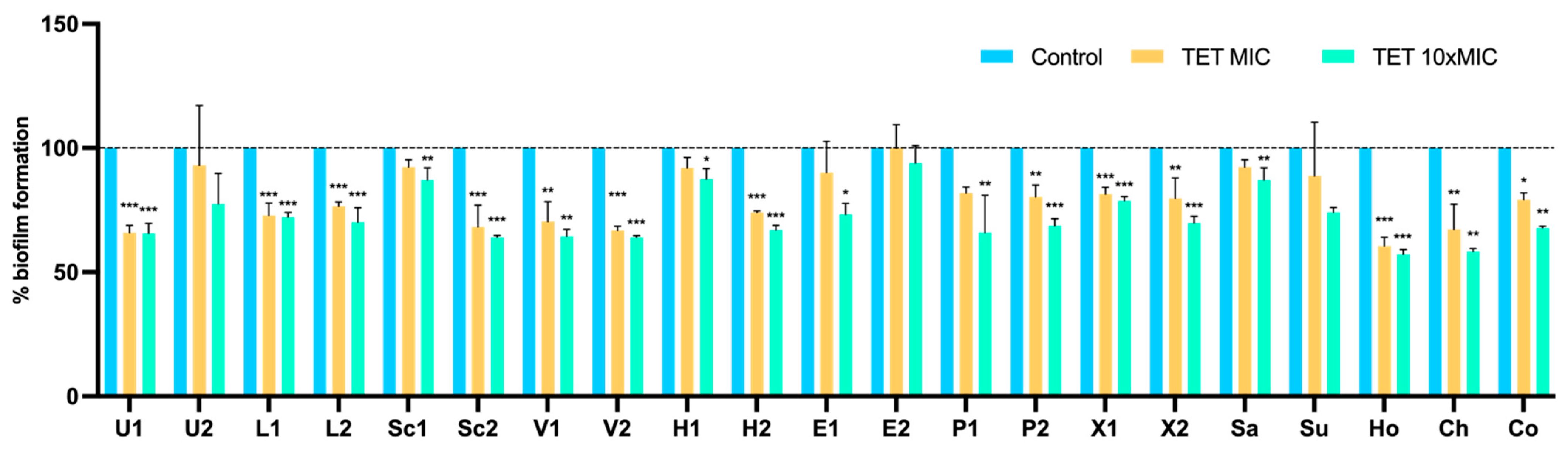
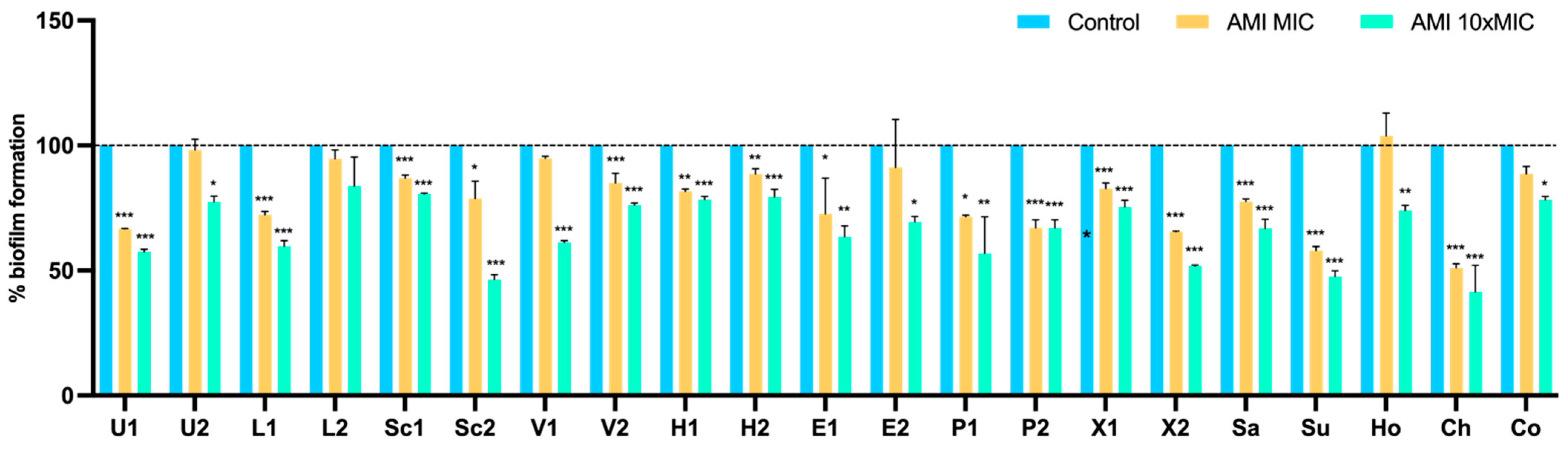
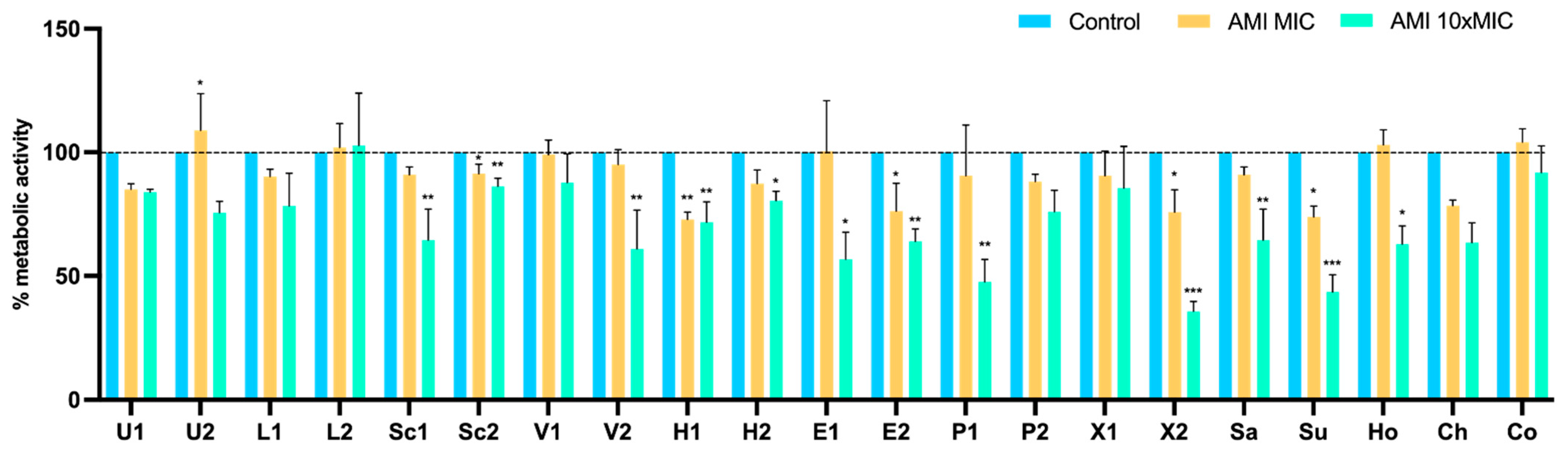
3.4. Effect of Antimicrobials on 24 h-Old Biofilms

To assess whether biofilm-specific resistance influences the action of conventional antimicrobials, the MICs of tetracycline and amikacin were determined for 24 h-old biofilms of 21 isolates. The MICs for these isolates ranged from 0.052 to 64 μg/mL for tetracycline and from 0.5 to 64 μg/mL for amikacin. Then, the capacity of these antimicrobials to reduce pre-established 24-h-old biofilms was evaluated using the microtiter biofilm assay at concentrations of MIC and ten times MIC (10 × MIC). Results were normalized according to the 48-h-old biofilm mass recorded for each strain tested grown without the presence of antimicrobials. As shown in Figure 5, biofilms of 18 out of the 21 isolates suffered a significant biomass reduction with 10 isolates having a very highly significant biomass reduction (p < 0.001). Although all isolates, except for E2, suffered a biofilm mass reduction when treated with tetracycline at MIC concentration, the reduction was only statistically significant in 13 isolates.
Results for the 24 h-old biofilm treatment with amikacin are shown in Figure 6. After amikacin at 10 × MIC, biofilm mass was reduced in almost all isolates (except for L2), with a highly significant reduction in 15 isolates (p < 0.001). At the MIC concentration, amikacin was able to significantly reduce the biofilm of 15 isolates. In contrast, there was an increase in biomass in strain Ho, corresponding to S. hominis, after treatment with amikacin at MIC. Nevertheless, this increase was not statistically significant. Overall, tetracycline at 10 × MIC had a higher capacity to reduce biofilm mass than amikacin; however, the difference was not significant (p = 0.055).

Metabolic Activity

The XTT assay was used to measure the cellular metabolic activity as an indicator of cell viability, after treatment with tetracycline and amikacin at concentrations of MIC and 10 × MIC. The results were normalized according to the 48 h-old biofilm of each tested isolate, which were grown without the presence of antimicrobials. After treatment with tetracycline at MIC and 10 × MIC, there was a significant reduction in metabolic activity in 5 and 9 isolates, respectively (Figure 7). In contrast, at the MIC concentration, the biofilm cells of eight isolates increased the metabolic activity. The effect of amikacin on the metabolic activity of biofilms are shown in Figure 8. The metabolic activity of biofilm cells was significantly reduced in 12 isolates at 10 × MIC. In four isolates (U2, L2, Ho, and Co) was also observed an increase in the metabolic activity at the MIC concentration, which corresponds to the results of biomass reduction.

4. Discussion

In the last decades, CoNS have emerged as multidrug-resistant nosocomial pathogens constituting a major threat. CoNS species differ greatly from each other and there is no such thing as typical CoNS [34]. Furthermore, although some CoNS species have a specific host, human-associated CoNS species are also regularly isolated from animals and vice versa [5]. Contrary to what was previously thought, CoNS species can cause infections in healthy hosts without apparent risk factors [34]. Furthermore, CoNS infections are often associated with the biofilm formation since these organisms have the ability to colonize abiotic and biotic surfaces [2,35]. Nevertheless, biofilm formation in CoNS species is still not well characterized.

4.1. Biofilm Formation

In our study, we investigated the biofilm-forming capacity of S. pseudintermedius and 12 CoNS species, including S. lentus, S. sciuri, S. urealyticus, S. vitulinus, S. xylosus, S. haemolyticus, S. epidermidis, S. cohnii, S. succinus, S. saprophyticus, S. hominis, and S. chromogenes. Among all these species, some are primarily human-associated, such as S. epidermidis, S. saprophyticus, and S. haemolyticus, and others are primarily animal associated [5]. All staphylococci had the ability to form biofilm in the plastic surface of the microplate. Among the most prevalent species isolated from animals, S. urealyticus, which is an animal-associated species, produced significantly stronger biofilms than the other species. Bino et al. studied the biofilm formation of several CoNS isolated from horses and reported that all species were biofilm producers [36]. In the same study, S. urealyticus were classified as strong biofilm producers [36]. Among S. lentus, S. sciuri and the CoPS S. pseudintermedius, there were no significant differences in biofilm production, but all were considered strong biofilm producers since the percentage mean of biofilm production was above 100%. S. lentus is often isolated from livestock and people with occupational exposure to livestock while S. sciuri has a wider host range [2,37,38,39,40]. In the study of Kala et al., the majority of S. lentus and S. sciuri isolated from pigs were biofilm producers [41]. S. pseudintermedius capacity to form biofilms has been more extensively studied than in the CoNS since they are the main colonizers and pathogens of dogs [23]. Studies have shown that S. pseudintermedius have the ability to mostly form medium to strong biofilms [42,43]. Other factors may influence biofilm production such as antimicrobial resistance and clonal lineage of the isolates. After performing multilocus sequence typing, Osland et al. showed that S. pseudintermedius belonging to sequence type (ST) 71 former stronger biofilms than strains belonging to other STs. Our S. pseudintermedius isolates collected from canine infections belonged to ST123, which differs from ST71 by a one-point mutation on the sar locus, which may explain the high capacity of our isolates to form biofilm. Among the least prominent CoNS species included in this study, S. heamolyticus produced less biofilm biomass and S. xylosus produced more biomass than the other CoNS. However, these differences were not statistically significant. Previous studies regarding the biofilm formation of S. haemolyticus have yielded different results among them, with studies reporting high rates of biofilm formation and others low to medium rates [44,45]. A study conducted with milk samples showed that S. xylosus was moderate to strong biofilm formers whereas S. epidermidis had the lowest ability to form biofilms [46]. Another study investigated the biofilm-forming ability of CoNS isolated from horses and the highest values were measured for S. xylosus strains [36].

4.2. Antimicrobial Resistance and Biofilm Formation

There are studies showing an association between antimicrobial resistance and the biofilm-forming capacity of staphylococci strains [47,48]. However, there are also other studies that did not find any association [9]. In our study, MDR staphylococci produced more biofilm biomass than non-MDR isolates (p < 0.05). Therefore, we also investigated if the antimicrobial resistance to individual antibiotics influenced the biofilm production. Strains resistant to cefoxitin, and therefore, resistant to methicillin, and to tetracycline produced more biofilm biomass than susceptible isolates (p < 0.001). These results may show the importance of individual antimicrobial resistance in the pathogenesis of biofilm-producing strains. Sheikh et al. also found an association between biofilm formation and antimicrobial resistance to most antimicrobials classes, except for oxazolidinones [49]. Koksal et al. showed that methicillin resistance was significantly higher in biofilm-positive isolates [50]. Studies investigating the association between biofilm formation and resistance antimicrobials in CoNS isolates are very scarce. Nevertheless, studies conducted with S. aureus also reported a positive relationship between biofilm formation and tetracycline and cefoxitin resistance [51,52,53].

4.3. Effect of Antimicrobials on 24 h-Old Biofilms

The biofilm matrix can prevent antimicrobials from entering and reaching their molecular targets by different mechanisms, such as modification/degradation of the antimicrobial, drug tolerance, chelation, and precipitation [54]. It is very important to investigate the biofilm resistance to antimicrobials since the values of MIC do not generally correlate with the minimal biofilm eradication concentration (MBEC). The biofilm formation mechanisms and the effect of antimicrobials on staphylococci have been mainly investigated in S. lugdunensis and S. epidermidis but little is known about other CoNS species [34]. In our study, we evaluated the biofilm reduction in 12 species of CoNS using tetracycline and amikacin at concentrations of MIC and 10 × MIC. As excepted, none of the antimicrobials, even at high concentrations, were able to eliminate staphylococci biofilms. Tetracycline at 10 × MIC was able to significantly reduce the biofilm mass of 18 isolates except for one S. urealyticus, one S. epidermidis, and S. succinus isolates. Not much is known about the biofilm resistance of S. urealyticus and S. succinus biofilms but studies on S. epidermidis have shown that concentrations higher than 10 × MIC are necessary for biofilm eradication [55,56]. In a recent study, the biofilm biomass of S. epidermidis suffered a 55% reduction after treatment with 10 × MIC [55]. Moreover, in the same study, 24 h-old biofilms of S. chromogenes and S. haemolyticus were also reduced by 30% and 29%, respectively, which are in accordance with our results [55]. Brady et al. have also showed that MBEC concentrations were 10–1000 times greater than that of the MIC breakpoints, with MBEC for tetracycline surpassing 256 µg/mL [56]. Accordingly, in the study by Flemming et al., the isolates tested under planktonic growth conditions were susceptible to tetracycline, but even after the biofilm treatment with 500 mg/L of tetracycline, a significant number of living cells were still detected [57]. Regarding the efficacy of amikacin, almost all isolates’ biofilm suffered a significant reduction in biomass after treatment with 10 × MIC. However, at the MIC concentration, there was an enhancement of biofilm production of S. hominis. The biofilm reduction exceeded 50% in S. sciuiri, S. succinus, and S. chromogenes. A study have reported amikacin MBECs between threefold to 1000-fold higher than the MIC in staphylococci biofilms [58,59]. Amikacin belongs to the aminoglycosides class of antimicrobials and some studies reporting the effect of other aminoglycosides on biofilm reduction have shown that even high doses of these antimicrobials, even higher than 1024 µg/mL, were not sufficient to eliminate CoNS biofilms [60,61]. In a study conducted with S. pseudintermedius, a >667-fold difference between the MIC and MBIC was observed [62]. The antimicrobial molecule and the mechanism of action can also play a role in the action of antimicrobials upon biofilms. Therefore, the penetration of amikacin in biofilms may be difficult since aminoglycosides are large polar molecules [63]. Our results also show the failure of amikacin and tetracycline to eradicate biofilms is completely independent of any staphylococci species or origin. These differences found between MIC and MBEC may explain the frequent failure in the treatment of CoNS infections with conventional antibiotics [60]. In fact, it is known that bacteria within biofilms are shielded against the action of antimicrobials due to the matrix that serves as a barrier hampering antimicrobial penetration, the reduced metabolic functions of the biofilm cells, the elevated mutation rates of staphylococci within biofilms, and antimicrobial tolerance [34,64,65].
The CV method to quantify biofilm biomass has been extensively used and is based on the CV bond negatively charged surface molecules and polysaccharides in the extracellular matrix of biofilms. However, in this method both dead and alive cells are stained [32]. Therefore, after antimicrobial treatment, we measured the metabolic activity of viable cells using the XTT method. Most biofilm isolates that suffered a significant biomass reduction also showed a lower metabolic activity. A study conducted by Flemming et al. have reported that biofilm treated with tetracycline had a lower metabolic activity even though a significant number of biofilm cells were still alive [57]. However, the metabolic activity of some strains, particularly those that did not undergo a significant reduction, was enhanced at the MIC concentration. This may be due to an increase in the number of viable cells, an increase in the metabolic activity of cells—in an attempt to resist the antimicrobial action—or the cells might have been at the proliferative stage, with a reduced extracellular matrix [64].

5. Conclusions

Our study showed that all CoNS and S. pseudintermedius isolated from animals are able to form biofilms. S. urealyticus strains, which had been isolated from poultry for human consumption, produced more biofilm than other staphylococcus species. We also found an association between biofilm-forming capacity and antimicrobial resistance, particularly to important antimicrobials such as cefoxitin. Our study also reinforces previous findings that CoNS in a biofilm mode are highly resistant to antimicrobials. Neither amikacin nor tetracycline at 10 × MIC were able to eliminate biofilms. Therefore, research for new antimicrobial classes or alternatives to antimicrobials is urgently needed. These staphylococci strains with zoonotic potential had a high capacity to form biofilms and may pose a threat to human health. Thus, this underlines the need for implementation of new measures in the public health and veterinary sectors to prevent transmission of CoNS in the One Health context.

Supplementary Materials

The following supporting information can be downloaded at: https://www.mdpi.com/article/10.3390/pathogens11060689/s1, Table S1: Characteristics of the S. pseudintermedius and CoNS isolated from pets, livestock and wild animals.

Author Contributions

Conceptualization, V.S.; methodology, V.S., C.G.-M. and R.C.; software, V.S.; formal analysis, R.C. and C.A.-C.; investigation, V.S.; resources, J.E.P.; data curation, E.C.; writing—original draft preparation, V.S.; writing—review and editing, V.S.; visualization, R.C.; supervision, G.I. and P.P. All authors have read and agreed to the published version of the manuscript.

Funding

This work was funded by the R&D Project CAREBIO2: Comparative assessment of antimicrobial resistance in environmental biofilms through proteomics—towards innovative theranostic biomarkers, with reference NORTE-01-0145-FEDER-030101 and PTDC/SAU-INF/30101/2017, financed by the European Regional Development Fund (ERDF) through the Northern Regional Operational Program (NORTE 2020) and the Foundation for Science and Technology (FCT). This work was supported by the Associate Laboratory for Green Chemistry-LAQV, which is financed by national funds from FCT/MCTES (UIDB/50006/2020 and UIDP/50006/2020) and by the projects UIDB/CVT/00772/2020 and LA/P/0059/2020 funded by the Portuguese Foundation for Science and Technology (FCT). The Ministerio de Ciencia, Innovación y Universidades (Spain, grant number RTI2018-098267-R-C33) and the Junta de Castilla y León (Consejería de Educación, Spain, grant number LE018P20). Vanessa Silva is grateful to FCT (Fundacão para a Ciência e a Tecnologia) for financial support through the PhD grant SFRH/BD/137947/2018.

Institutional Review Board Statement

Not applicable.

Informed Consent Statement

Not applicable.

Data Availability Statement

Not applicable.

Conflicts of Interest

The authors declare no conflict of interest.

References

  1. Heilbronner, S. Staphylococcus lugdunensis. Trends Microbiol. 2021, 29, 1143–1145. [Google Scholar] [CrossRef] [PubMed]
  2. Becker, K.; Heilmann, C.; Peters, G. Coagulase-negative staphylococci. Clin. Microbiol. Rev. 2014, 27, 870–926. [Google Scholar] [CrossRef] [PubMed] [Green Version]
  3. Abdel-Moein, K.A.; Zaher, H.M. The Nasal Carriage of Coagulase-Negative Staphylococci Among Animals and Its Public Health Implication. Vector Borne Zoonotic Dis. 2020, 20, 897–902. [Google Scholar] [CrossRef] [PubMed]
  4. Kloos, W.E.; Bannerman, T.L. Update on clinical significance of coagulase-negative staphylococci. Clin. Microbiol. Rev. 1994, 7, 117–140. [Google Scholar] [CrossRef]
  5. Heilmann, C.; Ziebuhr, W.; Becker, K. Are coagulase-negative staphylococci virulent? Clin. Microbiol. Infect. 2019, 25, 1071–1080. [Google Scholar] [CrossRef]
  6. Michels, R.; Last, K.; Becker, S.L.; Papan, C. Update on Coagulase-Negative Staphylococci—What the Clinician Should Know. Microorg. 2021, 9, 830. [Google Scholar] [CrossRef]
  7. Saber, H.; Jasni, A.S.; Jamaluddin, T.Z.M.T.; Ibrahim, R. A Review of Staphylococcal Cassette Chromosome mec (SCCmec) Types in Coagulase-Negative Staphylococci (CoNS) Species. Malays. J. Med. Sci. 2017, 24, 7–18. [Google Scholar] [CrossRef]
  8. Asante, J.; Hetsa, B.A.; Amoako, D.G.; Abia, A.L.K.; Bester, L.A.; Essack, S.Y. Multidrug-resistant coagulase-negative staphylococci isolated from bloodstream in the uMgungundlovu district of KwaZulu-Natal Province in South Africa: Emerging pathogens. Antibiotics 2021, 10, 198. [Google Scholar] [CrossRef]
  9. Chon, J.-W.; Lee, U.J.; Bensen, R.; West, S.; Paredes, A.; Lim, J.; Khan, S.; Hart, M.E.; Phillips, K.S.; Sung, K. Virulence Characteristics of mecA-Positive Multidrug-Resistant Clinical Coagulase-Negative Staphylococci. Microorg. 2020, 8, 659. [Google Scholar] [CrossRef]
  10. Michalik, M.; Nowakiewicz, A.; Trościańczyk, A.; Kowalski, C.; Podbielska-Kubera, A. Multidrug resistant coagulase-negative Staphylococcus spp. isolated from cases of chronic rhinosinusitis in humans. Study from Poland. Acta Microbiol. Immunol. Hung. 2022, 69, 68–76. [Google Scholar] [CrossRef]
  11. Silva, V.; Caniça, M.; Ferreira, E.; Vieira-Pinto, M.; Saraiva, C.; Pereira, J.E.; Capelo, J.L.; Igrejas, G.; Poeta, P. Multidrug-Resistant Methicillin-Resistant Coagulase-Negative Staphylococci in Healthy Poultry Slaughtered for Human Consumption. Antibiot. 2022, 11, 365. [Google Scholar] [CrossRef]
  12. Kizerwetter-Świda, M.; Chrobak-Chmiel, D.; Rzewuska, M. High-level mupirocin resistance in methicillin-resistant staphylococci isolated from dogs and cats. BMC Vet. Res. 2019, 15, 238. [Google Scholar] [CrossRef]
  13. Pexara, A.; Solomakos, N.; Govaris, A. Occurrence, antibiotic resistance and enteroxigenicity of Staphylococcus spp. in tonsils of slaughtered pigs in Greece. Lett. Appl. Microbiol. 2020, 71, 394–399. [Google Scholar] [CrossRef]
  14. Marincola, G.; Liong, O.; Schoen, C.; Abouelfetouh, A.; Hamdy, A.; Wencker, F.D.R.; Marciniak, T.; Becker, K.; Köck, R.; Ziebuhr, W. Antimicrobial resistance profiles of coagulase-negative staphylococci in community-based healthy individuals in Germany. Front. Public Heal. 2021, 9, 796. [Google Scholar] [CrossRef]
  15. Schilcher, K.; Horswill, A.R. Staphylococcal Biofilm Development: Structure, Regulation, and Treatment Strategies. Microbiol. Mol. Biol. Rev. 2020, 84, e00026-19. [Google Scholar] [CrossRef]
  16. Argemi, X.; Hansmann, Y.; Prola, K.; Prévost, G. Coagulase-negative staphylococci pathogenomics. Int. J. Mol. Sci. 2019, 20, 1215. [Google Scholar] [CrossRef] [Green Version]
  17. Silva, V.; Capelo, J.L.; Igrejas, G.; Poeta, P. Molecular Mechanisms of Antimicrobial Resistance in Staphylococcus aureus Biofilms BT—Emerging Modalities in Mitigation of Antimicrobial Resistance. In; Akhtar, N., Singh, K.S., Prerna, G.D., Eds.; Springer International Publishing: Cham, Switzerland, 2022; pp. 291–314. ISBN 978-3-030-84126-3. [Google Scholar]
  18. Geoghegan, J.A.; Foster, T.J. Cell wall-anchored surface proteins of Staphylococcus aureus: Many proteins, multiple functions. In Staphylococcus Aureus; Springer: Cham, Switzerland, 2015; pp. 95–120. [Google Scholar]
  19. Wojtyczka, R.D.; Orlewska, K.; Kępa, M.; Idzik, D.; Dziedzic, A.; Mularz, T.; Krawczyk, M.; Miklasińska, M.; Wąsik, T.J. Biofilm formation and antimicrobial susceptibility of Staphylococcus epidermidis strains from a hospital environment. Int. J. Environ. Res. Public Health 2014, 11, 4619–4633. [Google Scholar] [CrossRef] [Green Version]
  20. Folliero, V.; Franci, G.; Dell’Annunziata, F.; Giugliano, R.; Foglia, F.; Sperlongano, R.; De Filippis, A.; Finamore, E.; Galdiero, M. Evaluation of Antibiotic Resistance and Biofilm Production among Clinical Strain Isolated from Medical Devices. Int. J. Microbiol. 2021, 2021, 9033278. [Google Scholar] [CrossRef]
  21. Govan, J.R.; Deretic, V. Microbial pathogenesis in cystic fibrosis: Mucoid Pseudomonas aeruginosa and Burkholderia cepacia. Microbiol. Rev. 1996, 60, 539–574. [Google Scholar] [CrossRef]
  22. Singh, S.; Singh, S.K.; Chowdhury, I.; Singh, R. Understanding the Mechanism of Bacterial Biofilms Resistance to Antimicrobial Agents. Open Microbiol. J. 2017, 11, 53–62. [Google Scholar] [CrossRef] [Green Version]
  23. Silva, V.; Oliveira, A.; Manageiro, V.; Caniça, M.; Contente, D.; Capita, R.; Alonso-Calleja, C.; Carvalho, I.; Capelo, J.L.; Igrejas, G.; et al. Clonal Diversity and Antimicrobial Resistance of Methicillin-Resistant Staphylococcus pseudintermedius Isolated from Canine Pyoderma. Microorg. 2021, 9, 482. [Google Scholar] [CrossRef]
  24. Silva, V.; Lopes, A.F.; Soeiro, V.; Caniça, M.; Manageiro, V.; Pereira, J.E.; Maltez, L.; Capelo, J.L.; Igrejas, G.; Poeta, P. Nocturnal Birds of Prey as Carriers of Staphylococcus aureus and Other Staphylococci: Diversity, Antimicrobial Resistance and Clonal Lineages. Antibiot. 2022, 11, 240. [Google Scholar] [CrossRef]
  25. Silva, V.; Alfarela, C.; Caniça, M.; Manageiro, V.; Nóvoa, M.; Leiva, B.; Kress, M.; Capelo, J.L.; Poeta, P.; Igrejas, G. A One Health Approach Molecular Analysis of Staphylococcus aureus Reveals Distinct Lineages in Isolates from Miranda Donkeys (Equus asinus) and Their Handlers. Antibiot. 2022, 11, 374. [Google Scholar] [CrossRef]
  26. Silva, V.; Pereira, J.E.; Maltez, L.; Ferreira, E.; Manageiro, V.; Caniça, M.; Capelo, J.L.; Igrejas, G.; Poeta, P. Diversity of methicillin-resistant staphylococci among wild Lepus granatensis: First detection of mecA-MRSA in hares. FEMS Microbiol. Ecol. 2020, 96, fiz204. [Google Scholar] [CrossRef]
  27. Santos, V.; Gomes, A.; Ruiz-Ripa, L.; Mama, O.M.; Sabença, C.; Sousa, M.; Silva, V.; Sousa, T.; Vieira-Pinto, M.; Igrejas, G.; et al. Methicillin-Resistant Staphylococcus aureus CC398 in Purulent Lesions of Piglets and Fattening Pigs in Portugal. Microb. Drug Resist. 2020, 26, 850–856. [Google Scholar] [CrossRef]
  28. Silva, V.; de Sousa, T.; Gómez, P.; Sabença, C.; Vieira-Pinto, M.; Capita, R.; Alonso-Calleja, C.; Torres, C.; Capelo, J.L.; Igrejas, G.; et al. Livestock-Associated Methicillin-Resistant Staphylococcus aureus (MRSA) in Purulent Subcutaneous Lesions of Farm Rabbits. Foods 2020, 9, 439. [Google Scholar] [CrossRef] [Green Version]
  29. Silva, V.; Vieira-Pinto, M.; Saraiva, C.; Manageiro, V.; Reis, L.; Ferreira, E.; Caniça, M.; Capelo, J.L.; Igrejas, G.; Poeta, P. Prevalence and Characteristics of Multidrug-Resistant Livestock-Associated Methicillin-Resistant Staphylococcus aureus (LA-MRSA) CC398 Isolated from Quails (Coturnix Coturnix Japonica) Slaughtered for Human Consumption. Animals 2021, 11, 2038. [Google Scholar] [CrossRef]
  30. Silva, V.; Gabriel, S.I.; Borrego, S.B.; Tejedor-Junco, M.T.; Manageiro, V.; Ferreira, E.; Reis, L.; Caniça, M.; Capelo, J.L.; Igrejas, G.; et al. Antimicrobial Resistance and Genetic Lineages of Staphylococcus aureus from Wild Rodents: First Report of mecC-Positive Methicillin-Resistant S. aureus (MRSA) in Portugal. Animals 2021, 11, 1537. [Google Scholar] [CrossRef]
  31. Oniciuc, E.-A.; Cerca, N.; Nicolau, A.I. Compositional Analysis of Biofilms Formed by Staphylococcus aureus Isolated from Food Sources. Front. Microbiol. 2016, 7, 390. [Google Scholar] [CrossRef] [Green Version]
  32. Peeters, E.; Nelis, H.J.; Coenye, T. Comparison of multiple methods for quantification of microbial biofilms grown in microtiter plates. J. Microbiol. Methods 2008, 72, 157–165. [Google Scholar] [CrossRef] [Green Version]
  33. González-Machado, C.; Capita, R.; Riesco-Peláez, F.; Alonso-Calleja, C. Visualization and quantification of the cellular and extracellular components of Salmonella Agona biofilms at different stages of development. PLoS ONE 2018, 13, e0200011. [Google Scholar] [CrossRef] [PubMed]
  34. Becker, K.; Both, A.; Weißelberg, S.; Heilmann, C.; Rohde, H. Emergence of coagulase-negative staphylococci. Expert Rev. Anti. Infect. Ther. 2020, 18, 349–366. [Google Scholar] [CrossRef] [PubMed]
  35. Morgenstern, M.; Post, V.; Erichsen, C.; Hungerer, S.; Bühren, V.; Militz, M.; Richards, R.G.; Moriarty, T.F. Biofilm formation increases treatment failure in Staphylococcus epidermidis device-related osteomyelitis of the lower extremity in human patients. J. Orthop. Res. 2016, 34, 1905–1913. [Google Scholar] [CrossRef] [PubMed]
  36. Bino, E.; Lauková, A.; Ščerbová, J.; Kubašová, I.; Kandričáková, A.; Strompfová, V.; Miltko, R.; Belzecki, G. Fecal coagulase-negative staphylococci from horses, their species variability, and biofilm formation. Folia Microbiol. 2019, 64, 719–726. [Google Scholar] [CrossRef]
  37. De Martino, L.; Lucido, M.; Mallardo, K.; Facello, B.; Mallardo, M.; Iovane, G.; Pagnini, U.; Tufano, M.A.; Catalanotti, P. Methicillin-Resistant Staphylococci Isolated from Healthy Horses and Horse Personnel in Italy. J. Vet. Diagnostic Investig. 2010, 22, 77–82. [Google Scholar] [CrossRef] [Green Version]
  38. Ugwu, C.C.; Gomez-Sanz, E.; Agbo, I.C.; Torres, C.; Chah, K.F. Characterization of mannitol-fermenting methicillin-resistant staphylococci isolated from pigs in Nigeria. Braz. J. Microbiol. 2015, 46, 885–892. [Google Scholar] [CrossRef] [Green Version]
  39. Zhang, Y.; Agidi, S.; LeJeune, J.T. Diversity of staphylococcal cassette chromosome in coagulase-negative staphylococci from animal sources. J. Appl. Microbiol. 2009, 107, 1375–1383. [Google Scholar] [CrossRef]
  40. Nemeghaire, S.; Vanderhaeghen, W.; Argudín, M.A.; Haesebrouck, F.; Butaye, P. Characterization of methicillin-resistant Staphylococcus sciuri isolates from industrially raised pigs, cattle and broiler chickens. J. Antimicrob. Chemother. 2014, 69, 2928–2934. [Google Scholar] [CrossRef] [Green Version]
  41. Kalai, S.; Roychoudhury, P.; Dutta, T.K.; Subudhi, P.K.; Chakraborty, S.; Barman, N.N.; Sen, A. Multidrug resistant staphylococci isolated from pigs with exudative epidermitis in North eastern Region of India. Lett. Appl. Microbiol. 2021, 72, 535–541. [Google Scholar] [CrossRef]
  42. Singh, A.; Walker, M.; Rousseau, J.; Weese, J.S. Characterization of the biofilm forming ability of Staphylococcus pseudintermedius from dogs. BMC Vet. Res. 2013, 9, 93. [Google Scholar] [CrossRef] [Green Version]
  43. Osland, A.M.; Vestby, L.K.; Fanuelsen, H.; Slettemeås, J.S.; Sunde, M. Clonal diversity and biofilm-forming ability of methicillin-resistant Staphylococcus pseudintermedius. J. Antimicrob. Chemother. 2012, 67, 841–848. [Google Scholar] [CrossRef] [Green Version]
  44. Pinheiro, L.; Brito, C.I.; de Oliveira, A.; Pereira, V.C.; da Cunha de Souza, M.d.L.R. Staphylococcus epidermidis and Staphylococcus haemolyticus: Detection of biofilm genes and biofilm formation in blood culture isolates from patients in a Brazilian teaching hospital. Diagn. Microbiol. Infect. Dis. 2016, 86, 11–14. [Google Scholar] [CrossRef] [Green Version]
  45. Shrestha, L.B.; Bhattarai, N.R.; Khanal, B. Comparative evaluation of methods for the detection of biofilm formation in coagulase-negative staphylococci and correlation with antibiogram. Infect. Drug Resist. 2018, 11, 607. [Google Scholar] [CrossRef] [Green Version]
  46. Tremblay, Y.D.N.; Lamarche, D.; Chever, P.; Haine, D.; Messier, S.; Jacques, M. Characterization of the ability of coagulase-negative staphylococci isolated from the milk of Canadian farms to form biofilms. J. Dairy Sci. 2013, 96, 234–246. [Google Scholar] [CrossRef] [Green Version]
  47. Cabrera-Contreras, R.; Morelos-Ramírez, R.; Galicia-Camacho, A.N.; Meléndez-Herrada, E. Antibiotic resistance and biofilm production in Staphylococcus epidermidis strains, isolated from a tertiary care hospital in Mexico City. Int. Sch. Res. Not. 2013, 2013, 918921. [Google Scholar] [CrossRef] [Green Version]
  48. Singhai, M.; Malik, A.; Shahid, M.; Malik, M.A.; Goyal, R. A study on device-related infections with special reference to biofilm production and antibiotic resistance. J. Glob. Infect. Dis. 2012, 4, 193–198. [Google Scholar] [CrossRef]
  49. Farajzadeh Sheikh, A.; Asareh Zadegan Dezfuli, A.; Navidifar, T.; Fard, S.S.; Dehdashtian, M. Association between biofilm formation, structure and antibiotic resistance in Staphylococcus epidermidis isolated from neonatal septicemia in southwest Iran. Infect. Drug Resist. 2019, 12, 1771–1782. [Google Scholar] [CrossRef] [Green Version]
  50. Koksal, F.; Yasar, H.; Samasti, M. Antibiotic resistance patterns of coagulase-negative staphylococcus strains isolated from blood cultures of septicemic patients in Turkey. Microbiol. Res. 2009, 164, 404–410. [Google Scholar] [CrossRef]
  51. Jimi, S.; Miyazaki, M.; Takata, T.; Ohjimi, H.; Akita, S.; Hara, S. Increased drug resistance of meticillin-resistant Staphylococcus aureus biofilms formed on a mouse dermal chip model. J. Med. Microbiol. 2017, 66, 542. [Google Scholar] [CrossRef]
  52. Shah, M.S.; Qureshi, S.; Kashoo, Z.; Farooq, S.; Wani, S.A.; Hussain, M.I.; Banday, M.S.; Khan, A.A.; Gull, B.; Habib, A.; et al. Methicillin resistance genes and in vitro biofilm formation among Staphylococcus aureus isolates from bovine mastitis in India. Comp. Immunol. Microbiol. Infect. Dis. 2019, 64, 117–124. [Google Scholar] [CrossRef]
  53. Sun, X.; Lin, Z.; Hu, X.; Yao, W.; Bai, B.; Wang, H.; Li, D.; Chen, Z.; Cheng, H.; Pan, W.; et al. Biofilm formation in erythromycin-resistant Staphylococcus aureus and the relationship with antimicrobial susceptibility and molecular characteristics. Microb. Pathog. 2018, 124, 47–53. [Google Scholar] [CrossRef] [PubMed]
  54. Flemming, H.-C.; Wingender, J.; Szewzyk, U.; Steinberg, P.; Rice, S.A.; Kjelleberg, S. Biofilms: An emergent form of bacterial life. Nat. Rev. Microbiol. 2016, 14, 563–575. [Google Scholar] [CrossRef] [PubMed]
  55. Francisco, M.S.; Rossi, C.C.; Brito, M.A.V.P.; Laport, M.S.; Barros, E.M.; Giambiagi-deMarval, M. Characterization of biofilms and antimicrobial resistance of coagulase-negative Staphylococcus species involved with subclinical mastitis. J. Dairy Res. 2021, 88, 179–184. [Google Scholar] [CrossRef] [PubMed]
  56. Brady, A.J.; Laverty, G.; Gilpin, D.F.; Kearney, P.; Tunney, M. Antibiotic susceptibility of planktonic- and biofilm-grown staphylococci isolated from implant-associated infections: Should MBEC and nature of biofilm formation replace MIC? J. Med. Microbiol. 2017, 66, 461–469. [Google Scholar] [CrossRef] [Green Version]
  57. Flemming, K.; Klingenberg, C.; Cavanagh, J.P.; Sletteng, M.; Stensen, W.; Svendsen, J.S.; Flægstad, T. High in vitro antimicrobial activity of synthetic antimicrobial peptidomimetics against staphylococcal biofilms. J. Antimicrob. Chemother. 2009, 63, 136–145. [Google Scholar] [CrossRef]
  58. Mendoza-Olazarán, S.; Morfín-Otero, R.; Villarreal-Treviño, L.; Rodríguez-Noriega, E.; Llaca-Díaz, J.; Camacho-Ortiz, A.; González, G.M.; Casillas-Vega, N.; Garza-González, E. Antibiotic Susceptibility of Biofilm Cells and Molecular Characterisation of Staphylococcus hominis Isolates from Blood. PLoS ONE 2015, 10, e0144684. [Google Scholar] [CrossRef]
  59. Al-Ouqaili, M.T.S. Biofilm antimicrobial susceptibility pattern for selected antimicrobial agents against planktonic and sessile cells of clinical isolates of staphylococci using MICs, BICs and MBECs. Asian J Pharm 2018, 12. [Google Scholar]
  60. Qu, Y.; Daley, A.J.; Istivan, T.S.; Garland, S.M.; Deighton, M.A. Antibiotic susceptibility of coagulase-negative staphylococci isolated from very low birth weight babies: Comprehensive comparisons of bacteria at different stages of biofilm formation. Ann. Clin. Microbiol. Antimicrob. 2010, 9, 16. [Google Scholar] [CrossRef] [Green Version]
  61. De Oliveira, A.; Cataneli Pereira, V.; Pinheiro, L.; Moraes Riboli, D.F.; Benini Martins, K.; Ribeiro de Souza da Cunha, M.D. Antimicrobial Resistance Profile of Planktonic and Biofilm Cells of Staphylococcus aureus and Coagulase-Negative Staphylococci. Int. J. Mol. Sci. 2016, 17, 1423. [Google Scholar] [CrossRef] [Green Version]
  62. Walker, M.; Singh, A.; Nazarali, A.; Gibson, T.W.G.; Rousseau, J.; Weese, J.S. Evaluation of the impact of methicillin-resistant Staphylococcus pseudintermedius biofilm formation on antimicrobial susceptibility. Vet. Surg. 2016, 45, 968–971. [Google Scholar] [CrossRef]
  63. Glinka, M.; Wojnowski, W.; Wasik, A. Determination of aminoglycoside antibiotics: Current status and future trends. TrAC Trends Anal. Chem. 2020, 131, 116034. [Google Scholar] [CrossRef]
  64. Silva, V.; Almeida, L.; Gaio, V.; Cerca, N.; Manageiro, V.; Caniça, M.; Capelo, J.L.; Igrejas, G.; Poeta, P. Biofilm Formation of Multidrug-Resistant MRSA Strains Isolated from Different Types of Human Infections. Pathogens 2021, 10, 970. [Google Scholar] [CrossRef]
  65. Ryder, V.J.; Chopra, I.; O’Neill, A.J. Increased mutability of Staphylococci in biofilms as a consequence of oxidative stress. PLoS ONE 2012, 7, e47695. [Google Scholar] [CrossRef] [Green Version]
Figure 1. Biofilm-forming capacity of (a) the most prevalent staphylococci species; (b) the least prevalent species of staphylococci among animal isolates; and (c) by animal. The symbols represent the biomass mean of the biofilm formed in independent tests of the individual isolates. The red lines represent the average biofilm mass formed by all isolates. Statistical significance was determined using Tukey’s multiple comparison test (* p < 0.05 and *** p < 0.001).
Pathogens 11 00689 g001
Figure 2. Mean biofilm formation among multidrug-resistant (MDR) and non-multidrug-resistant (non-MDR) isolates. The red lines represent the average biofilm mass formed by all isolates. Statistical significance was determined using Student’s t-tests (* p < 0.05).
Pathogens 11 00689 g002
Figure 3. The images correspond to three-dimensional reconstructions obtained by CLSM and processed with IMARIS 9.1 software, including the virtual projections of the shadows on the right. U: S. urealyticus; L: S. lentus; Sc: S. sciuri; V: S. vitulinus; H: S. haemolyticus; E: S. epidermidis; P: S. pseudintermedius; X: S. xylosus; Sa: S. saprophyticus; Su: S. succinus; Ho: S. hominis; Ch: S. chromogenes; Co: S. cohnii.
Pathogens 11 00689 g003
Figure 4. Biovolume in the observation field of 16,078.2 μm2 (a), maximum height (b), maximum height, percentage of surface area covered (c), and roughness (d) of biofilms formed from the 13 selected CoNS and S. pseudinteremedius isolates. Statistical significance was determined using Tukey’s multiple comparison test. The values marked with the same letter are not statistically significant as determined by the Tukey’s post hoc test (p < 0.05). U: S. urealyticus; L: S. lentus; Sc: S. sciuri; V: S. vitulinus; H: S. haemolyticus; E: S. epidermidis; P: S. pseudintermedius; X: S. xylosus; Sa: S. saprophyticus; Su: S. succinus; Ho: S. hominis; Ch: S. chromogenes; Co: S. cohnii.
Pathogens 11 00689 g004
Figure 5. Effect of tetracycline on the biofilm biomass reduction of 21 isolates at concentrations of MIC and 10 × MIC. Data are presented as the mean ± standard deviation for four independent replicates. Statistical significance was determined using Dunnett’s multiple comparison test (* p < 0.05; ** p < 0.01; *** p < 0.001). U: S. urealyticus; L: S. lentus; Sc: S. sciuri; V: S. vitulinus; H: S. haemolyticus; E: S. epidermidis; P: S. pseudintermedius; X: S. xylosus; Sa: S. saprophyticus; Su: S. succinus; Ho: S. hominis; Ch: S. chromogenes; Co: S. cohnii; 1: produces the most biofilm; 2: produces the least biofilm.
Pathogens 11 00689 g005
Figure 6. Effect of amikacin on the biofilm biomass reduction of 21 isolates at concentrations of MIC and 10 × MIC. Data are presented as the mean ± standard deviation for four independent replicates. Statistical significance was determined using Dunnett’s multiple comparison test (* p < 0.05; ** p < 0.01; *** p < 0.001). U: S. urealyticus; L: S. lentus; Sc: S. sciuri; V: S. vitulinus; H: S. haemolyticus; E: S. epidermidis; P: S. pseudintermedius; X: S. xylosus; Sa: S. saprophyticus; Su: S. succinus; Ho: S. hominis; Ch: S. chromogenes; Co: S. cohnii; 1: produces the most biofilm; 2: produces the least biofilm.
Pathogens 11 00689 g006
Figure 7. Metabolic activity of staphylococci biofilms before and after treatment with tetracycline at concentrations of MIC and 10 × MIC. The results are expressed as the percentage of metabolic activity. Statistical significance was determined using Dunnett’s multiple comparison test (* p < 0.05; ** p < 0.01; *** p < 0.001). U: S. urealyticus; L: S. lentus; Sc: S. sciuri; V: S. vitulinus; H: S. haemolyticus; E: S. epidermidis; P: S. pseudintermedius; X: S. xylosus; Sa: S. saprophyticus; Su: S. succinus; Ho: S. hominis; Ch: S. chromogenes; Co: S. cohnii; 1: produces the most biofilm; 2: produces the least biofilm.
Pathogens 11 00689 g007
Figure 8. Metabolic activity of staphylococci biofilms before and after treatment with amikacin at concentrations of MIC and 10 × MIC. The results are expressed as the percentage of metabolic activity. Statistical significance was determined using Dunnett’s multiple comparison test (* p < 0.05; ** p < 0.01; *** p < 0.001). U: S. urealyticus; L: S. lentus; Sc: S. sciuri; V: S. vitulinus; H: S. haemolyticus; E: S. epidermidis; P: S. pseudintermedius; X: S. xylosus; Sa: S. saprophyticus; Su: S. succinus; Ho: S. hominis; Ch: S. chromogenes; Co: S. cohnii; 1: produces the most biofilm; 2: produces the least biofilm.
Pathogens 11 00689 g008
Table 1. Mean (M), standard deviation (SD), and univariate effects on biofilm formation when susceptible and resistant to each antibiotic.
AntibioticResistant
M ± SD
Susceptible
M ± SD
p
Penicillin123.469 ± 38.554116.446± 28.2140.262
Cefoxitin134.263 ± 47.678115.547 ± 26.949<0.001
Ciprofloxacin112.162 ± 22.167123.607 ± 37.0290.016
Gentamycin121.581 ± 43.431122.230 ± 35.5920.462
Tobramycin121.041 ± 37.159122.504 ± 36.8480.398
Kanamycin116.791 ± 38.061123.691 ± 36.4530.120
Erythromycin127.907 ± 43.901116.695 ± 27.8160.11
Clindamycin127.057 ± 42.489116.182 ± 27.6730.014
Tetracycline131.639 ± 44.944114.379 ± 26.382<0.001
Chloramphenicol129.674 ± 42.052116.704 ± 27.5750.039
Fusidic acid140.587 ± 48.318113.390 ± 25.542<0.001
Trimethoprim-
sulfamethoxazole
103.112 ± 13.315125.025 ± 37.153<0.001
Publisher’s Note: MDPI stays neutral with regard to jurisdictional claims in published maps and institutional affiliations.

