Article

Prevalence of Tick-Borne Pathogens in Questing Ixodes ricinus and Dermacentor reticulatus Ticks Collected from Recreational Areas in Northeastern Poland with Analysis of Environmental Factors

by
Anna Grochowska
1,*,
Justyna Dunaj-Małyszko
1,
Sławomir Pancewicz
1,
Piotr Czupryna
1,
Robert Milewski
2,
Piotr Majewski
3 and
Anna Moniuszko-Malinowska
1
1
Department of Infectious Diseases and Neuroinfections, Medical University of Białystok, 15-540 Białystok, Poland
2
Department of Statistics and Medical Informatics, Medical University of Białystok, 15-295 Białystok, Poland
3
Department of Microbiological Diagnostics and Infectious Immunology, Medical University of Białystok, 15-269 Białystok, Poland
*
Author to whom correspondence should be addressed.
Pathogens 2022, 11(4), 468; https://doi.org/10.3390/pathogens11040468
Submission received: 18 March 2022 / Revised: 5 April 2022 / Accepted: 13 April 2022 / Published: 14 April 2022

Abstract

:
Ticks, such as Ixodes ricinus and Dermacentor reticulatus, act as vectors for multiple pathogens posing a threat to both human and animal health. As the process of urbanization is progressing, those arachnids are being more commonly encountered in urban surroundings. In total, 1112 I. ricinus (n = 842) and D. reticulatus (n = 270) ticks were collected from several sites, including recreational urban parks, located in Augustów and Białystok, Poland. Afterwards, the specimens were examined for the presence of Borrelia spp., Babesia spp., Anaplasma phagocytophilum, Rickettsia spp., Bartonella spp., and Coxiella burnetii using the PCR method. Overall obtained infection rate reached 22.4% (249/1112). In total, 26.7% (225/842) of I. ricinus was infected, namely with Borrelia spp. (25.2%; 212/842), Babesia spp. (2.0%; 17/842), and A. phagocytophilum (1.2%; 10/842). Among D. reticulatus ticks, 8.9% (24/270) were infected, specifically with Babesia spp. (7.0%; 19/270), A. phagocytophilum (1.1%; 3/270), and Borrelia burgdorferi s.l. (0.7%; 2/270). No specimen tested positively for Rickettsia spp., Bartonella spp., or Coxiella burnetii. Co-infections were detected in 14 specimens. Results obtained in this study confirm that I. ricinus and D. reticulatus ticks found within the study sites of northeastern Poland are infected with at least three pathogens. Evaluation of the prevalence of pathogens in ticks collected from urban environments provides valuable information, especially in light of the growing number of tick-borne infections in humans and domesticated animals.

1. Introduction

Over the past few decades, the phenomenon of urbanization has increased significantly worldwide. Currently, more than 50% of the human population lives in urban areas, and by 2050, this number is expected to rise to 75% [1]. The transformation of wild landscapes into cities and recreational areas causes major changes in the distribution of wildlife. Ticks are an example of arthropods that adapted to the new conditions, increasing the risk of human exposure to tick-borne pathogens. Those arachnids are typically associated with forests, meadows, and other rural landscapes. However, in recent decades, reports of their presence in urban surroundings are becoming increasingly frequent [1,2,3]. The presence of well-known tick-borne pathogens, such as Borrelia burgdorferi sensu lato, Anaplasma phagocytophilum, or Babesia spp., among others, has been detected in ticks collected from recreational areas in multiple studies across Europe [4,5,6,7,8,9,10,11].
Alongside host availability, environmental conditions, such as temperature and humidity, are the most important factors influencing the activity, development, and survival of the ticks [12]. Higher temperatures and humidity contribute to faster development, shorter lifecycle, and increased activity of ticks [13]. However, a warmer climate may contribute to decreased humidity and thus causing higher tick mortality rates [12,13].
Both cities chosen for this study are located in the Podlaskie Voivodeship. This region has one of the highest incidence rates of Lyme disease in Poland, and it is twice as high as the national average [14], hence research regarding ticks occurring within this area is of great importance.
This study aimed to assess the prevalence of six chosen tick-borne pathogens: Borrelia spp., A. phagocytophilum, Babesia spp., Rickettsia spp., Coxiella burnetii, and Bartonella spp. in questing Ixodes ricinus and Dermacentor reticulatus ticks collected from recreational areas of Białystok and Augustów, Poland, as well as to analyze the influence of the climatic factors on tick infection rates.

2. Materials and Methods

2.1. Collection of Ticks

Questing I. ricinus and D. reticulatus ticks were sampled with the usage of the flagging method, from recreational sites within the cities of Białystok (2017–2019) and Augustów (2018–2019), both located in the Podlaskie Voivodeship. During each sampling, air temperature and relative air humidity were measured several times and the average value was documented.
In Białystok, the collection took place in the Zwierzyniecki Forest Nature Reserve (53°6′45″ N, 23°9′41″ E), which is dominated by hornbeam, oak, pine, and birch trees. This area is located approximately 2 km from the city center and in the immediate vicinity of the University of Bialystok campus. It is commonly used for recreational purposes, such as hiking, jogging, dog walking, and biking, among others. The sampling of I. ricinus and D. reticulatus ticks took place in July and August of 2017 and from April to October in 2018–2019. D. reticulatus ticks included in this study were collected in the year 2019, while those from 2018 were analyzed previously [15]. The frequency of tick sampling in Białystok was approximately once a week for up to 3 h, depending on weather conditions.
In Augustów, two sites were chosen for tick collection. The first location was the “Królowa Woda” resort (53°49′27.3″ N, 22°58′41.3″ E), located on Lake Sajno. The second location was the Public Beach (53°51′14.9″ N, 22°59′03.9″ E), located on Necko Lake. Both areas are surrounded by a forest dominated by pine and spruce trees. However, birches, maples, hornbeams, lindens, and alders may also be found. These are popular recreational sites, with designated swimming areas, water equipment rentals, gastronomic premises, and connections to walking and bike paths. The ticks were collected in the spring and autumn of 2018–2019, over several trips for up to 6 h.
Obtained individuals were placed separately in Eppendorf tubes. Later, each one was identified for species and stage [16] and stored at +4 °C for up to 1 week, until further DNA extraction.

2.2. DNA Isolation

Collected ticks were crushed individually in a mortar with the addition of 1.5 mL of PBS (without Ca2+ and Mg2+ ions). Obtained homogenate was centrifuged. Afterwards, 300 µL of supernatant was used to perform DNA extraction (EurX DNA Isolation Kit, Gdańsk, Poland) in accordance with the manufacturer’s instructions. Finally, 100 µL of obtained DNA extracts were stored at −20 °C until further analyses.

2.3. PCR Amplification

All PCR reactions were performed on the SensoQuest LabCycler (SensoQuest, Göttingen, Germany). Obtained DNA isolates were pooled by five (15 µL of each). If a pool tested positive, components were tested again, separately, in order to obtain the exact number of infected specimens.
Further PCR and electrophoresis, as well as sequencing analysis for the detection of chosen pathogens, were performed according to the methods previously described by Grochowska et al. [15].
For identification of Borrelia spp., a 120-bp fragment of the 16S rRNA gene encoding small ribosomal subunit was amplified. PCR was performed with the Borrelia burgdorferi PCR kit (GeneProof, Brno, Czech Republic) for in vitro diagnostics. The reaction program was designed in compatibility with GeneProof instruction with its own modifications and consisted of the following steps: UDG decontamination at 37 °C for 2 min, initial denaturation at 95 °C for 10 min, amplification for 45 cycles (denaturation at 95 °C for 5 s, annealing at 60 °C for 40 s, extension at 72 °C for 20 s), and final extension at 72 °C for 2 min.
For A. phagocytophilum DNA detection, a nested PCR, targeting a fragment of 16S rDNA gene encoding small ribosomal 16S RNA subunit, was used. Reactions were performed with the Anaplasma PCR kit (Blirt-DNA Gdańsk, Gdańsk, Poland), according to the manufacturer’s instructions.
Identification of Babesia spp. was performed using a fragment of the 18S rDNA gene, encoding a small ribosomal subunit, localized on conservative region V4. PCR was performed with Taq PCR Core Kit (Qiagen, Hilden, Germany) with the use of a pair of highly specific primers (Sigma-Aldrich, Schnelldorf, Germany): 18S rDNA BAB-F2 sense 5′-GAC ACA GGG AGG TAG TGA CAA G-3′ and 18S rDNA BAB-R2 antisense 5′-CTA AGA ATT TCA CCT CTG ACA GT-3′ [17,18,19,20].
For Rickettsia spp., Bartonella spp., and C. burnetii identification, the Vet PCR RICKETTSIA, The Hum PCR BARTONELLA, and The Hum PCR Coxiella burnetii detection kits (BioIngenTech, Concepción, Chile) were used, respectively. All reactions were performed in accordance with manufacturer’s instructions.
Electrophoresis on 2% agarose gel (Sigma-Aldrich, Darmstadt, Germany) stained with ethidium bromide (5 µg/mL; Syngene, Frederick, MD, USA) was used to separate the amplicons, as described by Grochowska et al. [15].
Samples positive for Borrelia spp. and Babesia spp. were sequenced by Macrogen (Amsterdam, The Netherlands). In total, 5 µL of obtained amplification products were mixed with specific primers: BIG BOR-F1 (5 µL, 50 mM) and BIG BOR-R1 (5 µL, 50 mM) for Borrelia spp. and those used previously for PCR for Babesia spp. Prepared samples were sent to Macrogen, where they were sequenced from both sides. All positive A. phagocytophilum amplicons were purified with the Wizard® SV Gel and PCR Clean-Up System (Promega, Madison, WIS, USA) and subjected to Sanger sequencing at a commercial facility (Macrogen Europe, Maastricht, The Netherlands).
Afterwards, the results were compared with sequences deposited in the GenBank using the BLAST program. Sequences with the highest compatibility were recorded.

2.4. Evolutionary Relationships of Taxa

The evolutionary history of the various Borrelia and Babesia genospecies was inferred by using the Neighbor-Joining method [21]. The evolutionary distances were computed using the Tamura-Nei method [22] were are in the units of the number of base substitutions per site. Evolutionary analyses were conducted in MEGA X [23] with subsequent phylogenetic tree visualization using iTOL v61 [24].
This analysis involved
  • 118 nucleotide sequences for Borrelia isolated from I. ricinus in Białystok (1264 bp),
  • 94 nucleotide sequences for Borrelia isolated from I. ricinus in Augustów (1243 bp),
  • 17 nucleotide sequences for Babesia isolated from I. ricinus (289 bp),
  • 19 nucleotide sequences for Babesia isolated from D. reticulatus (275 bp).

2.5. Statistical Analysis of Previous and Present Research

This study is the expansion of the previous study, focusing on D. reticulatus ticks collected in Białystok in 2018 [15]. Since the specimens were analyzed for the presence of the same six pathogens and were obtained in the same area, it was decided to combine the results and perform statistical analysis on a larger study group, including all collection years, in order to obtain more accurate results.
Aforementioned research included 368 D. reticulatus ticks collected in the Zwierzyniecki Forest Nature Reserve in Białystok, Poland, from April to October 2018. Among those, 9.2% were infected with Babesia spp., 0.8% with A. phagocytophilum, and 0.3% with B. burgdorferi s.l.
Statistical analysis was performed using the Statistica 12.0 program (StatSoft, Tulsa, OK, USA).
The Mann-Whitney test was used to assess the prevalence of pathogens in relation to temperature (above and below 20 °C) and humidity (above and below 80%), both with division to the sampling season (April–July, August–October). Overall infection rate, as well as the prevalence of individual pathogens between the two tick species, and developmental stages were also compared using the same test.
Additionally, logistic regression analysis was performed in order to compare the influence of multiple factors.
Statistical significance was established as p < 0.05.

3. Results

In total, 1112 ticks were collected from the study areas. The majority of them (842), specifically 460 from Białystok and 382 from Augustów, were classified as I. ricinus (239 females, 207 males, 319 nymphs, 77 larvae). The remaining 270 individuals (252 from Białystok and 18 from Augustów) were identified as D. reticulatus (162 females, 100 males, 8 nymphs) (Table 1). Environmental conditions (temperature and humidity) recorded during collection of the ticks are presented in Figure 1.
Presence of tick-borne pathogens was confirmed in 22.4% (249/1112) of the ticks. Total infection rate for I. ricinus was 26.7% (225/842; 85 females, 60 males, 79 nymphs, 1 larva). The most prevalent pathogen was Borrelia spp. (25.2%; 212/842), followed by Babesia spp. (2.0%; 17/842) and A. phagocytophilum (1.2%; 10/842). Among 270 D. reticulatus, 8.9% (24/270) were infected, namely with Babesia spp. (7.0%; 19/270), A. phagocytophilum (1.1%; 3/270), and Borrelia spp. (0.7%; 2/270) (Figure 2). No specimen tested positively for Rickettsia spp., Bartonella spp., or C. burnetii (Table 2). The most prevalent pathogen in both sampling sites was Borrelia spp., followed by Babesia spp. in Białystok and A. phagocytophilum in Augustów (Figure 3).

3.1. Sequencing Analysis
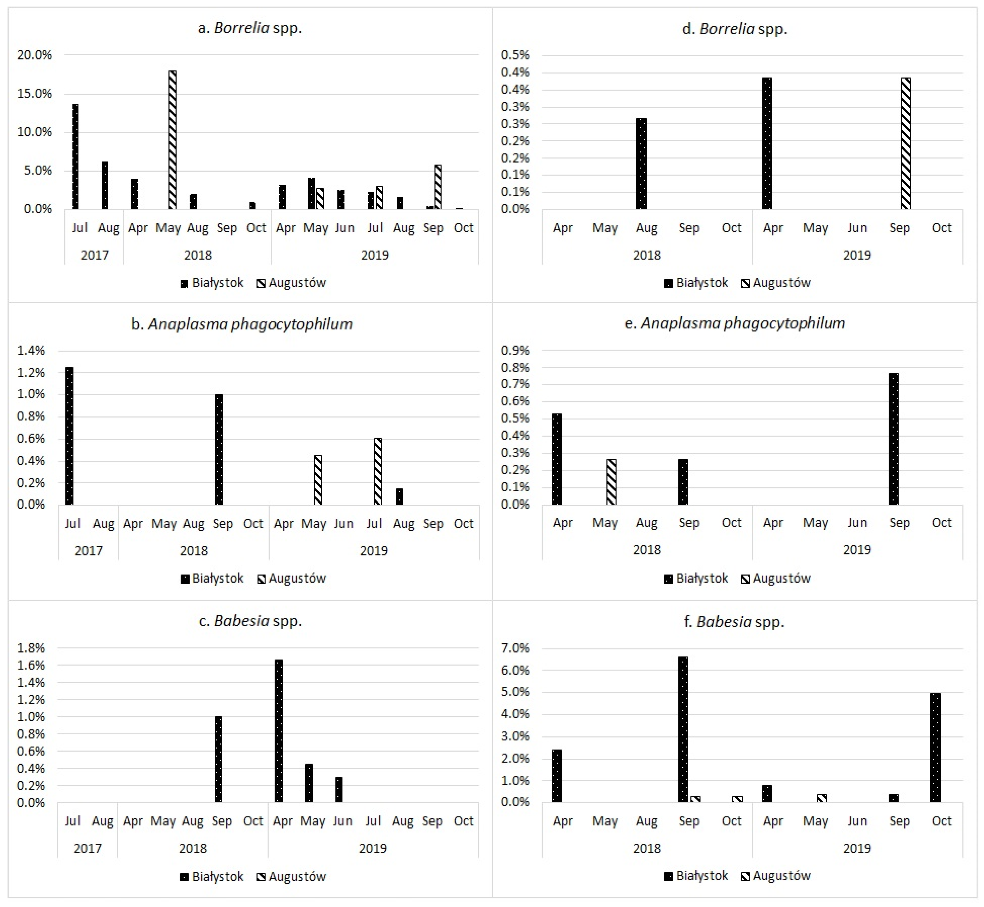
In Białystok, among 118 Borrelia-positive I. ricinus ticks, the majority was identified as Borrelia afzelii (65.3%; 77/118) with a similarity ranging from 86.88% to 98.69% to bacteria isolated both from humans in Austria and Germany (GenBank: CP009058.1, CP018262.1) and from ticks in Russia and France (CP009212.1, MW301927.1). Seventeen ticks (14.4%) showed 89.43–98.61% identity to Borrelia garinii isolated from different tick species from France, Spain (GenBank: CP028861.1, DQ147793.1), and Russia (GenBank: EF488989.1, KY312011.1, KY312012.1), as well as from human blood in China (GenBank: AY342031.1). Ten sequences (8.4%) were 83.97–98.25% identical to Borrelia burgdorferi sensu stricto, found in I. ricinus in the United Kingdom (GenBank: X98233.1) and in Peromyscus leucopus in the USA (GenBank: CP031412.1). The next 10 sequences (8.4%) were identified as Borrelia miyamotoi with a similarity range of 95.04–98.94% (GenBank: CP046389.1; Ixodes eggs, Czech Republic), while the remaining four sequences (3.4%) showed 92.49–97.05% identity to Borrelia lusitaniae (GenBank: AB091820.1; I. ricinus, Turkey). One Borrelia-positive D. reticulatus tick showed 96.86% similarity to B. afzelii (GenBank: CP009058.1). As for Babesia spp. sequencing, 15 out of 17 (88.2%) I. ricinus ticks showed 87.10–99.47% identity to Babesia microti isolated from ticks and small mammals from Turkey, China, Thailand, and Germany (GenBank: MH628094.1, KY649348.1, MG199182.1, MN355504.1, KP055650.1), while two were 91.83% and 99.36% identical to Babesia venatorum (GenBank: KR003828.1). Among Babesia-positive D. reticulatus ticks, 15 (93.8%; 15/16) were identified as Babesia canis, with 86.60–98.98% similarity to small and medium mammals from Lithuania, Poland, Serbia, Iran, Ukraine, Turkey, Romania, and Italy (GenBank: MN078319.1, MK872807.1, MH702200.1, MN173223.1, MN704759.1, MK934420.1, MG569903.1, KU821654.1, KT844899.1), as well as ticks from Ukraine and Poland (GenBank: MT346582.1, MF797820.1). One sequence showed 93.69% identity to B. microti (GenBank: KP055650.1, Myodes glareolus, Germany).
As for samples from Augustów, 34 out of 94 Borrelia-positive I. ricinus ticks were 82.89–98.25% identical to B. garinii (GenBank: CP028861.1, DQ147793.1, EF488989.1, KY312011.1; MW301936.1: I. ricinus, France). The next 29 sequences showed 88.80–99.10% similarity to B. afzelii (GenBank: CP009058.1, CP018262.1, MW301927.1), while 15 were 93.29–98.93% identical to B. burgdorferi s.s. (GenBank: CP031412.1; CP002228.1 and CP017201.1: humans, USA). Borrelia valaisiana was identified in nine samples, with similarity ranging from 91% to 98.24% to Ixodes ticks from France and Russia (GenBank: MW301935.1, CP009117.1). One sequence was 97.05% identical to B. lusitaniae found in I. ricinus in Turkey (GenBank: AB091820.1), and finally, six sequences were 96.83–98.94% similar to B. miyamotoi (GenBank: CP046389.1). One Borrelia-positive D. reticulatus female was identified as B. garinii with 89.43% similarity (GenBank: CP028861.1). Sequencing analysis for Babesia-positive samples (three male D. reticulatus) identified all of them as B. canis with 89.50–96.83% similarity (GenBank: MN704759.1, MK934420.1).
Sequencing of samples positive for A. phagocytophilum showed 100% similarity with the A. phagocytophilum strain Webster (188/188 bp) (GenBank: NR_044762.1).

3.2. Co-Infections

Overall, simultaneous presence of two different pathogens was detected in 14 I. ricinus ticks (1.7%; 14/842), among which 13 were collected in Białystok (2.8%; 13/460). The most prevalent co-infection was B. afzelii and B. microti, confirmed in 10 specimens (6 females, 3 males, 1 nymph). The remaining three samples, all females, were coinfected with B. afzelii and B. venatorum, B. burgdorferi s.s. and B. microti, as well as B. garinii and B. microti, respectively. Presence of B. afzelii and A. phagocytophilum was confirmed in one male tick from Augustów.

3.3. Phylogenetic Analysis

The results of the phylogenetic analysis are presented in a graphical form in Figure 4, Figure 5, Figure 6 and Figure 7.

3.4. Statistical Analysis

Statistical analysis revealed significant differences in several categories. Data from previous and present research used in the evaluation is presented in Figure 8.

3.4.1. Mann-Whitney Test

Statistically significant results were obtained in the following categories (Table 3).

Overall Infection Rate
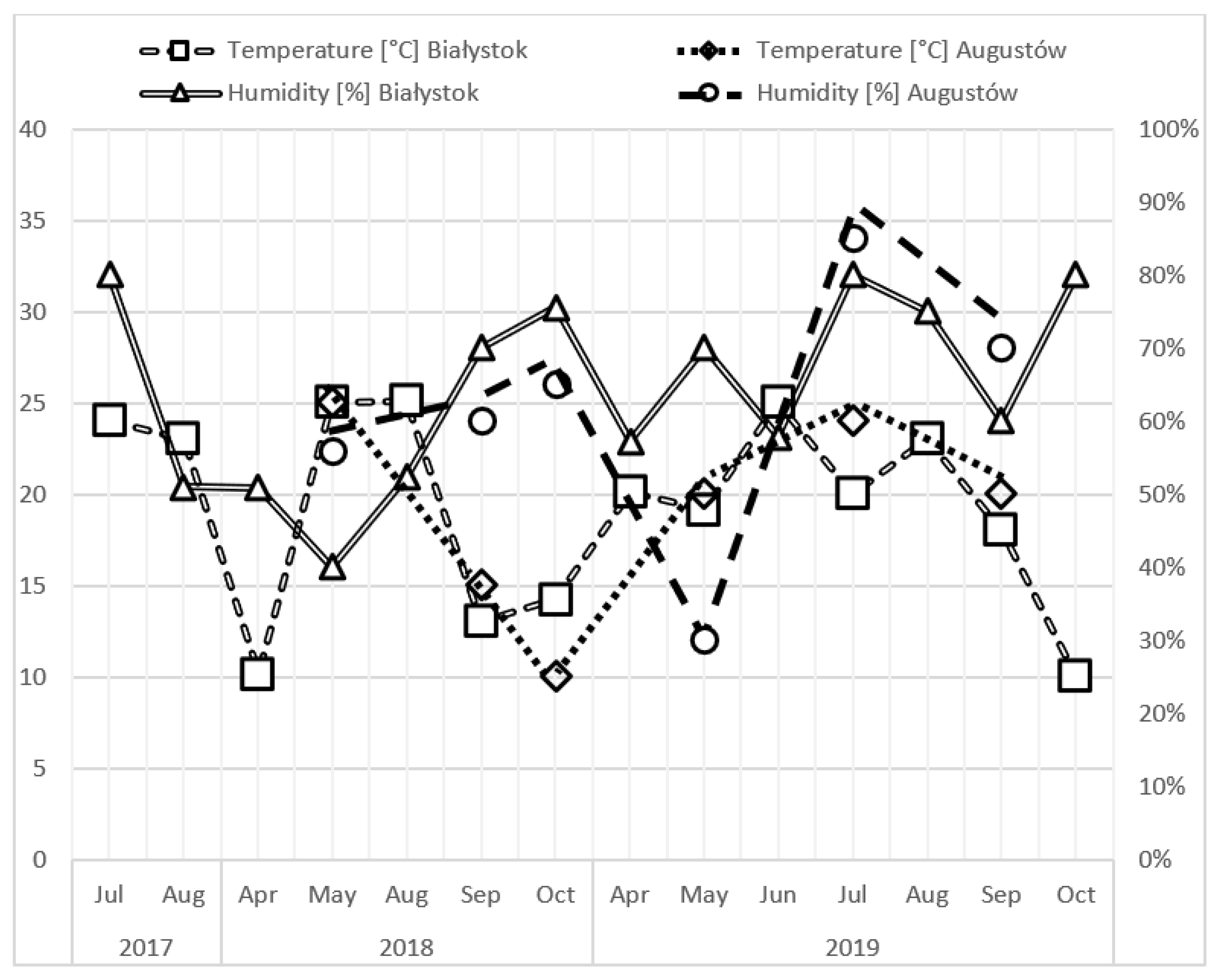
Comparative analysis revealed a statistically significant difference between Borrelia spp. infection rate in I. ricinus and D. reticulatus, with higher prevalence in I. ricinus ticks. Moreover, significantly more D. reticulatus ticks were infected with Babesia spp.

Air Temperature

For April–July, a statistically significant difference was confirmed in Borrelia spp. infection rates in ticks collected in over 20 °C temperature. Opposite results were obtained for Babesia spp. The same relations in both pathogens were observed in August–October.

Relative Air Humidity

The comparative analysis revealed a higher amount of Babesia spp. infections in ticks collected during periods of air relative humidity below 80% humidity in April–July. In contradiction, Borrelia spp. was found more frequently in ticks sampled in over 80% humidity in August–October.

Sampling Season

Comparative analysis of infection rates in individual developmental stages in relation to the sampling season revealed statistically significant differences for Borrelia spp. It was established that more adults were positive for this pathogen if collected in April–July, while higher prevalence was noticed for nymphs sampled in August–October.

3.4.2. Multivariate Logistic Regression Model

Multivariate logistic regression analysis showed that, for Borrelia spp. infections, D. reticulatus ticks were 97.16 times less likely to be infected with this pathogen as compared to I. ricinus. Moreover, the chance of detecting Borrelia spp. increased by 1.35 times in the successive sampling years and decreased by 1.59 times in males and nymphs, as compared to females (Table 4).

4. Discussion

Up until the 1980s, reports on infections in ticks in urban landscapes were incidental. Ever since then, the number of publications on this subject rose, presumably due to rapid development of recreational areas and green tourism, as well as progressing global urbanization [2].
Sequencing analysis of Borrelia-positive I. ricinus ticks identified the majority as B. afzelii and B. garinii, both in ticks collected in Białystok and Augustów. Those two Borrelia species are predominant in Europe [25], which was confirmed in other studies, including those from urban areas [6,7,26,27,28]. In this study, B. lusitaniae was confirmed in four I. ricinus ticks. Other than the current study, its presence was detected in two studies focusing on urban surroundings [7,27].
Interestingly, 6.4–8.4% of I. ricinus ticks were positive for B. miyamotoi, the causative agent of relapsing fever. Literature data regarding this spirochete presence in urban surrounding is scarce. However, B. miyamotoi was detected in such studies in Poland (4.7%) [26] and Switzerland (4.2%) [7]. Krause et al. suggests that B. miyamotoi may be prevalent in endemic borreliosis areas [29]. Human cases of B. miyamotoi infection were first reported in Russia in 2011 [30] and were since then described in multiple studies across Europe, the USA, and Japan [29,31,32].
It is worth emphasizing once again that Lyme disease incidence in the study region (107.7 per 100,000 people) is twice as high as the average in Poland (53.7 per 100,000 people) [14]. It is also worth noting that overall B. burgdorferi s.l. infection rates (23.5% and 23.0% for Białystok and Augustów, respectively) obtained in this study were also higher than the mean prevalence of B. burgdorferi s.l. in I. ricinus ticks in Europe (12.3%). Strnad et al. highlight that infection rates appear to increase significantly from western to eastern Europe [33].
B. afzelii was detected in only two D. reticulatus ticks (0.7%). This spirochete was also identified in other studies from Poland, although only in those from rural areas (0.09–1.6%) [34,35,36]. Low prevalence of B. burgdorferi s.l. in D. reticulatus ticks was confirmed in multiple studies in Europe [37,38,39,40], which may suggest that D. reticulatus ticks are ineffective vectors for this pathogen. In their study, Rudolf et al. examined the effect of D. reticulatus salivary glands and midgut extract on the growth, motility, and morphology of B. garinii in vitro. It was revealed that the extracts inhibited the growth of the spirochete [41].
The statistical analysis revealed higher median Borrelia spp. infection rate in ticks collected in temperatures above 20 °C in both seasons. It is known that the questing activity of I. ricinus nymphs and adults ranges from March to October [42], with a peak in AprilMay [43]. In a previous study that collected data on I. ricinus ticks from urban areas in Europe, it was revealed that temperature over 20 °C was connected to greater B. burgdorferi s.l. prevalence [44]. A relationship between higher mean temperatures and an increase in Lyme disease incidence was also observed by other studies [45,46]. As Keith et al. note, this may be further connected to the increase of human recreational activity in the warmer weather, thus higher tick exposure [45]. Babesia spp. infections were detected more frequently in temperatures below 20 °C and <80% humidity. In this study, D. reticulatus ticks were found to be primarily infected with Babesia spp. Additionally, all of the specimens were adults, who are most active during early spring (March-April) and autumn (September-October) [47], which, in Poland, are associated with lower temperatures. In comparison to I. ricinus ticks, D. reticulatus show higher resilience to colder environmental conditions [47,48].
In this study, the presence of A. phagocytophilum was confirmed in 0.7–1.8% I. ricinus ticks. Similar prevalence was reported in other Polish cities (1.7–3%) [11,49]. In Europe, A. phagocytophilum was detected in urban areas in Germany (1.7–3.8%) [10,27,50], Ukraine (5.2%) [5], Czech Republic (0–5.2%) [4,6], Switzerland (1.4%) [8], and Slovakia (3.1–7.2%) [51]. In a comprehensive study conducted by Derdakova et al. in various habitats across Slovakia, the Czech Republic, and Austria, it was found that the mean prevalence of A. phagocytophilum was 3.8% [52].
Overall, 1.1% of D. reticulatus ticks were infected with A. phagocytophilum in the current study, which is consistent with previous findings [15]. Similar results were obtained in Kyiv, Ukraine (0–1%) [5,53], while in the outskirts of Berlin, Germany, none of the collected D. reticulatus ticks were positive for this pathogen [39]. Comparable values were reported in studies conducted in rural areas of Poland and Serbia (0–1.1% and 1.9%, respectively) [36,40,54]. Results obtained in the current study most likely reflect the availability and population density of A. phagocytophilum hosts, such as rodents, hedgehogs, ungulates, foxes, and birds [5,11,51], which are necessary for the completion of the A. phagocytophilum life cycle, since this bacterium is not transmitted transovarially [55].The majority of Babesia in I. ricinus ticks were identified as B. microti in this study. Similar results were obtained by Wójcik-Fatla et al. in their research on recreational sites of eastern Poland [56]. It is worth noting that other studies focused on urban areas identified B. venatorum as the most prevalent [7,49,57]. Interestingly, one of the identified B. microti sequences (KP055650.1) is 100% identical to the pathogenic Jena/Germany strain. However, as stressed by Obiegala et al., it does not mean that the newly detected sequence is also pathogenic [58].
B. canis was the predominant pathogen identified in D. reticulatus ticks in this study (6.7% of all specimens), similar to the previous study (6.8%) [15]. Comparable results (4.18%) were obtained by Mierzejewska et al., who studied D. reticulatus ticks collected from multiple localities in eastern, central, and western Poland. In that study, B. canis was found only in ticks collected in the Eastern part of the country [34]. Noteworthy, Eastern Poland belongs to the European macro-region for D. reticulatus presence [59]. It is reflected by reported canine babesiosis cases. In a study conducted by Dwużnik et al., the authors collected data from 42 veterinary clinics from Eastern and Western Poland and reported 1558 cases of canine babesiosis. Interestingly, the majority (1532) of them came from clinics in the Eastern part of the country [60]. B. canis was also detected in a number of different studies on D. reticulatus ticks from Ukraine, Latvia, Lithuania, Slovakia, and Poland (0.63–3.4%) [53,56,61,62,63]. Notably, two studies from Poland and Serbia reported exceptionally high B. canis prevalence (21.3% and 20.8%, respectively) [35,40].
In this study, one D. reticulatus was infected with B. microti (0.4%), which is consistent with previous findings (0.8%) [15]. Other studies from Poland report 0.04–4.5% infection rate [34,36,54,64]. It is worth noting that the detected sequence was the same potentially pathogenic sequence as described in I. ricinus. Although it is known that D. reticulatus ticks rarely feed on humans [65], they significantly contribute to the circulation of pathogens, including those potentially harmful to humans, in the environment.
No I. ricinus ticks tested positively for C. burnetii, the causative agent of Q fever. Similar values (0–0.2%) were reported in studies conducted in Poland [66,67], Switzerland [68], Austria [69], and Sweden [70]. A notably higher infection rate (15.9%) was obtained by Szymańska-Czerwińska et al. in I. ricinus ticks collected from forests in south-eastern Poland [71]. In other European countries, obtained prevalence in rural areas was 0–4.9% in Slovakia [72,73,74], 1.7% in Belarus [38], and 1.9% in Germany [75].
Similarly, C. burnetii was not detected in any of the tested D. reticulatus ticks, which is consistent with results obtained in other studies [38,67,72,76]. Low prevalence of C. burnetii was detected in studies from Slovakia (2.1%) [74] and Serbia (3.7%) [40].
Bartonella spp. was not detected in any I. ricinus ticks investigated in this study. Similar values were reported in other European studies, both in urban Germany [77,78] and rural areas [38,40,79]. This pathogen has been reported in other research conducted in Poland with 1.7–4.8% prevalence, although all infected ticks were collected either from vegetation in rural areas or from animals [63,80,81]. In the current study, no D. reticulatus ticks were infected with Bartonella spp. Comparable results were obtained in research focused on urban areas: 0.5% in Warsaw, Poland [80] and 1.0% in Kyiv, Ukraine [53], as well as in rural surroundings: 0.6% in Belarus [38] and 0% in Serbia [40].
In this study, no I. ricinus tested positively for Rickettsia spp. In comparison, the presence of this pathogen was confirmed in urban parks in Warsaw (2.9–7.7%) [11,66]. In other European countries, infection rates reported in ticks from city landscapes were also higher, ranging from 5.7% to 16.5% [4,7,8,53,82,83]. Exceptionally high prevalence was confirmed within urban areas of Hanover and Hamburg, Germany (50.8% and 52.5%, respectively) [10,84].
Interestingly, also no D. reticulatus ticks were found to be infected with Rickettsia spp. in the current study. This pathogen has been reported with a high prevalence rate (40.7–56.7%) in natural sites in Poland [34,36,66,85,86,87]. Studies from urban areas in Kyiv, Ukraine, revealed 10.1–35.7% infection rate [5,53]. In other European countries, reported infection rates in natural sites were similar (14–21.4%) [65,88]. Given such discrepancies between Rickettsia spp. prevalence obtained in this and other studies, further research in the study area, focusing on this pathogen, is needed.

5. Conclusions

In conclusion, the molecular investigation carried out in this study confirms that I. ricinus and D. reticulatus ticks present within urban areas of the northeastern Poland are infected with at least three pathogens: Borrelia spp., A. phagocytophilum, and Babesia spp. Moreover, results reveal that the prevalence of B. burgdorferi s.l. is equal or even higher than in natural ecosystems. As this is the first study on ticks in cities of northeastern Poland, it provides valuable information for tick-borne pathogen surveillance.

Author Contributions

Conceptualization, all; methodology, J.D.-M.; validation, all; formal analysis, P.C., R.M. and P.M.; investigation, A.G. and J.D.-M.; resources, A.G. and J.D.-M.; data curation, A.G.; writing—original draft preparation, A.G.; writing—review and editing, A.G., J.D.-M. and A.M.-M.; visualization, A.G.; supervision, S.P. and A.M.-M.; project administration, J.D.-M. and A.M.-M.; funding acquisition, A.G. All authors have read and agreed to the published version of the manuscript.

Funding

This research was funded by Medical University of Bialystok, Poland, grant number N/ST/MN/18/001/1145.

Institutional Review Board Statement

The animal study protocol was approved by the Bioethics Committee of Medical University of Białystok (permit number: R-I-002/341/2018).

Data Availability Statement

The data sets used and/or analyzed during the current study are available from the corresponding author on request.

Conflicts of Interest

The authors declare no conflict of interest.

References

  1. Rizzoli, A.; Silaghi, C.; Obiegala, A.; Rudolf, I.; Hubalek, Z.; Foldvari, G.; Plantard, O.; Vayssier-Taussat, M.; Bonnet, S.; Spitalska, E.; et al. Ixodes ricinus and its transmitted pathogens in urban and peri-urban areas in Europe: New Hazards and Relevance for Public Health. Front. Public Health 2014, 2, 251. [Google Scholar] [CrossRef] [PubMed]
  2. Akimov, I.A.; Nebogatkin, I.V. Ixodid ticks (Acari, Ixodidae) in urban landscapes. A Review. Vestn. Zool. 2016, 50, 155–162. [Google Scholar] [CrossRef] [Green Version]
  3. Heylen, D.; Lasters, R.; Adriaensen, F.; Fonville, M.; Sprong, H.; Matthysen, E. Ticks and tick-borne diseases in the city: Role of Landscape Connectivity and Green Space Characteristics in a Metropolitan Area. Sci. Total Environ. 2019, 670, 941–949. [Google Scholar] [CrossRef] [PubMed]
  4. Venclíková, K.; Mendel, J.; Betášová, L.; Blažejová, H.; Jedličková, P.; Straková, P.; Hubálek, Z.; Rudolf, I. Neglected tick-borne pathogens in the Czech Republic, 2011–2014. Ticks Tick. Borne. Dis. 2016, 7, 107–112. [Google Scholar] [CrossRef] [PubMed]
  5. Didyk, Y.M.; Blaňárová, L.; Pogrebnyak, S.; Akimov, I.; Peťko, B.; Víchová, B. Emergence of tick-borne pathogens (Borrelia burgdorferi sensu lato, Anaplasma phagocytophilum, Rickettsia raoultii and Babesia microti) in the Kyiv urban parks, Ukraine. Ticks Tick. Borne. Dis. 2017, 8, 219–225. [Google Scholar] [CrossRef]
  6. Kybicová, K.; Baštová, K.; Malý, M. Detection of Borrelia burgdorferi sensu lato and Anaplasma phagocytophilum in questing ticks Ixodes ricinus from the Czech Republic. Ticks Tick. Borne. Dis. 2017, 8, 483–487. [Google Scholar] [CrossRef]
  7. Lommano, E.; Bertaiola, L.; Dupasquier, C.; Gern, L. Infections and coinfections of questing Ixodes ricinus ticks by emerging zoonotic pathogens in Western Switzerland. Appl. Environ. Microbiol. 2012, 78, 4606–4612. [Google Scholar] [CrossRef] [Green Version]
  8. Oechslin, C.P.; Heutschi, D.; Lenz, N.; Tischhauser, W.; Péter, O.; Rais, O.; Beuret, C.M.; Leib, S.L.; Bankoul, S.; Ackermann-Gäumann, R. Prevalence of tick-borne pathogens in questing Ixodes ricinus ticks in urban and suburban areas of Switzerland. Parasites Vectors 2017, 10, 558. [Google Scholar] [CrossRef] [Green Version]
  9. Kowalec, M.; Szewczyk, T.; Welc-Falȩciak, R.; Siński, E.; Karbowiak, G.; Bajer, A. Ticks and the city—Are there any differences between city parks and natural forests in terms of tick abundance and prevalence of spirochaetes? Parasites Vectors 2017, 10, 573. [Google Scholar] [CrossRef]
  10. Blazejak, K.; Janecek, E.; Strube, C. A 10-Year Surveillance of Rickettsiales (Rickettsia spp. and Anaplasma phagocytophilum) in the city of Hanover, Germany, reveals Rickettsia spp. as emerging pathogens in ticks. Parasites Vectors 2017, 10, 588. [Google Scholar] [CrossRef] [Green Version]
  11. Welc-Falȩciak, R.; Kowalec, M.; Karbowiak, G.; Bajer, A.; Behnke, J.M.; Siński, E. Rickettsiaceae and Anaplasmataceae infections in Ixodes ricinus ticks from urban and natural forested areas of Poland. Parasites Vectors 2014, 7, 121. [Google Scholar] [CrossRef] [PubMed] [Green Version]
  12. Estrada-Peña, A.; Ayllón, N.; de la Fuente, J. Impact of climate trends on tick-borne pathogen transmission. Front. Physiol. 2012, 3, 64. [Google Scholar] [CrossRef] [PubMed] [Green Version]
  13. Ogden, N.H.; Lindsay, L.R. Effects of climate and climate change on vectors and vector-borne diseases: Ticks are Different. Trends Parasitol. 2016, 32, 646–656. [Google Scholar] [CrossRef]
  14. National Institute of Public Health. Infectious Diseases and Poisonings in Poland in 2019. Available online: http://wwwold.pzh.gov.pl/oldpage/epimeld/2019/Ch_2019.pdf (accessed on 13 October 2021).
  15. Grochowska, A.; Dunaj, J.; Pancewicz, S.; Czupryna, P.; Majewski, P.; Wondim, M.; Tryniszewska, E.; Moniuszko-Malinowska, A. Detection of Borrelia burgdorferi s.l., Anaplasma phagocytophilum and Babesia spp. in Dermacentor reticulatus ticks found within the city of Białystok, Poland-first data. Exp. Appl. Acarol. 2021, 85, 63–73. [Google Scholar] [CrossRef] [PubMed]
  16. Nowak-Chmura, M. Fauna Kleszczy (Ixodida) Europy Środkowej; Wydawnictwo Naukowe Uniwersytetu Pedagogicznego: Kraków, Poland, 2013; ISBN 978-83-7271-829-7. [Google Scholar]
  17. Dunaj, J.; Moniuszko-Malinowska, A.; Święcicka, I.; Andersson, M.; Czupryna, P.; Rutkowski, K.; Zambrowski, G.; Zajkowska, J.; Grygorczuk, S.; Kondrusik, M.; et al. Tick-borne infections and co-infections in patients with non-specific symptoms in Poland. Adv. Med. Sci. 2018, 63, 167–172. [Google Scholar] [CrossRef] [PubMed]
  18. Moniuszko-Malinowska, A.; Święcicka, I.; Dunaj, J.; Zajkowska, J.; Czupryna, P.; Zambrowski, G.; Chmielewska–Badora, J.; Żukiewicz-Sobczak, W.; Swierzbinska, R.; Rutkowski, K.; et al. Infection with Babesia microti in humans with non-specific symptoms in north east Poland. Infect. Dis. 2016, 48, 537–543. [Google Scholar] [CrossRef] [PubMed]
  19. Katargina, O.; Geller, J.; Vasilenko, V.; Kuznetsova, T.; Järvekülg, L.; Vene, S.; Lundkvist, Ã.; Golovljova, I. Detection and characterization of Babesia species in Ixodes ticks in Estonia. Vector-Borne Zoonotic Dis. 2011, 11, 923–928. [Google Scholar] [CrossRef]
  20. Pichon, B.; Kahl, O.; Hammer, B.; Gray, J.S. Pathogens and host DNA in Ixodes ricinus nymphal ticks from a German Forest. Vector Borne Zoonotic Dis. 2006, 6, 382–387. [Google Scholar] [CrossRef]
  21. Saitou, N.; Nei, M. The Neighbor-Joining Method: A New Method for Reconstructing Phylogenetic Trees. Mol. Biol. Evol. 1987, 4, 406–425. [Google Scholar] [CrossRef]
  22. Tamura, K.; Nei, M. Estimation of the Number of Nucleotide Substitutions in the Control Region of Mitochondrial DNA in humans and chimpanzees. Mol. Biol. Evol. 1993, 10, 512–526. [Google Scholar] [CrossRef]
  23. Kumar, S.; Stecher, G.; Li, M.; Knyaz, C.; Tamura, K. MEGA X: Molecular Evolutionary Genetics Analysis across Computing Platforms. Mol. Biol. Evol. 2018, 35, 1547–1549. [Google Scholar] [CrossRef] [PubMed]
  24. Letunic, I.; Bork, P. Interactive Tree Of Life (ITOL) v5: An Online Tool for Phylogenetic Tree Display and Annotation. Nucleic Acids Res. 2021, 49, W293–W296. [Google Scholar] [CrossRef] [PubMed]
  25. Reye, A.L.; Hübschen, J.M.; Sausy, A.; Muller, C.P. Prevalence and Seasonality of Tick-Borne Pathogens in Questing Ixodes ricinus ticks from Luxembourg. Appl. Environ. Microbiol. 2010, 76, 2923–2931. [Google Scholar] [CrossRef] [Green Version]
  26. Kiewra, D.; Stańczak, J.; Richter, M. Ixodes ricinus ticks (Acari, Ixodidae) as a vector of Borrelia burgdorferi sensu lato and Borrelia miyamotoi in Lower Silesia, Poland—Preliminary study. Ticks Tick. Borne. Dis. 2014, 5, 892–897. [Google Scholar] [CrossRef] [PubMed]
  27. May, K.; Jordan, D.; Fingerle, V.; Strube, C. Borrelia burgdorferi sensu lato and co-infections with Anaplasma phagocytophilum and Rickettsia spp. in Ixodes ricinus in Hamburg, Germany. Med. Vet. Entomol. 2015, 29, 425–429. [Google Scholar] [CrossRef]
  28. Pangrácová, L.; Derdáková, M.; Pekárik, L.; Hviščová, I.; Víchová, B.; Stanko, M.; Hlavatá, H.; Peťko, B. Ixodes ricinus abundance and its infection with the tick-borne pathogens in urban and suburban areas of eastern Slovakia. Parasites Vectors 2013, 6, 238. [Google Scholar] [CrossRef] [Green Version]
  29. Krause, P.J.; Narasimhan, S.; Wormser, G.P.; Rollend, L.; Fikrig, E.; Lepore, T.; Barbour, A.; Fish, D. Human Borrelia Miyamotoi Infection in the United States. N. Engl. J. Med. 2013, 368, 291–293. [Google Scholar] [CrossRef] [Green Version]
  30. Platonov, A.E.; Karan, L.S.; Kolyasnikova, N.M.; Makhneva, N.A.; Toporkova, M.G.; Maleev, V.V.; Fish, D.; Krause, P.J. Humans infected with relapsing fever spirochete Borrelia miyamotoi, Russia. Emerg. Infect. Dis. 2011, 17, 1816–1823. [Google Scholar] [CrossRef]
  31. Kubiak, K.; Szczotko, M.; Dmitryjuk, M. Borrelia miyamotoi—an emerging human tick-borne pathogen in Europe. Microorganisms 2021, 9, 154. [Google Scholar] [CrossRef]
  32. Sato, K.; Takano, A.; Konnai, S.; Nakao, M.; Ito, T.; Koyama, K.; Kaneko, M.; Ohnishi, M.; Kawabata, H. Human Infections with Borrelia miyamotoi, Japan. Emerg. Infect. Dis. 2014, 20, 1391–1393. [Google Scholar] [CrossRef]
  33. Strnad, M.; Honig, V.; Ruzek, D.; Grubhoffer, L.; Rego, R.O.M. Europe-wide meta-analysis of Borrelia burgdorferi sensu lato prevalence in questing Ixodes ricinus ticks. Appl. Environ. Microbiol. 2017, 83, e00609-17. [Google Scholar] [CrossRef] [PubMed] [Green Version]
  34. Mierzejewska, E.J.; Pawełczyk, A.; Radkowski, M.; Welc-Falęciak, R.; Bajer, A. Pathogens vectored by the tick, Dermacentor reticulatus, in endemic regions and zones of expansion in Poland. Parasites Vectors 2015, 8, 490. [Google Scholar] [CrossRef] [PubMed] [Green Version]
  35. Dzięgiel, B.; Kubrak, T.; Adaszek, Ł.; Dębiak, P.; Wyłupek, D.; Bogucka-kocka, A.; Lechowski, J.; Winiarczyk, S. Prevalence of Babesia canis, Borrelia burgdorferi sensu lato, and Anaplasma phagocytophilum in hard ticks collected from meadows of Lubelskie Voivodship (Eastern Poland). Bull. Vet. Inst. Pulawy 2014, 58, 29–33. [Google Scholar] [CrossRef] [Green Version]
  36. Zając, V.; Wójcik-Fatla, A.; Sawczyn, A.; Cisak, E.; Sroka, J.; Kloc, A.; Zając, Z.; Buczek, A.; Dutkiewicz, J.; Bartosik, K. Prevalence of infections and co-infections with 6 pathogens in Dermacentor reticulatus ticks collected in eastern Poland. Ann. Agric. Environ. Med. 2017, 24, 26–32. [Google Scholar] [CrossRef] [PubMed]
  37. Król, N.; Kiewra, D.; Szymanowski, M.; Lonc, E. The role of domestic dogs and cats in the zoonotic cycles of ticks and pathogens. preliminary studies in the Wroclaw Agglomeration (SW Poland). Vet. Parasitol. 2015, 214, 208–212. [Google Scholar] [CrossRef]
  38. Reye, A.L.; Stegniy, V.; Mishaeva, N.P.; Velhin, S.; Hübschen, J.M.; Ignatyev, G.; Muller, C.P. Prevalence of tick-borne pathogens in Ixodes ricinus and Dermacentor reticulatus ticks from different geographical locations in Belarus. PLoS ONE 2013, 8, e54476. [Google Scholar] [CrossRef] [Green Version]
  39. Richter, D.; Kohn, C.; Matuschka, F.-R. Absence of Borrelia spp., Candidatus Neoehrlichia mikurensis, and Anaplasma phagocytophilum in questing adult Dermacentor reticulatus ticks. Parasitol. Res. 2013, 112, 107–111. [Google Scholar] [CrossRef]
  40. Tomanovic, S.; Chochlakis, D.; Radulovic, Z.; Milutinovic, M.; Cakic, S.; Mihaljica, D.; Tselentis, Y.; Psaroulaki, A. Analysis of pathogen co-occurrence in host-seeking adult hard ticks from Serbia. Exp. Appl. Acarol. 2013, 59, 367–376. [Google Scholar] [CrossRef]
  41. Rudolf, I.; Hubalek, Z. Effect of the salivary gland and midgut extracts from Ixodes ricinus and Dermacentor reticulatus (Acari: Ixodidae) on the growth of Borrelia garinii in vitro. Folia Parasitol. 2003, 50, 159–160. [Google Scholar] [CrossRef] [Green Version]
  42. Gray, J.S.; Dautel, H.; Estrada-Peña, A.; Kahl, O.; Lindgren, E. Effects of climate change on ticks and tick-borne diseases in Europe. Interdiscip. Perspect. Infect. Dis. 2009, 2009, 593232. [Google Scholar] [CrossRef]
  43. Steinbrink, A.; Brugger, K.; Margos, G.; Kraiczy, P.; Klimpel, S. The evolving story of Borrelia burgdorferi sensu lato transmission in Europe. Parasitol. Res. 2022, 121, 781–803. [Google Scholar] [CrossRef] [PubMed]
  44. Grochowska, A.; Milewski, R.; Pancewicz, S.; Dunaj, J.; Czupryna, P.; Milewska, A.J.; Róg-Makal, M.; Grygorczuk, S.; Moniuszko-Malinowska, A. Comparison of tick-borne pathogen prevalence in Ixodes ricinus ticks collected in urban areas of Europe. Sci. Rep. 2020, 10, 6975. [Google Scholar] [CrossRef] [PubMed]
  45. Keith, K.; Årestedt, K.; Tjernberg, I. The relationship between the laboratory diagnosis of Lyme neuroborreliosis and climate factors in Kalmar County Sweden—An overview between 2008 and 2019. Eur. J. Clin. Microbiol. Infect. Dis. 2022, 41, 253–261. [Google Scholar] [CrossRef] [PubMed]
  46. Bennet, L.; Halling, A.; Berglund, J. Increased incidence of Lyme borreliosis in southern Sweden following mild winters and during warm, humid summers. Eur. J. Clin. Microbiol. Infect. Dis. 2006, 25, 426–432. [Google Scholar] [CrossRef] [Green Version]
  47. Földvári, G.; Široký, P.; Szekeres, S.; Majoros, G.; Sprong, H. Dermacentor reticulatus: A Vector on the Rise. Parasites Vectors 2016, 9, 314. [Google Scholar] [CrossRef] [Green Version]
  48. Tokhov, Y.M.; Lutsuk, S.N.; Dyachenko, Y.V. Phenology of Ixodid ticks of the genus Dermacentor in the Central Ciscaucasia. Entomol. Rev. 2014, 94, 426–433. [Google Scholar] [CrossRef]
  49. Stańczak, J.; Cieniuch, S.; Lass, A.; Biernat, B.; Racewicz, M. Detection and quantification of Anaplasma phagocytophilum and Babesia spp. in Ixodes ricinus ticks from urban and rural environment, northern Poland, by Real-Time Polymerase Chain Reaction. Exp. Appl. Acarol. 2015, 66, 63–81. [Google Scholar] [CrossRef] [Green Version]
  50. Mehlhorn, H.; Mehlhorn, T.; Müller, M.; Vogt, M.; Rissland, J. Tick survey for prevalent pathogens in peri-urban recreation sites in Saarland and Rhineland-Palatinate (Germany). Parasitol. Res. 2016, 115, 1167–1172. [Google Scholar] [CrossRef]
  51. Svitálková, Z.; Haruštiaková, D.; Mahríková, L.; Berthová, L.; Slovák, M.; Kocianová, E.; Kazimírová, M. Anaplasma phagocytophilum prevalence in ticks and rodents in an urban and natural habitat in south-western Slovakia. Parasites Vectors 2015, 8, 276. [Google Scholar] [CrossRef] [Green Version]
  52. Derdáková, M.; Václav, R.; Pangrácova-Blaňárová, L.; Selyemová, D.; Koči, J.; Walder, G.; Špitalská, E. Candidatus Neoehrlichia mikurensis and its co-circulation with Anaplasma Phagocytophilum in Ixodes ricinus ticks across ecologically different habitats of Central Europe. Parasites Vectors 2014, 7, 160. [Google Scholar] [CrossRef] [Green Version]
  53. Rogovskyy, A.; Batool, M.; Gillis, D.C.; Holman, P.J.; Nebogatkin, I.V.; Rogovska, Y.V.; Rogovskyy, M.S. Diversity of Borrelia spirochetes and other zoonotic agents in ticks from Kyiv, Ukraine. Ticks Tick. Borne. Dis. 2018, 9, 404–409. [Google Scholar] [CrossRef] [PubMed]
  54. Opalińska, P.; Wierzbicka, A.; Asman, M. The PCR and nested PCR detection of Borrelia burgdorferi sensu lato, Anaplasma phagocytophilum and Babesia microti in Dermacentor reticulatus F. Collected in a new location in Poland (Trzciel, Western Poland). Acta Parasitol. 2016, 61, 849–854. [Google Scholar] [CrossRef] [PubMed]
  55. Huhn, C.; Winter, C.; Wolfsperger, T.; Wuppenhorst, N.; Strasek Smrdel, K.; Skuballa, J.; Pfaffle, M.; Petney, T.; Silaghi, C.; Dyachenko, V.; et al. Analysis of the population structure of Anaplasma phagocytophilum using multilocus sequence typing. PLoS ONE 2014, 9, e93725. [Google Scholar] [CrossRef] [PubMed] [Green Version]
  56. Wójcik-Fatla, A.; Zając, V.; Sawczyn, A.; Cisak, E.; Dutkiewicz, J. Babesia spp. in questing ticks from eastern Poland: Prevalence and Species Diversity. Parasitol. Res. 2015, 114, 3111–3116. [Google Scholar] [CrossRef] [Green Version]
  57. Hamšíková, Z.; Kazimírová, M.; Haruštiaková, D.; Mahríková, L.; Slovák, M.; Berthová, L.; Kocianová, E.; Schnittger, L. Babesia spp. in ticks and wildlife in different habitat types of Slovakia. Parasites Vectors 2016, 9, 292. [Google Scholar] [CrossRef] [Green Version]
  58. Obiegala, A.; Pfeffer, M.; Pfister, K.; Karnath, C.; Silaghi, C. Molecular examinations of Babesia microti in rodents and rodent-attached ticks from urban and sylvatic habitats in Germany. Ticks Tick. Borne. Dis. 2015, 6, 445–449. [Google Scholar] [CrossRef]
  59. Karbowiak, G. The occurrence of the Dermacentor reticulatus tick--its expansion to new areas and possible causes. Ann. Parasitol. 2014, 60, 37–47. [Google Scholar]
  60. Dwużnik-Szarek, D.; Mierzejewska, E.J.; Rodo, A.; Goździk, K.; Behnke-Borowczyk, J.; Kiewra, D.; Kartawik, N.; Bajer, A. Monitoring the expansion of Dermacentor reticulatus and occurrence of canine babesiosis in Poland in 2016–2018. Parasit. Vectors 2021, 14, 267. [Google Scholar] [CrossRef]
  61. Karbowiak, G.; Vichová, B.; Slivinska, K.; Werszko, J.; Didyk, J.; Peťko, B.; Stanko, M.; Akimov, I. The infection of questing Dermacentor reticulatus ticks with Babesia canis and Anaplasma phagocytophilum in the Chernobyl Exclusion Zone. Vet. Parasitol. 2014, 204, 372–375. [Google Scholar] [CrossRef]
  62. Svehlová, A.; Berthová, L.; Sallay, B.; Boldiš, V.; Sparagano, O.A.E.; Spitalská, E. Sympatric occurrence of Ixodes ricinus, Dermacentor reticulatus and Haemaphysalis concinna ticks and Rickettsia and Babesia species in Slovakia. Ticks Tick. Borne. Dis. 2014, 5, 600–605. [Google Scholar] [CrossRef]
  63. Zając, V.; Wójcik-Fatla, A.; Dutkiewicz, J.; Szymańska, J. Bartonella henselae in eastern Poland: The Relationship between Tick Infection Rates and the Serological Response of Individuals Occupationally Exposed to Tick Bites. J. Vector Ecol. 2015, 40, 75–82. [Google Scholar] [CrossRef] [PubMed] [Green Version]
  64. Wójcik-Fatla, A.; Bartosik, K.; Buczek, A.; Dutkiewicz, J. Babesia microti in adult Dermacentor reticulatus ticks from eastern Poland. Vector Borne Zoonotic Dis. 2012, 12, 841–843. [Google Scholar] [CrossRef] [PubMed]
  65. Dautel, H.; Dippel, C.; Oehme, R.; Hartelt, K.; Schettler, E. Evidence for an increased geographical distribution of Dermacentor reticulatus in Germany and detection of Rickettsia Sp. RpA4. Int. J. Med. Microbiol. 2006, 296, 149–156. [Google Scholar] [CrossRef]
  66. Chmielewski, T.; Andrzejewski, K.; Maczka, I.; Fiecek, B.; Radlinska, M.; Tylewska-Wierzbanowska, S. Ticks infected with bacteria pathogenic to humans in municipal parks in Warsaw. Przegl. Epidemiol. 2011, 65, 577–581. [Google Scholar] [PubMed]
  67. Tylewska-Wierzbanowska, S.; Kruszewska, D.; Chmielewski, T.; Zukowski, K.; Zabicka, J. Ticks as a reservoir of Borrelia burgdorferi and Coxiella burnetii on Polish terrain. Przegl. Epidemiol. 1996, 50, 245–251. [Google Scholar] [PubMed]
  68. Pilloux, L.; Baumgartner, A.; Jaton, K.; Lienhard, R.; Ackermann-Gäumann, R.; Beuret, C.; Greub, G. Prevalence of Anaplasma phagocytophilum and Coxiella burnetii in Ixodes ricinus ticks in Switzerland: An Underestimated Epidemiologic Risk. New Microbes New Infect. 2019, 27, 22–26. [Google Scholar] [CrossRef] [PubMed]
  69. Schötta, A.M.; Wijnveld, M.; Stockinger, H.; Stanek, G. Approaches for reverse line blot-based detection of microbial pathogens in Ixodes ricinus ticks collected in Austria and impact of the chosen method. Appl. Environ. Microbiol. 2017, 83, e00489-17. [Google Scholar] [CrossRef] [Green Version]
  70. Wallménius, K.; Pettersson, J.H.O.; Jaenson, T.G.T.; Nilsson, K. Prevalence of Rickettsia spp., Anaplasma phagocytophilum, and Coxiella burnetii in adult Ixodes ricinus ticks from 29 study areas in central and southern Sweden. Ticks Tick. Borne. Dis. 2012, 3, 100–106. [Google Scholar] [CrossRef]
  71. Szymańska-Czerwińska, M.; Monika Galińska, E.; Niemczuk, K.; Zasepa, M. Prevalence of Coxiella burnetii infection in foresters and ticks in south-eastern Poland and comparison of diagnostic methods. Ann. Agric. Environ. Med. 2013, 20, 699–704. [Google Scholar]
  72. Smetanová, K.; Schwarzová, K.; Kocianová, E. Detection of Anaplasma phagocytophilum, Coxiella burnetii, Rickettsia spp., and Borrelia burgdorferi s. l. in ticks, and wild-living animals in western and middle Slovakia. Ann. N. Y. Acad. Sci. 2006, 1078, 312–315. [Google Scholar] [CrossRef]
  73. Minichová, L.; Hamšíková, Z.; Mahríková, L.; Slovák, M.; Kocianová, E.; Kazimírová, M.; Škultéty, Ľ.; Štefanidesová, K.; Špitalská, E. Molecular Evidence of Rickettsia spp. in Ixodid ticks and rodents in suburban, natural and rural habitats in Slovakia. Parasites Vectors 2017, 10, 158. [Google Scholar] [CrossRef] [PubMed] [Green Version]
  74. Špitalská, E.; Sparagano, O.; Stanko, M.; Schwarzová, K.; Špitalský, Z.; Škultéty, Ľ.; Havlíková, S.F. Diversity of Coxiella-like and Francisella-like endosymbionts, and Rickettsia spp., Coxiella burnetii as pathogens in the tick populations of Slovakia, Central Europe. Ticks Tick. Borne. Dis. 2018, 9, 1207–1211. [Google Scholar] [CrossRef] [PubMed]
  75. Hildebrandt, A.; Franke, J.; Schmoock, G.; Pauliks, K.; Krämer, A.; Straube, E. Diversity and coexistence of tick-borne pathogens in central Germany. J. Med. Entomol. 2011, 48, 651–655. [Google Scholar] [CrossRef] [PubMed]
  76. Pluta, S.; Hartelt, K.; Oehme, R.; Mackenstedt, U.; Kimmig, P. Prevalence of Coxiella burnetii and Rickettsia spp. in ticks and rodents in southern Germany. Ticks Tick. Borne. Dis. 2010, 1, 145–147. [Google Scholar] [CrossRef]
  77. Mancini, F.; Di Luca, M.; Toma, L.; Vescio, F.; Bianchi, R.; Khoury, C.; Marini, L.; Rezza, G.; Ciervo, A. Prevalence of tick-borne pathogens in an urban park in Rome, Italy. Ann. Agric. Environ. Med. 2014, 21, 723–727. [Google Scholar] [CrossRef] [Green Version]
  78. Schorn, S.; Pfister, K.; Reulen, H.; Mahling, M.; Silaghi, C. Occurrence of Babesia spp., Rickettsia spp. and Bartonella spp. in Ixodes ricinus in Bavarian public parks, Germany. Parasites Vectors 2011, 4, 135. [Google Scholar] [CrossRef] [Green Version]
  79. Raileanu, C.; Moutailler, S.; Pavel, I.; Porea, D.; Mihalca, A.D.; Savuta, G.; Vayssier-Taussat, M. Borrelia diversity and co-infection with other tick borne pathogens in ticks. Front. Cell. Infect. Microbiol. 2017, 7, 36. [Google Scholar] [CrossRef] [Green Version]
  80. Podsiadly, E.; Karbowiak, G.; Tylewska-Wierzbanowska, S. Presence of Bartonella spp. in Ixodidae Ticks. Clin. Microbiol. Infect. 2009, 15, 120–121. [Google Scholar] [CrossRef] [Green Version]
  81. Sytykiewicz, H.; Karbowiak, G.; Werszko, J.; Czerniewicz, P.; Sprawka, I.; Mitrus, J. Molecular screening for Bartonella henselae and Borrelia burgdorferi sensu lato co-existence within Ixodes ricinus populations in central and eastern parts of Poland. Ann. Agric. Environ. Med. 2012, 19, 451–456. [Google Scholar]
  82. Špitalská, E.; Boldiš, V.; Derdáková, M.; Selyemová, D.; Rusňáková Tarageľová, V. Rickettsial infection in Ixodes ricinus ticks in urban and natural habitats of Slovakia. Ticks Tick. Borne. Dis. 2014, 5, 161–165. [Google Scholar] [CrossRef]
  83. Szekeres, S.; Docters van Leeuwen, A.; Rigó, K.; Jablonszky, M.; Majoros, G.; Sprong, H.; Földvári, G. Prevalence and diversity of human pathogenic Rickettsiae in urban versus rural habitats, Hungary. Exp. Appl. Acarol. 2016, 68, 223–226. [Google Scholar] [CrossRef] [PubMed]
  84. May, K.; Strube, C. Prevalence of Rickettsiales (Anaplasma phagocytophilum and Rickettsia spp.) in hard ticks (Ixodes ricinus) in the city of Hamburg, Germany. Parasitol. Res. 2014, 113, 2169–2175. [Google Scholar] [CrossRef] [PubMed]
  85. Stańczak, J. Detection of Spotted Fever Group (SFG) Rickettsiae in Dermacentor reticulatus (Acari: Ixodidae) in Poland. Int. J. Med. Microbiol. 2006, 296, 144–148. [Google Scholar] [CrossRef] [PubMed]
  86. Stańczak, J.; Biernat, B.; Racewicz, M.; Zalewska, M.; Matyjasek, A. Prevalence of different Rickettsia spp. in Ixodes ricinus and Dermacentor reticulatus ticks (Acari: Ixodidae) in north-eastern Poland. Ticks Tick. Borne. Dis. 2018, 9, 427–434. [Google Scholar] [CrossRef] [PubMed]
  87. Wójcik-Fatla, A.; Cisak, E.; Zajac, V.; Sroka, J.; Sawczyn, A.; Dutkiewicz, J. Study on tick-borne Rickettsiae in eastern Poland. I. Prevalence in Dermacentor reticulatus (Acari: Amblyommidae). Ann. Agric. Environ. Med. 2013, 20, 276–279. [Google Scholar]
  88. Nijhof, A.M.; Bodaan, C.; Postigo, M.; Nieuwenhuijs, H.; Opsteegh, M.; Franssen, L.; Jebbink, F.; Jongejan, F. Ticks and associated pathogens collected from domestic animals in the Netherlands. Vector-Borne Zoonotic Dis. 2007, 7, 585–595. [Google Scholar] [CrossRef]
Figure 1. Temperature and humidity recorded during collection of the ticks in Białystok and Augustów.
Pathogens 11 00468 g001
Figure 2. The total number of tick-borne pathogen compositions among different developmental stages of sampled Dermacentor reticulatus and Ixodes ricinus ticks collected in Białystok and Augustów in years 2017–2019.
Pathogens 11 00468 g002
Figure 3. The total number of pathogen compositions with division to the sampling site.
Pathogens 11 00468 g003
Figure 4. Phylogenetic analysis of the Borrelia spp. sequences obtained from Ixodes ricinus in Białystok.
Pathogens 11 00468 g004
Figure 5. Phylogenetic analysis of the Borrelia spp. sequences obtained from Ixodes ricinus in Augustów.
Pathogens 11 00468 g005
Figure 6. Phylogenetic analysis of the Babesia spp. sequences obtained from I. ricinus.
Pathogens 11 00468 g006
Figure 7. Phylogenetic analysis of the Babesia spp. sequences obtained from D. reticulatus.
Pathogens 11 00468 g007
Figure 8. Prevalence of Borrelia spp., Babesia spp., and Anaplasma phagocytophilum in collected Ixodes ricinus (ac) and Dermacentor reticulatus (df) ticks, divided by the sampling month and site.
Pathogens 11 00468 g008
Table 1. The number of Ixodes ricinus and Dermacentor reticulatus ticks collected from the study areas.
Ixodes ricinus Dermacentor reticulatus
Collection SiteSampling YearFemalesMalesNymphsLarvaeTotalFemalesMalesNymphsTotal
Białystok201733416-80----
201816142-32----
2019127927950348150948252
Total1761478750460150948252
Augustów
“Królowa Woda” resort2018181535-6854-9
Public Beach ---------
“Królowa Woda” resort201931414-311--1
Public Beach 42311832728362-8
Total636023227382126-18
Table 2. The percentage and number of pathogens detected in Ixodes ricinus and Dermacentor reticulatus ticks collected during the study.
Ixodes ricinusDermacentor reticulatus
Collection SiteSampling YearBorBabApTotalBorBabApTotal
Białystok201720%
(16/80)
0%
(0/80)
1.3%
(1/80)
21.3%
(17/80)
----
201821.9%
(7/32)
3.1%
(1/32)
3.1%
(1/32)
28.1%
(9/32)
----
201927.3% (95/348)4.6%
(16/348)
0.3%
(1/348)
28.4% (99/348)0.4%
(1/252)
6.4%
(16/252)
0.8%
(2/252)
7.6%
(19/252)
Total25.7% (118/460)3.7%
(17/460)
0.7%
(3/460)
27.2% (125/460)0.4%
(1/252)
6.4%
(16/252)
0.8%
(2/252)
7.6%
(19/252)
Augustów
“Królowa Woda”
resort
201826.5%
(18/68)
0%
(0/68)
0%
(0/68)
26.5%
(18/68)
0%
(0/9)
22.2%
(2/9)
11.1%
(1/9)
33.3%
(3/9)
“Królowa Woda”
resort
201912.9%
(4/31)
0%
(0/31)
3.2%
(1/31)
16.1%
(5/31)
0%
(0/1)
0%
(0/1)
0%
(0/1)
0%
(0/1)
Public Beach25.4% (72/283)0%
(0/283)
2.1%
(6/283)
27.0% (77/283)12.5%
(1/8)
12.5%
(1/8)
0%
(0/8)
25%
(2/8)
Total24.6% (94/382)0%
(0/382)
1.8%
(7/382)
26.2% (100/382)5.5%
(1/18)
16.7%
(3/18)
5.5%
(1/18)
27.7%
(5/18)
Bor—Borrelia spp., Bab—Babesia spp., Ap—Anaplasma phagocytophilum.
Table 3. Comparison of tick-borne pathogen infection rates in Ixodes ricinus and Dermacentor reticulatus ticks, according to seasonal variety, air temperature, and relative air humidity.
CategoryVariablep Value
Overall infection rate between
Dermacentor reticulatus and Ixodes ricinus
Borrelia spp.0.001
Babesia spp.0.001
Temperature in April-JulyBorrelia spp.0.003
Babesia spp.0.001
Temperature in August-OctoberBorrelia spp.0.001
Babesia spp.0.001
Relative air humidity in April-JulyBabesia spp.0.001
Relative air humidity in August-OctoberBorrelia spp.0.001
Sampling season (April-July, August-October)Borrelia spp. in females0.001
Borrelia spp. in males0.001
Borrelia spp. in nymphs0.046
Table 4. Multivariate logistic regression model for Borrelia spp. infection rate.
ParameterOdds Ratiop Value95% Confidence Interval
Tick species0.010.0010.0030.033
Sampling year1.350.0241.041.76
Developmental stage0.630.0010.530.75
Publisher’s Note: MDPI stays neutral with regard to jurisdictional claims in published maps and institutional affiliations.

