
Toxoplasma gondii Infections in Animals and Humans in Southern Africa: A Systematic Review and Meta-Analysis


Abstract
:1. Introduction
2. Results
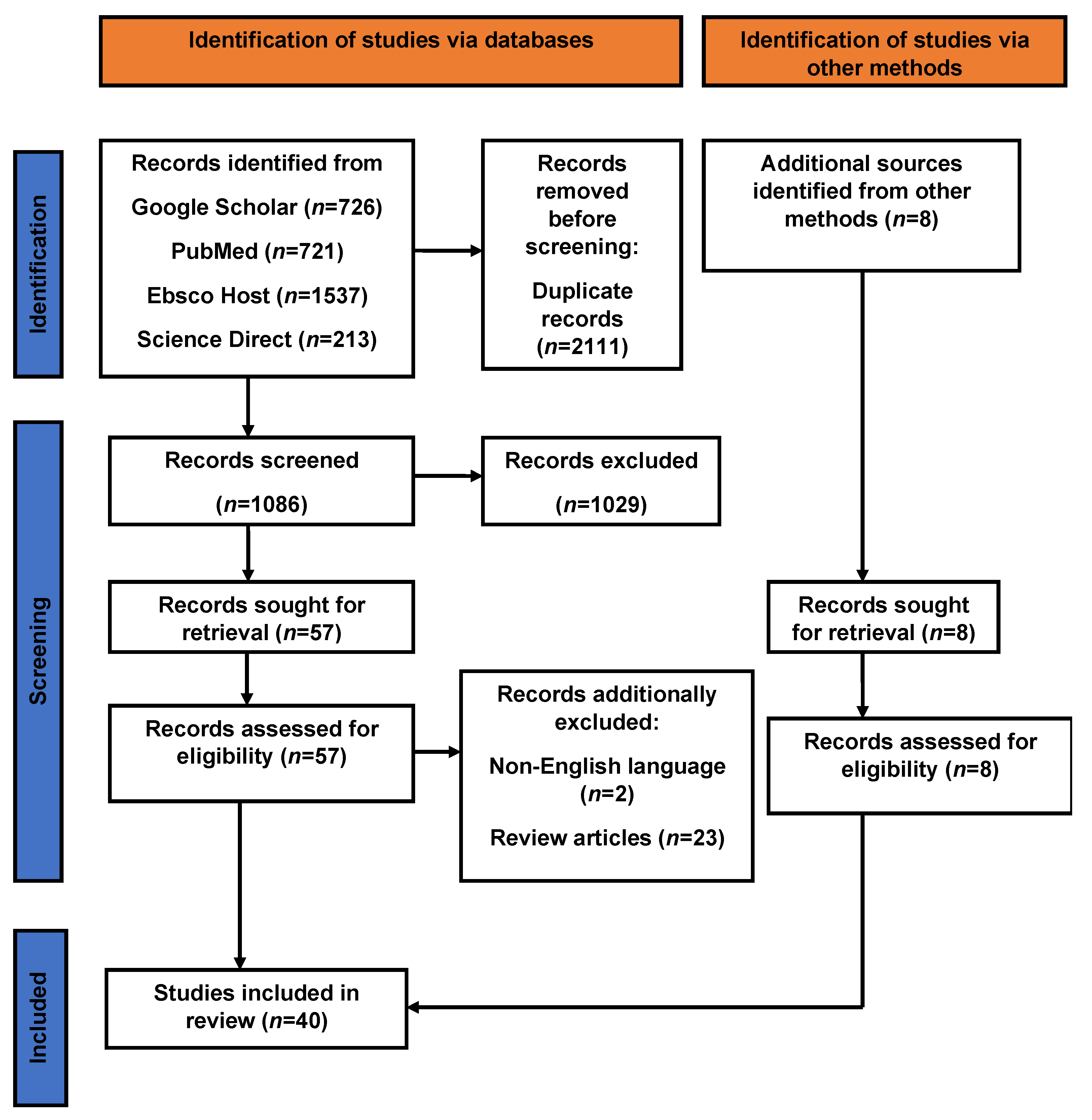
2.1. Systematic Review
2.2. Quality Assessment of Articles and Diagnostic Tests Used
2.3. Results from the Meta-Analysis
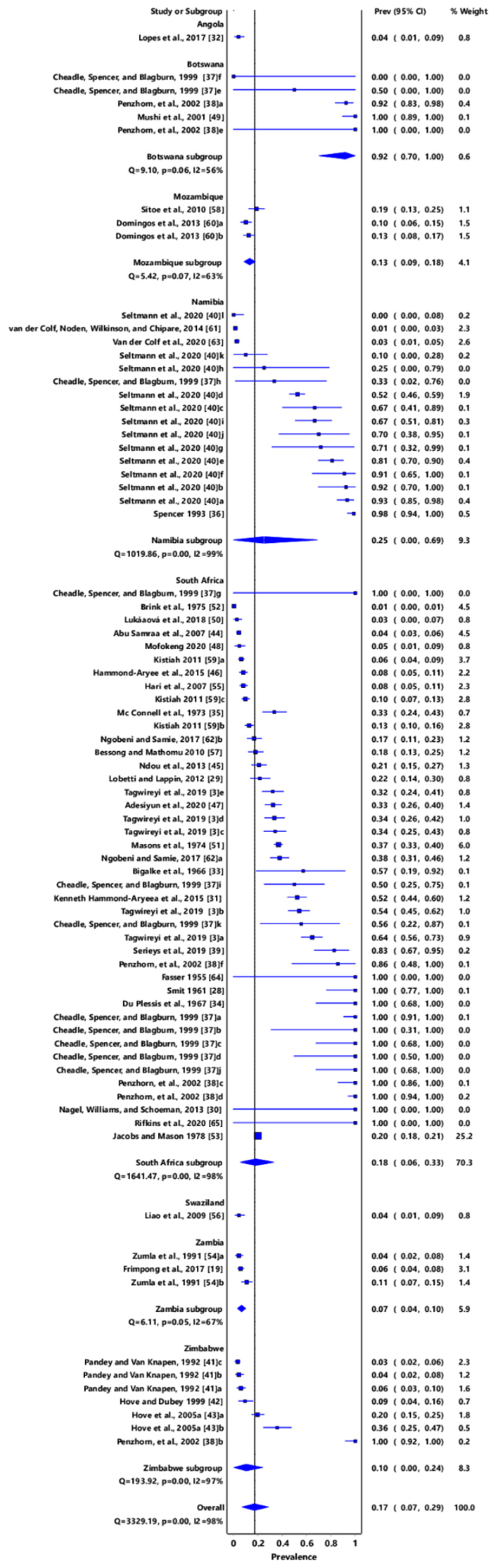
2.3.1. Pooled Prevalence and Heterogeneity

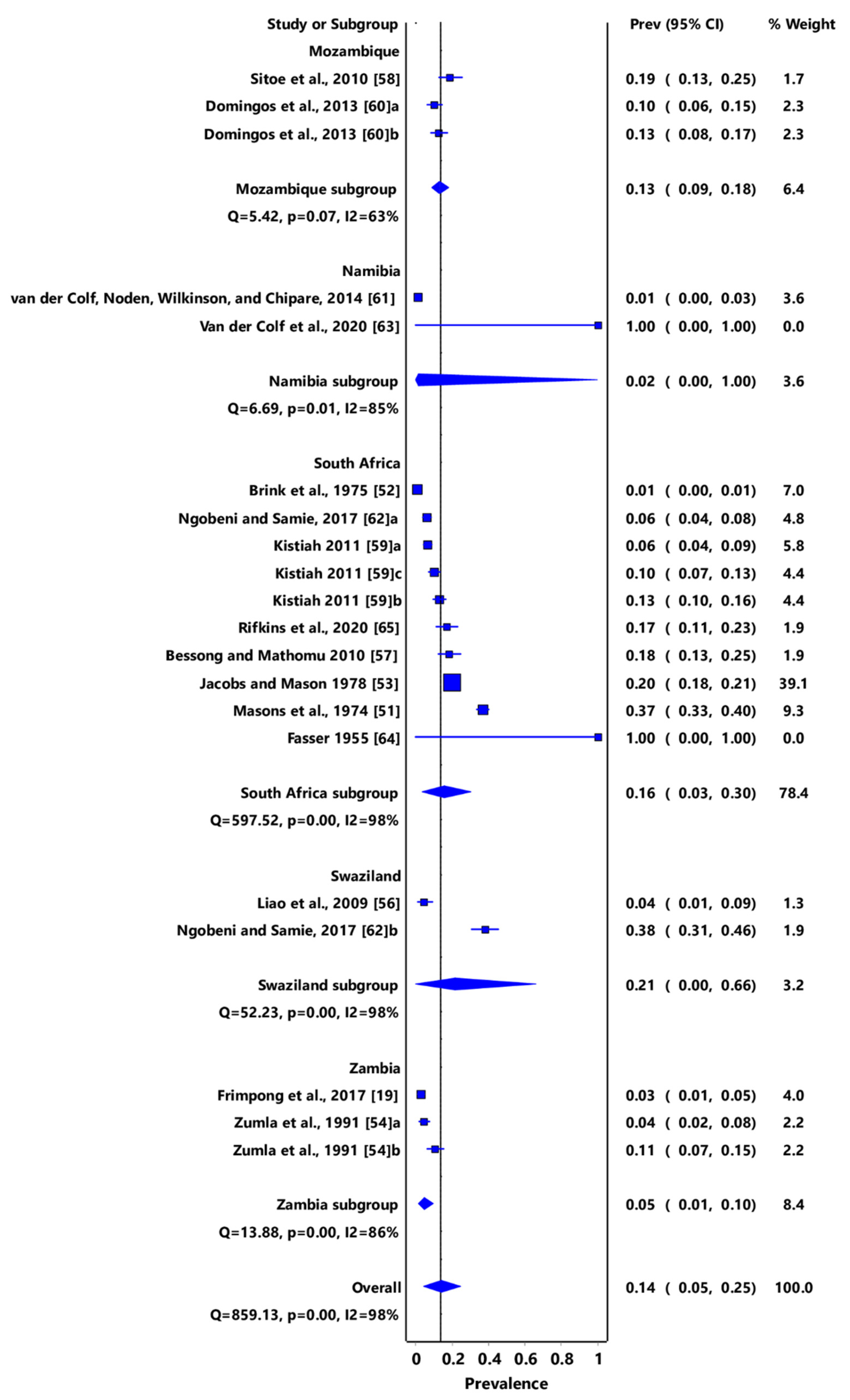
2.3.2. Toxoplasma gondii Infections in Humans in Southern African Countries
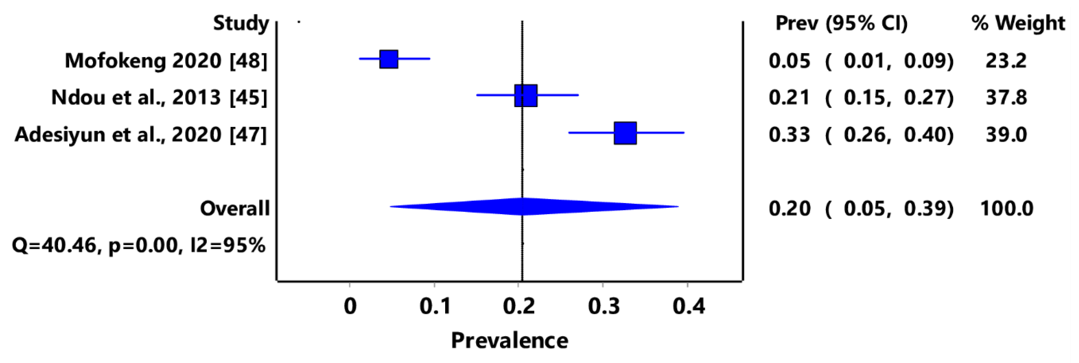
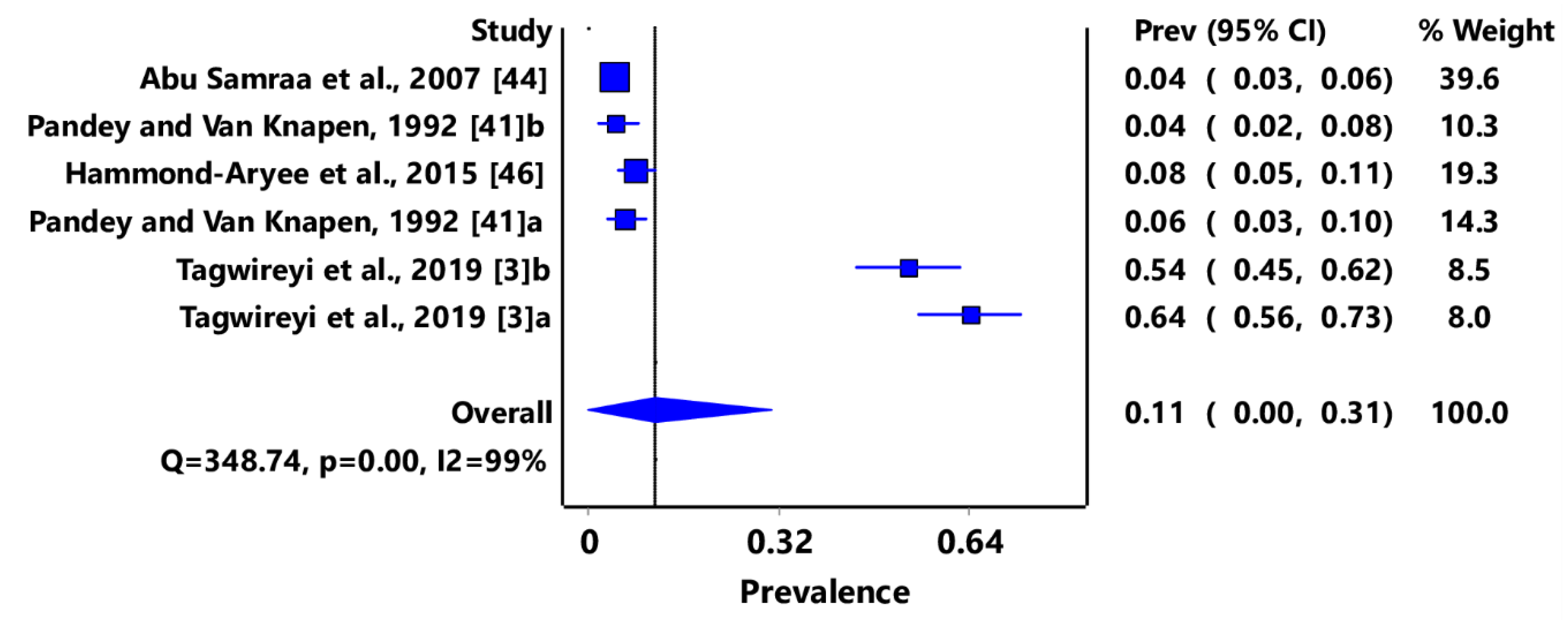
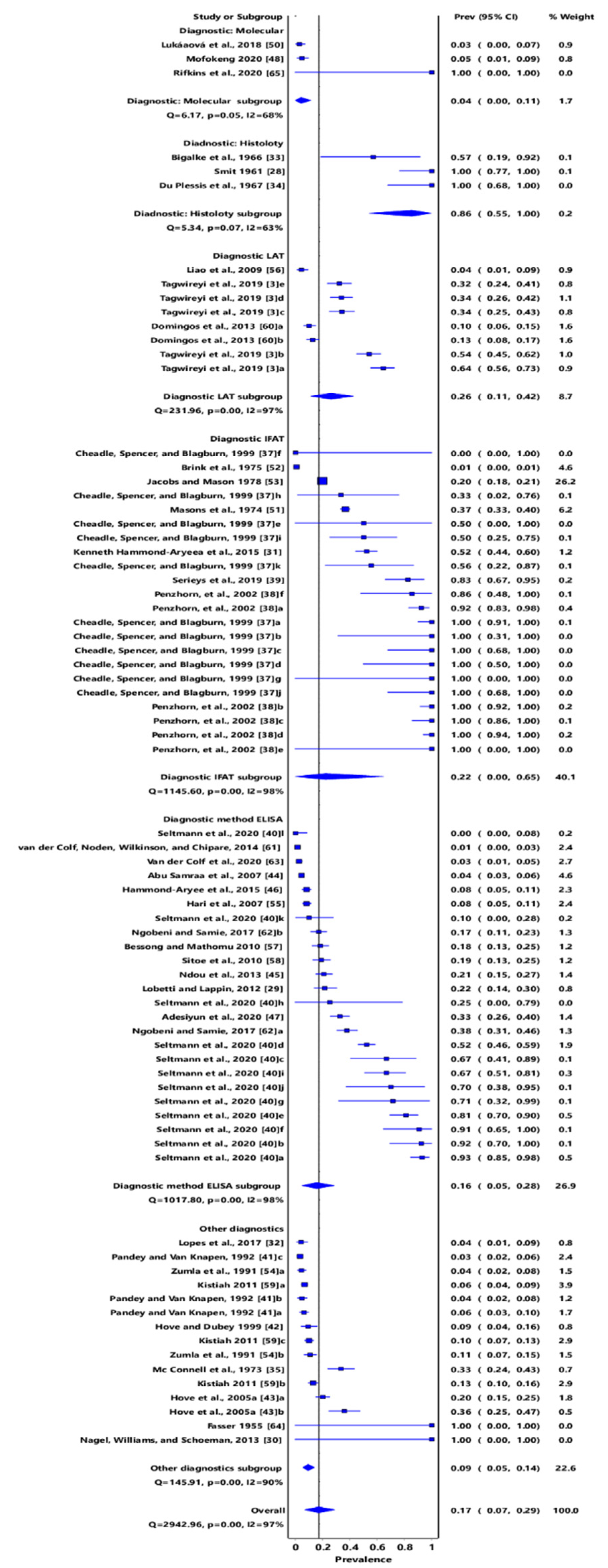
2.3.3. Pooled Prevalence and Heterogeneity of Diagnostic Tests
3. Discussion
4. Methods
4.1. Search Strategy
- Inclusion and exclusion criteria
- Data extraction and quality assessment
4.2. Data Analysis
5. Conclusions and Recommendation
Supplementary Materials
Author Contributions
Funding
Institutional Review Board Statement
Informed Consent Statement
Data Availability Statement
Acknowledgments
Conflicts of Interest
References
- Tonouhewa, A.B.N.; Akpo, Y.; Sessou, P.; Adoligbe, C.; Yessinou, E.; Hounmanou, Y.G.I.; Assogba, M.N.; Youssao, I.; Farougou, S. Toxoplasma gondii infection in meat animals from Africa: Systematic review and meta-analysis of sero-epidemiological studies. Vet. World 2017, 10, 194–208. [Google Scholar] [CrossRef] [PubMed]
- Attias, M.; Teixeira, D.E.; Benchimol, M.; Vommaro, R.C.; Crepaldi, P.H.; De Souza, W. The life cycle of Toxoplasma gondii reviewed using animations. Parasits Vectors 2020, 13, 588. [Google Scholar] [CrossRef]
- Tagwireyi, W.M.; Etter, E.; Neves, L. Seroprevalence and associated risk factors of Toxoplasma gondii infection in domestic animals in southeastern South Africa. Onderstepoort J. Vet. Res. 2019, 86, 1–6. [Google Scholar] [CrossRef] [PubMed]
- Dubey, J.; Bhatia, C.; Lappin, M.; Ferreira, L.; Thorn, A.; Kwok, O. Seroprevalence of Toxoplasma gondii and Bartonella spp. antibodies in cats from Pennsylvania. J. Parasitol. 2009, 95, 578–580. [Google Scholar] [CrossRef] [PubMed]
- Stelzer, S.; Basso, W.; Silván, J.B.; Ortega-Mora, L.M.; Maksimov, P.; Gethmann, J.; Conraths, F.J.; Schares, G. Toxoplasma gondii infection and toxoplasmosis in farm animals: Risk factors and economic impact. Food Waterborne Parasitol. 2019, 15, e00037. [Google Scholar] [CrossRef] [PubMed]
- Chiari, C.A.; Neves, D.P. Human toxoplasmosis acquired by ingestion of goat’s milk. Mem. Inst. Oswaldo Cruz. 1984, 79, 337–340. [Google Scholar] [CrossRef] [Green Version]
- Lindsay, D.S.; Collins, M.V.; Mitchell, S.M.; Cole, R.A.; Flick, G.J.; Wetch, C.N.; Rosypal, A.C.; Flick, G.J.; Zajac, A.M.; Lindquist, A.; et al. Survival of Toxoplasma gondii oocysts in eastern oysters (Crassostrea virginica). J. Parasitol. 2004, 90, 1054–1057. [Google Scholar] [CrossRef]
- Coupe, A.; Howe, L.; Shapiro, K.; Roe, W.D. Comparison of PCR assays to detect Toxoplasma gondii oocysts in green-lipped mussels (Perna canaliculus). Parasitol. Res. 2019, 118, 2389–2398. [Google Scholar] [CrossRef]
- Monteiro, T.R.M.; Rocha, K.S.; Silva, J.; Mesquita, G.S.S.; Rosário, M.K.S.; Ferreira, M.F.S.; Honorio, B.E.; Melo, H.F.; Barros, F.N.; Scofield, A.; et al. Detection of Toxoplasma gondii in Crassostrea spp. oysters cultured in an estuarine region in eastern Amazon. Zoonoses Public Health 2019, 66, 296–300. [Google Scholar] [CrossRef]
- Montazeri, M.; Galeh, T.M.; Moosazadeh, M.; Sarvi, S.; Dodangeh, S.; Javidnia, J.; Sharif, M.; Daryani, A. The global serological prevalence of Toxoplasma gondii in felids during the last five decades (1967–2017): A systematic review and meta-analysis. Parasits Vectors 2020, 13, 82. [Google Scholar] [CrossRef]
- Mose, J.M.; Kagira, J.M.; Kamau, D.M.; Maina, N.W.; Ngotho, M.; Karanja, S.M. A review on the present advances on studies of toxoplasmosis in eastern Africa. BioMed Res. Int. 2020, 2020, e7135268. [Google Scholar] [CrossRef] [PubMed]
- Abdoli, A.; Dalimi, A.; Movahedin, M. Impaired reproductive function of male rats infected with Toxoplasma gondii. Andrologia 2012, 44, 679–687. [Google Scholar] [CrossRef] [PubMed]
- Saki, J.; Sabaghan, M.; Arjmand, R.; Teimoori, A.; Rashno, M.; Saki, G.; Shojaee, S. Spermatogonia apoptosis induction as a possible mechanism of Toxoplasma gondii-induced male infertility. Iran. J. Basic Med. Sci. 2020, 23, 1164. [Google Scholar] [PubMed]
- Zhou, Y.; Lu, Y.; Hu, Y. Experimental study of influence of Toxoplasma tachyzoites on human sperm motility parameters in vitro. Chin. J. Zoonoses 2003, 19, 47–49. [Google Scholar]
- Terpsidis, K.I.; Papazahariadou, M.G.; Taitzoglou, I.A.; Papaioannou, N.G.; Georgiadis, M.P.; Theodoridis, I.T. Toxoplasma gondii: Reproductive parameters in experimentally infected male rats. Exp. Parasitol. 2009, 121, 238–241. [Google Scholar] [CrossRef]
- Gebremedhin, E.Z.; Agonafir, A.; Tessema, T.S.; Tilahun, G.; Medhin, G.; Vitale, M.; Di Marco, V. Some risk factors for reproductive failures and contribution of Toxoplasma gondii infection in sheep and goats of Central Ethiopia: A cross-sectional study. Res. Vet. Sci. 2013, 95, 894–900. [Google Scholar] [CrossRef]
- Edwards, J.F.; Dubey, J. Toxoplasma gondii abortion storm in sheep on a Texas farm and isolation of mouse virulent atypical genotype T. gondii from an aborted lamb from a chronically infected ewe. Vet. Parasitol. 2013, 192, 129–136. [Google Scholar] [CrossRef]
- Dubey, J.P. Toxoplasmosis in sheep—The last 20 years. Vet. Parasitol. 2009, 163, 1–14. [Google Scholar] [CrossRef]
- Frimpong, C.; Makasa, M.; Sitali, L.; Michelo, C. Seroprevalence and determinants of toxoplasmosis in pregnant women attending antenatal clinic at the university teaching hospital, Lusaka, Zambia. BMC Infect. Dis. 2017, 17, 10. [Google Scholar] [CrossRef] [Green Version]
- Hammond-Aryee, K.; Esser, M.; Van Helden, P.D. Toxoplasma gondii seroprevalence studies on humans and animals in Africa. South Afr. Fam. Pract. 2014, 56, 119–124. [Google Scholar] [CrossRef] [Green Version]
- Ramírez, M.D.L.L.G.; Orozco, L.V.S.; Ramírez, C.G.T. The laboratory diagnosis in Toxoplasma infection. Toxoplasmosis 2017, 6, 89–104. [Google Scholar]
- Montoya, J.G. Laboratory Diagnosis of Toxoplasma gondii infection and Toxoplasmosis. J. Infect. Dis. 2002, 185, 73–82. [Google Scholar] [CrossRef] [PubMed] [Green Version]
- Daka, V.M. Seroprevalence and Risk Factors of Toxoplasmosis in Individuals Attending Chipokotamayamba Clinic in Ndola, Zambia. Ph.D. Thesis, University of Zambia, Lusaka, Zambia, April 2015. [Google Scholar]
- Muhie, Y.; Keskes, S. Toxoplasmosis: Emerging and Re-Emerging zoonosis. Afr. J. Appl. Microbiol. 2014, 3, 1–11. [Google Scholar]
- EFSA. Surveillance and monitoring of Toxoplasma in humans, food, and animals; scientific opinion of the panel on biological hazards. EFSA J. 2007, 583, 1–64. [Google Scholar]
- Vogel, M.; Schwarze-Zander, C.; Wasmuth, J.-C.; Spengler, U.; Sauerbruch, T.; Rockstroh, J.K. The treatment of patients with HIV. Dtsch Ärztebl. Int. 2010, 107, 507–516. [Google Scholar] [CrossRef]
- Overton, T.; Bennet, P. Toxoplasmosis in Pregnancy. Fetal Matern. Med. Rev. 2010, 8, 11–18. [Google Scholar] [CrossRef]
- Smit, J.D. Toxoplasmosis in dogs in South Africa: Seven case reports. J. South Afri. Vet. Assoc. 1961, 32, 339–348. [Google Scholar]
- Lobetti, R.; Lappin, M.R. Prevalence of Toxoplasma gondii, Bartonella species and haemoplasma infection in cats in South Africa. J. Feline Med. Surg. 2012, 14, 857–862. [Google Scholar] [CrossRef]
- Nagel, S.S.; Williams, J.H.; Schoeman, J.P. Fatal disseminated toxoplasmosis in an immunocompetent cat. J. South Afri. Vet. Assoc. 2013, 84, 1–6. [Google Scholar] [CrossRef] [Green Version]
- Hammond-Aryee, K.; Esser, M.; Van Helden, L.; Van Helden, P. A high seroprevalence of Toxoplasma gondii antibodies in a population of feral cats in the Western Cape province of South Africa. South Afr. J. Infect. Dis. 2015, 30, 141–144. [Google Scholar]
- Lopes, A.P.; Oliveira, A.C.; Granada, S.; Rodrigues, F.T.; Papadopoulos, E.; Schallig, H.; Dubey, J.P.; Cardoso, L. Antibodies to Toxoplasma gondii and Leishmania spp. in domestic cats from Luanda, Angola. Vet. Parasitol. 2017, 239, 15–18. [Google Scholar] [CrossRef] [PubMed] [Green Version]
- Bigalke, R.D.; Tustin, R.C.; Du Plessis, J.L.; Basson, P.A.; McCully, R.M. The isolation of Toxoplasma gondii from ferrets in South Africa. South Afr. Vet. Assoc. 1966, 37, 243–247. [Google Scholar]
- Du Plessis, J.L.; Bigalke, R.D.; Gurnell, T. An outbreak of toxoplasmosis in chinchillas in South Africa. J. South Afr. Vet. Assoc. 1967, 38, 79–83. [Google Scholar]
- Mc Connell, E.E.; Basson, P.A.; Wolstenholme, B.; De Vos, V.; Malherbe, H.H. Toxoplasmosis in free-ranging chacma baboons (Papio ursinus) from the Kruger National Park. Trans. R. Soc. Trop. Med. Hyg. 1973, 67, 851–855. [Google Scholar] [CrossRef]
- Spencer, J.A.; Markel, P. Serological survey of sera from lions in Etosha National Park. South Afr. J. Wildl. Res. 1993, 23, 60–61. [Google Scholar]
- Cheadle, M.A.; Spencer, J.A.; Blagburn, B.L. Seroprevalences of Neospora caninum and Toxoplasma gondii in nondomestic felids from southern Africa. J. Zoo Wildl. Med. 1999, 30, 248–251. [Google Scholar]
- Penzhorn, B.L.; Stylianides, E.; Van Vuuren, M.; Alexander, K.; Meltzer, D.G.A.; Mukarati, N. Seroprevalence of Toxoplasma gondii in free-ranging lion and leopard populations in southern Africa. South Afr. J. Wildl. Res. 2002, 32, 163–165. [Google Scholar]
- Serieys, L.E.; Hammond-Aryee, K.; Bishop, J.; Broadfield, J.; O’Riain, M.J.; van Helden, P.D. High seroprevalence of Toxoplasma gondii in an urban caracal (Caracal caracal) population in South Africa. J. Wildl. Dis. 2019, 55, 951–953. [Google Scholar] [CrossRef] [Green Version]
- Seltmann, A.; Schares, G.; Aschenborn, O.H.K.; Heinrich, S.K.; Thalwitzer, S.; Wachter, B.; Czirják, G.A. Species-specific differences in Toxoplasma gondii, Neospora caninum and Besnoitia besnoiti seroprevalence in Namibian wildlife. Parasit. Vectors 2020, 13, 7. [Google Scholar] [CrossRef]
- Pandey, V.S.; Van Knapen, F. The seroprevalence of toxoplasmosis in sheep, goats, and pigs in Zimbabwe. Ann. Trop. Med. Parasitol. 1992, 86, 313–315. [Google Scholar] [CrossRef]
- Hove, T.; Dubey, J.P. Prevalence of Toxoplasma gondii antibodies in sera of domestic pigs and some wild game species from Zimbabwe. J. Parasitol. 1999, 85, 372–373. [Google Scholar] [CrossRef] [PubMed]
- Hove, T.; Lind, P.; Mukaratirwa, S. Seroprevalence of Toxoplasma gondii infection in domestic pigs reared under different management systems in Zimbabwe. Onderstepoort J. Vet. Res. 2005, 72, 231–237. [Google Scholar] [CrossRef] [PubMed]
- Abu Samraa, N.; McCrindle, C.M.E.; Penzhorn, B.L.; Cenci-Goga, B. Seroprevalence of toxoplasmosis in sheep in South Africa. J. South Afr. Vet. Assoc. 2007, 78, 116–120. [Google Scholar] [CrossRef] [PubMed] [Green Version]
- Ndou, R.V.; Maduna, N.M.; Dzoma, B.M.; Nyirenda, M.; Motsei, L.E.; Bakunzi, F.R. A seroprevalence survey of Toxoplasma gondii amongst slaughter cattle in two high throughput abattoirs in the NorthWest Province of South Africa. J. Food Agric. Environ. 2013, 11, 338–339. [Google Scholar]
- Hammond-Aryee, K.; Van Helden, P.D. The prevalence of antibodies to Toxoplasma gondii in sheep in the Western Cape, South Africa: Research communication. Onderstepoort J. Vet. Res. 2015, 82, 1–5. [Google Scholar] [CrossRef] [PubMed] [Green Version]
- Adesiyun, A.A.; Knobel, D.L.; Thompson, P.N.; Wentzel, J.; Kolo, F.B.; Kolo, A.O.; Conan, A.; Simpson, G.J. Sero-epidemiological study of selected zoonotic and abortifacient pathogens in cattle at a wildlife-livestock interface in South Africa. Vector-Borne Zoonotic Dis. 2020, 20, 258–267. [Google Scholar] [CrossRef]
- Mofokeng, L.S.; Taioe, O.M.; Smit, N.J.; Thekisoe, O.M. Parasites of veterinary importance from domestic animals in uMkhanyakude district of KwaZulu-Natal province. J. South Afr. Vet. Assoc. 2020, 91, 1–11. [Google Scholar] [CrossRef]
- Mushi, E.Z.; Binta, M.G.; Chabo, R.G.; Ndebele, R.; Panzirah, R. Seroprevalence of Toxoplasma gondii and Chlamydia psittaci in domestic pigeons (Columba livia domestica) at Sebele, Gaborone, Botswana. Onderstepoort J. Vet. Res. 2001, 68, 159–161. [Google Scholar]
- Lukášová, R.; Kobédová, K.; Halajian, A.; Bártová, E.; Murat, J.B.; Rampedi, K.M.; Luus-Powell, W.J. Molecular detection of Toxoplasma gondii and Neospora caninum in birds from South Africa. Acta Trop. 2018, 178, 93–96. [Google Scholar] [CrossRef]
- Mason, P.R.; Jacobs, M.R.; Fripp, P.J. Serological survey of toxoplasmosis in the Transvaal. South Afr. Med. J. 1974, 48, 1707–1709. [Google Scholar]
- Brink, J.D.; de Wet, J.S.; van Rensburg, A.J. A serological survey of toxoplasmosis in the Bloemfontein area. South Afr. Med. J. 1975, 49, 1441–1443. [Google Scholar]
- Jacobs, M.R.; Mason, P.R. Prevalence of Toxoplasma antibodies in southern Africa. South Afr. Med. J. 1978, 53, 619–621. [Google Scholar] [PubMed]
- Zumla, A.; Savva, D.; Wheeler, R.B.; Hira, S.K.; Luo, N.P.; Kaleebu, P.; Sempala, S.K.; Johnson, J.D.; Holliman, R. Toxoplasma serology in Zambian and Ugandan patients infected with the human immunodeficiency virus. Trans. R. Soc. Trop. Med. Hyg. 1991, 85, 227–229. [Google Scholar] [CrossRef]
- Hari, K.R.; Modi, M.R.; Mochan, A.H.D.; Modi, G. Reduced risk of Toxoplasma encephalitis in HIV-infected patients—A prospective study from Gauteng, South Africa. Int. J. STD AIDS 2007, 18, 555–558. [Google Scholar] [CrossRef] [PubMed]
- Liao, C.W.; Lee, Y.L.; Sukati, H.; D’lamini, P.; Huang, Y.C.; Chiu, C.J.; Liu, Y.H.; Chou, C.M.; Chiu, W.T.; Du, W.Y.; et al. Seroprevalence of Toxoplasma gondii infection among children in Swaziland, southern Africa. Ann. Trop. Med. Parasitol. 2009, 103, 731–736. [Google Scholar] [CrossRef] [PubMed]
- Bessong, P.O.; Mathomu, L.M. Seroprevalence of HTLV1/2, HSV1/2 and Toxoplasma gondii among chronic HIV-1 infected individuals in rural north-eastern South Africa. Afr. J. Microbiol. Res. 2010, 4, 2587–2591. [Google Scholar]
- Sitoe, S.P.B.L.; Rafael, B.; Meireles, L.R.; Andrade Jr, H.F.D.; Thompson, R. Preliminary report of HIV and Toxoplasma gondii occurrence in pregnant women from Mozambique. Revista Inst. Med. Trop. São Paulo 2010, 52, 291–295. [Google Scholar] [CrossRef] [Green Version]
- Kistiah, K.; Winiecka-Krusnell, J.; Barragan, A.; Karstaedt, A.; Frean, J. Seroprevalence of Toxoplasma gondii infection in HIV-positive and HIV-negative subjects in Gauteng, South Africa. South Afr. J. Epidemiol. Infect. 2011, 26, 225–228. [Google Scholar] [CrossRef] [Green Version]
- Domingos, A.; Ito, L.S.; Coelho, E.; Lúcio, J.M.; Matida, L.H.; Ramos, A.N. Seroprevalence of Toxoplasma gondii IgG antibody in HIV/AIDS-infected individuals in Maputo, Mozambique. Rev. Saude Publica 2013, 47, 890–896. [Google Scholar] [CrossRef] [Green Version]
- van der Colf, B.E.; Noden, B.H.; Wilkinson, R.; Chipare, I. Low seroprevalence of antibodies to Toxoplasma gondii in blood donors in central Namibia. South Afr. J. Infect. Dis. 2014, 29, 101–104. [Google Scholar] [CrossRef]
- Ngobeni, R.; Samie, A. Prevalence of Toxoplasma gondii IgG and IgM and associated risk factors among HIV-positive and HIV-negative patients in Vhembe district of South Africa. Afr. J. Infect. Dis. 2017, 11, 1–9. [Google Scholar] [PubMed] [Green Version]
- Van der Colf, B.E.; Ntirampeba, D.; Van Zyl, G.U.; Noden, B.H. Seroprevalence of Toxoplasma gondii infection among pregnant women in Windhoek, Namibia, in 2016. South Afr. J. Infect. Dis. 2020, 35, 1–7. [Google Scholar] [CrossRef] [PubMed]
- Rifkin, R.F.; Vikram, S.; Ramond, J.B.; Cowan, D.A.; Jakobsson, M.; Schlebusch, C.M.; Lombard, M. Ancient DNA of Rickettsia felis and Toxoplasma gondii implicated in the death of a hunter-gatherer boy from South Africa, 2000 years ago. BioRxiv 2020. [Google Scholar] [CrossRef]
- Fasser, E. Congenital toxoplasmosis in South Africa-a review and case report. South Afr. Med. J. 1955, 29, 684–688. Available online: https://www.biorxiv.org/content/biorxiv/early/2020/07/23/2020.07.23.217141.full.pdf (accessed on 16 January 2022).
- Dubey, J.P.; Beattie, C. Toxoplasmosis of Animals and Man, 2nd ed.; CRC Press: Boca Raton, FL, USA, 2010. [Google Scholar]
- Webster, J.P. Dubey, J.P. Toxoplasmosis of Animals and Humans; CRC Press: Boca Raton, FL, USA, 2010. [Google Scholar]
- Rouatbi, M.; Amairia, S.; Amdouni, Y.; Boussaadoun, M.A.; Ayadi, O.; Al-Hosary, A.A.T.; Rekik, M.; Abdallah, R.B.; Aoun, K.; Darghouth, M.A. Toxoplasma gondii infection and toxoplasmosis in North Africa: A review. Parasite 2019, 26, e2019006. [Google Scholar] [CrossRef] [PubMed] [Green Version]
- Elmore, S.A.; Jones, J.L.; Conrad, P.A.; Patton, S.; Lindsay, D.S.; Dubey, J. Toxoplasma gondii: Epidemiology, feline clinical aspects, and prevention. Trends Parasitol. 2010, 26, 190–196. [Google Scholar] [CrossRef] [Green Version]
- Hatam-Nahavandi, K.; Calero-Bernal, R.; Rahimi, M.T.; Pagheh, A.S.; Zarean, M.; Dezhkam, A.; Ahmadpour, E. Toxoplasma gondii infection in domestic and wild felids as public health concerns: A systematic review and meta-analysis. Sci. Rep. 2021, 11, 9509. [Google Scholar] [CrossRef]
- Dubey, J.P.; Murata, F.H.; Cerqueira-Cézar, C.K.; Kwok, O.C. Recent epidemiologic and clinical Toxoplasma gondii infections in wild canids and other carnivores: 2009–2020. Vet. Parasitol. 2021, 290, 109337. [Google Scholar] [CrossRef]
- Wei, X.Y.; Gao, Y.; Lv, C.; Wang, W.; Chen, Y.; Zhao, Q.; Gong, Q.L.; Zhang, X.X. The global prevalence and risk factors of Toxoplasma gondii among foxes: A systematic review and meta-analysis. Microb. Pathog. 2021, 150, 104699. [Google Scholar] [CrossRef]
- Odeniran, P.O.; Omolabi, K.F.; Ademola, I.O. A meta-analysis of Toxoplasma gondii seroprevalence, genotypes and risk factors among food animals in West African countries from public health perspectives. Prev. Vet. Med. 2020, 176, e104925. [Google Scholar] [CrossRef]
- Costa, G.H.N.; Cabral, D.D.; Varandas, N.P.; de Almeida Sobral, E.; de Almeida Borges, F.; Castagnolli, K.C. Frequency of anti-Neospora caninum and anti-Toxoplasma gondii antibodies in bovine sera from the states of São Paulo and Minas Gerais. Sem. Ciên. Agrárias 2001, 22, 61–66. [Google Scholar] [CrossRef]
- Khalil, K.M.; Abdel Gadir, A.E. Prevalence of Toxoplasma gondii antibodies in camels and their herders in three ecologically different areas in Sudan. J. Camel. Pract. Res. 2007, 14, 11–13. [Google Scholar]
- Baril, L.; Ancelle, T.; Goulet, V.; Thulliez, P.; Tirard-Fleury, V.; Carme, B. Risk factors for Toxoplasma infection in pregnancy: A case-control study in France. Scand. J. Infect. Dis. 1999, 31, 305–309. [Google Scholar] [PubMed]
- Belluco, S.; Simonato, G.; Mancin, M.; Pietrobelli, M.; Ricci, A. Toxoplasma gondii infection and food consumption: A systematic review and meta-analysis of case-controlled studies. Crit. Rev. Food Sci. Nutr. 2018, 58, 3085–3096. [Google Scholar] [CrossRef] [PubMed]
- Jones, J.L.; Dargelas, V.; Roberts, J.; Press, C.; Remington, J.S.; Montoya, J.G. Risk factors for Toxoplasma gondii infection in the United States. Clin. Infect. Dis. 2009, 49, 878–884. [Google Scholar] [CrossRef] [PubMed] [Green Version]
- Mancianti, F.; Nardoni, S.; D’Ascenzi, C.; Pedonese, F.; Mugnaini, L.; Franco, F.; Papini, R. Seroprevalence, detection of DNA in blood and milk, and genotyping of Toxoplasma gondii in a goat population in Italy. BioMed Res. Int. 2013, 2013, 905326. [Google Scholar] [CrossRef] [PubMed] [Green Version]
- Sadek, O.; Abdel-Hameed, Z.M.; Kuraa, H.M. Molecular detection of Toxoplasma gondii DNA in raw goat and sheep milk with discussion of its public health importance in Assiut Governorate. Assiut Vet. Med. J. 2015, 61, 166–177. [Google Scholar]
- Sroka, J.; Kusyk, P.; Bilska-Zając, E.; Karamon, J.; Dutkiewicz, J.; Wójcik-Fatla, A.; Zajac, V.; Stojecki, K.; Rózycki, M.; Cencek, T. Seroprevalence of Toxoplasma gondii infection in goats from the south-west region of Poland and the detection of T. gondii DNA in goat milk. Folia Parasitol. 2017, 64, 023. [Google Scholar] [CrossRef]
- Foroutan, M.; Fakhri, Y.; Riahi, S.M.; Ebrahimpour, S.; Namroodi, S.; Taghipour, A.; Spotin, A.; Gamble, H.R.; Rostami, A. The global seroprevalence of Toxoplasma gondii in pigs: A systematic review and meta-analysis. Vet. Parasitol. 2019, 269, 42–52. [Google Scholar] [CrossRef]
- Dubey, J.P.; Jones, J.L. Toxoplasma gondii infection in humans and animals in the United States. Int. J. Parasitol. 2008, 38, 1257–1278. [Google Scholar] [CrossRef]
- Bamba, S.; Halos, F.; Tarnagda, Z.; Alanio, A.; Macé, E.; Moukoury, S.; Sangaré, I.; Guiguemdé, R.; Costa, J.M.; Bretagne, S. Seroprevalence of Toxoplasma gondii and direct genotyping using mini sequencing in free-range pigs in Burkina Faso. Int. J. Food Microbiol. 2016, 230, 10–15. [Google Scholar] [CrossRef]
- Dubey, J.; Pena, H.; Cerqueira-Cézar, C.; Murata, F.; Kwok, O.; Yang, Y.; Gennari, S.M.; Su, C. Epidemiologic significance of Toxoplasma gondii infections in chickens (Gallus domesticus): The past decade. Parasitology 2020, 147, 1263–1289. [Google Scholar] [CrossRef]
- Dubey, J.P.; Murata, F.H.A.; Cerqueira-Cézar, C.K.; Kwok, O.C.H.; Su, C. Epidemiologic significance of Toxoplasma gondii infections in turkeys, ducks, ratites, and other wild birds: 2009–2020. Parasitology 2021, 148, 1–30. [Google Scholar] [CrossRef] [PubMed]
- Bigna, J.J.; Tochie, J.N.; Tounouga, D.N.; Bekolo, A.O.; Ymele, N.S.; Youda, E.L.; Sime, P.S.; Nansseu, J.R. Global, regional, and country seroprevalence of Toxoplasma gondii in pregnant women: A systematic review, modelling and meta-analysis. Sci. Rep. 2020, 10, 12102. [Google Scholar] [CrossRef] [PubMed]
- Dubey, J.P. Toxoplasmosis of Animals and Humans, 2nd ed.; CRC Press Inc.: Boca Raton, FL, USA; New York, NY, USA, 2010. [Google Scholar]
- Rajendran, C.; Su, C.; Dubey, J.P. Molecular genotyping of Toxoplasma gondii from Central and South America revealed high diversity within and between populations. Infect. Genet. Evol. 2012, 12, 359–368. [Google Scholar] [CrossRef] [PubMed]
- Mahmoud, H.; Saedi Dezaki, E.; Soleimani, S.; Baneshi, M.R.; Kheirandish, F.; Ezatpour, B.; Zia-Ali, N. Seroprevalence and risk factors of Toxoplasma gondii infection among healthy blood donors in southeast of Iran. Parasite Immunol. 2015, 37, 362–367. [Google Scholar] [CrossRef]
- Robert-Gangeux, F.; Darde, M.-L. Epidemiology and diagnostic strategies for Toxoplasmosis. Clin. Microbiol. Rev. 2012, 25, 264–296. [Google Scholar] [CrossRef] [Green Version]
- Smith, N.C.; Goulart, C.; Hayward, J.A.; Kupz, A.; Miller, C.M.; Van Dooren, G.C. Control of human toxoplasmosis. Int. J. Parasitol. 2021, 51, 95–121. [Google Scholar] [CrossRef]
- Dubey, J.P.; Lappin, M.R.; Thulliez, P. Long-term antibody responses of cats fed Toxoplasma gondii tissue cysts. J. Parasitol. 1995, 81, 887–893. [Google Scholar] [CrossRef]
- Hove, T.; Lind, P.; Mukaratirwa, S. Seroprevalence of Toxoplasma gondii infection in goats and sheep in Zimbabwe. Onderstepoort J. Vet. Res. 2005, 72, 267–272. [Google Scholar] [CrossRef]
- Sharma, R.; Parker, S.; Al-Adhami, B.; Bachand, N.; Jenkins, E. Comparison of tissues (heart vs. brain) and serological tests (MAT, ELISA and IFAT) for detection of Toxoplasma gondii in naturally infected wolverines (Gulo gulo) from the Yukon, Canada. Food Waterborne Parasitol. 2019, 15, e00046. [Google Scholar] [CrossRef] [PubMed]
- Munn, Z.; Moola, S.; Lisy, K.; Riitano, D.; Tufanaru, C. Methodological guidance for systematic reviews of observational epidemiological studies reporting prevalence and cumulative incidence data. Int. J. Evid.-Based Healthc. 2015, 13, 147–153. [Google Scholar] [CrossRef] [PubMed]
- Barendregt, J.J.; Doi, S.A. MetaXL User Guide. Available online: http://www.epigear.com/index_files/MetaXL%20User%20Guide.pdf (accessed on 16 January 2022).


| Study Country | Host Species | n | Np | (%) | Diagnostic Test | Study Period | Quality Index Score | References |
|---|---|---|---|---|---|---|---|---|
| South Africa | Dogs | 7 | 7 | 100 | Histology | 1955–1961 | 0.7 | Smit 1961 [28] |
| South Africa | Cats | 102 | 22 | 21.6 | ELISA | 2012 | 0.6 | Lobetti and Lappin, 2012 [29] |
| South Africa | Cats | 1 | 1 | 100 | Histology and PCR | 2012 | 0.9 | Nagel, Williams, and Schoeman, 2013 [30] |
| South Africa | Cats | 159 | 83 | 52.2 | IFAT | 2013–2014 | 0.8 | Kenneth Hammond-Aryeea et al., 2015 [31] |
| Angola | Cats | 102 | 4 | 3.9 | MAT | 2014–2016 | 0.7 | Lopes et al., 2017 [32] |
| South Africa | Cats | 109 | 35 | 32.1 | LAT | 2016 | 0.9 | Tagwireyi et al., 2019 [3] |
| Study Area | Host Species | n | Np | (%) | Diagnostic Test | Study Period | Quality Index Score | References |
|---|---|---|---|---|---|---|---|---|
| South Africa | Ferrets | 7 | 4 | 42.9 | Histology | 1966 | 0.5 | Bigalke et al., 1966 [33] |
| South Africa | Chinchilla | 5 | 5 | 100 | Histology | 1966 | 0.5 | Du Plessis et al., 1967 [34] |
| South Africa | Baboons | 90 | 30 | 11.7 | IFAT, CF, Wolstenholme’s modification, Sabin–Feldman dye test | 1969–1971 | 0.8 | Mc Connell et al., 1973 [35] |
| Namibia | Lions | 66 | 65 | 98 | Indirect Immunofluorescence Assay | 1989–1991 | 0.6 | Spencer 1993 [36] |
| South Africa | Lions | 18 | 18 | 100 | IFAT | 1984–1996 | 0.8 | Cheadle, Spencer, and Blagburn, 1999 [37]a |
| South Africa | Leopard | 2 | 2 | 100 | IFAT | 1984–1996 | 0.8 | Cheadle, Spencer, and Blagburn, 1999 [37]b |
| South Africa | Lions | 5 | 5 | 100 | IFAT | 1984–1996 | 0.8 | Cheadle, Spencer, and Blagburn, 1999 [37]c |
| South Africa | Lions | 3 | 3 | 100 | IFAT | 1984–1996 | 0.8 | Cheadle, Spencer, and Blagburn, 1999 [37]d |
| Botswana | Leopard | 2 | 1 | 50 | IFAT | 1984–1996 | 0.8 | Cheadle, Spencer, and Blagburn, 1999 [37]e |
| Botswana | Cheetah | 1 | 0 | 0 | IFAT | 1984–1996 | 0.8 | Cheadle, Spencer, and Blagburn, 1999 [37]f |
| Namibia | Lions | 1 | 1 | 100 | IFAT | 1984–1996 | 0.8 | Cheadle, Spencer, and Blagburn, 1999 [37]g |
| Namibia | Cheetah | 6 | 2 | 33.3 | IFAT | 1984–1996 | 0.8 | Cheadle, Spencer, and Blagburn, 1999 [37]h |
| South Africa | Cheetah | 16 | 8 | 50 | IFAT | 1984–1996 | 0.8 | Cheadle, Spencer, and Blagburn, 1999 [37]i |
| South Africa | Lions | 5 | 5 | 100 | IFAT | 1984–1996 | 0.8 | Cheadle, Spencer, and Blagburn, 1999 [37]j |
| South Africa | Lions | 9 | 5 | 55.6 | IFAT | 1984–1996 | 0.8 | Cheadle, Spencer, and Blagburn, 1999 [37]k |
| Botswana | Lions | 53 | 49 | 92 | IFAT | 2002 | 0.5 | Penzhorn et al., 2002 [38]a |
| Zimbabwe | Lions | 21 | 21 | 100 | IFAT | 2002 | 0.5 | Penzhorn et al., 2002 [38]b |
| South Africa | Lions | 12 | 12 | 100 | IFAT | 2002 | 0.5 | Penzhorn et al., 2002 [38]c |
| South Africa | Lions | 30 | 30 | 100 | IFAT | 2002 | 0.5 | Penzhorn et al., 2002 [38]d |
| Botswana | Leopard | 1 | 1 | 100 | IFAT | 2002 | 0.5 | Penzhorn et al., 2002 [38]e |
| South Africa | Leopard | 7 | 6 | 86 | IFAT | 2002 | 0.5 | Penzhorn et al., 2002 [38]f |
| South Africa | Caracal | 29 | 24 | 83 | IFAT | 2014–2017 | 0.9 | Serieys et al., 2019 [39] |
| Namibia | African Lion | 59 | 55 | 93.2 | ELISA | 2002–2015 | 0.6 | Seltmann et al., 2020 [40]a |
| Namibia | Brown hyena | 19 | 12 | 92.3 | ELISA | 2002–2015 | 0.6 | Seltmann et al., 2020 [40]b |
| Namibia | Caracal | 15 | 10 | 66.7 | ELISA | 2002–2015 | 0.6 | Seltmann et al., 2020 [40]c |
| Namibia | Cheetah | 250 | 131 | 52.4 | ELISA | 2002–2015 | 0.6 | Seltmann et al., 2020 [40]d |
| Namibia | Leopard | 58 | 47 | 81 | ELISA | 2002–2015 | 0.6 | Seltmann et al., 2020 [40]e |
| Namibia | Spotted hyena | 11 | 10 | 90.9 | ELISA | 2002–2015 | 0.6 | Seltmann et al., 2020 [40]f |
| Namibia | African wild dog | 7 | 5 | 71.4 | ELISA | 2002–2015 | 0.6 | Seltmann et al., 2020 [40]g |
| Namibia | Bat eared fox | 4 | 1 | 25 | ELISA | 2002–2015 | 0.6 | Seltmann et al., 2020 [40]h |
| Namibia | Black backed jackal | 39 | 26 | 66.7 | ELISA | 2002–2015 | 0.6 | Seltmann et al., 2020 [40]i |
| Namibia | Honey badger | 10 | 7 | 70 | ELISA | 2002–2015 | 0.6 | Seltmann et al., 2020 [40]j |
| Namibia | Blue-wildebeest | 20 | 2 | 10 | ELISA | 2002–2015 | 0.6 | Seltmann et al., 2020 [40]k |
| Namibia | Springbok | 20 | 0 | 0 | ELISA | 2002–2015 | 0.6 | Seltmann et al., 2020 [40]l |
| Study Country | Host Species | n | Np | (%) | Diagnostic Test | Study Period | Quality Index Score | References |
|---|---|---|---|---|---|---|---|---|
| Zimbabwe | Sheep | 216 | 13 | 8.8 | LAT and ELISA | 1992 | 0.7 | Pandey and Van Knapen, 1992 [41]a |
| Zimbabwe | Goats | 156 | 7 | 7.1 | LAT and ELISA | 1992 | 0.7 | Pandey and Van Knapen, 1992 [41]b |
| Zimbabwe | Pigs | 311 | 10 | 4.2 | LAT and ELISA | 1992 | 0.7 | Pandey and Van Knapen, 1992 [41]c |
| Zimbabwe | Pigs | 97 | 9 | 9.3 | MAT | 1995 | 0.7 | Hove and Dubey 1999 [42] |
| Zimbabwe | Pigs | 238 | 47 | 19.75 | IFAT and ELISA | 2000–2002 | 0.8 | Hove et al., 2005a [43]a |
| Zimbabwe | Pigs | 70 | 25 | 35.71 | IFAT and ELISA | 2000–2002 | 0.8 | Hove et al., 2005a [43]b |
| South Africa | Sheep | 600 | 26 | 4.3 | ELISA | 2007 | 0.9 | Abu Samraa et al., 2007 [44] |
| South Africa | Cattle | 178 | 37 | 20.8 | ELISA | 2012 | 0.8 | Ndou et al., 2013 [45] |
| South Africa | Sheep | 292 | 23 | 7.9 | ELISA | 2014 | 0.9 | Hammond-Aryee et al., 2015 [46] |
| South Africa | Sheep | 121 | 78 | 64.5 | LAT | 2016 | 0.9 | Tagwireyi et al., 2019 [3]a |
| South Africa | Goats | 128 | 69 | 53.9 | LAT | 2016 | 0.9 | Tagwireyi et al., 2019 [3]b |
| South Africa | Pigs | 106 | 36 | 34 | LAT | 2016 | 0.9 | Tagwireyi et al., 2019 [3]c |
| South Africa | Cattle | 184 | 60 | 32.6 | ELISA | 2013 | 0.8 | Adesiyun et al., 2020 [47] |
| South Africa | Cattle | 109 | 5 | 4.6 | PCR | 2019 | 0.8 | Mofokeng 2020 [48] |
| Study Area | Host Species | n | Np | (%) | Diagnostic Test | Study Period | Quality Index Score | References |
|---|---|---|---|---|---|---|---|---|
| Botswana | Pigeons | 16 | 16 | 100 | Indirect Haemaglutination Test (IHT) | 2001 | 0.4 | Mushi et al., 2001 [49] |
| South Africa | Birds | 110 | 3 | 2.7 | PCR | 2014–2015 | 0.7 | Lukášová et al., 2018 [50] |
| South Africa | Chickens | 137 | 46 | 33.6 | LAT | 2016 | 0.9 | Tagwireyi et al., 2019 [3]d |
| Study Area | Human Description | n | Np | (%) | Diagnostic Test | Study Period | Quality Index Score | References |
|---|---|---|---|---|---|---|---|---|
| South Africa | People from different ethnic groups | 806 | 296 | 37 | IFAT | 1974 | 0.8 | Masons et al., 1974 [51] |
| South Africa | Reproductive age women | 600 | 3 | 0.5 | IFAT | 1975 | 0.8 | Brink et al., 1975 [52] |
| Southern Africa | Blood donors from diverse ethnic groups | 3379 | 665 | 20 | IFAT | 1978 | 0.8 | Jacobs and Mason 1978 [53] |
| Zambia | HIV-positive individuals | 187 | 8 | 4.3 | LAT and DT | 1991 | 0.8 | Zumla et al., 1991 [54]a |
| Zambia | HIV-negative individuals | 189 | 20 | 10.6 | LAT and DT | 1991 | 0.8 | Zumla et al., 1991 [54]b |
| South Africa | HIV-positive individuals | 307 | 25 | 8 | ELISA | 2007 | 0.9 | Hari et al., 2007 [55] |
| Swaziland | Apparently healthy children | 113 | 5 | 4.4 | LAT | 2009 | 0.8 | Liao et al., 2009 [56] |
| South Africa | HIV-positive individuals | 160 | 29 | 18.1 | ELISA | 2007–2008 | 0.8 | Bessong and Mathomu 2010 [57] |
| Mozambique | HIV-positive patients | 150 | 28 | 18.7 | ELISA | 2010 | 0.7 | Sitoe et al., 2010 [58] |
| South Africa | Immunocompetent individuals | 497 | 32 | 6.4 | Pastorex Toxo latex particle agglutination test and BioMèrieux ToxoScreen DA test | 2011 | 0.8 | Kistiah 2011 [59]a |
| South Africa | HIV-negative patients | 376 | 48 | 12.8 | Pastorex Toxo latex particle agglutination test and BioMèrieux ToxoScreen DA test | 2011 | 0.8 | Kistiah 2011 [59]b |
| South Africa | HIV-positive patients | 376 | 37 | 9.8 | Pastorex Toxo latex particle agglutination test and BioMèrieux ToxoScreen DA test | 2011 | 0.8 | Kistiah 2011 [59]c |
| Mozambique | HIV-positive men | 200 | 20 | 39.3 | LAT | 2010 | 0.7 | Domingos et al., 2013 [60]a |
| Mozambique | HIV-positive women | 200 | 25 | 50.9 | LAT | 2010 | 0.7 | Domingos et al., 2013 [60]b |
| Namibia | Blood donor | 312 | 4 | 1.3 | ELISA | 2011–2012 | 0.8 | van der Colf, Noden, Wilkinson, and Chipare, 2014 [61] |
| Zambia | Pregnant women | 411 | 24 | 5.9 | OnSite Toxo IgG/IgM Combo Rapid test | 2015 | 0.8 | Frimpong et al., 2017 [19] |
| South Africa | HIV-positive individuals | 161 | 61 | 38 | ELISA | 2012–2013 | 0.7 | Ngobeni and Samie, 2017 [62]a |
| South Africa | HIV-negative individuals | 161 | 27 | 16.7 | ELISA | 2012–2013 | 0.7 | Ngobeni and Samie, 2017 [62]b |
| Namibia | Pregnant women | 344 | 9 | 2.61 | ELISA | 2016 | 0.9 | Van der Colf et al., 2020 [63] |
Publisher’s Note: MDPI stays neutral with regard to jurisdictional claims in published maps and institutional affiliations. |
© 2022 by the authors. Licensee MDPI, Basel, Switzerland. This article is an open access article distributed under the terms and conditions of the Creative Commons Attribution (CC BY) license (https://creativecommons.org/licenses/by/4.0/).
Share and Cite
Omonijo, A.O.; Kalinda, C.; Mukaratirwa, S. Toxoplasma gondii Infections in Animals and Humans in Southern Africa: A Systematic Review and Meta-Analysis. Pathogens 2022, 11, 183. https://doi.org/10.3390/pathogens11020183
Omonijo AO, Kalinda C, Mukaratirwa S. Toxoplasma gondii Infections in Animals and Humans in Southern Africa: A Systematic Review and Meta-Analysis. Pathogens. 2022; 11(2):183. https://doi.org/10.3390/pathogens11020183
Chicago/Turabian StyleOmonijo, Adejumoke O., Chester Kalinda, and Samson Mukaratirwa. 2022. "Toxoplasma gondii Infections in Animals and Humans in Southern Africa: A Systematic Review and Meta-Analysis" Pathogens 11, no. 2: 183. https://doi.org/10.3390/pathogens11020183